III./7.5. Therapy Introduction
In this chapter, the treatment of nervous system tumors, including symptomatic therapies, surgical options, various types of radiation therapy, chemotherapy, and finally some new therapeutic options will be reviewed.
Symptomatic treatment
Why is the treatment of brain edema important?
Central nervous system tumors cause symptoms partly by damaging the function of their environment and partly by their space-occupying effect (due to the tumor itself or to the perifocal edema) causing increased intracranial pressure.
The reduction of the intracranial pressure, space-occupying effect and the perifocal edema may be needed before and after causal therapy (surgery, radiotherapy, chemotherapy), by the administration of diuretics (e.g. furosemide), osmotically active agents (glycerin, mannitol), or steroids (e.g. dexamethasone, methylprednisolone). Potassium supplements should be given with diuretics and steroids, and H2-blockers or antacids (e.g. sucralfat) with steroids to protect the mucous membrane of the stomach.
Steroid treatment and its risks
Long-term steroid treatment may be needed, when side effects such as gastrointestinal bleeding, electrolyte shifts, hyperglycemia, infections (especially Candida infections), peripheral edema, glaucoma and osteoporosis should be taken into account. Steroids can also cause nervous system side effects, such as myopathy, mood changes, insomnia, psychosis, and tremor. In most cases, these symptoms are reversible and slowly resolve when the dose is reduced. Because of these side effects, steroid dose should be gradually reduced and eventually discontinued if possible when neurological symptoms improve.
Patients with malignant brain tumors have a higher risk of venous thrombosis, therefore the administration of preventive doses of
anticoagulants (heparin derivatives) is required, particularly in patients with limited mobility.
When and which
antiepileptic drug should be chosen?
If epilepsy occurs, antiepileptic drugs should be given (e.g.
carbamazepine). Antiepileptic drugs are sometimes administered with preventive purposes if the tumor is located in an epileptogenic area (i.e.
central, subcortical, temporal localization) before the various treatments and interventions (e.g. biopsy, surgery, stereotactic radiosurgery), and in multiple progressive metastases. If antiepileptic drugs are administered, blood level and liver function monitoring may be necessary in some cases.
It is important to keep in mind that the most commonly used
antiepileptic drugs, such as carbamazepine and phenytoin metabolize via the same pathway as many chemotherapeutic agents do, so they may reduce the effects of chemotherapy. The use of levetiracetam should be considered in patients receiving chemotherapy who also need
antiepileptic treatment.
Surgical therapy
In most cases, particularly in tumors with a marked space-occupying effect and increased intracranial pressure, the first step is surgical removal. With today's modern surgical techniques (microscope, microsurgical technique, neuro-navigation, intraoperative ultrasound, CT / MRI, intraoperative monitoring), the majority of tumors - particularly extraparenchymal tumors – can be removed without damaging brain function. In infiltrative tumors, radicality is often limited and depends on the location of the tumor. Functional MRI, intraoperative monitoring, and awake surgery contribute to the significant improvement in the surgery of eloquent areas and neighboring brain regions.
Different cancers require different technical solutions
In general, intracranial surgery is made under general anesthesia with intubation, with the help of an operating microscope. The microscope provides the desired magnification and the optimal light exposure.
The neuro-navigation system uses a computer to project the location of instruments used during surgery onto the pre-operative CT / MR images, allowing optimal exploration and localization.
Fig. 29: Neuronavigation. The tip of the tool held by the surgeon is projected onto the monitor and fused with the images made before the operation.
When assessing the images, the shift of brain occurring during surgery should be taken into account. Intraoperative CT and MRI devices are also available, which may further improve the safety and efficacy of surgery.
Due to modern surgical techniques requiring smaller exploration areas and causing less tissue damage (i.e. minimally invasive techniques), surgical stress has decreased and recovery time has become shorter.
Sellar tumors (e.g. pituitary adenomas) can be reached through the nose using the paraseptal or transphenoid exploration with an operating microscope or endoscope.
In benign tumors, radical surgical removal is curative. In infiltrative malignant tumors, the goal is to reduce tumor size and to achieve maximum radicality while preserving function.
In malignant tumors, surgery is followed by radiation therapy and/or chemotherapy. After oncological therapy, regular follow-up CT / MRI examinations are made. If recurrence is seen, further surgery may be considered depending on the patient's condition and the radiological image. It is always an individual decision made by the onco-team, taking into account the expected "quality of life".
Stereotactic radiosurgery
With stereotactic radiosurgery, well-defined IC areas can be destroyed with a large dose of radiation, without damaging the surrounding structures and having to open the skull.
This can be achieved with the same procedure described at stereotactic biopsy. The coordinates of the area to be treated are determined, then this area is irradiated with low radiation beams from multiple directions using a special equipment. The beams intersect in the target area, where the energy is the highest, and decreases rapidly with distance.
Stereotactic radiosurgery is suitable for the treatment of circumscribed tumors smaller than 2.5 to 3 cm. As there is only one high beam dose (10-45 Gy marginal dose), radiation sensitivity of the target area does not play a significant role in the development of the effect. Technically, this procedure may be performed with a gamma knife, or with a specially adapted linear accelerator (LINAC).
The gamma knife consists of several Co-isotope radiation sources placed along a spherical surface, focusing to one point. The patient's head is placed into the center of the device using a stereotactic method, with focus of the beams adjusted on the area to be treated.
Fig. 30: Gamma knife, operating principle (A) and application (B, C).
Lesions located intracranially, on the base of the skull and in the sinuses can be treated with gamma knife.
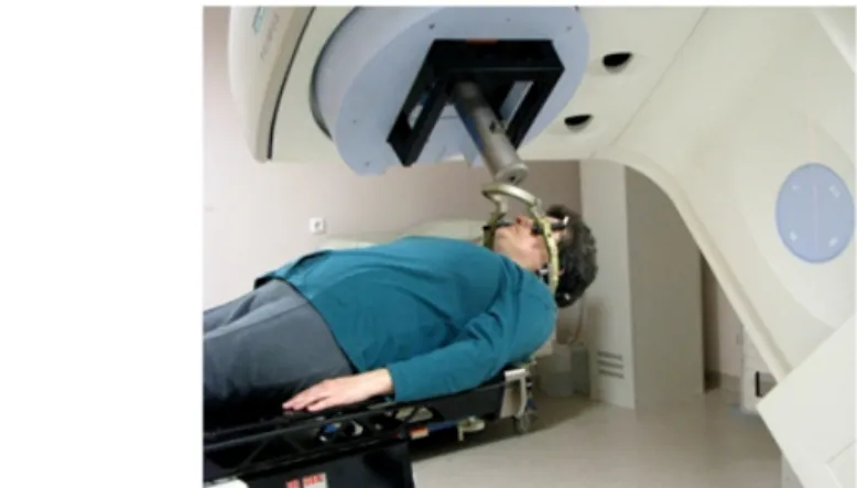
In the linear accelerator system (LINAC), radiation beams emitted from a source moving along a spherical surface are focused on the treatment area determined also by using a stereotactic method.
Fig. 31: Application of the LINAC system. In the linear accelerator, the area to be treated is located in the intersection of the treatment table and the axis of rotation of the radiation
source.
The mechanical accuracy of the LINAC system is within 1 mm,
allowing the treatment not only of intracranial, but also of spinal lesions with a specially designed system.
"Cyberknife" technology is also to be mentioned, which is a small linear accelerator installed into a robot arm so that the beam can move in any direction into the target. With this device, extracranial targets can also be treated.
Stereotactic radiosurgery can be performed by proton beam procedures as well. The dose distribution of these systems is very good, but they are expensive and require a cyclotron.
All stereotactic radiosurgery procedures use a coordinate system fixed to the head. There are stereotactic frames fixed in a non-invasive way. The biological effects of radiosurgery are independent of the device used, but it is important to note that the largest and most rapid drop of radiation dose outside the targeted volume is achieved with the proton beam technique.
Radiation surgery therapy should ideally be performed in a special center, with a neurosurgeon, oncologist, radiation therapist and physicist in close co-operation.
Stereotactic radiosurgery is particularly suitable for the treatment of brain metastases, vestibular schwannomas, meningiomas, and pituitary adenomas. In addition to tumors, angiomas can also be treated
successfully, and even functional neurosurgical procedures can be done using this method (e.g. in trigeminal neuralgia).
It is important to note that the desired effect, regression of the tumor occurs slowly after radiosurgery. Therefore, this therapy is not suitable for tumors causing severe symptoms, uncontrolled by drugs. The role of radiosurgery is limited in the treatment of diffuse gliomas. In brain metastases, the expected radiographic changes appear after 2-3 months, whereas in vestibular schwannomas it may take 4-5 years. In irradiated angiomas, vascular occlusion appears 3 years after treatment.
With the introduction of radiosurgery, the treatment of certain tumors has changed significantly. It has become first-line therapy for small neuromas, meningiomas, metastases, arteriovenous malformations, and lesions inaccessible for an open neurosurgical intervention have also become treatable with this procedure. Radiosurgery plays an important role in treating meningiomas, especially those growing on the skull base, or recurrent and residual tumors.
More information can be found on the treatment of intracranial tumors, including radiosurgery at the following website:
http://www.braintumortreatment.org/
Radiotherapy
The efficacy of radiotherapy is based on the different radiation sensitivity of normal and abnormal tissues. Since IC tumors contain rapidly dividing cells, they are suitable for radiotherapy, but the radiation sensitivity of white matter is a dose-limiting factor.
Radiotherapy of malignant brain and spinal tumors is usually warranted after surgery. The dose applied ranges between 40-60 Gy in daily
fractions of 2 Gy.
In brain metastases, whole brain radiotherapy (WBRT) is used in combination with surgery or radiotherapy, but it may be used alone in multiple metastases. In brain metastases of small cell lung cancer, WBRT is the treatment of first choice, as this type of tumor is very radiosensitive. For WBRT, 30 Gy radiation dose is usually given in 10 - 15 fractions. In case of long survival, cognitive impairment often occurs.
In some tumors (germinoma, lymphoma, some types of glioma), fractionated radiotherapy may be the primary treatment after the confirmation of the histological result.
Re-irradiation of recurrent brain tumors – because of the increased risk of radiation necrosis - is rarely an option. It is individually decided depending on the type of the first radiation treatment, the time elapsed, and tumor histology. The dose of second radiation is generally lower than of the first treatment.
Brachytherapy: With this procedure, different isotopes can be placed into the tumor and kept there until needed. This method is actually an internal radiation therapy, which has the advantage that the surrounding healthy tissue receives only a minimal radiation dose. It may be used for the treatment of cystic craniopharyngeomas, some gliomas, and
metastases. This treatment is however not widely available because of the relatively high rate of complications, and the invasive mode of introduction of the isotopes.
Chemotherapy
For the chemotherapy of brain tumors, special methods are needed to help the drug in penetrating the blood-brain barrier and reaching the tumor in adequate concentration. For this purpose, lipophilic, low molecular weight agents are used. In some areas of the tumors, the blood-brain barrier is damaged where chemotherapeutic agents are able to penetrate, but more distant tumor cells can only be reached by penetrating the barrier. Using various agents (e.g. mannitol,
bradykinine), the blood-brain barrier can be temporarily opened and high drug concentrations can be achieved around the tumor cells.
Chemotherapeutic drugs may be given systemically (intravenously or orally), or locally, directly into the tumor during surgery or later through an implanted catheter. They may also be delivered in an intra-arterial way, into the internal carotid artery, or in a superselective way, to the main feeding artery of the tumor through an inserted catheter. In meningeal tumor dissemination, intrathecal therapy may be used.
Chemotherapy is usually an adjuvant therapy in recurrent tumors after radiotherapy, but in some cases - after a biopsy or surgery – it is used before or during radiotherapy.
In a recurrent malignant glioma or glioblastoma, if the patient is in a good general condition, the standard treatment comprises radio- chemotherapy after the surgery or biopsy. This means that a patient under radiotherapy will also receive continuous treatment with
temozolamide. After the end of irradiation, chemotherapy continues in higher doses, but only for 5 days every month. Follow-up MRI is done every 3 months, and temozolamide treatment continues until tumor progression (Stupp protocol).
In primary brain lymphomas, the first-line treatment is high-dose methotrexate.
New therapeutic options in brain tumors
The expected survival of brain malignancies is far below the progress achieved in general oncology, despite the growing neurosurgical, radiation and chemotherapy potential. Therefore, new therapeutic possibilities are being explored in neuro-oncology, such as molecular targeted therapy, immunotherapy and gene therapy, which also play an increasing role in general oncology. Clinical trials are ongoing.
Molecular therapy
Low molecular weight tyrosine kinase inhibitors given in monotherapy gave only modest results, but there are ongoing trials with treatment combined with chemotherapeutic agents known to be effective in central nervous system tumors (temozolamide, CCNU). Tyrosine kinase
inhibitors may be promising in the treatment of brain metastases of lung adenocarcinoma showing EGFR receptor overexpression. There are also trials with the combination of angiogenesis inhibitors (thalidomide) and alkylating agents (BCNU, temozolamide).
Immunotherapy
Treatment with immuno-conjugates (rituximab) of primary brain lymphomas was assessed. There are also ongoing trials examining tumor vaccines, and dendritic cellular immunotherapy. The TGF-beta-2 production of gliomas cause local immunosuppression, thus the
anticancer immune response may be enhanced with TGF-beta- 2-inhibitor therapy. Results comparable with temozolamide have been reported with TGF-beta-2 antisense oligonucleotides.
Gene therapy
Successful studies have been published on the use of herpes simplex timidine kinase gancyclovir gene therapy. The timidine kinase gene placed into the cavity around the tumor using a virus vector
phosphorylates the gancyclovir administered systematically after surgery into cytotoxic nucleotide analogues, which are incorporated into the DNA and exert an inhibitory effect in proliferating tumor cells.
The complex treatment of brain tumors
Brain tumors, especially malignant primary brain tumors and metastatic tumors require a complex treatment. The treatment plan of patients with a histological diagnosis is determined by an onco-team. The onco-team consists of a neurosurgeon, an oncologist, a radiotherapist, and the possibility of consultation with a neuroradiologist, a neurologist, and a neuro-pathologist.
Treatment of spinal tumors
According to their location, spinal tumors can be extradural or intradural (intramedullary or extramedullary).
Surgery plays the major role in the management of spinal tumors, radiation and chemotherapy options are limited. The radical removal of benign tumors is a curative procedure.
Among extradural tumors, metastases are the most common, but rarely intramedullary metastasis may also occur. The primary goal of the surgical procedure is the decompression of the spinal cord, but it is also important to maintain or to restore spinal stability.
Spinal cord compression causes paraparesis. In case of paraplegia caused by a malignant tumor, especially if it has been present for more than 24 hours, functional improvement is not expected after
decompression. In disseminated cancer patients, the treatment of vertebral metastases does not prolong survival, but significantly affects the quality of life. In vertebral metastases associated with significant pain that do not cause spinal cord compression, vertebroplasty (filling the vertebra percutaneously with plastic) or radiation therapy may be an option.
Chondromas and chordomas are extradural tumors, often located in the craniocervical transition. Their treatment is a considerable surgical challenge. Proton irradiation plays an important role in treating residual or recurrent tumors in this region. Tumors located on the clivus are often best accessed via a transoral exploration.
Nervous system manifestations of hematologic diseases, such as
lymphomas, are located extradurally. Radiotherapy is used to treat these tumors.
Micro-surgical removal is recommended in extramedullary intradural neuromas and meningiomas. The earlier technique of laminectomy, causing spinal deformities, has been replaced by the semi-hemi laminectomy and split laminectomy techniques. In semi-hemi
laminectomy, the two lamina portions facing each other are removed, so part of the lamina and the spinous process is preserved. This can be done in several segments simultaneously to achieve enough space for intraspinal surgery. In split laminectomy, the spinous process is halved and the intraspinal space can be reached by pulling the laminas apart. In both techniques, stability of the spine is preserved after surgery.
In intramedullary tumors (ependymomas, astrocytomas), surgical removal is recommended if symptoms are progressive. Depending on the histological result, radiotherapy may also be considered.