Tumors
JANET Ε. HARKER
Zoological Laboratory, University of Cambridge, Cambridge, England
I. Introduction 191 II. Naturally Occurring Tumors 192
A. Tumors Caused by Endoparasites 192 B. Tumors Caused by Ectoparasites 193 III. Hereditary Tumors in Drosophila 193
A. Benign Tumors or Pseudotumors 193
B. Malignant Tumors 197 C. Factors Affecting Incidence 199 IV. Experimentally Induced Tumors 203
A. Nerve Severance 203 B. Hormonal Imbalance 204
V. Discussion 207 References 210
I . INTRODUCTION
T h e study of tumors in insects is still at an elementary stage. T h e primary barrier to study is the great difficulty of evaluating the status of "growths" or swellings in insects: any irritation results in an accu
mulation of blood cells around the affected region, and blood cells (hemocytes) may quite normally take on strange shapes and invade healthy tissues of all types. T h e number of hemocytes in the blood may also vary from time to time as sessile cells are mobilized into the blood stream, so that the sudden appearance of cells in any region, despite an apparent scarcity in the hemolymph, does not necessarily indicate any multiplication of cells.
Even in the case of the much-studied vertebrates no satisfactory definition of a tumor cell has yet been proposed, so it is hardly sur
prising that there is not yet even an attempted definition of insect tumor 191
cells. T h e general definition of neoplasms as proliferations in which the cells grow in a new and different way might be thought applicable, but owing to the versatility of insect cells even this can have little, if any, meaning.
Types of irregularities which are frequently, though not necessarily, found in vertebrate tumor cells are (1) an increased nuclear volume, (2) nuclei with irregular contours, (3) double nucleoli, (4) anomalies of mitosis, and (5) basophilic cytoplasm. Any of these characteristics can be seen in insect cells in a variety of circumstances, such as wound healing, or in tissues affected by hormones.
At this stage, then, it may be advisable to proceed along the lines which have, historically, been followed in vertebrate studies; that is, to investigate cases which appear to be abnormal proliferations and to wait for an accumulated body of knowledge before any concise defi
nitions are attempted.
I I . NATURALLY OCCURRING T U M O R S
Considering the number of insects examined in systematic collec
tions, and in research and teaching laboratories, it is extraordinary how few naturally occurring tumorlike structures have been described. Ex
cept for those in Drosophila, which are dealt with in the next section, as far as is known only two have been described in the past twenty-five years. In the case of Drosophila, on the other hand, a great variety of tumors has been described; it is possible that these insects have a greater tendency than most insects for tumor development, but it seems likely that more would be found in other insects were they given the same degree of attention, or if mutations were as carefully preserved.
Scharrer and Lochhead (1950) list the records of the spontaneous tumors which have been found in other insects, but the only one in which any histological detail is known occurs in a lepidopteran larva, Pygaera. T h e tumors affect the male only, but the female carries the gene for the abnormal growth. T h e tumors are either free floating, or attached to the gut, testis, ganglia, or muscles. In some tumors there are giant cells, and multipolar divisions occur: the center of the tumor consists of necrotic cells (Federley, 1936).
A. Tumors Caused by Endoparasites
Abnormal growths have been observed in the European sawfly, Gilpinia hercyniae Hartig, after infection of the larval midgut epi
thelium by a virus (Bird, 1949). Abnormal cell proliferation occurs in the region of the regenerative nidi, and the resultant growth pushes out into the body cavity. When infection occurs just before pupation
very large numbers of tumors appear, consisting of necrotic and pig
mented centers, a layer of large, virus-infected cells, and, on the out
side, layers of proliferating cells. Tissues other than those of the midgut are not invaded by the proliferating cells, and the tumor may disappear during metamorphosis. Not enough detail of these growths is known for any judgment to be made as to whether or not they are caused
by a hemocytic reaction to the virus infection, but the fact that their occurrence is a sex-linked character, and that the regenerative nidi are concerned with the initial reaction, suggests that the proliferation is not entirely due to an injury reaction.
Salt (personal communication) has drawn attention to a number of observations by parasitologists concerning swellings in insects caused by the presence of internal parasites. In some cases hemocytes are involved, but in others there is no doubt that the tissues themselves hypertrophy (Marchal, 1906; Pantel, 1910) and show a resemblance to tissues which have, in other contexts, been termed tumorous. Not enough evidence is available for any further conclusions to be drawn.
B. Tumors Caused by Ectoparasites
Tumors resulting from the presence of an ectoparasitic chironomid are known in mayfly larvae (Codreanu, 1935, 1939). T h e so-called
"syncytial tumor," which seems to be produced from the blood cells, is said to contain cells with increased cytoplasm, abnormally large nu
clei and nucleoli, and cells which show abnormal mitosis. All these features can be seen in normal wound healing (Wigglesworth, 1937), but in addition to these symptoms small pockets of similar cells appear in other regions of the host, for example in the ovary, and may do so even when the parasite has been removed at an early stage. There is not enough evidence for any diagnosis of the nature of these abnormal tissues to be made.
I I I . HEREDITARY T U M O R S IN Drosophila A. Benign Tumors or Pseudotumors
T h e occurrence in Drosophila of what have been called either be
nign tumors or pseudotumors is well known, and most studies on insect tumors have centered on this species.
Melanotic masses may appear, according to the genotype, in all, or any one, of the three stages larva, pupa, and adult. T h e position of the dark masses also seems to be fairly specific for any stock: they may occur in the head and thorax, or in the abdomen; in the latter they are nearly always associated with some specific organ such as the fat body or tracheae.
1. Histology
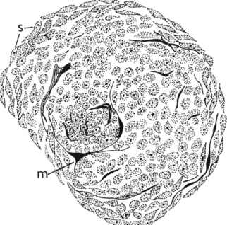
Russell (1940) studied the formation of tumors in five stocks of Drosophila. Tumors in the st sr strain first appear at 72 to 75 hours of larval life, and consist of a nodule of very tiny cells coated with a layer of melanin (chemically confirmed by Härtung and Tillinghast, 1949); spindle-shaped cells are present on the outside of the node, oriented parallel to its surface, and thin tongues of melanin may extend from the periphery down between the cells of the nodule (Fig. 1 ) . Older tumors show a space in the center, no doubt due to the break-
FIG. 1. Pseudotumor from abdomen of Drosophila larva showing presence of spindle cells (s) and beginning of melanization (ra). (Redrawn from Stark, 1919.)
down of the cells which have been encircled. T h e rate of tumor development in this stock seems to be impressive; Russell cites the case of a larva which had one tumor when first examined, two after 4 hours, three after 6 hours, and four at 8 hours. This rate of appearance sounds unlikely for true tumor development, even allowing for the brief life cycle of the insect, but what Russell calls the development of the tumors is in fact the darkening of the tumors, so it may be only the process of melanization which is proceeding at this rate.
In this connection an observation of Salt (1956) is of interest:
he found that the accumulation of blood cells around a foreign body in a stick insect decreases after a time and fragments of melanin molded to the shape of the foreign body move away from the main mass, a
phenomenon indicating a withdrawal of the hemocytes from the center of the clump. T h i s type of movement might be involved in the ap
pearance of melanized cells in regions away from the initial site of the "black bodies," so that multiplication of these bodies may not always be due to further aggregations.
T h e presence of melanin makes detailed examination of the tumors difficult, but Kaplan (1955) was able to retard melanization in the tu-e stock by keeping second-ins tar larvae at 32 °C for 48 hours; the forma
tion of the pseudotumors could then be followed. Spindle-shaped cells appear in the hemolymph at about 72 hours and begin to aggregate about a specific organ. After about 12 hours the cell clusters are ar
ranged in a continuous strand around the organ. T h e number of cells now increases and melanization begins; cells isolated from the edge of the tumor at this time show a yellowish tinge at their periphery.
In time the entire mass of spindle cells melanizes, but although the surrounded organ becomes necrotic no melanization has ever been seen in these cells. T h e melanotic masses have been termed "inert"
by a number of authors, but tissue cultures have been made of the melanized region and show the presence of living cells. Castiglioni
(1956) and Barigozzi (1958) traced the origin of the spindle cells, in the stock e 144, to the pericardial body (or lymph gland). They noted that, although two types of cell in the pericardial body divide mito- tically, the largest cells, containing remarkably large chromosomes, never do so: it is these cells which they consider to give rise to the spindle cells. T h e pericardial bodies form a series of lobes on either side of the aorta and in nontumorous stocks cells are thought to be released from the gland by migration through the gland sheath: in the tumorous stock e 144, however, the anterior lobe of the gland disintegrates, re
leasing the cells. Waddington (in Campbell, 1959) implies, on the other hand, that the normal gland ruptures toward the end of both the second and the third instars.
Since the spindle cells appear to be concerned in the formation of both benign and malignant (page 198) tumors, they have been inves
tigated in some detail, but results are contradictory and confusion is added by the varied terminology which has been applied to the cells found in the hemolymph. Leaving aside the question of whether all the hemocytes originate in the pericardial body and are thus oikocytes
(Shatoury and Waddington, 1957), the names given to the cells in
volved in tumors can be to some extent correlated. Shatoury and Wad
dington refer to hexagons and spheroids, and these appear to correspond to Rizki's (1957a) crystal cells and plasmocytes, and Burdette's (1950) polygonal and fusiform cells.
In nontumorous stocks Rizki (1957a) observed a transformation of plasmocytes to lamellocytes at either the very end of the third larval instar or in the white puparium stage. He further noted that these lamellocytes are, in side view, spindle shaped. T h e absence of spindle-shaped cells in the normal larva, except at this time, is con
firmed by Shatoury (1955b) (but see Section V ) . In the stock tuw, in which tumors occur in the caudal fat body (Wilson et al, 1955), the plasmocyte-lamellocyte transformation begins with the approach of the second molt, that is, an instar earlier than normal (Rizki, 1957b).
Very soon after the appearance of the lamellocytes, the encapsulation of a number of tissues takes place, and it is only when the lamellocytes have aggregated that melanization occurs; free-floating lamellocytes do not melanize.
2. Tumor Extract and Hemolymph Injections
Castiglioni and Beati (1954) made reciprocal injections of hemo
lymph into a variety of tumorous and wild-type stocks. T h e i r results show that injected hemolymph can affect the tumor incidence: the higher the percentage incidence of the donor stock, the higher is the resulting tumor incidence in the host. It is possible that the injected cells may keep the property of aggregating and melanizing and thus
"black bodies" found in the host could be due to the injected cells, not to host cells. Cell counts show that the tumorous stocks used in these experiments had a significantly larger number of cells than did the tumorless stocks, and therefore a considerable number of cells may have been involved in the injection.
A series of observations have been made by Friedman, Burton, and their associates on the effect of injecting extracts from tumorous larvae into tumorless larvae. Recently one of their collaborators, Mitchell, has stated that he wishes to be dissociated from these joint publications as Friedman and Burton were unable, when presented with coded sam
ples, to distinguish between the effects of injected buffer solutions and extract solutions (Mitchell, 1961). In view of this, the results of extract experiments must be treated with caution until more evidence is forthcoming, but a brief summary of the work is given below.
Injection of crude extracts obtained from larvae of the tu-e strain is said to induce tumors in a very high proportion of the larvae of non tumorous strains: no tumors appear when extracts are obtained from nontumorous larvae. When the injected extracts are crude the tumors formed are free-floating, melanized bodies; but if the extract has been purified 12,000-fold by protein-fractionation procedures, in
jection is said to be followed by the appearance of invasive tumors
of the pharynx, ring gland, brain, anterior midgut, and muscle. T h e purification process is thought by the authors to remove substances which inactivate, or modify, the tumor-inducing factor. Cells from tumors induced by the purified extract are said to invade normal tissues in culture, and a striking increase in the virulence of the tumor-inducing factor apparently occurs during culture and in serial transmissions (Burton et al, 1956a, b; Burton and Friedman, 1956; Friedman and Burton, 1956).
T h e proteolytic enzyme papain has a significant effect on the tumor- inducing factor, whereas neither chymotrypsin nor trypsin have any effect, indicating that the factor contains a native, not a denatured, protein. Deoxyribonuclease and ribonuclease reduce the activity of the extracts, and analysis of the ultraviolet absorption spectrum shows the presence of up to 5 percent nucleic acid. T h e factor is stable up to 35°C, and between pH 6.5 and 8.0.
Because of the factor's apparent pathogenic characteristics, the pro
tein, nucleic acid, and lipid content, and the way in which its virulence increases in serial transmissions, Friedman et al. (1957) suggested that the factor is a virus.
T h e inductive activity of the extract is said to appear at the 36th hour of larval development, and by 144 hours no activity could be found. T h e peak of activity was shown to be at about 72 hours.
T h e effect of the extract is claimed to vary with both the age of the donor and that of the host, the greatest activity occurring when the host and donor are fairly close in age, particularly as the host becomes older (Burton, 1955). Harnly (1955) points out that differences in the effectiveness of an extract in hosts of various ages could be due to more material being injected into larger larvae (the method of injec
tion involved introducing as much fluid as the larvae could contain), but although larvae continue to grow after 72 hours, the degree of effectiveness in older larvae is said to decrease, perhaps owing to a loss of competence of the tissues.
B. Malignant Tumors
T h e first so-called lethal tumor to be carefully investigated (Stark, 1919) was one found in the strain L ( l ) 17 by Bridges (1916), but the death of the larvae has since been shown to be caused by a malformation of the midgut, the melanotic bodies themselves apparently not affecting
the larvae (Russell, 1940). In this strain it seems very likely that the melanotic bodies are in fact the result of a hemocytic reaction to the ab
normality in the midgut.
A few tumors which might be regarded as malignant have, however,
been described. In the stock lethal malignant (/-m), as in the benign tumor stocks already described, the first sign of abnormality concerns the lymph glands (or pericardial bodies) (Shatoury, 1955a, b ) . In this stock the lymph glands increase to several times their normal size at the end of the third instar, that is, just prior to the pupal molt. T h e lymph glands then rupture, and the freed cells are swept into the hemolymph and circulated to the imaginal buds, which they surround and invade. Cells can be seen inside the peripodial cavity and among the disc cells; at a later stage the disc cells become necrotic. Other cells from the lymph gland are swept into the neural sinus and accumulate along the ventral nerve cord. Later on, cells accumulate at the posterior end of the body and invade the fat bodies. T h e invading cells are said to be both hexagons and spheroids (crystal cells and plasmocytes).
Shatoury refers to the invasion of the nerve cord as metastasis, and to that of the caudal fat body as a secondary attack. There does not seem to be any clear evidence, however, that all three are other than a primary invasion, the timing of their appearance being due to the course taken by the blood circulation rather than to secondary growth.
Some larvae, in which the cells surrounding the imaginal buds and nerve cord melanize very rapidly, show no invasion of the fat body, and Shatoury quotes these cases as additional evidence that the fat bodies are invaded by cells coming from the primary tumors. It is still pos
sible, however, that only a limited number of cells is produced by the lymph gland, and if they aggregate sufficiently at the first invasion site (which might cause rapid melanization), none will be left to affect the more posterior site. In all these arguments it is assumed that the cells involved in the tumor production are lymph gland cells, but this in itself is a questionable assumption, as is discussed in Section V.
A third type of lymph gland cell, a platelet, has been described by Shatoury; such cells are said to increase in size in tumorous larvae and in some cases to encapsulate the tumorous cells which then disintegrate.
After tissues invaded by tumor cells become necrotic, the tumor cells undergo further changes, some of the spheroids transforming into spindle cells (cf. pseudotumors) ; spheroids which do not become spin
dle cells clump together and are themselves surrounded by spindle cells.
T h e spindle cells melanize, while the enclosed spheroids undergo lysis.
Shatoury comments that the incidence of the resulting "black bodies"
is very low: their possible relationship to pseudotumors is discussed at the end of this chapter.
In the stock l-m, the testis is also sometimes attacked, the first sign of abnormality being the presence of strongly basophilic cells in the vicinity of both the spermatogonia and the terminal cells. T h e
abnormal cells are more or less triangular with a basal extension, and the nuclei are very large. T h e surrounding tissue eventually becomes necrotic. In some larvae the testes show premature development of mature sperms, and these are basophilic in contrast to normal pupal or imaginal sperms.
T h e midgut is also affected at the time of the lymph gland hyper
trophy (Shatoury and Waddington, 1957). T h e cells lying immedi
ately against the basement membrane begin to multiply, and the epi
thelial cells lining the gut lumen swell, become strongly basophilic, and eventually break free into the gut cavity. Later the basement cells begin to melanize, and when this occurs the proliferation of the diges
tive epithelium ceases. T h e authors suggest that the cells between the basement membrane and the epithelium are derived from the lymph gland.
Gastric tumors have also been investigated in the stock lethal-no- imaginal bud (l-nib), in which differentiation of the imaginal discs is inhibited and pupation does not take place. At the end of the second instar the first pair of lymph glands degenerate instead of undergoing their normal increase in size. T h e degeneration of the gland is followed by a proliferation of the midgut basement cells, the midgut epithelium hypertrophies and finally becomes cytolized to an undifferentiated, partly melanized structure: the basement cells eventually become mel
anotic. T h e hindgut and salivary gland may similarly become tumorous.
T h e degeneration of the lymph gland at the end of the second instar is followed eventually by regression of the ring gland, the components becoming cytolized by the middle of the third instar.
A third stock, lethal-no-differentiation (l-nd), in which the imaginal discs do not differentiate into their adult form, also produces tumorlike bodies (Shatoury, 1955c). In the middle of the third instar a number of cells in the lymph gland degenerate, and soon afterward the imaginal wing bud mesoderm, which lies underneath the epithelium, begins to proliferate to such an extent that it grows through the epithelium and spreads into the peripodial sac and from there may grow into the hemocoel.
C. Factors Affecting Incidence 1. Genetic Factors
Both malignant and benign tumors in Drosophila are genotypically induced, the second chromosome apparently exerting a considerable in
fluence (Bridges, 1916; Härtung, 1942; Herskowitz and Burdette, 1951;
Russell, 1940; Stark, 1919; Wilson, 1947). Shatoury's l-m factor is lo
cated on the first chromosome, and other strains are known in which an
action is exerted by the fourth chromosome. T h e stock tu-er has a suppressor gene on chromosome I I I which suppresses the factor for the production of tumors which is itself located on chromosome I I (Glass and Plaine, 1952).
According to Barigozzi (1958) the production of melanotic masses in the stocks tuA2 and t u B3 depends on three factors which are genet
ically affected as follows: (1) the release of cells from the lymph gland—
controlled by all three major chromosomes; (2) the presence of a high proportion of large hemolymph cells—under the multichromosomal con
trol; (3) production of melanin—controlled by chromosome I I .
A cytoplasmic effect in the production of tumors is also known (Kanehisa, 1954), the offspring having a higher tumor incidence when the female parent shows a high incidence. Gardner (1959) also found a maternal affect and suggested that a recessive tu-l gene acts by con
ditioning the egg while it is developing inside the female, the egg thus becoming more susceptible to the action of the tumor-producing gene tu-3. Barigozzi et al. (1958) replaced all the chromosomes of the tumor
ous stocks tu-A2, tu-B3, tu-C4, and tu-D with chromosomes from tumor
less stocks. After chromosome replacement, tumors still occurred in all stocks.
2. Irradiation
Härtung (1942) X-irradiated Drosophila eggs of three strains having a normal tumor incidence of approximately 15 percent. T h e incidence increased at irradiation values above 500 r, a peak incidence occurring at 1500 r. From 1500 r to 5000 r the incidence decreased until it was below that of the controls. X-irradiation of eggs from nontumorous stocks occasionally caused tumors, but the incidence was low. Plaine and Glass (1952) found that the incidence of tumors in X-irradiated embryos of the suppressor-erupt stock increased with an increase in oxygen concentration.
King and Burnette (1957) increased the incidence of ovarian tumors 26-fold by irradiating with 4000 r of C o6 0 gamma rays.
T h e effect of irradiation on larvae does not seem to have been studied: this might be a profitable line of investigation since the blood cells of some insects are known to be affected by X rays.
3. Oxygen
Abrahamson and Fanale (1959) showed that anoxia lasting for 1 to 2 hours produced pseudotumors in adults of a strain in which tumors had not previously been observed. T h e tumors appeared from within a few minutes after treatment to up to 48 hours and occurred in the
anterior abdominal region. T h e females were more susceptible than the males, and adults of age 0 to 12 hours showed the lowest incidence.
Without more evidence it is not possible to judge how far the melanotic masses are due to an injury reaction, rather than to tumor production.
4. Carcinogens
Substances known to be carcinogenic for vertebrates have, on the whole, little effect on insects. External application, as far as is known, never produces tumors (Demerec et ah, 1949).
Benign melanotic tumors are said to be produced in Drosophila after introduction of compounds of arsenic and boron, mercuric chloride, silver and sodium fluoride (Rapport, 1939; Sang and McDonald, 1954).
There does not seem to be any evidence, however, that the "black bodies" produced are in any way related to abnormal growth; indeed it seems likely that the toxic substances cause local injuries which be
come melanized in the normal course of wound healing.
Aerosol solutions of a number of vertebrate carcinogens, including nitrogen mustard, benzypyrene, napthylamine, and 1,2,5,6-dibenzanthra- cene, have been shown to cause mutations and chromosomal rearrange
ments in Drosophila. Fahmy and Fahmy in a long series of papers have discussed the cytogenetic action of carcinogens and tumor inhibitors on Drosophila, but there is no evidence of tumor production despite the effect on the genetic constitution (for references see Fahmy and Fahmy, 1956).
Incidentally, injection of folic acid into Pieris pupae produces pro
liferations at the site of injection: these proliferations are said to be melanomas (L'Helias, 1957), but further study is needed to ensure that they are not produced by wound healing plus a hemocytic reaction against the injected material.
5. Nutrition
Nutritional factors have a strong influence on the incidence of tumors in Drosophila larvae.
Poor nutrition, or overcrowding which results in poor nutrition, generally lowers the tumor incidence; in Goldsmith and Friedman's (1949) experiments, overcrowding reduced the incidence from 85 per
cent to 54 percent. T h e effect was not related to differential survival rates, delay in pigmentation, or sexual differences (Herskowitz and Bur- dette, 1951). T h e developmental period lengthens when there is a reduction of yeast in the food (Friedman et al., 1955a), and it is possible
that changes in developmental rates are closely concerned with the effect of nutrition on tumor incidence, although it should be noted that
when the rate of development is increased by raising the temperature, tumor incidence also decreases (Härtung, 1947).
T h e critical period for nutritional effects appears to begin at about the 3rd day of larval life; it will be recalled that it is at this age that the spindle cells are first seen to appear in many tumorous strains.
More critical analysis of nutritional factors has shown that a variety of vitamins are probably involved. In the tu-e strain a variety of vita
mins in the food medium increase the tumor incidence; this tumor- promoting effect is inhibited by the respective analogs pyrithiamine, dethiobotin, L-picotonic acid, isonicotinyl hydrazide, and 3-acetylpyri- dine (Friedman et al., 1955b). T h e evidence concerning vitamin B1 2 is contradictory; Mittler (1954) recorded a doubling of the percentage of tumors when it was added to the food, whereas Briones (1949) found that the incidence decreased on yeast-enriched medium. T h e discrepancy in these findings may be related to the concentrations used.
Although larvae cannot live on a medium lacking in tryptophan, a high concentration of /-tryptophan in the food increases the tumor incidence in most strains (Hinton et al., 1951; Mittler, 1952a, b). Sim- monds and Gardner (1958) report, however, that the incidence of tumorous head decreased when larvae were fed on a tryptophan medium.
In the stock suppressor-erupt the tumor incidence is increased by feeding supplementary tryptophan or kynurenine (Plaine and Glass,
1955). In this stock there are suppressor genes to the genes responsible for tumor formation, and it is interesting that the action of these suppressor genes, as well as being affected by tryptophan, are blocked by X rays and modified by oxygen and hydrogen peroxide. Ionizing radiations affect metabolic processes susceptible to oxidation by perox
ides, unless there is an adequate peroxide-destroying system, and the first step in the oxidative degradation of tryptophan in insects is a coupled peroxidase-oxidase reaction leading to the formation of formyl- kynurenine, which in turn is converted to kynurenine (Kikkawa, 1953).
It may be that the various agents which inhibit the suppressor system act on this peroxidative step (Plaine and Glass, 1955).
What is known about tryptophan metabolism in insects has been discovered almost entirely from biochemical and genetic studies of pigmentation in the eyes: the formation of kynurenine has already been mentioned, and this is converted to 3-hydroxykynurenine and finally to the brown pigment ommochrome. Mutants are known in which various steps in the series are blocked, and it should be possible to discover more about the effect of tryptophan on tumor incidence by studying
these mutants. Surprisingly little work has been done in this promising field, although a beginning has been made.
T h e vermilion mutant (designated by the change v+ —v) lacks one of the enzymes responsible for the conversion of tryptophan to kynure
nine, and tryptophan accumulates in this stock. Another mutant, cin- nibar-eye (en) accumulates kynurenine, the step between this and 3- hydroxykynurenine being blocked, and a third mutant, scarlet-eye (s) is thought to be blocked at the stage between brown chromogen and brown chromophobe (Green, 1949) . In vermilion metabolism can pro
ceed if kynurenine is injected, and in cinnibar synthesis is restored if 3- hydroxykynurenine is injected.
Kanehisa (1956) crossed a tumorous strain with vermilion, cinnibar, and scarlet, and found that, when the progeny were fed supplementary tryptophan, tumor incidence was greater than that in flies which had been crossed with the wild-type eye color. From Kanehisa's tables it can be seen that the tumor incidence increases in the order vermilion, cinnibar, scarlet. When tryptophan was added at least some presumably was metabolized by the wild-type cross, whereas in vermilion none would be converted even to the kynurenine stage, which might indicate that tryptophan itself is at least partly responsible for the increased incidence.
Beadle et al. (1938) have, however, shown the presence of a very small quantity of the v+ substance in vermilion, this increasing at least a hundredfold after partial starvation: therefore it is unwise at this stage to rule out the presence of kynurenine in vermilion eye. Incidence was higher still in the cn flies after tryptophan feeding, and in these flies either tryptophan or kynurenine could accumulate; the highest incidence occurred in scarlet-eye in which 3-hydroxykynurenine in addition to the other substance might be present. Since, presumably, any of these steps are reversible it is not known at the moment where the critical step lies.
Further metabolic pathways are also open to kynurenine and 3-hydroxy
kynurenine: although not a great deal is known about further metabolism in insects kynurenic and xanthurenic acids have been found in Dro
sophila (Gilmour, 1961).
IV. EXPERIMENTALLY INDUCED T U M O R S
A. Nerve Severance
Severance of the recurrent nerve in the cockroach Leucophaea causes the development of tumors in organs that are innervated by this nerve, i.e., the foregut, anterior midgut, and salivary organs (Scharrer, 1945,
1948). Whether the nerve is cut anterior or posterior to the brain or in the thorax, makes no difference to the results. From 70 to 80 percent of the operated adults and nymphs develop tumors in Scharrer's experi-
merits, and although there does not seem to be a sex difference in the tumor incidence, the males die at an earlier stage than the females. T h e survival rates of tumor-bearing castrates of either sex, however, are about equal and lie midway between those of male and female. Analysis of the fat content of the tumor-bearing animals shows that males have a lower than normal fat content, whereas many of the females still have a normal content, and ovariectomized females have in some cases a higher than normal content (Scharrer, 1949).
Histology. In the anterior midgut the first sign of abnormal growth appears in the ventricular region, the wall increasing in thickness; the intestinal wall later thickens and the digestive epithelium and muscle layer are replaced by a multiple layer of cells. Melanization does not occur until a late stage, when perhaps it may be associated with an injury reaction. Giant cells occur in the tumor region, and there are few, if any, mitoses. Necrosis does not occur until a late stage. Although blood cells may be involved in the late stages of the tumor the epithelial cells appear to be the site of the primary effect.
Tumors occurring in the foregut (which are rare) have the appear
ance of sarcomas, whereas the tumors of the salivary reservoir give the appearance of epithelial tumors.
T h e tumors are invasive and have been seen penetrating the body wall.
B. Hormonal Imbalance 1. Allatectomy
In the stick insect Carausius atypical growths appear when the corpora allata are removed from early nymphal stages, the growths being related particularly to mesodermal structures. Amitotic division is seen in that region of the gut from which the Malpighian tubes arise and in the wall of the oviduct; the corpora cardiaca show similar changes and giant nuclei are present. Reimplantation of the corpora allata prevents the abnormal tissue reaction.
When embryonic tissues are implanted into such allatectomized insects, they too show abnormal growth and giant cells appear; when embryonic mesoderm is implanted, it invades the host mesoderm.
Similar effects occur when embryonic tissue is implanted into hosts if extra allata have been implanted at the same time. No such effects are recorded when embryonic implants are made into normal hosts (Pflug
felden 1948).
No further information is available about this type of abnormal growth. T h e implantation, or removal, of corpora allata has been performed many times in a wide range of insects in the course of
experiments on development, yet as far as is known no similar ab
normalities have been observed in other insects.
2. Imposed Secretory Cycles
Tumors have been induced in the cockroach Periplaneta by imposing extra secretory cycles (Harker, 1958). In this cockroach there is a group of neurosecretory cells in the subesophageal ganglion which secrete with a diurnal rhythm having an approximately 24-hour period; the cells will continue to secrete rhythmically even when the ganglion is implanted into another insect. T h e time of secretion from the neurosecretory cells is related to the environmental conditions of light and darkness to which the animal has been exposed, although the phase of the rhythm, once set, will persist in constant conditions, or in an implant, for some days.
When subesophageal ganglia taken from insects which have been kept in light during the night and darkness during the day are implanted into cockroaches living in normal conditions of light and darkness, a fresh implant being made daily for at least 4 days, tumors appear in the midgut, and occasionaly in the forgut, of the host animal. No tumors appear when the neurosecretory cells of the implanted ganglia secrete at the same time as those of the host, nor when an extract of five ganglia is injected daily during the time of the host's secretory period. T h e site of implantation is not related to the region in which the tumors form.
T h e tumors metastasize and are transplantable. Once tumors have been formed it is possible to control their growth by implanting ganglia in which the neurosecretory cells are secreting in time with those of the host, but when the implants are removed the tumor begins to grow once more.
Histology. In the midgut the first sign of abnormality occurs about 2 days after the first implantation of an "out-of-phase" ganglion. T h e clear pattern of the epithelial cells and regenerative nidi becomes slightly distorted, and the number of mitoses in the nidi at least doubles.
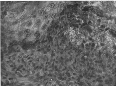
T h e cells from the nidi appear to be pushed down, or to migrate, into the connective tissue layer which lies between the epithelial layer and the circular muscle surrounding the gut. After 14 to 18 days the epithelial layer has broken down and the tumor is then well developed, consisting of small cells in which the thin layer of cytoplasm stains densely with hematoxylin. T h e cells form a whorled pattern, and tracheae invade the tumorous mass (Fig. 2).
T h e metastases appear to develop from cells moving in the connective layer; they occur in the fore- and hindgut. Tumors have also been seen in the salivary glands; these may be formed from cells which have been moved in the hemocoel, but it is possible that the glands have been
FIG. 2. Section of invasive tumor in gut of Periplaneta. T h e tumor cells are small and in places grow in whorls.
FIG. 3. T u m o r cells in the midgut of Periplaneta, invading the region between the regenerative nidi and the epithelium.
affected directly by the subesophageal ganglion secretion, these tumors taking longer to develop than those in the midgut.
T h e sequence of development of a secondary tumor differs from that of the primary tumor. Cells are first seen invading the connective tissue layer below the epithelium of the midgut, and the connective tissue layer rapidly increases in size until it is about quadrupled. T h e epithelial layer does not show any abnormal appearance for some days, but it then begins to break down (Fig. 3). It is not until the epithelium is affected that any increase in mitosis in nidi occurs. At the time of breakdown of the epithelium, blood cells begin to accumulate along the muscle layer and the swelling becomes invaded by tracheae.
T h e nidi cells appear to be those which give rise to the primary tumor, but the secondary tumor seems to arise from cells migrating from the implanted tumor tissue and invading the same region as that invaded by metastases, the nidi cells being unaffected by their presence except insofar as they fulfill their normal function of replacing the epithelium when it is eventually affected by the presence of the tumor.
Melanization seldom occurs in these tumors, except at the stage when the blood cells are accumulating in large masses around the affected area; at this time the melanization would appear to be a reaction against the wound or irritant.
V. DISCUSSION
Although the difficulties of classifying conditions of abnormal growth in insects are such as to discourage definitions concerning insect tumors, any concept of a tumor implies multiplication of cells. In the case of the so-called benign tumors of Drosophila the question must therefore be raised whether any multiplication of cells is involved. All the evidence suggests that benign, or pseudo, tumors are due to an aggregation of hemocytes, either as a group or around specific tissues, and that this in turn stimulates the hemocytic reaction of melanization. There does not seem to be any evidence that the hemocytes in the blood stream are growing abnormally fast or in any disorganized manner, although this evidence might be obtained with further study.
Shatoury and Waddington (1957) and Waddington (in Campbell, 1959) argue, however, that all the cells involved in both benign and malignant Drosophila tumors originate from the lymph gland, that all the cells known as hemocytes are in fact the product of this gland, and that they are produced by abnormal growth. This hypothesis is based on the evidence that similar types of cell can be seen in the lymph gland and in pockets of cells lying in the hemocoel but associated with specific tissues, and that certain changes take place in the cells in all these
places at the same time. Shatoury (1955b) further maintains that there are no free cells in the blood except at the time of rupture of the lymph glands. In evaluating this hypothesis at least three other pieces of evidence need to be considered: (1) Rizki (1957a) has clearly demon
strated the presence of free cells in the blood at all times of larval life;
(2) in other insects hemopoetic organs release cells from time to time, but all the cells in the blood at any one time do not necessarily come from any one of these; (3) there is very good evidence that hemocytes penetrate certain regions at specific times so that similarities between cells in the lymph glands and other tissues could arise because of penetration by blood cells. There is not sufficient evidence to warrant drawing any final conclusion, but it should be mentioned that if the lymph glands are indeed producing tumorous cells then the primary tumor occurs specifically in the lymph gland in both malignant and benign forms, and both the described "black bodies" and invasive tumors are metastases.
Wherever the abnormalities originate, there seems to be no doubt that blood cells are rapidly implicated. What is causing their aggregation and melanization is not known, but it has been suggested that one factor involved is the abnormal time of production of the lamellocyte spindle cells associated with all the Drosophila tumors; Rizki has observed that in normal larvae spindle cells are present only just prior to the molt to the pupal stage. Cells resembling spindle cells are associated with wounds in other insects (Day, 1952; Wigglesworth, 1959), but Rizki could find no spindle cells associated with a healed area in Drosophila.
Consideration of Wigglesworth's observations, however, suggests that if the epidermal cells are involved in a wound the hemocytic reaction may differ from that when hemocytes alone are responsible for encapsulating a "foreign body"; if fragments of wax are injected into Drosophila larvae, an examination of the blood cells encapsulating the wax (Fig. 4) reveals the presence of spindle cells at any stage of the larval instar
(Harker, unpublished). Therefore it is likely that the presence of spindle cells is not the first stage in production of pseudotumors, but the result of an abnormality which is already present.
There is then a very close resemblance between pseudotumors and
"foreign body" reactions: in each case blood cells aggregate, spindle cells form, and the aggregate melanizes. Salt (1961) has proposed a hypo
thesis concerning hemocytic reactions in the following terms: " T h e haemocytes of an insect react to any surface which lacks the properties of the surface which they themselves lay down, or contribute to, about the organs and tissues of the insect of which they form part." In the pseudotumor aggregate, the blood cells appear to be reacting to each
other, so that if Salt's hypothesis holds true one must turn again to the possibility that cells coming from the lymph glands initiate the reaction by presenting an unsuitable surface. T h e fact that the lymph gland appears to be the one organ in Drosophila which cannot be transplanted without causing a vigorous hemycytic reaction from a host supports this suggestion; on the other hand the lymph gland in a tumorous larva does not seem to be encapsulated.
Should the lymph gland cells be tumorous, their ability to promote a reaction by the blood cells, or indeed by any cells, is not in line with current knowledge about vertebrate tumors, in which the lack of reaction
FIG. 4. Encapsulated wax fragment (dissolved during preparation of section) from abdomen of Drosophila showing spindle cells (s).
by other cells to their presence is a striking characteristic. If, on the other hand, the hemocytes are reacting to each other and aggregating, then they too differ noticeably from vertebrate tumor cells, which undergo a change in surface properties leading to absence of adhesives.
Oster (1954), however, has produced evidence which may indicate that the larval hemocytes are not capable of sealing off the pseudo
tumors. He combined a tumor gene with a gene, "giant," which causes an extra molt in the larva, and in this cross the tumors increased in size during the extra instar.
Malignant tumors in Drosophila are characterized by invasion of tissues by cells from the hemocoel, and here again there is considerable difficulty in distinguishing between a hemocytic reaction and multipli
cation of cells. Since there is good evidence that in normal larvae a
particular group of cells may attract hemocytes when no other tissues are doing so, and that the activities of hemocytes are strongly affected by hormones, a study of the hormonal relationship of tumorous flies might give further information about tumorigenesis. In this connection perhaps it is significant that the ring gland of the l-nib stock regresses soon after tumor formation and that in other strains the timing of events in the life cycle appears to be abnormal.
Turning to the tumors found in Orthoptera, again we find indications that hormonal upset is related to their occurrence. This certainly seems to be the case for those tumors formed in the midgut of Periplaneta after the insect has been exposed to a secretion from the subesophageal ganglion at an abnormal time of day. There may not, however, appear to be very much connection between hormones and the type of tumor formed after section of the recurrent nerve, but it has been shown
(Harker, 1960) that section of the recurrent nerve in Periplaneta upsets the rhythm of secretion of the subesophageal ganglion: in fact the two types of tumor production may be very closely related. Direct involve
ment of a hormone from the corpora allata seems to be indicated in the tumors described by Pflugfelder in the stick insect.
Pflugfeldens stick insect tumor, and Harker's Periplaneta tumor both appear to be endocrine-dependent, conditioned tumors, but the Peri
planeta tumor does seem to differ from other known conditioned tumors in that the timing of the presence of the hormone, not the hormone concentration, is the controlling factor; further study may show that some apparently autonomous tumors can be controlled in a similar way.
REFERENCES
Abrahamson, S., and Fanale, F . P. 1959. T h e induction by anoxia of melanotic masses in Drosophila. Genetics, 44, 497-512.
Barigozzi, C. 1958. Melanotic tumours in Drosophila. J. Cellular Comp. Physiol., 52, 371-381.
Barigozzi, C , Castiglioni, M. C , and di Pasquale, A. 1958. Morphogenesis of melanotic tumors (pseudotumors) and its genetical control in three wild stocks of Drosophila melanogaster. Experientia, 14, 443-444.
Beadle, G. W., Tatum, E . L . , and Clancy, C. W . 1938. Food level in relation to rate of development and eye-pigmentation in Drosophila melanogaster. Biol. Bull., 75, 447-462.
Bird, F . T . 1949. Tumors associated with a virus infection in an insect. Nature, 163, 777-778.
Bridges, C. B . 1916. Non-disjunction and the chromosome theory. Genetics, 1, 1-52.
Briones, Η. M. 1949. Tumores y alimentacion. Biologica Fasc, 10, 55-67.
Burton, L . 1955. Carcinogenic effects of an extractable larval tumor agent. Trans.
N.Y. Acad. Sei., 17, 301-308.
Burdette, W . J . 1950. Studies on Drosophila tumors. Cancer Research, 10, 209-215.
Burton, L . 1955. Carcinogenic effects of an extractable larval tumor agent. Trans.
N.Y. Acad. Sei., 17, 301-308.
Burton, L . , and Friedman, F. 1956. Detection of tumor-inducing factors in Dro
sophila. Science, 124, 220-221.
Burton, L . , Friedman, F., and Mitchell, Η. K. 1956a. T h e purification of an inherited tumor-inducing factor in Drosophila melanogaster. Cancer Research, 16, 880-884.
Burton, L.; Harnly, Μ. H., and Kopac, M. J . 1956b. T h e activity of a tumor factor factor in Drosophila development. Cancer Research 16, 402-407.
Campbell, F . L . , ed. 1959. "Physiology of Insect Development." Univ. Chicago Press, Chicago, Illinois.
Castiglioni, M. C. 1956. Fenogenetica degli pseudotumori in Drosophila melano
gaster e D. simulans. Ricerca sei. Suppl., convegno di Genetica (Pavia 1955), pp. 125-130.
Castiglioni, M. C , and Beati, S. 1954. Production of pseudotumors in Drosophila after injection of haemolymph. Experientia, 10, 501-502.
Codreanu, R. 1935. Neoplasie maligne dans l'hemocoele des £ph£meres sous Taction de Symbiocladius rithrogenae, Chironomide ectoparasite. Compt. rend. acad. sei., 201, 102-104.
Codreanu, R. 1939. Recherches biologiques sur un chironomide Symbiocladius rithrogenae, ectoparasite "cancerigene" des Ephemeres torrenticoles. Arch. zool.
exptl. et gen., 81, 1-283.
Day, Μ. F. 1952. Wound healing in the gut of the cockroach Periplaneta. Australian J. Sei. Research, B5, 282-289.
Demerec, M., Wallace, B., Witkin, Ε . M., and Bertani, G. 1949. T h e gene. Carnegie Inst. Wash. Year Book, 48, 156-158.
Fahmy, O. G., and Fahmy, M. J . 1956. Cytogenetic analysis of the action of carcinogens and tumor inhibitors in Drosophila melanogaster. V. / . Genet., 54, 146-164.
Federley, H. 1936. Sex-limited hereditary cancer in Lepidopterous larvae. Hereditas, 22, 193-216.
Friedman, F., and Burton, L . 1956. Benign and invasive tumors induced in Drosophila by an inherited tumor-inducing factor. Cancer Research, 16, 1059- 1061.
Friedman, F., Harnly, Μ. H., and Kopac, M. J . 1955a. T h e effects of vitamin variations upon tumor incidence of a tu-e strain of Drosophila melanogaster.
Cancer Research, 15, 375-381.
Friedman, F., Harnly, Μ. H., and Kopac, M. J . 1955b. T h e effects of vitamin analogues upon tumor penetrance in a tu-e strain of Drosophila melanogaster.
Cancer Research, 15, 382-389.
Friedman, F., Burton, L . , and Mitchell, Η. K. 1957. Characteristics of an inherited tumor-inducing factor in Drosophila melanogaster. Cancer Research, 17, 208-214.
Gardner, E . J . 1959. Genetic mechanism of maternal effect for tumorous head in Drosophila melanogaster. Genetics, 44, 471-481.
Gilmour, D. 1961. "The Biochemistry of Insects," 343 pp. Academic Press, New York.
Glass, B., and Plaine, H. L . 1952. T h e role of oxygen concentration in determining the effectiveness of X-rays on the action of specific genes in Drosophila melano
gaster. Proc. Natl. Acad. Sei. U.S., 38, 697-705.
Goldsmith, Ε . D., and Friedman, F. 1949. T h e effect of crowding on the penetrance of an hereditary melanoma of Drosophila melanogaster. Cancer Research, 9, 604.
Green, Μ. M. 1949. A study of tryptophane in eye colour mutants of Drosophila.
Genetics, 34, 564-572.
Harker, J . E . 1958. Experimental production of midgut tumors in Periplaneta americana L . / . Exptl. Biol., 35, 251-259.
Harker, J . E . 1960. Endocrine and nervous factors in insect circadian rhythms.
Cold Spring Harbor Symposia Quant. Biol., 25, 279-287.
Harnly, Μ. H. 1955. T h e tu-e gene action and tumor development. Trans. N.Y.
Acad. Set., 17, 309-311.
Härtung, Ε . W . 1942. T h e effects of roentgen radiation on tumor incidence in Drosophila melanogaster. Cancer Research, 2, 837-840.
Härtung, Ε . W . 1947. Some effects of temperature on tumor incidence in several strains of Drosophila melanogaster. J. Exptl. Zool., 106, 223-232.
Härtung, Ε . W., and Tillinghast, M. G. 1949. T h e nature of the pigmented sheath in Drosophila tumors. Science, 109, 565-566.
Herskowitz, I. H., and Burdette, W . J . 1951. Some genetic and environmental influences on the incidence of a melanotic tumor in Drosophila. J. Exptl. ZooL, 117, 499-521.
Hinton, T., Noyes, D. T., and Ellis, J . 1951. Amino acids and growth factors in a chemically defined medium for Drosophila. Physiol. ZooL, 24, 335-353.
Kanehisa, T . 1954. Some aspects of the maternal effects in the expression of tumor in Drosophila virilis. Annotationes Zool. Japon., 27, 201-207.
Kanehisa, T . 1956. Relation between the formation of melanotic tumors and tryptophane metabolism involving eye-colour in Drosophila. Annotationes Zool.
Japon., 29, 97-100.
Kaplan, M. L . 1955. Histogenesis of the tu-e melanoma in Drosophila. Trans. N.Y.
Acad. Sei., 17, 289-293.
Kikkawa, H. 1953. Biochemical genetics of Bombyx mori (Silkworm). Advances in Genet., 5, 107-140.
King, R. C. and Burnette, R. G. 1957. Hereditary ovarian tumors in Drosophila melanogaster. Science, 126, 562.
L'Helias, C. 1957. Tumeurs d'insectes provoquees par l'acide folique. Compt. rend.
acad. sei., 244, 1678-1680.
Marchal, P. 1906. Les Platygasters. Arch. zool. exptl. et gen., 6, 485-640.
Mitchell, Η. Κ. 1961. Tumor-inducing factor in Drosophila. Science, 133, 876.
Mittler, S. 1952a. Influence of nutrition upon appearance of tumors in tu50J stock of Drosophila melanogaster. Science, 115, 271-272.
Mittler, S. 1952b. Influence of amino acids upon incidence of tumors in tu50J stocks of Drosophila melanogaster. Science, 116, 657-659.
Mittler, S. 1954. Influence of vitamins upon incidence of tumors in tu50J stock of Drosophila melanogaster. Science, 120, 314.
Oster, I. I. 1954. Factors bearing on the non-malignancy of tumors in Drosophila.
Cancer Research, 14, 478-482.
Pantel, J . 1910. Recherches sur les Dipteres ä larves entomobies. I. Caracteres parasitiques aux points de vue biologique, ethologique et histologique. Cellule rec. cytol. histol., 26, 25-216.
Pflugfelder, Ο. 1948. Atypische Gewebsdifferenzierungen bei Stabheuschrecken. Ζ.
Krebsforsch., 56, 107.
Plaine, Η. L . , and Glass, Β. 1952. T h e effect of oxygen concentration upon the
induction by X-rays of melanotic tumors in Drosophila melanogaster. Cancer Research, 12, 829-833.
Plaine, H. L . , and Glass, B. 1955. Influence of tryptophan and related compounds upon the action of a specific gene and the induction of melanotic tumors in Drosophila melanogaster. J. Genet., 53, 244-261.
Rapport, J . A. 1939. A specific morphosis in Drosophila induced by chemical compounds. Bull. biol. med. exptl. U.R.S.S., 7, 415-416.
Rizki, Μ. Τ . M. 1957a. Alterations in the haemocyte population of Drosophila melanogaster. J. Morphol., 100, 437-458.
Rizki, Μ. Τ . M. 1957b. T u m o r formation in relation to metamorphosis in Dro
sophila melanogaster. J. Morphol., 100, 459-472.
Russell, E. S. 1940. A comparison of benign and "Malignant" tumors in Drosophila melanogaster. J. Exptl. Zool., 84, 363-384.
Salt, G. 1956. I X . T h e reactions of a stick insect to an alien parasite. Proc. Roy.
Soc. B146, 93-108.
Salt, G. 1961. T h e haemocytic reaction of insects to foreign bodies. In "The Cell and the Organism" (J. A. Ramsay and V. B. Wigglesworth, eds.), pp. 175-192.
Cambridge Univ. Press, London and New York.
Sang, J . H., and McDonald, J . M. 1954. Production of phenocopies in Drosophila using salts, particularly sodium metaborate. / . Genet., 52, 392-412.
Scharrer, B. 1945. Experimental tumors after nerve section in an insect. Proc. Soc.
Exptl. Biol. Med., 60, 184.
Scharrer, B. 1948. Malignant characteristics of experimentally induced tumors in the insect, Leucophaea maderae (Orthoptera). Anat. Record, 100, 774-775.
Scharrer, B. 1949. T u m o r mortality and sex in Leucophaea maderae (Orthoptera).
Anat. Record, 105, 624.
Scharrer, B., and Lochhead, M. S. 1950. Tumors in the invertebrates. Cancer Research, 10, 403-418.
Shatoury, Η. H. El. 1955a. T h e structure of the lymph glands of Drosophila larvae.
Wilhelm Roux' Arch. Entwicklungsmech. Organ., 147, 489-495.
Shatoury, Η. H. El. 1955b. A genetically controlled malignant tumor in Drosophila.
Wilhelm Roux' Arch. Entwicklungsmech. Organ., 147, 496-522.
Shatoury, Η. H. El. 1955c. Lethal no-differentiation and the development of the imaginal discs during the larval stage in Drosophila. Wilhelm Roux' Arch.
Entwicklungsmech. Organ., 147, 523-538.
Shatoury, Η. H. EL, and Waddington, C. H. 1957. T h e development of gastric tumors in Drosophila larvae. / . embryol. et exptl. morphol., 5, 143-152.
Simmonds, J . R., and Gardner, E . J . 1958. T h e effect of tryoptophan on the penetrance of tumorous head in crosses among selected stock of Drosophila malanogaster. Genetics, 43, 164-171.
Stark, Μ. B. 1919. An hereditary tumor. / . Exptl. Zool, 27, 509-529.
Wigglesworth, V. B. 1937. Wound healing in an insect (Rhodnius prolixus Hemiptera). / . Exptl. Biol., 14, 364-381.
Wigglesworth, V. B. 1959. Insect blood cells. Ann. Rev. Entomol., 4, 1-16.
Wilson, L . P. 1947. T h e effect of dinitrophenol and excess amino acids upon melanotic growth in Drosophila. Anat. Record, 99, 60.
Wilson, L . P., King, R. C , and Lowry, J . L . 1955. Studies on the tuw strain of Drosophila melanogaster. Growth, 19, 215-244.