III./8.4.2: Spinal trauma Introduction
Causes: motor vehicle accidents, falls, sport injuries, industrial accidents
The prevalence of spinal column trauma is 64/100,000, associated with neurological dysfunction in 10-30% of cases. Accidents may cause both vertebral column and spinal cord injury. The estimated number of new cases is 12,000/year, and approximately 250,000 people live with the permanent consequences of spinal cord injury in the U.S. Half of the injuries affect the cervical, and the other half the lumbar part of the spine. The average age of individuals who suffer a spinal injury is 32 years, and 55% of them are 15-30 years old, 80% of them are males. The most vulnerable parts of the spine are the border zones of rigid and flexible regions, such as the craniocervical, cervicothoracic, and thoracolumbar zones. In polytrauma, the injury extends over several regions of the spine; the incidence of polytrauma is 20-25%. Spinal cord trauma may result from motor vehicle accidents, falls, sport injuries, industrial accidents, and assaults or in some cases gunshot wounds.
Minor traumas may also have consequences if the spine is weakened, such as in osteoporosis or in osseal tumors (e.g. multiple myeloma).
The outcome of spinal cord injuries depends on adequate neurological and radiological assessment, knowledge of biomechanics, appropriate surgical treatment, emergency care, and effective rehabilitation.
Key words: spinal trauma, direct spinal cord injury, indirect spinal cord injury
III./8.4.2.1 Injury of the spinal cord
Important to avoid secondary damage
In a case of suspected spinal cord injury, to avoid further neurological deterioration, the patient should be moved with caution. Spinal cord injury is a medical emergency requiring special pre-hospital care (stabilization of blood pressure and oxygenization are vital for the adequate blood perfusion of the spinal cord) and transport to the hospital, to avoid secondary damage until definite treatment is available.
The symptoms vary depending on the level of the injury: spinal cord, conus, epiconus, cauda syndrome or radicular signs may occur.
Acute transverse spinal cord injury, spinal shock
Transverse lesion, spinal shock
Damage to the spinal cord results in motor, sensory and autonomic dysfunction, due to the lesion of all ascending and descending pathways.
Hypesthesia or anesthesia affecting all sensory modalities develops below the level of the lesion. The motor deficit due to corticospinal tract injury involves both lower extremities (paraparesis) or all four
extremities (quadriparesis). In the acute phase, flaccid muscle tone, areflexia, and no pyramidal signs are seen, which is explained by the phenomenon of spinal shock. Spinal shock is the transient depression of all spinal cord functions below the level of the lesion, including reflexes and autonomic function. Signs of autonomic dysfunction include paralytic ileus, vasoparalysis due to the disturbance of sympathetic innervation with drop of blood pressure and cardiac shock, and
hypoactive (flaccid) bowel and bladder (urinary retention eventually with
overflow incontinence). Spinal shock usually lasts for several hours to weeks, and then the reflex function of the spinal cord below the level of lesion gradually returns: spasticity, brisk deep tendon reflexes, pyramidal signs appear, indicating upper motoneuron lesion.
Unilateral injury Partial spinal cord injuries
Brown-Séquard syndrome
The hemisection of the spinal cord is a rare disorder, mainly caused by trauma (e.g. gunshot or stab wound). It is associated with ipsilateral proprioceptive sensory loss (proprioceptive tract lesion) and weakness (corticospinal tract lesion), with contralateral loss of pain and
temperature sensation (spinothalamic tract lesion).
Anterior cord syndrome Anterior injury
Central lesion
Conus injury
Concussion
Cauda lesion
The clinical signs are similar to those of the anterior spinal artery syndrome. It is caused either by the compression of the anterior part of the spinal cord or the interruption of blood supply from the anterior spinal artery. Below the level of lesion, motor function, pain and temperature sensation are lost.
Central cord syndrome
It is often associated with flexion type of injuries of the cervical spine.
Weakness of the upper limbs and, to a lesser extent, of the lower limbs is characteristic in this injury. Spinothalamic sensory loss with intact proprioceptive sensation is also typical.
Conus medullaris syndrome
Sensory loss (numbness) in the perianal region and the inner thighs (saddle anesthesia), and loss of bladder control (retention with overflow incontinence) appear without weakness of the legs or diminished stretch reflexes.
Spinal cord concussion
It causes a temporary neurological deficit, similar to cerebral concussion.
No permanent structural damage or neurological deficit develops.
Cauda equina syndrome
Its symptoms include radicular pain in several dermatomes, flaccid paralysis of the lower limbs with loss of deep tendon reflexes and overflow incontinence.
III./8.4.2.2 Vertebral fractures
Fracture of the craniocervical junction
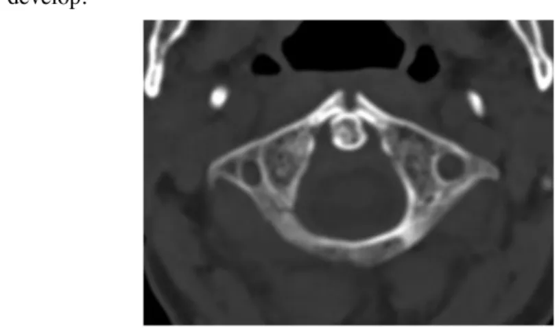
Jefferson’s fracture (fracture of C1 vertebra)
Two-thirteen percent of cervical fractures are of the Jefferson’s type. It is often caused by an axial impact of the head, when the head is pushed towards the spine. The atlas is caught between the occipital condyles and its arches break. The fracture of the arches may be single or multiple (56%). Jefferson’s fracture refers to the simultaneous fracture of the anterior and posterior arches. Symptoms include stiff neck and pain
when moving neck, without neurological signs. Structural instability may develop.
Fig. 7: C1 fracture on axial CT scan
Dens fracture Common among
elderly patients
Motor vehicle accidents or falls are responsible for most of the C2 fractures. They are common in the elderly. C2 fractures have a fatal outcome in 25-40% of cases due to medulla compression. The dens can be break off or the body of the C2 vertebra may break. There are several types of dens fracture. In most cases, it leads to structural instability requiring stabilization. Dens fracture is associated with neck pain radiating to the occipital region, which is exacerbated with neck movements. It is typical that the patient holds his/her head by his/her hands. The tear of alar ligaments may occur without fracture, which results in atlanto-axial subluxation and dislocation.
Fig. 8: Dens fracture on CT scan, reconstructed sagittal plane
Hangman’s fracture (C2 – axis trauma)
It represents 7% of cervical fractures. This type of fracture occurs during hanging, hence its name. The body of the C1 vertebra slides forward on the body of C2, which leads to the fracture of the arches of C2. A sudden hyperextension of the neck during motor vehicle accidents may cause C2 fracture with spinal cord injury.
Treatment is based on the AO classification
A widely accepted classification of subaxial cervical (C3-7I) and thoracolumbar fractures was developed by the Association for Osteosynthesis (AO). The vertebral column is divided into two main structural sections, to the anterior column (the vertebral bodies, the annulus fibrosus, the intervertebral discs, and the anterior and posterior longitudinal ligaments) and to the posterior column (pedicles, transverse processes, facet joints, laminae, and spinous processes). The fractures are categorized according to the affected spinal column and the mechanism
of injury.
There are three main (A, B, C) and several subtypes of spinal injuries according to the morphology and the mechanism of the fracture. These subtypes are based on the type and the extent of instability.
A type is common at thoracolumbar level
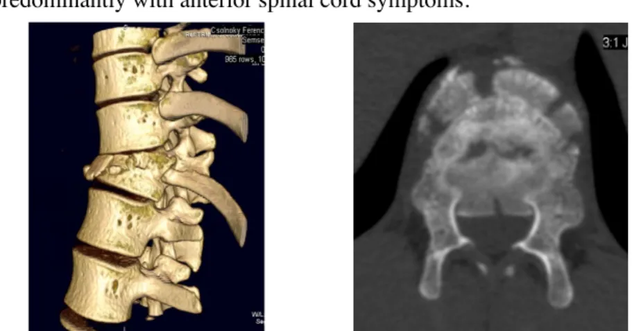
In “A” type injury, axial compressive forces lead to vertebral compression and a burst fracture, which results in segment shrinkage.
When the force strikes the spine from the vertical direction (e.g. diving into shallow water, or falling on the head in motor vehicle accidents), burst fracture of vertebral bodies may occur. Half of these injuries are associated with sever neurological deficit below the level of the injury, predominantly with anterior spinal cord symptoms.
Fig. 9: 3D reconstruction of an A type fracture of the Th12 vertebra on CT scan
Fig. 10: Burst fracture with bone fragments in the spinal canal of the L2 vertebra on axial CT scan
B type is subaxial In “B” type injury, distractive forces (flexion-extension mechanism) lead to the tear of parts of the segment with consequent segment elongation. A typical example is the whiplash injury in motor vehicle accidents, when the cervical spine suffers hyperflexion then
hyperextension. Injuries due to hyperextension, especially in old age, are often associated with a central cord syndrome and hematomyelia.
Fig. 11: Spinal cord compression caused by vertebral luxation at the level of C6-7 vertebrae on MRI
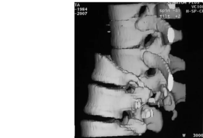
“C” type injuries are caused by shearing forces, leading to either shrinkage or elongation of the given segment.
Fig. 12: L1 fracture with dislocation on 3D reconstructed CT scan
Direct effects (due to gunshot or stab wounds) result in open injuries.
The degree of instability of the spine increases from A to C type,
according to AO classification. The risk of neurological deficit correlates strongly with the degree of instability. Fifteen percent of the subaxial cervical injuries belong to type A, 50% to type B, and the remaining to type C.
Important: the extent of instability
Two thirds of thoracolumbar injuries are type A; the rest is either type B or C. The stable type A fractures comprise one third of all fractures.
20-22% of thoracolumbar injuries lead to neurological deficit; its incidence is 14% in A, 32% in B and 55% in type C fracture.
According to the AO classification, the three principles of surgical intervention in spinal trauma are reposition, decompression and stabilization.
III./8.4.2.3 Emergency in spinal trauma
High dose of intravenous steroid
Acute spinal injury is often accompanied with severe neurological deficit. In case of spinal cord compression, functional outcome of the injury depends mainly on the time of surgical intervention. Hence, spinal cord trauma is an absolute emergency. Secondary effects of the injury may be prevented with
high dose (1-2 g) intravenous corticosteroid until the surgery.