eart valve disease is a considerable medical problem worldwide. The current treatment methods for heart valve disease include the application of mechanical and tissue valves. Nonetheless, the present substitute heart valves suffer important drawbacks. The practical disadvantages of mechanical heart valves include life-long anticoagulation therapy with the risk of anticoagulant-related hemorrhage, in- creased risk of infections, and thromboembolism caused by their non-physiological surface and flow abnormalities.1,2 Being non-viable constructs, all clinically accessible replacement heart valves, apart from the pulmonary autograft, lack the capabil- ity to grow, repair, or remodel.3 Glutaraldehyde-fixed biologi-
cal valves, and particularly cryopreserved human allografts, do not need anticoagulation treatment but suffer from progres- sive tissue deterioration, including early calcification and gen- erally poor longevity because of the preserved viability of the cells with immunogenic rejection potential.4–6 Despite innova- tive developments to improve the biological valve prostheses durability, long-term results remain unsatisfactory.7 To date, none of the currently available heart valve prostheses provide long-term stability, ideal hemodynamic performance, and re- modeling capability.
Encouraging alternatives to the current heart valve prosthe- ses are being investigated in the area of cardiac tissue engi-
H
Received April 23, 2012; revised manuscript received July 18, 2012; accepted August 21, 2012; released online September 21, 2012 Time for primary review: 27 days
Department of Cardiac Surgery, Heart Center (A.W., B. Schmack, T.O., P.S., E.B., S.L., I.P., N.C., C.S., S.K., U.T., M.K., G.S.), Depart- ment of Pathology (B. Straub), University of Heidelberg, Heidelberg, Germany; Heart Center (P.S., T.R.) and 2nd Department of Pathol- ogy (R.I.), Semmelweis University Budapest, Budapest, Hungary
This study was supported by a grant from the National Development Agency of Hungary (TÀMOP-4.2.2-08/1/KMR-2008-004).
Mailing address: Alexander Weymann, MD, Department of Cardiac Surgery, Experimental Laboratory of Cardiac Surgery, Head; “Whole Heart Tissue Engineering and Advanced Cell Technologies” workgroup, Heart and Marfan Center – University of Heidelberg, Im Neuenheimer Feld 110, 69120 Heidelberg, Germany. E-mail: weymann.alexander@googlemail.com
ISSN-1346-9843 doi: 10.1253/circj.CJ-12-0540
All rights are reserved to the Japanese Circulation Society. For permissions, please e-mail: cj@j-circ.or.jp
Carsten Schies; Sevil Korkmaz, PhD; Ursula Tochtermann, MD;
Matthias Karck, MD, PhD; Gábor Szabó, MD, PhD
Background: Heart valve tissue engineering represents a concept for improving the current methods of valvular heart disease therapy. The aim of this study was to develop tissue engineered heart valves combining human um- bilical vein endothelial cells (HUVECs) and decellularized human heart valve matrices.
Methods and Results: Pulmonary (n=9) and aortic (n=6) human allografts were harvested from explanted hearts from heart transplant recipients and were decellularized using a detergent-based cell extraction method. Analysis of decellularization success was performed with light microscopy, transmission electron microscopy and quantitative analysis of collagen and elastin content. The decellularization method resulted in full removal of native cells while the mechanical stability and the quantitative composition of the neoscaffolds was maintained. The luminal surface of the human matrix could be successfully recellularized with in vitro expanded HUVECs under dynamic flow condi- tions. The surface appeared as a confluent cell monolayer of positively labeled cells for von Willebrand factor and CD 31, indicating their endothelial nature.
Conclusions: Human heart valves can be decellularized by the described method. Recellularization of the human matrix resulted in the formation of a confluent HUVEC monolayer. The in vitro construction of tissue-engineered heart valves based on decellularized human matrices followed by endothelialization using HUVECs is a feasible and safe method, leading to the development of future clinical strategies in the treatment of heart valve disease. (Circ J 2013; 77: 207 – 216)
Key Words: Decellularization; Extracellular matrix; Heart valves; Tissue engineering
neering (TE). Despite various scientists reporting promising results, TE of human heart valves is still at the beginning of its evolution.3,5,8–10 The best possible scaffold architecture and ad- vanced cell technologies are needed to develop a TE product that will possibly become an applicable valve prosthesis. TE may help to construct viable valve substitutes that feature life- time durability and the opportunity to use in vitro fabrication of living tissue replacements while holding potential for re- generation.11 Various models featuring polymer biodegradable, biological allogeneic, and xenogeneic decellularized scaffolds have been already developed.5,12–15 Our group already reported the successful in vivo application of TE pulmonary heart valves based on decellularized xenograft matrices3,8 and generation of 3-dimensional myocardial neoscaffolds by decellularization of whole hearts.9
In this paper, we will present the in vitro TE of human heart valve neoscaffolds using human umbilical cord endothelial cells (HUVECs) under simulated physiological conditions. More- over, the influence of the decellularization protocol on the biochemical composition and ultrastructure of the human scaf- folds is characterized to assess the surface properties for con- secutive seeding with HUVECs.
Methods
All patients gave written informed consent, allowing the study to be approved by the institutional review board abiding to a protocol for human tissue use (Transplant Registry).
Fresh Human Heart Valves
Human pulmonary (n=9) and aortic valves (n=6) were freshly harvested from explanted hearts of patients suffering from isch- emic cardiomyopathy, but having no history of heart valve dis- ease, undergoing heart transplantation (age: 52.3±12.5 years;
6 males/3 females). Additionally, transesophageal ultrasound examination of every heart transplant recipient was done to rule out any pathology of the pulmonary or aortic valve. The heart valves were dissected from the heart under sterile condi- tions, leaving only a short subvalvular myocardial cuff. After the separation of adhesive tissue, all heart valves were exam- ined macroscopically to exclude any other pathology not de- tected by echocardiography. Three aortic valves were ex- cluded because of calcification of the commissures. Next, the heart valves were rinsed with phosphate-buffered saline (PBS) at 4°C and embedded in 1% penicillin-streptomycin (PAA Laboratories, Cölbe, Germany) augmented Earle’s Medium 199 (PAA Laboratories).
Decellularization Procedure
Human heart valves were decellularized through continuous shaking in 1% sodium dodecyl sulfate (SDS) and 0.05% so- dium azide (NaN3) in PBS (PAA Laboratories) at room tem- perature for 48 h. The solution was exchanged every 6 h. At the end of the decellularization protocol, the heart valves were washed with PBS for 12 h to remove residual detergents and cell debris. Before seeding, the valves were incubated in cul- ture medium composed of Endothelial Cell Growth Medium (PromoCell, Heidelberg, Germany), 10% fetal bovine serum (FBS, Life Technologies, Darmstadt, Germany) and 1% peni- Figure 1. Schematic of the bioreactor compartments and perfusion flow diagram. The pulsatile pump creates circulation of the medium with variable flow and perfusion pressures.
blood cells and coagulum. Next, the vein was filled with 20 ml of 0.1% collagenase-dispase (Boehringer Mannheim, Germany) in Hanks’ balanced salt solution (Gibco, Grand Island, NY, USA). Following 20 min of incubation at 37°C, the cell efflu- ent was collected by flushing the vessel with 20 ml of Medium 131 (Gibco) supplemented with 10% fetal calf serum (FCS;
Gibco) to inhibit further enzymatic activity. After 10 min of centrifugation (12,000 rpm), the pelleted cells were cultured in 25-cm2 flasks at 37°C with 5% CO2. The HUVECs were pas- saged when 80–90% confluent and were used between pas- sages 3 and 6. The expected endothelial phenotype of the cultured cells was analyzed microscopically (cobblestone ap- pearance typical of endothelial cells) and immunohistochemi- cally for expression of von Willebrand factor (vWF).
DNA Quantification
DNA was isolated from 200-mg freeze-dried, decellularized valve tissue and processed for spectrophotometric quantifica- tion to determine the concentration of residual DNA in the decellularized group in comparison with a control group. The total amount of DNA was purified using a silica-membrane- based method, following the manufacturer’s instructions, and later quantified by spectrophotometry (QIAamp DNA Mini Kit, Qiagen, Basel, Switzerland).
Perfusion Bioreactor
The cell seeding experiments were performed using a custom- made dynamic bioreactor system, kept in a humidified incu- bator at 37°C, allowing pulsatile circulation. The bioreactor system consisted of a valve inlet, a pulsatile pump (Stöckert, Freiburg, Germany), and a connected medium reservoir (Figure 1), which was manufactured to be placed in a 37°C conventional incubator (HERAcell 240, Thermo Scientific, Bremen, Germany). The valve inlet allowed for different sized valves to be used by suturing the valves in the support mate- rial (CarboMedics Carbo-Seal Valsalva, Austin, TX, USA).
The entire closed loop flow system, which circulates a medium volume of 1.5 L, was constructed from polycarbonate and is completely transparent to allow direct observation. Silicon tubing was used to connect all parts. The decellularized heart valve was perfused continuously with medium from the reser- voir using a pulsatile pump (Stöckert) under humidified incu- bator conditions (37°C, 5% CO2).
The pressure was controlled constantly using a pressure transducer (Medex Smith Medical) and monitored by a com- puter system guaranteeing optimal fluid flow and a continuous perfusion pressure report. Fresh gas (average 95% air, 5% CO2) was transported via a roller pump into the medium reservoir (Duran, Wertheim, Germany). The gas exchange occurred by constant medium surface oxygenation inside the medium res- ervoir. Sterile filters (Sterifix 0.2 μm; B. Braun Melsungen AG, Germany) were interposed at the top of the medium reservoir to avoid contamination during oxygenation of the circulating medium. The culture medium was replaced daily to provide fresh cell nutrients and to eliminate decomposition products
from the evolving valve tissue. The culture medium also un- derwent blood-gas analysis (Rapidlab 860, Siemens, Mannheim, Germany) twice daily. The pH level in the circulating culture medium was adjusted by flexible variation of the CO2 supply.
Fabrication of Neoendothelium Under Dynamic Tissue Cultivation Conditions
Decellularized human heart valves were placed in cell culture bottles containing a suspension of HUVECs and 50 ml of cul- ture medium supplemented with L-ascorbic acid 2-phosphate (0.25 mg/ml; Sigma-Aldrich, Steinheim, Germany), to promote extracellular matrix (ECM) production. The medium used for cultivation and consecutive tissue fabrication included 1% gen- tamicin. Subsequently, the leaflets were seeded with a density of 5–6×10⁶ cells (HUVEC passage 3–6)/cm2. The bottles were gassed with 5% CO2/air for 20 s and exposed to rotation at 5 rpm for 24 h in an atmosphere of 5% CO2 at 37°C, exposing the entire valve surface to achieve optimal attachment condi- tions. Afterwards, the heart valve neoscaffolds were mount- ed in the bioreactor chamber and perfusion was started at 15 ml/min. The medium circulation through the bioreactor complex was sustained for 4 weeks to a maximum flow of 3.5 L/min.
Analysis of Heart Valve Neoscaffolds
After in vitro endothelialization, characteristic samples of all valve leaflets were investigated by the following methods.
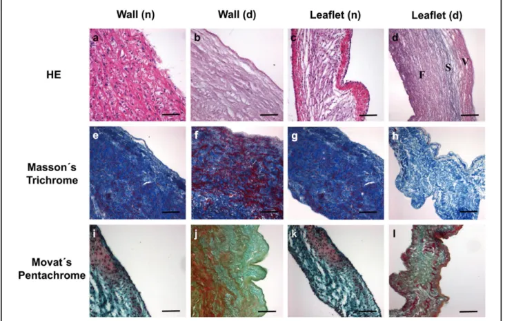
Histology and Immunohistochemistry Heart valve leaflets were dissected in the central cusp area through the adjoining arterial wall and fixed in 10% phosphate-buffered formalin and imbedded in paraffin. Sections (5-μm) were stained with he- matoxylin-eosin (HE) to determine if residual nuclear struc- tures could be identified after decellularization. Masson’s tri- chrome stain was used to distinguish the cells from the sur- rounding connective tissue (Sigma-Aldrich), and the Movat’s pentachrome stain (Mastertechs, Lodi, CA, USA) was used to visualize different ECM components such as collagen, elastin, and proteoglycans. All 3 cusps from each heart valve were evaluated.
Frozen tissue specimens were examined for ECM integrity:
collagen I (clone Coll-1, Sigma-Aldrich), collagen IV (clone Figure 2. Transmission electron microscopy demonstrating retained longitudinally running collagen fibrils with proteogly- cans after decellularization treatment.
CI22; Dako, Hamburg, Germany); evidence of endothelial cells:
vWF (polyclonal rabbit IgG; Dako), and CD 31 (Polyclonal Rabbit IgG, Santa Cruz Biotechnology, Heidelberg, Germany).
The fluorescence method of immunohistochemistry used has been described elsewhere.13 Based on the intensity and arrange- ment of labeling, histological evaluations of all stained sam- ples were implemented in a blinded mode. The degree of re- endothelialization and preservation of ECM was evaluated by 2 independent pathologists. Tissue sections were analyzed using routine bright-field microscopy and fluorescence microscopy (Olympus Optical Co, BX 51 and CKX 41 microscopes).
Images were acquired with the CellA Soft Imaging System (Olympus Soft Imaging Solutions®, Germany).
Transmission Electron Microscopy (TEM)
Tissue specimens (1 mm3) were fixed in 2.5% glutaraldehyde and embedded in Epon. Ultrathin sections were prepared for microscopy according to standard procedure. Electron micros- copy was performed using a Zeiss analytical EM 902 (Zeiss, Oberkochen, Germany) coupled with a Pro-Scan digital cam- era (Tröndle, Munich, Germany).
Quantification of Collagen and Elastin Content
To quantify collagen and elastin, Biocolor assays (Biocolor, Carrickfergus, UK) were performed in decellularized heart valve samples and compared with native human heart valve tissue.
Collagen was extracted with 0.5 mol/L acetic acid (Sigma- Aldrich) and 1:50,000 protease inhibitor cocktail at 4°C. Elastin was extracted with 100°C 0.25 mol/L oxalic acid (Sigma- Aldrich). Samples and calibrators were treated with the respec- tive dyes and later quantified by spectrophotometry.
Measurement of Mechanical Stability
Mechanical stability of heart valve tissue was analyzed using a static material testing instrument (Zwick Roell, Ulm, Germany).
Decellularized heart valve samples were stretched until com- plete tearing in the longitudinal and circumferential directions.
Passive tensile strength was constantly registered during the displacement. As a control, samples from untreated native heart valves were used.
Statistical Analysis
Quantitative analysis and mechanical stability data are expressed as mean ± standard error of the mean (SEM). Student’s t-test was used to determine significant differences between the un- treated native heart valve and decellularized neoscaffold groups
in terms of mechanical stability. A value of P<0.05 was con- sidered statistically significant.
Results
Human heart valves were decellularized by the detergent treat- ment for 48 h, and consecutively seeded with HUVECs. Under bioreactor conditions, a confluent monolayer of endothelial cells formed on the decellularized human heart valves.
Analysis of Valve Morphology, Histology and Decellularization Treatment
Decellularization of human heart valves according to the pro- tocol described here resulted in complete removal of cellular components, as shown by standard histology, immunohisto- chemistry, and TEM. Histological examination of aortic and pulmonary heart valves did not show any significant differences after decellularization. However, during the decellularization process, the heart valves became translucent, consistently los- ing their natural color. TEM examination demonstrated large amounts of collagen-fiber bundle networks lacking intracel- lular components (Figure 2). The initial structural configura- tion with intact leaflet and vascular wall morphology was main- tained. The endothelial cell layer was completely removed.
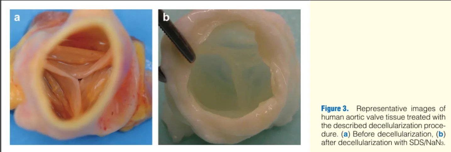
The decellularized cusps appeared smooth-walled, comparable to native heart valve leaflets (Figure 3).
Residual total DNA content of the neoscaffolds was notably decreased in comparison with the control group. After decel- lularization, the DNA content was reduced to 21.8 ng DNA/mg tissue vs. 450.7 ng DNA/mg tissue in the control group, indi- cating a 95.2% reduction in DNA content.
Morphological and histological analyses revealed an opti- mally preserved 3-dimensional neoscaffold composition (HE stain) with complete maintenance of the basement membrane as depicted through collagen IV staining. Further examination using Masson’s trichrome, Movat’s pentachrome and collagen I stain visualized different ECM components such as mesh-like collagen formations, elastin and proteoglycans (Figures 4,5).
In conclusion, cell nuclei and other intracellular components were successfully extracted from native heart valve tissue, while preserving the typical ECM composition on both the histological and ultrastructural level.
Isolation and Characterization of HUVECs
HUVECs were successfully isolated from human umbilical cord tissue. As indicated through positive immunostaining with anti-
Figure 3. Representative images of human aortic valve tissue treated with the described decellularization proce- dure. (a) Before decellularization, (b) after decellularization with SDS/NaN3.
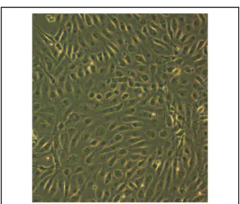
vWF factor, they retained their endothelial character in suc- cessive passages. At passages 3–6, chosen for all of our experi- ments based on cell numbers and appropriate cell characteristics, the cells were characterized. They showed good in-vitro pro- liferation and formed confluent, cobblestone-like monolayers after 3 weeks (Figure 6).
Perfusion Bioreactor System
During our experiments, the perfusion bioreactor worked ex- ceptionally well with constant flow. No leakages from the vari- ous bioreactor compartments were observed. All experiments were performed in the bioreactor system without any infection and under normal aerobic cell metabolic conditions. Gas con- centration analysis of the circulating medium showed physi- ological conditions of pO2, pCO2, and pH.
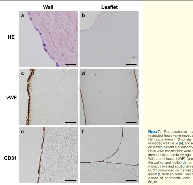
Analysis of Tissue-Engineered Heart Valve Neoscaffolds Both aortic and pulmonary heart valve neoscaffolds demon- strated cell attachment on both sides of the leaflet surface fol- lowing 3 weeks in dynamic culture conditions. Moreover, after reendothelialization the luminal surfaces of the aortic and pul- monary heart valve neoscaffolds were similarly covered. Im- munohistological analysis revealed that the cells on the scaf- fold expressed vWF and were CD31-positive, indicating a de
novo endothelial layer (Figure 7). Under dynamic, pulsatile flow at a rate of 3.5 L/min, the endothelial cells converged and formed a complete monolayer. The original endothelial cells of the heart valve leaflets were replaced by HUVECs and they had spread and divided on the neoscaffold surface. The HUVECs fully coated the leaflet surface and were oriented to- ward the flow direction. The leaflet coating consisted of typi- cal cobblestone-like endothelial cells covering the basal lam- ina of both sides of the cusp, including the pulmonary and aortic wall. The leaflets appeared intact and flexible without fenes- trations or adhesions and presented a pale, shiny surface, com- parable to native tissue samples. There was no cusp thickening or reduction of cusp mobility upon gross inspection, and the heart valve neoscaffolds demonstrated acceptable resistance and stability, comparable to that of native tissue, posing no hindrance to suture application.
Analysis of Collagen and Elastin Content
Collagen and elastin content of decellularized heart valve sam- ples was compared with that of untreated native heart valve tissue, which was taken as 100% (Figure S1). Quantification of the collagen and elastin content was 87.70±2.86% and 85.15± 2.81%, respectively, compared with native heart valve tissue.
Figure 4. Heart valve tissue structure after decellularization. Hematoxylin-eosin (HE) staining of (a) native heart valve wall and (b) after decellularization, and native leaflet before (c) and after decellularization treatment (d; [F, fibrosa section; S, spongiosa sec- tion; V, ventricularis section of the valve leaflet is completely retained]) from aortic valve tissue. Masson’s trichrome stain (e–h) from pulmonary valve tissue and Movat’s Pentachrome stain (i–l) from aortic valve tissue show preservation of the extracellular matrix after the decellularization procedure. Bars=50 μm; n=native, d=decellularized. Movat’s pentachrome stain: nuclei (dark purple to black), elastic fibers (purple to black), collagen (yellow), glycosaminoglycans (green), mucin (blue), cytoplasm (pink to brownish-red). Masson’s trichrome stain: cytoplasm (red), collagen (blue), nuclei (dark-brown).
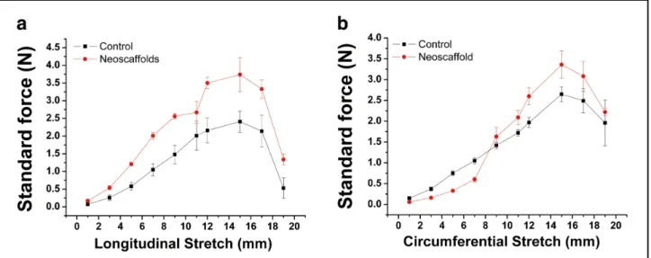
Analysis of Mechanical Stability
Figure 8 demonstrates the stress-strain curves of the decellu- larized heart valve neoscaffolds. Using the decellularization protocol described here, the decellularized heart valve neoscaf- folds showed similar mechanical stability as untreated native heart valve tissue for longitudinal (a) and circumferential (b) stretch (P>0.05).
Discussion
TE through endothelial cell transfer onto specially designed neoscaffolds is a promising experimental approach to dimin- ishing the drawbacks of allografts, non-living xenografts, and prosthetic heart valves. Our group has already reported on clin- ical experience with in-vivo-developed TE pulmonary heart valves, based on decellularized xenograft matrices3,8 and the creation of decellularized hearts as potential neoscaffolds for whole heart TE.9
The present investigation was based on total decellulariza- tion of human heart valve scaffolds, dissected from explanted hearts of heart transplant recipients. Repopulation under bio- reactor conditions was accomplished through in-vitro reseed- ing with HUVECs, which in turn formed a functional endothe- lial cell layer on the surface of the decellularized heart valve neoscaffolds.
It has been shown that decellularized heart valve neoscaf- folds implanted in a sheep model were subject to calcification Figure 5. Collagen I stain of decellularized heart valve wall (a) and leaflet (b) demonstrating retained collagen I formation and collagen IV stain (c) of wall, and (d) leaflet from aortic valve tissue depicting complete preservation of the basement membrane.
Bars=50 μm.
Figure 6. Bright-field microscopy (×60) of human umbilical vein endothelial cells (HUVECs) with their typical cobblestone- like appearance after 3 weeks of culture in a tissue culture flask.
and endocarditis without prior reendothelialization.15 Hence, decellularized heart valve neoscaffolds should be endothelial- ized before further manipulation to prevent the risks of calci- fication and infection. We exclusively used an allograft ECM because it would not introduce cross-species immunological conflict. For example, repopulation with recipient cells could convert the allograft tissue to autologous tissue having a syn- geneic origin, and the potential ability to remodel.
Scaffold Source
In this study, we used decellularized human heart valve neoscaf- folds as biologically active structures because they not only provide a 3-dimensional (3D) supporting matrix, but also im- portant ECM proteins such as collagen, elastin, and proteogly- cans, parts missing in synthetic polymer materials.9 We are convinced that the key to successful TE lies in the preserva- tion of the anatomical structure. Our human scaffold provides such a structure, as well as a higher immune histocompatibil- ity compared with synthetic materials.
In contrast is the application of biodegradable polymers with
limited mechanical properties and variable degradation times for heart valve TE. The advantages of these synthetic-based scaffolds are mainly their biocompatibility, availability and abil- ity to be reproduced with predictable chemistry,16,17 but they do not approximate the composition of a natural matrix. On the other hand, synthetic-based scaffolds are expensive and potentially immunogenic, and additionally, suffer from toxic degradation and inflammatory reaction.9 Usually, there are 2 major drawbacks: (1) synthetic scaffold sources cannot express the important bioactive molecules and ligands that are neces- sary for neoscaffold maturation and (2) effective diffusion of vital cell nutrients is aggravated.17
Furthermore, synthetic polymer scaffolds are rigid, throm- bogenic, and induce an overshooting fibrosis that can induce leaflet retraction with insufficiency following implantation.18–20 Preliminary studies have proven that biodegradable polyglac- tin-PGA copolymer matrices used to produce vascular scaf- folds are unstable and cause aneurysm formation.21
There have also been reported efforts from other research- ers of the use of synthetic scaffold components in heart valve
Figure 7. Representative images of reseeded heart valve neoscaffolds.
Hematoxylin-eosin (HE) staining of reseeded wall tissue (a), and reseed- ed leaflet (b) from a pulmonary valve.
Heart valve neoscaffolds were stained immunohistochemically against von Willebrand factor (vWF) (brown) in the wall (c) and leaflet (d) from a pul- monary valve and additionally against CD31 (brown-red) in the wall (e) and leaflet (f) from an aortic valve for evi- dence of endothelial cells. Bars=
50 μm.
TE,22–24 but none of these concepts has achieved the criterion of safe human implantation.
Decellularization Treatment
For the first time, a mono-regime SDS/NaN3 decellularization protocol has been successfully used for human heart valve scaf- fold cell elimination. The ionic detergent, SDS, and the ionic solid, NaN3, have already demonstrated excellent cell remov- al properties with preservation of major structural ECM mol- ecules.9,25,26 In our investigation, decellularization with SDS/
NaN3 resulted in a biological neoscaffold free of any visible cellular material with maintenance of the heart valve architec- ture and geospatial arrangement of its matrix. The 3D fibrous network of lengthwise folded collagen fibers and bundles in- side the cusps, as well as other ECM components, were well- conserved and properly arranged, suggesting that the tissue’s matrix structures were not significantly compromised. In both aortic and pulmonary heart valves the normal structure was optimally preserved. Furthermore, the applied decellulariza- tion protocol was able to conserve cusp coaptation and mac- roscopic valve geometry. In the present study, both the colla- gen and elastin content and the mechanical properties of the decellularized heart valve neoscaffolds demonstrated similar characteristics to untreated controls. Taken together, these find- ings are favorable for continued recellularization of the neoscaf- fold once implanted in vivo.
Other investigators have used enzymatic decellularization treatments for heart valve TE and demonstrated scaffold in- jury with basal membrane damage.25,27 Other decellularization protocols include the use of glutaraldehyde, which leads to structural deterioration in vivo. It is imperative to avoid a mul- timodal aggressive decellularization treatment.28
In our study, we did not achieve complete reduction of nu- clear material. However, we observed the removal of virtually all DNA material from the decellularized heart valves, based on histological staining and TEM results. The impossibility that any decellularization method will remove 100% of all cell components is well known.9 There has yet to be a study prov- ing a direct correlation between these nuclear fragments and immunological rejection.28
The final biologic neoscaffolds had the majority of their cel- lular content removed, potentially enabling safe implantation.
Cell Source
In this evaluation, endothelial cells were successfully isolated from human umbilical cord tissue through enzymatic digestion.
As the cells were transplanted onto the luminal surface of the decellularized human neoscaffolds, they formed a monolayer of neoendothelium on the valve leaflets, as has already been shown in animal models.29 In the present study, we showed that after reendothelialization the luminal surface of human aortic and pulmonary heart valve neoscaffolds was identically covered with a solid layer of HUVECs. The formation of a confluent endothelial cell layer is essential in the development of a TE heart valve, because it provides an antithrombogenic surface and physiological continuous remodeling.21
Endothelial cells are key players in the regulation of impor- tant processes such as inflammation, angiogenesis, immune re- action, and transport of molecules. Moreover, they are able to produce various genes through different signaling pathways and contain receptors, which are important for cell metabolism and homeostasis.16
Additional research has shown that a population of mesen- chymal stem cell-like cells exist in human umbilical cord tis- sue and cord blood.30,31 Umbilical cords are readily available and contain different juvenile cell types for regeneration ther- apy. The possibility of cryopreserving a patient’s own umbili- cal cord tissue after birth would allow for the preparation of TE heart valve grafts for future autologous use. Available al- lografts could be incubated with appropriate cells from the cryopreserved umbilical cord until maturity to become living and functional scaffolds and later be implanted as an autolo- gous tissue heart valve. Furthermore, degeneration of these au- tologous heart valves based on immunological reactions may be reduced.
Conceivable future studies using progenitor cells from um- bilical cord tissue could become a new approach to cardiac valve TE. This advance would overcome the lack of living au- tologous replacements with regeneration potential.
Figure 8. Stress-strain curve of tissue-engineered heart valve neoscaffolds. All data are expressed as mean ± SEM.
continuously observed, allowing for defects and contamina- tions to be detected. Furthermore, the reactor can be easily modified using a sample-mounting adapter to take other car- diovascular constructs; for example, to colonize vascular grafts with different cells prior to implantation.
It is common knowledge that cardiovascular cells such as endothelial cells are influenced by local fluid dynamics. Me- chanical shear stress is induced through the modification of the bioreactor flow, circulating volume, and pressure. This influ- ences the growth, orientation, and phenotypic remodeling of the endothelial cells.32–35 It has been shown that a rapid increase in bioreactor flow causes significant damage of the reseeded endothelium and complete cell wash-off from cusps.12 This is especially common near the valve leaflet, where turbulent flow is present.
To avoid cell detachment, we slowly adjusted the endothe- lial cells to physiological flow levels using incremental increas- es in the flow rate. This strategy allowed endothelial cells to attach and fully align to the valve scaffold. Nevertheless, the optimal flow conditions for cell adhesion, differentiation, and proliferation are still unknown and may vary between various bioreactor constructions.
Study Limitations
Whether or not the human heart valve neoscaffolds, produced using the methodology presented here, will provide the micro- environment needed for further cellular differentiation, has to be evaluated in future studies. This implicates the invasion of valvular interstitial cells into the heart valve scaffold, either in vivo after implantation or in vitro by means of cultivated cells.
Conclusion
To our knowledge, this is the first report of the generation of a TE human heart valve neoscaffold with preserved ECM com- position containing an in vitro reseeded HUVEC neoendothe- lium as a peripartal cell source.
The most important results of the presented work are: (1) fabrication of TE human heart valve neoscaffolds reseeded with HUVECs is feasible; (2) there is evidence that the architecture of the ECM is retained after treatment with SDS/NaN3 and is comparable to native tissue, based on our light- and electron- microscopic, quantitative and mechanical analyses; and (3) HUVECs can be easily extracted and preserved, which allows for individuals to store their own samples for possible future autologous use. Although the results shown here are limited, this investigation demonstrates the ability of our bioreactor system to develop the optimal in-vitro environment for TE heart valve neoscaffolds. The bioreactor enables consistent and uni- form delivery of cells, nutrients, gases, and hydrodynamic shear forces.
The presented approach could be used to transform human allografts into autologous tissue heart valves. Our work has the potential for an important clinical contribution, which is a strong argument for evaluating our approach experimentally in addi-
acknowledged.
Source of Funding
This study was supported by the Land Baden-Württemberg, Germany, the Medical Faculty of the University of Heidelberg, Germany (to S.K.) and by a grant from the National Development Agency of Hungary (TÀMOP- 4.2.2-08/1/KMR-2008-004).
Disclosures None.
References
1. Mayer JE Jr. Uses of homograft conduits for right ventricle to pul- monary artery connections in the neonatal period. Semin Thorac Car- diovasc Surg 1995; 7: 130 – 132.
2. Schoen FJ, Levy RJ. Founder’s award, 25th Annual Meeting of the Society for Biomaterials, perspectives. Providence, Rhode Island, April 28–May 2, 1999. Tissue heart valves: Current challenges and future research perspectives. J Biomed Mater Res 1999; 47: 439 – 465.
3. Weymann A, Dohmen PM, Grubitzsch H, Dushe S, Holinski S, Konertz W. Clinical experience with expanded use of the Ross pro- cedure: A paradigm shift? J Heart Valve Dis 2010; 19: 279 – 285.
4. Yacoub M, Rasmi NR, Sundt TM, Lund O, Boyland E, Radley- Smith R, et al. Fourteen-year experience with homovital homografts for aortic valve replacement. J Thorac Cardiovasc Surg 1995; 110:
186 – 193; discussion 193 – 194.
5. da Costa FD, Dohmen PM, Lopes SV, Lacerda G, Pohl F, Vilani R, et al. Comparison of cryopreserved homografts and decellularized porcine heterografts implanted in sheep. Artif Organs 2004; 28: 366 – 6. da Costa FD, Dohmen PM, Duarte D, von Glenn C, Lopes SV, Filho 370.
HH, et al. Immunological and echocardiographic evaluation of de- cellularized versus cryopreserved allografts during the Ross opera- tion. Eur J Cardiothorac Surg 2005; 27: 572 – 578.
7. Homann M, Haehnel JC, Mendler N, Paek SU, Holper K, Meisner H, et al. Reconstruction of the RVOT with valved biological conduits:
25 years experience with allografts and xenografts. Eur J Cardiotho- rac Surg 2000; 17: 624 – 630.
8. Dohmen PM, Lembcke A, Holinski S, Kivelitz D, Braun JP, Pruss A, et al. Mid-term clinical results using a tissue-engineered pulmonary valve to reconstruct the right ventricular outflow tract during the Ross procedure. Ann Thorac Surg 2007; 84: 729 – 736.
9. Weymann A, Loganathan S, Takahashi H, Schies C, Claus B, Hirschberg K, et al. Development and evaluation of a perfusion de- cellularization porcine heart model: Generation of 3-dimensional myo- cardial neoscaffolds. Circ J 2011; 75: 852 – 860.
10. Dohmen PM, Lembcke A, Hotz H, Kivelitz D, Konertz WF. Ross operation with a tissue-engineered heart valve. Ann Thorac Surg 2002;
74: 1438 – 1442.
11. Vesely I. Heart valve tissue engineering. Circ Res 2005; 97: 743 – 755.
12. Lichtenberg A, Cebotari S, Tudorache I, Sturz G, Winterhalter M, Hilfiker A, et al. Flow-dependent re-endothelialization of tissue-en- gineered heart valves. J Heart Valve Dis 2006; 15: 287 – 293; discus- sion 293 – 294.
13. Cebotari S, Mertsching H, Kallenbach K, Kostin S, Repin O, Batrinac A, et al. Construction of autologous human heart valves based on an acellular allograft matrix. Circulation 2002; 106: I63 – I68.
14. Hoerstrup SP, Kadner A, Melnitchouk S, Trojan A, Eid K, Tracy J, et al. Tissue engineering of functional trileaflet heart valves from human marrow stromal cells. Circulation 2002; 106: I143 – I150.
15. Steinhoff G, Stock U, Karim N, Mertsching H, Timke A, Meliss RR, et al. Tissue engineering of pulmonary heart valves on allogenic acellular matrix conduits: In vivo restoration of valve tissue. Circula- tion 2000; 102: III50 – III55.
16. Rabkin-Aikawa E, Mayer JE Jr, Schoen FJ. Heart valve regeneration.
Adv Biochem Eng Biotechnol 2005; 94: 141 – 179.
17. Pankajakshan D, Krishnan LK. Design of fibrin matrix composition to enhance endothelial cell growth and extracellular matrix deposi- tion for in vitro tissue engineering. Artif Organs 2009; 33: 16 – 25.
18. Shinoka T, Breuer CK, Tanel RE, Zund G, Miura T, Ma PX, et al.
Tissue engineering heart valves: Valve leaflet replacement study in a lamb model. Ann Thorac Surg 1995; 60: S513 – S516.
19. Shinoka T, Ma PX, Shum-Tim D, Breuer CK, Cusick RA, Zund G, et al. Tissue-engineered heart valves: Autologous valve leaflet re- placement study in a lamb model. Circulation 1996; 94: II164 – II168.
20. Shinoka T, Shum-Tim D, Ma PX, Tanel RE, Isogai N, Langer R, et al.
Creation of viable pulmonary artery autografts through tissue engi- neering. J Thorac Cardiovasc Surg 1998; 115: 536 – 545; discussion 545 – 546.
21. Mitchell SL, Niklason LE. Requirements for growing tissue-engi- neered vascular grafts. Cardiovasc Pathol 2003; 12: 59 – 64.
22. Sodian R, Hoerstrup SP, Sperling JS, Daebritz S, Martin DP, Moran AM, et al. Early in vivo experience with tissue-engineered trileaflet heart valves. Circulation 2000; 102: III22 – III29.
23. Sodian R, Sperling JS, Martin DP, Egozy A, Stock U, Mayer JE Jr, et al. Fabrication of a trileaflet heart valve scaffold from a polyhy- droxyalkanoate biopolyester for use in tissue engineering. Tissue Eng 2000; 6: 183 – 188.
24. Hoerstrup SP, Sodian R, Daebritz S, Wang J, Bacha EA, Martin DP, et al. Functional living trileaflet heart valves grown in vitro. Circula- tion 2000; 102: III44 – III49.
25. Grauss RW, Hazekamp MG, van Vliet S, Gittenberger-de Groot AC, DeRuiter MC. Decellularization of rat aortic valve allografts reduces leaflet destruction and extracellular matrix remodeling. J Thorac Car- diovasc Surg 2003; 126: 2003 – 2010.
26. Wainwright JM, Czajka CA, Patel UB, Freytes DO, Tobita K, Gilbert TW, et al. Preparation of cardiac extracellular matrix from an intact porcine heart. Tissue Eng Part C Methods 2010; 16: 525 – 532.
27. Kasimir MT, Rieder E, Seebacher G, Silberhumer G, Wolner E, Weigel G, et al. Comparison of different decellularization procedures of porcine heart valves. Int J Artif Organs 2003; 26: 421 – 427.
28. Gilbert TW, Sellaro TL, Badylak SF. Decellularization of tissues and organs. Biomaterials 2006; 27: 3675 – 3683.
29. Kasimir MT, Weigel G, Sharma J, Rieder E, Seebacher G, Wolner E, et al. The decellularized porcine heart valve matrix in tissue engi- neering: Platelet adhesion and activation. Thromb Haemost 2005;
94: 562 – 567.
30. Lee OK, Kuo TK, Chen WM, Lee KD, Hsieh SL, Chen TH. Isolation of multipotent mesenchymal stem cells from umbilical cord blood.
Blood 2004; 103: 1669 – 1675.
31. Lu LL, Liu YJ, Yang SG, Zhao QJ, Wang X, Gong W, et al. Isolation and characterization of human umbilical cord mesenchymal stem cells with hematopoiesis-supportive function and other potentials. Hae- matologica 2006; 91: 1017 – 1026.
32. Blackman BR, Garcia-Cardena G, Gimbrone MA Jr. A new in vitro model to evaluate differential responses of endothelial cells to simu- lated arterial shear stress waveforms. J Biomech Eng 2002; 124: 397 – 407.
33. Imberti B, Seliktar D, Nerem RM, Remuzzi A. The response of en- dothelial cells to fluid shear stress using a co-culture model of the arterial wall. Endothelium 2002; 9: 11 – 23.
34. Jin ZG, Ueba H, Tanimoto T, Lungu AO, Frame MD, Berk BC.
Ligand-independent activation of vascular endothelial growth factor receptor 2 by fluid shear stress regulates activation of endothelial nitric oxide synthase. Circ Res 2003; 93: 354 – 363.
35. Teebken OE, Bader A, Steinhoff G, Haverich A. Tissue engineering of vascular grafts: Human cell seeding of decellularised porcine ma- trix. Eur J Vasc Endovasc Surg 2000; 19: 381 – 386.
Supplementary Files Supplementary File 1
Figure S1. Quantitative analysis of collagen and elastin content of tissue-engineered heart valve neoscaffolds.
Please find supplementary file(s);
http://dx.doi.org/10.1253/circj.CJ-12-0540