in dogs using a combination of tibial tuberosity advancement procedure and autologous
mesenchymal stem cells/multipotent
mesenchymal stromal cells – A pilot study
SA Š A KOPRIVEC
1, MARKO NOVAK
1, STANISLAV BERNIK
1, METKA VOGA
2, LUKA MOHORI C
3and GREGOR MAJDI C
2p1Klinika Loka d.o.o,Skofja Loka, Slovenia
2Institute for Preclinical Sciences, Veterinary Faculty, University of Ljubljana, Gerbiceva 60, 1000 Ljubljana, Slovenia
3Animacel Biotechnology Ltd., Ljubljana, Slovenia
Received: January 9, 2020 • Accepted: December 21, 2020 Published online: March 2, 2021
ABSTRACT
In the present pilot study, we evaluated different supplemental therapies using autologous multipotent mesenchymal stromal cells (MMSCs) for the treatment of cranial cruciate ligament defects in dogs. We used tibial tuberosity advancement (TTA) and augmented it by supportive therapy with MMSCs in three patient groups. In the first patient group, the dogs were injected with MMSCs directly into the treated stifle one month after surgery. In the second group, MMSCs were delivered in a silk fibroin scaffold which was placed in the osteotomy gap during surgery. In the third group, MMSCs were first mixed with bone tissue and blood from the patient and delivered into the osteotomy gap during surgery.
In the control group, patients underwent the TTA procedure but did not receive MMSC treatment. In the group of patients who received cells in the silk fibroin scaffold during surgery, the osteotomy gap did not heal, presumably due to the low absorption of silk fibroin. Patients who received MMSCs mixed with bone tissue and blood during surgery into the osteotomy gap recovered clinically faster and had better healing of the osteotomy gap than dogs from the other two treated groups and from the control group, as assessed by clinical examination and quantification of radiographs. In conclusion, dogs that received stem cells directly into the osteotomy gap (Group 3) recovered faster compared to dogs from Groups 1 (MMSCs injected into the joint one month after surgery), 2 (cells implanted into the osteotomy gap in a silk fibroin scaffold), and the control group that did not receive additional MMSCs treatment.
KEYWORDS
dog, stifle joint, cranial cruciate ligament, tibial tuberosity advancement, autologous multipotent mesenchymal stromal cells, stem cells, fibroin, cancellous bone tissue scaffold
INTRODUCTION
Whether it is a one-time traumatic event or long-term irreversible degenerative changes, the rupture of the cranial cruciate ligament (CrCL) in dogs exerts significant adverse effects on the anatomy and physiology of the stifle joint (Doom et al., 2008). The main functions of the CrCL include preventing cranial translation of the tibia relative to the femur and preventing internal rotation or hyperextension of the stifle joint (Hayashi et al., 2010). CrCL damage is a major cause of hindlimb lameness and associated pain in dogs because it leads to cranial
Acta Veterinaria Hungarica
68 (2020) 4, 405–412 DOI:
10.1556/004.2020.00063
© 2021 Akademiai Kiado, Budapest
ORIGINAL RESEARCH PAPER
pCorresponding author.
E-mail:gregor.majdic@vf.uni-lj.si
subluxation of the tibia (Simpson, 2012). If left untreated, it can lead to progressive (secondary) osteoarthritis as the abnormal biomechanics affects the cartilage in the stifle joint (Cook, 2010). Several surgical techniques may be used to treat the damage to the CrCL. The most common are tibial plateau levelling osteotomy (TPLO) and tibial tuberosity advancement (TTA). Both alter the geometry of the tibia in relation to the femur and ultimately restore stability to the stifle (MacDonald et al., 2013). However, the TTA procedure developed by Montavon and Tepic (Montavon et al., 2004) is considered less invasive and surgically less complex compared to TPLO. Its main goal is to stabilise the joint and neutralise the cranial tibial thrust by per- forming an osteotomy of the tibial crest in the frontal plane to advance the patellar tendon to perpendicular to the tibial plateau (Lafaver et al., 2007). The TTA procedure had routinely been performed at Klinika Loka before the present pilot study was initiated. However, the current time to full recovery is approximately six months and includes the use of pain medication. Therefore, our main focus was to investigate alternatives to shorten the recovery time in dogs.
The use of stem cell/stromal cell therapy has grown significantly since its beginnings at the turn of the century (Koh and Piedrahita, 2014). It is a rapidly evolving field, both experimentally and clinically. There is growing interest in the use of stem cells to treat musculoskeletal injuries, particularly in certain groups of animals, such as racehorses and rescue dogs, where there is an urgent need to restore normal function as quickly as possible (Braun et al., 2010;
Fortier and Travis, 2011). In particular, multipotent mesenchymal stromal cells (MMSCs), often referred to as mesenchymal stem cells, have become popular thanks to their wide availability, the simplicity of their isolation, and their ability to differentiate into various tissue types (Minteer et al., 2013). Their therapeutic rolein vivohas not been fully elucidated, but their trophic, paracrine, anti-inflammatory and immunomodulatory effects have been shown to posi- tively influence disease progression (Baraniak and McDevitt, 2010; Fortier and Travis, 2011). By secreting regenerative bioactive molecules, they accelerate healing processes and minimise inflammation at the site of injury (Marx et al., 2015). Since local environmental signals also regulate the action of MMSCs, it is optimal for the cells to remain at the site of injury. Therefore, to keep them at the site of injury, it is necessary to use a carrier that can keep MMSCs in place and maximise their therapeutic potential (Baraniak and McDevitt, 2010; Krishna et al., 2016). Silk proteinfibroin is widely used for bone tissue engineering due to its favourable biomechanical properties and biocompatibility (Melke et al., 2016).
The aim of this study was to test whether MMSC sup- plementation therapy, in addition to the surgical TTA pro- cedure, could result in faster recovery of the stifle joint function in dogs after CrCL injuries. We wanted to provide our patients with a method that would allow for a faster recovery time, but also partially eliminate or reduce the need for postoperative pain management. One group of patients
(control) underwent only the traditional surgical procedure (TTA). Three other groups of dogs underwent the TTA surgery and received additional treatment with autologous MMSCs, either delivered to the affected bone during surgery or injected into the joint one month after surgery. The dogs were monitored for six months after surgery for lameness and radiographic monitoring was done to check the healing of the osteotomy gap.
MATERIALS AND METHODS
Patients
Sixteen dogs with signs of CrCL deficits were included in this study. All dogs were clinical patients of Klinika Loka d.o.o. Dog owners voluntarily participated in the study.
Owners were informed about the potential risks and benefits of the procedures used and they signed an informed consent form. Before the surgery, all patients underwent clinical examination to assess the extent and grade of lameness.
CrCL rupture was confirmed clinically using the cranial drawer test. Radiographic imaging was performed to confirm the presence of typical changes associated with CrCL rupture and to rule out other problems that might affect the course of surgery and postoperative treatment.
According to the legal opinion of the Slovenian Adminis- tration for Food Safety, Veterinary Affairs and Plant Pro- tection, no ethical approval was needed as all procedures were performed on clinical privately-owned patients with the owners' consent.
Organisation of the test groups
All dogs underwent the TTA surgery. In the control group (CG), the osteotomy gap in the tibia was filled with autol- ogous cancellous bone tissue and blood harvested from the ipsilateral proximal tibia during surgery, but no additional MMSC therapy was used. In Group 1 (G1), patients received an injection of autologous MMSCs directly into the joint (33106cells in 1 mL sterile PBS) one month after the TTA surgery. For patients in Group 2 (G2), autologous MMSCs were seeded onto a porous silkfibroin scaffold, and the scaffold with MMSCs was inserted into the surgically created osteotomy gap during TTA surgery. In Group 3 (G3), autologous MMSCs were mixed with the patient's blood, and cancellous bone tissue and this was inserted into the osteotomy gap during surgery. There was no specific key for assigning patients to test groups. The dogs were randomly assigned to the test groups by a person (S. K.) who was not involved in the clinical examination of the dogs.
Multipotent mesenchymal stromal cell preparation
In G1, G2 and G3 patient-derived (autologous) MMSCs were used. For patients in G1, MMSCs were isolated from adipose tissue, harvested from the stifle infrapatellar fat pad during the TTA surgery. The tissue was placed in a sterile
container with growth media and stored in cold until cell isolation. For patients from G2 and G3, the adipose tissue was harvested from the shoulder region two weeks before the surgical procedure. Our previous observations (Mohoric and Majdic, unpublished observation) have shown that there is no difference in the isolation success or therapeutic potential of canine MMSCs for the treatment of osteoar- thritis with regard to the origin of the cells (abdominal, shoulder or stifle fat pads). Therefore, cells from different locations were used to reduce the discomfort for patients.
MMSCs were isolated as previously described (Mohoric et al., 2016). Briefly, adipose tissue was dissected with a scalpel and incubated overnight with 1 mg/mL collagenase.
The digested tissue was centrifuged (3,000g, 5 min), the cell pellet resuspended in growth media containing DMEM and 10% fetal bovine serum, and seeded on 6-well plates. Cells were grown under standard conditions at 37 8C and 5%
CO2. At full confluence, cells were trypsinised and reseeded in T75 cell cultureflasks.
For use with patients in G1, MMSCs in T75 cell culture flasks were trypsinised, centrifuged, and washed repeatedly with sterile PBS. Cells were counted and a final concentra- tion of 33106 cells per ml was resuspended in sterile PBS and transferred to a sterile syringe.
For patients in G2, fibroin scaffolds were seeded with 4 million MMSCs each, which continued to grow and infiltrate the porous scaffolds immersed in growth media for five days under standard growth conditions as described above. Prior to use, the scaffolds were washed thoroughly with sterile PBS on a shaker several times within 24 h.
MMSCs for patients in G3 were prepared beforehand, as described for G1. In the operating room 3 3 106 MMSCs were mixed with the patient's blood and collected cancellous bone tissue in a sterile tube. The blood mixed with tissue and MMSCs was allowed to clot and then inserted in the newly formed osteotomy gap during surgery.
Pre-operative care and surgical procedure
Upon admission of the patient for surgery, the patient's history was taken and complete clinical evaluation was performed, including a lameness assessment. A blood sam- ple was collected for the analysis of whole blood panel and biochemical parameters. An intravenous catheter was inserted aseptically into the cephalic vein. General anaes- thesia was adjusted according to the patient's characteristics and maintained using sevoflurane. Radiographs of the affected joint were taken in the extended mediolateral pro- jection with strict condylar superimposition and cranio- caudal projection to measure and calculate the size of the titanium implants and cage (common tangent technique) and mark the osteotomy line.
The hindlimb was aseptically prepared for surgery, and a subpatellar medial mini arthrotomy was performed. The remnants of the CrCL were removed and meniscal damage was assessed. If deemed necessary, a partial or total caudal meniscectomy was performed. The TTA procedure was performed using commercially available equipment (KYON,
Z€urich), as described elsewhere (Montavon et al., 2004;
Lafaver et al., 2007).
Post-operative monitoring and evaluation of the results
Clinical examinations were performed 1, 3 and 6 months after surgery. Examinations included radiographic assess- ments for monitoring the bone healing process, evaluation of range of motion (flexion and extension) by goniometric measurements, lameness evaluation on a 5-point scale as described by Stein and Schmoekel (2008), general ortho- paedic inspection of the operated joint (signs of joint swelling and pain or other common post-surgery compli- cations were recorded) and owner satisfaction with the procedure was evaluated with a questionnaire.
Radiographic assessment of bone healing
For radiographic assessment of osteotomy healing we used a scoring system described by Guerrero et al. (2011).
Mediolateral radiographic projections at 1, 3 and 6 months after TTA surgery were compared between the test groups (G1, G2, G3 and GC). Score A was used to evaluate overall osteotomy healing, and score B was used to evaluate the healing at three anatomic sites of the newly formed osteot- omy gap. The three sites were defined as the area of the newly formed osteotomy proximal to the cage, between the cage and the plate, and distal to the plate. A rating scale of 0–4 was used for assessment of score A, as previously vali- dated by Hoffmann et al. (2006): 0 5 no osseous healing;
1 5 early bone formation but no bridging between the tibial tuberosity and tibial shaft; 25bridging bone forma- tion at one site; 3 5bridging bone formation at two sites;
45bridging bone formation at all three sites. Score B was used to evaluate bone healing at the same three sites (B1, B2 and B3) as mentioned above and in comparison to the density of the original bone. Here a scale of 0–3 was used in each region separately (05no bone healing; 15density of callus less than normal; 25 density equal to original bone;
3 5 density higher than the original bone). For statistical analysis, the scores from each of the three sites were added together.
Physiotherapy
Along with orthopaedic monitoring, a comprehensive physical therapy protocol was established to achieve a more rapid recovery of stifle function. Consultations with a trained physical therapist began two weeks after surgery, with regular follow-up visits at 1, 2, 3, 4, and 6 months after surgery. The exercise protocol consisted of stretching, passive range of motion (PROM) exercises, followed by active exercises and cooling down. Detailed instructions for home exercises were provided for owners.
Statistical analyses
Postoperative lameness score was compared between groups using the chi-squared test. Assessment of osteotomy gap
healing was analysed using one-way ANOVA with the group as the independent variable.
Ethical approval and consent to participate
Dog owners were informed about the potential risks and benefits of the procedures used and participated voluntarily.
All owners signed an informed consent form and were informed about all the procedures and potential risks. Ac- cording to the legal opinion of the Slovenian Administration for Food Safety, Veterinary Affairs and Plant Protection, no ethical permission was needed as all procedures were done on privately owned clinical patients with their owners' consent.
RESULTS
Patient characteristics
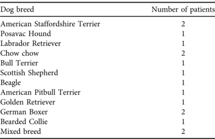
Sixteen dogs with evidence of CrCL deficiencies were included in the study. Four of the 16 patients had bilateral stifle difficulties and surgery was performed on both stifles, although not simultaneously. The age of the dogs at the time of surgery ranged from 2 years and one month to 9 years and one month. Twelve of the patients were female, and 4 were male. The weight range of all dogs (males and females) was from 25 to 50 kg. Their breeds are listed inTable 1. Of the 20 stifles operated on, nine were right and 11 were left.
Orthopaedic evaluation
All 16 patients (20 stifles) were examined 1, 3, and 6 months after surgery. Before surgery, they had all tested positive on the cranial drawer test. Also, before surgery and at each visit, the lameness score of the affected joint was evaluated on a scale from 0 to 4, with 0 representing no lameness, and 4 representing lameness with no-weight bearing (Stein and Schmoekel, 2008). The results are shown in Table 2. All patients had markedly improved lameness scores by the six months mark. However, there was a difference between the groups, as in CG, G1 and G2 the majority of dogs were still limping one month after surgery, whereas in G3 only 1 out of 5 dogs was still limping after one month.
This difference was statistically significant as determined by the chi-squared test (P < 0.05). Patient 4 of G3 was an exception, because the owner did not perform any physio- therapy exercises at home, contrary to the physiotherapist's instructions.
Radiographic evaluation
Radiographs of the affected limb(s) were taken before sur- gery and 1, 3, and 6 months after surgery. Radiographs taken at each orthopaedic examination after surgery were used to follow the progress of callus formation and osteotomy gap healing (Fig. 1).
Analysis of bone healing revealed that G2 (cells with scaffold) had statistically significantly lower scores A and B for bone healing in comparison to the other three groups at 1 month, 3 months and 6 months after surgery (P< 0.001;
Fig. 2). Interestingly, the score A was higher in G3 than in all other groups (including CG) at 1 month after surgery, confirming the results of the clinical examination and sug- gesting that MMSCs promoted bone healing in this group (P< 0.001, Fig. 2).
DISCUSSION
In the present pilot study, we evaluated a novel approach to the treatment of CrCL injuries by supplementing TTA Table 1.List of the breeds of canine patients included in the current
study
Dog breed Number of patients
American Staffordshire Terrier 2
Posavac Hound 1
Labrador Retriever 1
Chow chow 2
Bull Terrier 1
Scottish Shepherd 1
Beagle 1
American Pitbull Terrier 1
Golden Retriever 1
German Boxer 2
Bearded Collie 1
Mixed breed 2
Table 2.Assessment of limping scores on a scale from 0 to 4, according toStein and Schmoekel (2008)
Group Patient
Before surgery
1 month after surgery
3 months after surgery
6 months after surgery
CG 1 3 3 0 0
2ppa 4 2 0 0
3ppb 2 0 0 0
4ppc 3 1 0 0
5 2 2 1 1
G1 1ppd 3 1 0 0
2 2 1 0 4p
3 2 1 1–2 0
4ppc 2 1 0 0
5 4 1 0 0
G2 1 1 1 0 0
2ppa 3 1 0 0
3ppb 1 1 0 0
4 2 1 0 0
5 2 1 0 0
G3 1ppd 2 0 0 0
2 1 0 0 0
3 1 0 0 0
4 2 1 1 0
5 2 0 0 0
pComplications arose due to an uncontrolled sudden movement by the dog, limping disappeared after 2 weeks of implementing physiotherapy exercises.
ppLeft and right stifle joint belonging to the same animal, 4 patients (termed a, b, c and d).
surgery with autologous MMSC treatment. We tested three different approaches, with one group of dogs receiving MMSCs into the joint after surgery and two groups receiving MMSCs into the osteotomy gap during surgery. The goal of injecting cells into the joint after surgery was to take advantage of the immunomodulatory role of MMSCs in the same manner as in the treatment of osteoarthritis (Black et al., 2007, 2008; Mohoric et al., 2016; Srzentic Drazilov et al., 2018) and potentially accelerate patient recovery. In two other groups, we placed the cells in the osteotomy gap with the aim of promoting healing of the osteotomy gap.
Although lameness recovery was satisfactory and similar to the other groups, thefibroin-based scaffold used in G2 did not prove to be a good choice of material. Contrary to some previous reports (Horan et al., 2005; Cao and Wang, 2009;
Polo-Corrales et al., 2014; Choi et al., 2018), its inability to dissolve at the expected rate hindered the expansion of MMSC and native cells in the osteotomy gap and prevented callus formation.
CrCL trauma is a degenerative disease with multifactorial aetiopathogenesis (Doom et al., 2008). Because multiple causative agents are involved, a multifactorial treatment Fig. 2.Osteotomy gap healing was statistically significantly worse in Group G2 (the group receiving mesenchymal stem cells in silkfibroin scaffold) in comparison to the other three groups at all time points and in scores A and B (ppp,P< 0.001). Healing was faster in Group G3 (the group receiving mesenchymal stem cells in the osteotomy gap) as assessed by score A at 1 and 3 months after surgery (###,P< 0.001) Fig. 1.Tracking of callus formation 3 months after surgery. The differences between the different treatment groups are visible. There is a marked absence of bone callus in G2 (c), most likely due to inadequate scaffold degradation, which hindered the spread of cells from the cancellous bone exposed in the osteotomy gap. Callus formation in CG (a) is slightly thinner than in G3 (d), indicating a positive effect of
autologous mesenchymal stem cells on bone healing
approach is required to assess points of potential injury, and all biological and biomechanical factors must be considered when designing a treatment plan (Cook, 2010). Conven- tional surgical therapies for the treatment of CrCL rupture such as TTA or TPLO usually result in a good outcome in terms of functional restoration (Boudrieau, 2009; Ferreira et al., 2016). Regardless of the surgical technique used, a prolonged recovery time of up to 12 months can be expected (MacDonald et al., 2013; Krotscheck et al., 2016).
Numerous studies suggest that MMSC therapy can slow or stop degenerative processes. This has also been described in our previous study (Mohoric et al., 2016) and several other studies (Perez-Merino et al., 2015; Vega et al., 2015;
Lee et al., 2017; Gardin et al., 2018; Villatoro et al., 2018).
Autologous adipose tissue-derived MMSC therapy is already commercially available and successful in the treatment of various health problems in animals, e.g. osteoarthritis.
Srzentic-Drazilov et al. (2018) reported that great improve- ments were still seen in trot lameness and range of motion up to 4 years after the first MMSC therapy. In some cases, the use of MMSC can effectively eliminate the need for NSAID pain therapy (Black et al., 2007, 2008). Pain man- agement in general still revolves around the use of phar- macological agents, especially NSAIDs, which are very effective but costly and also have many adverse side effects, including gastrointestinal ulceration/perforation, renal and hepatic side effects, dose-dependent hepatotoxicity, etc.
(Rychel, 2010). A therapeutic approach that avoids the use of NSAID drugs would be beneficial.
As a cell therapy, MMSCs can be injected at the site of injured tissue, but there are studies confirming that their effect can be enhanced when the cells are associated with a scaffold base that holds them in place at the affected site (Shakouri-Motlagh et al., 2017). Therefore, the use of MMSCs in combination with a biodegradable scaffold might be the optimal support for standard treatment. Although it is challenging to accurately mimic cellular microniches, scaf- folds provide the necessary architectural support for cells to enter their natural dynamic cycle where they are inserted (Censi et al., 2012). Physically enclosing the cells does not appear to hinder the interaction of MMSCs with their environment. Because they are attached to the surface or reside within the porous scaffold (but still close to the sur- face), they can actively release substances that have a positive effect on their environment (Coutu et al., 2009). In addition, several studies show that combining stem cell therapy with the matrix scaffold is advantageous over direct cell injection and that the transplanted cells, by providing them with three-dimensional support, can increase bone differentiation and tissue repair (Liao et al., 2014). The results of our study also confirm this, as bone healing scores in the G3 group were superior to those of G1.
Many studies on scaffold–cell interactions and their use are being conducted in vitro, and we believe there is an increasing need to translate thesefindings into clinical trials.
There are many natural and synthetic materials being tested, including the silk protein fibroin, which has many beneficial effects for use in therapeutic applications. Fibroin
is considered a biodegradable, non-cytotoxic, non-inflam- matory three-dimensional porous material suitable for use with MMSCs (Cao and Wang, 2009).
We expected that the gradual degradation of fibroin would allow a constant release of MMSCs into the sur- rounding tissue. Based on previous reports, we assumed that the fibroin scaffold within the fissure would spontaneously degrade in 1–2 months at the latest, which would give the MMSCs sufficient stability to fully execute their healing potential while allowing the formation of new bone callus.
However, radiographs showed differences in bone healing processes between groups, because although in most dogs the bone gap was already filled with the newly formed bone tissue one month after surgery, in the group that had been implanted a silk fibroin scaffold seeded with MMSCs (G2), the bone did not heal properly and no new bone tissue was visible on radiographs. No callus formation occurred in this group, and the osteotomy gap remained empty six months after surgery. Contrary to several reports, this probably occurred because of the slow resorption of silk fibroin, which most likely physically prevented bone tissue from growing into the bone gap. Although the bone tissue was not replaced, the joint remained stable, and three months after surgery, the majority of dogs were no longer lame, even in this group. The lack of callus formation and reduced healing in the G2 patients did not result in any recognised clinical problems after the TTA procedure or appear to affect the recovery of stifle function as noted by the veterinarian during subsequent follow-up examinations. However, this was an unanticipated and highly undesirable side effect.
There is potentially a higher risk associated with possible future implant failure because of the lack of osseous support.
The effect of slower breakdown and reduced healing of the osteotomy gap on stifle function would need to be assessed in the long term. Despite that, there was no evidence of inflammation or other adverse effects of silk fibroin on native tissues in this group of patients, suggesting that silk fibroin does not induce an immune response in the body.
Our results indicate that fibroin is indeed a biocompatible material, and based on this observation, we would suggest its use in some other therapeutic applications where the rate of degradation is of less/no importance in achieving the desired therapeutic effect.
After experiencing difficulties with fibroin scaffold degradation, we modified the MMSC scaffold and decided to use an autologous cancellous bone graft scraped from within the proximal tibia through the osteotomy gap, providing a natural scaffold with a large surface area for the MMSCs to colonise. The cancellous bone tissue and MMSCs were mixed with fresh patient blood, allowed to clot, and then inserted into the gap. Radiographs of the joints of the pa- tients in G3 showed extensive callus formation within the gap.
In all four groups of dogs, almost all dogs (including the controls) recovered completely three months after surgery, with lameness still present in only three dogs in different groups. However, one month after surgery, there was a statistically significant difference between the groups, as the
majority of dogs in Groups 1 (5 of 5), 2 (5 of 5), and the control group (4 of 5) were still limping, while only one dog in Group 3 was still limping one month after surgery (and this dog did not follow the rehabilitation procedure prop- erly). These results, along with the radiographic studies, suggest that a combination of cancellous bone tissue, blood clots, and MMSCs accelerated recovery after TTA surgery.
Although it is difficult to speculate on what caused the faster recovery in this group, MMSCs have previously been shown to promote regeneration, and possibly the presence of MMSCs in the bone led to the faster formation of new, more stable bone tissue, which allowed the dogs in this group to use the injured limb fully earlier than the other groups.
As this was a pilot study, further studies will be needed to confirm our initial results. It would be essential to assess what is happening inside the joint at the macroscopic and possibly microscopic level. Most studies of CrCL injuries focus on the end stage of the disease, i.e. the already affected stifle joint after rupture, but more data are needed to identify early risk factors (Doom et al., 2008). In future efforts, it would also be beneficial to study the long-term effects of such interventions. There are only few in vivo studies examining clinical function after a longer period of time, mostly because it is difficult to secure the co-operation of the owners. Postoperative follow-up of patients is neces- sary to determine whether MMSC therapy has a long-term beneficial effect, such as slowing degenerative processes, as reported in our previous study (Mohoric et al., 2016).
Mohoric et al. (2016) reported that improvement of the articular surface in MMSC treatment of osteoarthritis pa- tients occurs later (e.g., 18 months postoperatively).
Several biases in this pilot study need to be addressed. The study could not be conducted in a completely blinded fashion because the patients who participated in the study were selected by the participating veterinarians based on an examination and control radiographs taken before the study began. This was done to rule out possible concomitant health problems that would confound the postoperative evaluation. However, the pilot study was a randomised controlled trial in the sense that the person assigning patients to the different test groups (S. K.) was not a veterinarian. We believe that the randomness in assigning patients to test groups nevertheless reduced some experimental bias. Also, blinding would be very difficult to achieve in this particular study, as a sample of subcutaneous fat was required for the patients in the MMSC-treated groups before surgery began.
A major limitation is also the small patient number, but the study was designed to obtain an early evaluation of the proposed procedure and not expose multiple patients to unknown potential side effects. Even though radiography is noninvasive and widely used in clinical practice (Guerrero et al., 2011), evaluation of bone healing is a subjective method which represents another minor limitation in our study.
In conclusion, the main goal of our pilot study was to evaluate whether a combination of TTA surgical procedure together with the use of MMSCs could shorten recovery time and provide a complete recovery of function. Although supplementation of MMSCs during or after TTA surgery in
dogs did not have a significant impact on the healing and recovery of dogs with CrCL deficiencies, dogs receiving stem cells inside the osteotomy gaps did show shorter recovery time in comparison to dogs without mesenchymal stem cells. A combination of MMSCs, bone tissue and blood seems to stimulate the regeneration of bone tissue after TTA surgery. Furthermore, fibroin scaffolds prevented the for- mation of bone tissue inside the osteotomy gaps, suggesting that in contrast to previous reports, fibroin scaffold might not be a suitable biomaterial for some tissue engineering applications.
ACKNOWLEDGEMENTS
This study was supported by ARRS grant P4-0053 and the European Fund for Regional Development KKIPP-94/2017.
S. K. has a postdoctoral grant from the European Social Fund 5442-1/2018/70.
REFERENCES
Baraniak, P. R. and McDevitt, T. C. (2010): Stem cell paracrine actions and tissue regeneration. Regen. Med.5, 121–143.
Black, L. L., Gaynor, J., Adams, C., Dhupa, S., Sams, A. E., Taylor, R., Harman, S., Gingerich, A. A. and Harman, R. (2008): Effect of intraarticular injection of autologous adipose-derived mesen- chymal stem and regenerative cells on clinical signs of chronic osteoarthritis of the elbow joint in dogs. Vet. Ther.9, 192–200.
Black, L. L., Gaynor, J., Gahring, D., Adams, C., Aron, D., Harman, S., Gingerich, D. A. and Harman, R. (2007): Effect of adipose- derived mesenchymal stem and regenerative cells on lameness in dogs with chronic osteoarthritis of the coxofemoral joints: a randomized, double-blinded, multicenter, controlled trial. Vet.
Ther.8, 272–284.
Boudrieau, R. J. (2009): Tibial plateau leveling osteotomy or tibial tuberosity advancement? Vet. Surg.38, 1–22.
Braun, J., Hack, A., Weis-Klemm, M., Conrad, S., Treml, S., Kohler, K., Walliser, U., Skutella, T. and Aicher, W. K. (2010):
Evaluation of the osteogenic and chondrogenic differentiation capacities of equine adipose tissue-derived mesenchymal stem cells. Am. J. Vet. Res.71, 1228–1236.
Cao, Y. and Wang, B. (2009): Biodegradation of silk biomaterials.
Int. J. Mol. Sci.10, 1514–1524.
Censi, R., Di Martino, P., Vermonden, T. and Hennink, W. E.
(2012): Hydrogels for protein delivery in tissue engineering.
J. Control. Release161, 680–692.
Choi, J. H., Kim, D. K., Song, J. E., Oliveira, J. M., Reis, R. L. and Khang, G. (2018): Silk fibroin-based scaffold for bone tissue engineering. Adv. Exp. Med. Biol.1077, 371–387.
Cook, J. L. (2010): Cranial cruciate ligament disease in dogs:
biology versus biomechanics. Vet. Surg.39, 270–277.
Coutu, D. L., Yousefi, A. M. and Galipeau, J. (2009): Three- dimensional porous scaffolds at the crossroads of tissue engineering and cell-based gene therapy. J. Cell. Biochem.108, 537–546.
Doom, M., de Bruin, T., de Rooster, H., van Bree, H. and Cox, E.
(2008): Immunopathological mechanisms in dogs with rupture of the cranial cruciate ligament. Vet. Immunol. Immunopathol.
125, 143–161.
Ferreira, M. P., Ferrigno, C. R., de Souza, A. N., Caquias, D. F. and de Figueiredo, A. V. (2016): Short-term comparison of tibial tuberosity advancement and tibial plateau levelling osteotomy in dogs with cranial cruciate ligament disease using kinetic analysis. Vet. Comp. Orthop. Traumatol.29, 209–213.
Fortier, L. A. and Travis, A. J. (2011): Stem cells in veterinary medicine. Stem Cell Res. Ther.2, 9.
Gardin, C., Ferroni, L., Bellin, G., Rubini, G., Barosio, S. and Zavan, B. (2018): Therapeutic potential of autologous adipose-derived stem cells for the treatment of liver disease. Int. J. Mol. Sci.19, 4064.
Guerrero, T. G., Makara, M. A., Katiofsky, K., Fluckiger, M. A., Morgan, J. P., Haessig, M. and Montavon, P. M. (2011):
Comparison of healing of the osteotomy gap after tibial tuberosity advancement with and without use of an autogenous cancellous bone graft. Vet. Surg.40, 27–33.
Hayashi, K., Lansdowne, J. L. and Dejardin, L. (2010): Cranial cruciate ligament and meniscal injuries in dogs. In: Bojrab, M. J.
and Monnet, E. (eds) Mechanisms of Disease in Small Animal Surgery. CRC Press, Boca Raton, FL, USA. pp. 646–654.
Hoffmann, D. E., Miller, J. M., Ober, C. P., Lanz, O. I., Martin, R. A.
and Shires, P. K. (2006): Tibial tuberosity advancement in 65 canine stifles. Vet. Comp. Orthop. Traumatol.19, 219–227.
Horan, R. L., Antle, K., Collette, A. L., Wang, Y., Huang, J., Moreau, J. E., Volloch, V., Kaplan, D. L. and Altman, G. H. (2005):In vitrodegradation of silkfibroin. Biomaterials26, 3385–3393.
Koh, S. and Piedrahita, J. A. (2014): From‘ES-like’cells to induced pluripotent stem cells: a historical perspective in domestic an- imals. Theriogenology81, 103–111.
Krishna, L., Dhamodaran, K., Jayadev, C., Chatterjee, K., Shetty, R., Khora, S. S. and Das, D. (2016): Nanostructured scaffold as a determinant of stem cell fate. Stem Cell Res. Ther.7, 188.
Krotscheck, U., Nelson, S. A., Todhunter, R. J., Stone, M. and Zhang, Z. (2016): Long term functional outcome of tibial tuberosity advancement vs. tibial plateau leveling osteotomy and extracapsular repair in a heterogeneous population of dogs.
Vet. Surg.45, 261–268.
Lafaver, S., Miller, N. A., Stubbs, W. P., Taylor, R. A. and Bou- drieau, R. J. (2007): Tibial tuberosity advancement for stabili- zation of the canine cranial cruciate ligament-deficient stifle joint: surgical technique, early results, and complications in 101 dogs. Vet. Surg.36, 573–586.
Lee, S. J., Ryu, M. O., Seo, M. S., Park, S. B., Ahn, J. O., Han, S. M., Kang, K-S., Bhang, D-H. and Youn, H-Y. (2017): Mesenchymal stem cells contribute to improvement of renal function in a canine kidney injury model. In Vivo31, 1115–1124.
Liao, Y., Zhang, X. L., Li, L., Shen, F. M. and Zhong, M. K. (2014):
Stem cell therapy for bone repair: a systematic review and meta- analysis of preclinical studies with large animal models. Br. J.
Clin. Pharmacol.78, 718–726.
MacDonald, T. L., Allen, D. A. and Monteith, G. J. (2013): Clinical assessment following tibial tuberosity advancement in 28 stifles at 6 months and 1 year after surgery. Can. Vet. J.-Rev. Vet. Can.
54, 249–254.
Marx, C., Silveira, M. D. and Nardi, N. B. (2015): Adipose-derived stem cells in veterinary medicine: characterization and thera- peutic applications. Stem Cells Dev.24, 803–813.
Melke, J., Midha, S., Ghosh, S., Ito, K. and Hofmann, S. (2016): Silk fibroin as biomaterial for bone tissue engineering. Acta Bio- mater.31, 1–16.
Minteer, D., Marra, K. G. and Rubin, J. P. (2013): Adipose-derived mesenchymal stem cells: biology and potential applications. In:
Weyand, B., Dominici, M., Hass, R., Jacobs, R. and Kasper, C.
(eds) Mesenchymal Stem Cells: Basics and Clinical Application I. Adv. Biochem. Eng./Biotechnol.129, pp. 59–71.
Mohoric, L., Zorko, B., Ceh, K. and Majdic, G. (2016): Blinded pla- cebo study of bilateral osteoarthritis treatment using adipose derived mesenchymal stem cells. Slovenian Vet. Res.53, 167–174.
Montavon, P. M., Damur, D. M. and Tepic, S. (2004): Tibial tu- berosity advancement (TTA) for the treatment of cranial cru- ciate disease in dogs: evidences, technique and initial clinical results. Paper presented at the 12th ESVOT Congress, Munich.
Perez-Merino, E. M., Uson-Casaus, J. M., Zaragoza-Bayle, C., Duque-Carrasco, J., Marinas-Pardo, L., Hermida-Prieto, M., Hermida-Prieto, M., Barrera-Chacon, R. and Gualtieri, M.
(2015): Safety and efficacy of allogeneic adipose tissue-derived mesenchymal stem cells for treatment of dogs with inflamma- tory bowel disease: endoscopic and histological outcomes. Vet.
J.206, 391–397.
Polo-Corrales, L., Latorre-Esteves, M. and Ramirez-Vick, J. E.
(2014): Scaffold design for bone regeneration. J. Nanosci.
Nanotechnol.14, 15–56.
Rychel, J. K. (2010): Diagnosis and treatment of osteoarthritis. Top.
Companion Anim. Med.25, 20–25.
Shakouri-Motlagh, A., O'Connor, A. J., Brennecke, S. P., Kalionis, B. and Heath, D. E. (2017): Native and solubilized decellular- ized extracellular matrix: a critical assessment of their potential for improving the expansion of mesenchymal stem cells. Acta Biomater.55, 1–12.
Simpson, S. (2012): Tibial tuberosity advancement technique explained. Vet. Times 2019, from https://www.vettimes.co.uk/
app/uploads/wp-post-to-pdf-enhanced-cache/1/tibial- tuberosity-advancement-technique-explained.pdf.
Srzentic Drazilov, S., Mrkovacki, J., Spasovski, V., Fazlagic, A., Pavlovic, S. and Nikcevic, G. (2018): The use of canine mesenchymal stem cells for the autologous treatment of oste- oarthritis. Acta Vet. Hung.66, 376–389.
Stein, S. and Schmoekel, H. (2008): Short-term and eight to 12 months results of a tibial tuberosity advancement as treatment of canine cranial cruciate ligament damage. J. Small Anim.
Pract.49, 398–404.
Vega, A., Martin-Ferrero, M. A., Del Canto, F., Alberca, M., Garcia, V., Munar, A., Orozco, L., Soler, R., Fuertes, J. J., Huguet, M., Sanchez, A. and Garcıa-Sancho, J. (2015): Treatment of knee osteoarthritis with allogeneic bone marrow mesenchymal stem cells. A randomized controlled trial: a randomized controlled.
Transplantation99, 1681–1690.
Villatoro, A. J., Hermida-Prieto, M., Fernandez, V., Farinas, F., Alcoholado, C., Rodriguez-Garcia, M. I., Marinas-Pardo, L. and Becerra, J. (2018): Allogeneic adipose-derived mesenchymal stem cell therapy in dogs with refractory atopic dermatitis:
clinical efficacy and safety. Vet. Rec.183, 654.