1
Effect of organic aquatic pollutants on cell adhesion and migration
-Applicability of cell adhesion and migration as ecotoxicological endpoints-
PhD thesis
Júlia Anna Láng
Pharmaceutical Sciences Doctoral School Semmelweis University
Supervisor: Dr. László Kőhidai M.D., C.Sc.
Official reviewers:
Dr. Orsolya Dobay, Ph.D.
Dr. Zoltán Kapui, Ph.D.
Head of the Final Examination Committee:
Dr. Miklós Tóth M.D., D.Sc.
Members of the Final Examination Committee:
Dr. Zsuzsanna Csukás M.D., Ph.D.
Dr. Ákos Sveiczer, Ph.D. ,
Budapest, 2013
2
3
Background
Cell migration is a basic cell biological response that plays crucial role in the physiological processes of both unicellular and multicellular organisms. The movement of unicellular organisms can be regulated by diverse physical and chemical cues provided by their environment. Similarly, cell migration occurring in multicellular organisms is a complex process modulated by in vivo physical and chemical stimuli.
The extremely high structural and physico-chemical diversity of the compounds that are able to influence the migration of cells raised the possibility that environmental pollutants occurring usually at very low concentrations might also be capable to modulate cell migration. This hypothesis as well as the outstanding sensitivity of the cell migratory response were confirmed by several studies focusing primarily on the bioremediation of soils contaminated with different environmental pollutants (e.g.
heavy metals or polycyclic aromatic hydrocarbons).
Massively produced and consumed human or veterinary pharmaceuticals constitute a relatively recent yet highly significant group of anthropogenic contaminants occurring predominantly in the aquatic compartment of our environment. They belong to the longstanding family of ―emerging contaminants‖ that are characterized by their very low environmental concentration (<10-8M). Moreover the methods used for their chemical identification or quantification as well as the bioassays used to evaluate their biological potency have not yet been standardized. Consequently, for the moment there is no harmonized regulatory background in the European Union concerning their admissible environmental concentration. The most frequently detected drug families are the non-steroidal anti-inflammatory drugs (NSAIDs) and antibiotics followed by other groups including -adrenergic antagonists and iodinated contrast agents.
Pharmaceuticals are designed to exert biological action. Consequently, once in the environment, they might act on molecules similar to their molecular targets (e.g.
receptors) in non-target organisms. This motivates the evaluation of their biological activity which also furnishes additional or complementary information compared to the chemical analysis (e.g. in terms of the bioavailability). Even if the available bioassays show high diversity regarding the applied models -ranging from the molecular level to the level of simplified ecosystems- or regarding the endpoints investigated, there are
4
only few techniques that are able to detect the biological effects elicited at the particularly low environmental concentrations (ng/l-g/l). Behavioral assays including those assessing migratory responses can be considered as promising alternatives because of their 1 or 2 order of magnitude higher sensitivity reported by the literature compared to the ―classical‖ endpoints such as cell death or proliferation. In the case of adhesion-dependent cell types impedance based techniques for example, represent promising options to measure cell migration or its first step, the cell adhesion in real time and exploit the above mentioned exceptional sensitivity. Briefly, these techniques are based on the electrical insulator property of the intact plasma membrane due to which the adhesion of cells to the electrode surface placed in an alternating current electric field is accompanied by an increase in the impedance. Similarly, cytotoxic effects resulting in a decreased cell adhesion or viability can be detected in real-time throughout the declining impedance. Thus whole cell impedimetric biosensors can be used for the real-time on-line monitoring of water quality.
Considering the aforementioned facts, the aim of our work was to study the effect of the most frequently detected classes of aquatic pollutant pharmaceuticals on the cell viability/proliferation and cell adhesion/migration using models representing different levels of the phylogeny. Moreover, we also aimed to investigate the applicability of two cutting-edge cell migration-based techniques that have not been tested previously in the field of ecotoxicology. On the one hand, we optimized the so called ―electric fence‖
setup for the impedimetric detection of cell migration. On the other hand we studied a relatively recently described migratory response elicited by the stiffness gradient of the cells’ microenvironment (durotaxis) as a potential new ecotoxicological endpoint.
Aims
In the first phase of our work we used the eukaryotic ciliate Tetrahymena pyriformis as a model. Firstly, we studied the ligand-specificity of the chemotactic response and the underlying intracellular signaling events using molecules applied by the industry as food additives or fragrances exhibiting a high degree of structural similarity. Our questions were:
Are the chemotactic profiles of structurally similar molecules (isomers) identical?
5
Is there any similarity between the chemotactic responses elicited by the studied ester fragrances and their potential residual tracer contaminants, the reagents used during their synthesis?
Do phospholipase-C (PLC) and phosphatidyl-inositol-3-kinase (PI3K) participate in the intracellular signaling process induced by chemoattractant esters?
In the next step we investigated the cell physiological effects of 14 massively consumed drugs that are frequently detected in the aquatic environment. The four prescription classes studied were: i) non-steroidal anti-inflammatory drugs, ii) antibiotics, iii) - blockers and iv) iodinated contrast agents. Our questions were:
Do the applied drugs influence proliferation or chemotaxis of the T. pyriformis at environmentally relevant concentrations?
Can we predict mixture toxicity of binary drug mixtures using the concept of concentration addition validated on several common test species?
In the final phase of our experiments with the ciliate model we applied the above detailed proliferation inhibition and chemotaxis assay combination in order to support technological optimization of the vacuum-UV photolysis of the diclofenac. Our questions were:
How does the treatment time influence the biological activity of the samples?
Is there any difference between the biological potency of samples obtained using O2-saturated or O2-deprived atmosphere during the photolysis?
In the following phase of our work we continued our experiments by investigating the effect of the aquatic pollutant pharmaceuticals on the viability of human cell lines. For this purpose we used 3 validated toxicological model cell lines: the HaCaT keratinocyte, the HepG2 hepatocellular carcinoma and the MCF7 mammary carcinoma cell lines. Our questions were:
Do the 14 applied pharmaceuticals affect cell viability? Does the sensitivity of the MTT cytotoxicity assay depend on the incubation time (24h, 48h or 72h) used?
How do the results of the colorimetric MTT assay and the impedimetric viability detection correlate with each other in the case of the 3 most cytotoxic drugs?
In the next step, we studied the effect of selected molecules belonging to the two most toxic prescription classes: the NSAIDs and the -adrenerg antagonists on the cell migration. Within the frame of these experiments we also investigated the applicability of two innovative cell migration measuring techniques that have not been used in the ecotoxicological research before. The first method was the use of the ―electric fence‖
6
setup during the impedance based evaluation of cell migration. We aimed to answer the following questions:
Are the 3 studied human cell lines suitable subjects in impedimetric cell migration assays?
Do the selected pharmaceutical affect cell migration at the highest environmental concentration (10-8M)?
On the other hand, within the frame of our durotaxis experiments conducted during my research stay at the Cavendish Laboratory of the University of Cambridge we investigated the capacity of environmental pollutant pharmaceuticals to alter this recently described form of migratory responses. Our questions were:
Do our model cell lines respond by durotaxis to changes in the apparent stiffness occurring in the hydrogel platform?
Can we prevent durotaxis by using specific inhibitors of different cytoskeletal elements (F-actin, microtubulues, myosin II)? Is inhibition of the durotaxis accompanied by simultaneous modification of cell adhesion and migratory capacity?
Are the selected clinically and environmentally relevant NSAIDs and -blockers able to inhibit durotaxis of HaCaT keratinocytes and that of the 3T3 mouse fibroblast cell line used as a reference?
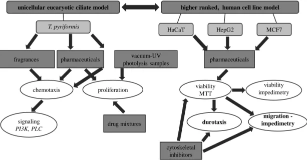
The most important experiments and investigations conducted during our work are summarized in Figure 1.
unicellular eucaryotic ciliate model higher ranked, human cell line model
HepG2 MCF7
HaCaT T. pyriformis
fragrances
signaling PI3K, PLC
pharmaceuticals
chemotaxis
vacuum-UV photolysis samples
proliferation
drug mixtures
pharmaceuticals
viability MTT
viability impedimetry
migration - impedimetry durotaxis
cytoskeletal inhibitors
Fig. 1: Overview of the applied model systems and most important biological assays
7
8
Methods
Chemical substances studied
The 7 esters used by the industry as food additives or fragrances in personal care products were synthesized and chemically characterized at the Research Group of Peptide Chemistry at the Eötvös Loránd University of Sciences.
The remaining 4 tastant aromatic aldehydes, the 14 frequently detected aquatic contaminant pharmaceutical as well as the 7 cytoskeleton inhibitors used during the durotaxis experiments were purchased from Sigma-Aldrich Ltd.
Vacuum-UV photolysis samples from the degradation of diclofenac were generated and chemically characterized at the Department of Inorganic and Analytical Chemistry and the Research Group of Environmental Chemistry of the University of Szeged.
Model cells
During the first phase of our work experiments were conducted using the nonpathogenic free living freshwater ciliate Tetrahymena pyriformis GL. The choice of this model organism was motivated by its practical advantages (e.g. relatively short life cycle of 150 min.), its ecological relevance as well as by its significant homology with cells of higher ranked organisms (e.g. in terms of receptors).
In the second phase of our work, we used 3 human cell lines that are well established toxicological models of different tissues/organs. The studied cell lines were: i) the HaCaT spontaneously transformed keratinocyte; ii) the HepG2 hepatocellular carcinoma cell line; iii) the MCF7 mammary gland carcinoma cell line. Embryonic mouse fibroblast 3T3 cell line was used as reference in the durotaxis assay.
Proliferation inhibition of the ciliate T. pyriformis
The proliferation inhibiting effect of the frequently detected pollutant pharmaceuticals and diclofenac vacuum-UV photolysis samples was evaluated using the cell number as assay endpoint following 24 h of exposure. Cell number was determined by the impedimetric CASY TT cell counter and analyzer system (Innovatis-Roche).
Chemotaxis assay
The chemotactic responses of the ciliate model were quantified using a two chamber capillary assay modified by the Chemotaxis Research Group. In this setup, the inner
9
chamber containing the test substance consisted in the sterile tips of a multichannel pipette, whereas the outer chamber containing the cell suspension consisted in the wells of a sterile 96-well plate. Incubation time was 20 min. as optimized by previous simulations ran by our Research Group.
Study of the intracellular signaling in T. pyriformis
We studied the activation of two key signaling molecules the phospholipase-C (PLC) and the phosphatidyl-inositol-3-kinase (PI3K) upon chemoattractant stimuli. PLC activation was quantified using immunocytochemistry and detected in flow cytometer (FACS-Calibur, Beckton-Dickinson). The contribution of PI3K to the signaling of chemotaxis was measured indirectly using two specific inhibitors: wortmannin and LY29004.
Colorimetric cell viability measurement in cell lines
Cytotoxic effect of aquatic contaminant pharmaceuticals and cytoskeletal inhibitors towards cell lines was evaluated using two colorimetric viability assays: the MTT (3- (4,5-dimetyl-tiazol-2-yl)-2,5-difenyl-tetrazolium bromide) method based on the reducing activity of mitochondrial dehydrogenases and the AlamarBlue method based on the intracellular reduction of resazurin blue.
Impedimetric cell viability and migration assay
The impedimetric detection of cell adhesion and migration is based on the electric insulator property of the intact cell membrane due to which adhesion of cell onto the surface of an electrode is accompanied by the increase of impedance in an alternating current field. Cell viability measurements were conducted in the xCELLigence SP (Roche) whereas cell migration studies took place in ECIS1600 (Applied BioPhysics) and ECIS Z (Applied BioPhysics) equipments. Within these devices two different experimental setups were used: the wound healing and the ―electric fence‖ options respectively. While in the former case we introduced a wound into a confluent cell layer by electrical means and followed the wound closure, in the case of the later method we prevented cell growth on the surface of the measuring electrode and registered cell migration onto the electrode after the inactivation of the ‖electric fence‖.
10 Durotaxis assay
We studied durotaxis using apparent stiffness gradient containing polyacrylamide hydrogels. Briefly, hydrogel was polymerized on the surface of a topographically patterned rigid glass substrate resulting in gel areas with different heights and consequently varying apparent stiffness. Evaluation of the stiffness dependent distribution of cells pretreated with cytoskeletal inhibitors or pharmaceuticals was performed using light microscopy (Zeiss LSM-510).
Statistics
Statistical evaluation of the data was performed using the OriginPro8® software.
Significance of the encountered difference between results of treated and untreated cells was determined using one-way ANOVA. Concentration-response curves were fitted using the four-parameter logistic function.
Results
Unicellular ciliate model
1. Firstly, the aim of our work was to analyze the sensitivity of the chemotactic response towards slight changes in the molecular structure. For this end we used 7 esters and 4 aromatic aldehydes showing high structural similarity. On the other hand, we also investigated the potential overlaps between the chemotactic profiles of the esters and the reagents used for their synthesis that can contaminate in trace amounts the esters.
Our results underpinned the high sensitivity of the chemotaxis of T. pyriformis regarding the subtle changes in the molecular structure. Chemotactic profile of isomer fragrances showed considerably similarity in the case of both C5H10O2 isomers and C7H14O2 isomers (e.g. the attractant peaks detected at 10-9 M and 10-6 M for both isoamyl-acetate and isobutyl-propionate or the attractant character of methyl-butyrate and methyl-isobutyrate at 10-9 M). However, some fundamental differences could also be observed between the chemotactic behaviors within each group of isomers (e.g. the repellant character of the isobutyl-proprionate at 10-12 M compared to the attractant nature of isoamyl-acetate at the same concentration). These findings were in accordance with results reported previously by our Research Group as T. pyriformis chemotaxis was found to be molecule-specific in the case of the 20 L-amino acids as well.
11
Similarly to what was mentioned above, comparison of the chemotactic profile of the esters with that of the respective reagents revealed some similarities but also considerable differences. This suggests the potential tracer contaminant reagents do not significantly influence chemotactic profile of a given ester.
2. Based on the above results we continued our work by stimulating T. pyriformis cells with adequate concentrations of chemoattractant esters in order to investigate activation of two key enzymes in the signaling of chemotaxis: phospholipase-C and phosphatidyl- inositol-3-kinase. Even though molecules involved in the chemosensory signaling pathway of the T. pyriformis and those involved in signaling events occurring in olfactory epithelia show high degree of homology, the role of the two studied enzymes in olfaction is still unclear.
According to our results stimulation of cells with chemoattractant esters did not resulted in significant activation of the PLC. Furthermore chemotactic response was not mediated by the activation of PI3K. These findings suggest that chemosensory signal transduction process elicited by these esters is not conveyed by phosphatidyl-inositol second messengers but other molecules such as c-AMP. Literature data also reported the c-AMP dependent signaling of these esters in rodent olfactory neurons.
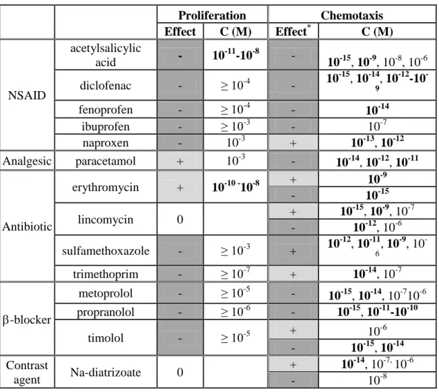
3. In the next step our work was focused on the cell physiological effects of 14 frequently detected aquatic contaminant pharmaceuticals. Our aim was predominantly to compare the sensitivity and predictive power of the classical toxiciological endpoint cell proliferation and the less regularly used migratory behavior. Available literature is abundant concerning acute toxicity of these drugs towards the most common ecotoxicological model organisms such as Vibrio fischeri, Daphnia magna or algal species. However, data are scars at the protozoon level or regarding the impact of these substances on the migratory behavior. Moreover, T. pyriformis is a highly relevant test species which is widely used in quantitative structure-activity relationship (QSAR) studies. In turn, this later is the predominantly applied approach for the environmental risk assessment of pollutants when experimental ecotoxicological data are missing.
Our results suggest that proliferation inhibiting effect of the investigated drugs towards T. pyriformis in the aquatic environment is unlikely as only acetylsalicylic acid exhibited significant (yet mild) proliferation inhibiting potential in the environmental
12
concentration range (Table 1.). The weak proliferation enhancing effect of the erythromycin at 10-10 - 10-8M is most probably not relevant from an ecotoxicological point of view.
On the contrary, chemotaxis results demonstrated that 13 from the 14 tested drugs were able to induce significant sublethal impact and alter migratory response in the environmental concentration range (emphasized by bold letters in Table 1.). This was in accordance with previous studies reporting outstanding sensitivity of the migratory behavior as a toxicological endpoint.
Table 1. Comparison of the proliferation and chemotaxis altering effect of the 14 drugs Proliferation Chemotaxis
Effect C (M) Effect* C (M)
NSAID
acetylsalicylic
acid - 10-11-10-8 -
10-15, 10-9, 10-8, 10-6 diclofenac - ≥ 10-4 - 10-15, 10-14, 10-12-10-
9
fenoprofen - ≥ 10-4 - 10-14
ibuprofen - ≥ 10-3 - 10-7
naproxen - 10-3 + 10-13, 10-12
Analgesic paracetamol + 10-3 - 10-14, 10-12, 10-11
Antibiotic
erythromycin + 10-10 -10-8 + 10-9
- 10-15
lincomycin 0 + 10-15, 10-9, 10-7
- 10-12, 10-6 sulfamethoxazole - ≥ 10-3 + 10-12, 10-11, 10-9, 10-
6
trimethoprim - ≥ 10-7 + 10-14, 10-7
-blocker
metoprolol - ≥ 10-5 - 10-15, 10-14, 10-710-6 propranolol - ≥ 10-6 - 10-15, 10-11-10-10
timolol - ≥ 10-5 + 10-6
- 10-15, 10-14 Contrast
agent Na-diatrizoate 0 + 10-14, 10-7, 10-6
- 10-8
―-―: chemorepellent; ―+‖: chemoattractant
Despite of its excellent sensitivity -because of the obtained non monotonic, multi-peak concentration-response curves- we found chemotaxis might be valuable as a qualitative rather than quantitative tool in the field of ecotoxicology. The multi-peak allure of the concentration-response curve can be explained by hormesis which is a general
13
phenomenon in toxicology. It was reported for various model organisms from different levels of phylogeny and with a wide range of endpoints. Although the underlying molecular mechanisms still need elucidation, it is supposed to be related to the simultaneous presence of low and high affinity receptor populations responding to the same ligand. Moreover saturation of chemotaxis receptor may also contribute to the non monotony of the concentration-response curves as under- or oversaturation of receptors can result in decreased chemotaxis.
4. In the aquatic environment pollutant pharmaceuticals do not occur individually but as part of a complex mixture. Thus in the following phase of our work we studied combined effects of 4 drugs in all the 6 possible binary mixtures. Selected drugs belonged to the 2 most toxic prescription classes: the NSAIDs (diclofenac and ibuprofen) and -blockers (metoprolol and propranolol). Our aim was to verify validity of the concept of concentration addition commonly used for the prediction of mixture toxicity based on the knowledge of individual effective concentrations. To this end, we compared the observed mixture toxicity to the sum of the individual proliferation inhibitions of the two mixture constituents at their respective actual concentrations.
Our results showed that concentration additivity could predict the mixture toxicity with acceptable precision in about 1/3 of the 96 combinations studied. In the remaining mixtures antagonism was the predominant type of interaction, the prevalence of which increased with the total drug concentration. Moreover, antagonism was significantly more frequent in the mixtures composed of drugs acting on the same molecular targets.
Synergism was observed in 7 of the 16 combination containing diclofenac and metoprolol. These finding suggest that the concept of concentration addition which could predict combined effect of these drugs on commonly used test species such as D.
magna or L. minor cannot be generalized to T. pyriformis. On the other hand, the observed additive and synergistic combined actions show that significant biological action can take place at low concentrations where individual effects would be negligible. Consequently possible mixture effects should be taken into consideration when assessing environmental risk of these molecules.
5. In our closing experiments with the ciliate model we applied the above described proliferation inhibition – chemotaxis assay combination to support the optimization of
14
the operating conditions (irradiation time, O2-saturation of the applied atmosphere) used during the vacuum-UV photolysis of the diclofenac.
Our results confirmed the adequacy of the suggested assay combination in the monitoring of the vacuum-UV photolysis as significant differences were detected in the biological activity of the samples taken at different time points as well as between samples taken using O2-saturated or O2-deprived condition. Moreover results of the proliferation inhibition assay were in agreement with data obtained by chemical analysis. Accordingly, until 300 s irradiation time the slight proliferation inhibiting potency (13%) of untreated samples increased to about 25% for the samples taken using O2-saturated condition, whereas it decreased to almost 0% for samples taken using O2- deprived condition. This might be explained by the significantly higher amount of the major aromatic byproduct 5-hydroxi-diclofenac formed in O2-saturated condition.
Significant difference could also be observed in the case of samples from 2400 – 3600 s irradiation time, as proliferation inhibiting potential of O2-saturated samples decreased from about 30% to almost 0%. On the contrary, proliferation inhibition exerted by O2- deprived samples stagnated around 20%. This finding may be correlated to the more efficient transformation of organic byproducts into inorganic molecules (mineralization) achieved using O2-saturated condition (approximately 70 % after 3000 s of irradiation).
Using O2-deprived condition in turn mineralization remained as low as 20-25 %.
In addition to the above results chemotaxis data confirmed that the mixture of the degradation products retained the chemorepellent character of the parent compound diclofenac. Furthermore, sublethal migratory behavior modifying effects could be observed even in the samples taken after 3600 s of irradiation using O2-saturated condition, where proliferation inhibition capacity became insignificant. Anyhow, for the deeper understanding of the obtained chemotactic responses further investigations with the individual degradation products are necessary.
Human cell lines
6. In the second phase of our work we continued our experiments with 3 human cell lines that are considered as validated toxicological models. In the beginning we used the common colorimetric MTT method to evaluate cytotoxic effect of the 14 pharmaceuticals. Then we also monitored the cytotoxicity of the three most potent drugs
15
in an impedimetric device in real-time. Previous studies comparing the impedimetric viability assay with the MTT method reported good correlation with the two techniques as well as a higher sensitivity achieved with the former assay. Moreover, impedance based techniques furnish additional information concerning the kinetics and the irreversibility of the cytotoxic effect.
Results of the MTT assay performed with the three cell lines demonstrated that the cell lines were less sensitive than the ciliate model; most probably because of the smaller relative exposure surface of the former cells. Cell lines responded with higher sensitivity only in the case of the antibiotic erythromycin and the analgesic paracetamol.
In addition, the ciliate and the HepG2 cell line turned out to be equally sensitive to the NSAID diclofenac and fenoprofen. Similarly to the results obtained with T. pyriformis, propranolol proved to be the most toxic pharmaceutical for all three cell lines. Literature explains this enhanced toxicity with the strong membrane stabilizing potency of propranolol that the other drugs lack. Obtained EC50 values were in the most cases independent of the exposure time applied (24 h, 48h, 72h) which suggests that the pharmaceuticals are rapidly taken up by the cells and metabolized into inactive products. Two exceptions were observed with diclofenac (HaCaT) and fenoprofen (MCF7) where EC50 values after 72h were about 50% of the values at 24h. This raises the possibility that these molecules are transformed intracellularly into more potent metabolites.
Comparison of the impedance based viability detection and the MTT assay confirmed a good agreement of the two methods regarding the relative toxicity of the studied compounds (metoprolol<diclofenac<propranolol). On the other hand in accordance with literature data a 1.5-2 fold better sensitivity was obtained using the impedimetric technique. Nevertheless, significant modification of the cell adhesion capacity was not observed when drugs were used at environmental concentrations (<10-8M).
7. As T. pyriformis chemotaxis assay showed that most pharmaceuticals were able to alter cell migration at environmentally relevant concentrations we continued our work with similar studies using the three human cell lines. Firstly, we used the ―electric fence‖ option of the ECIS device in order to characterize cell adhesion and migratory behavior of the cell lines and measure cell migration influencing capacity of three
16
selected pharmaceuticals (diclofenac, metorpolol and propranolol) at the highest environmentally relevant concentration (10-8 M).
Based on the obtained impedimetric curves and microscopic images taken from the surface of the electrodes HaCaT and MCF7 cell lines were suitable models for the use of the ―electric fence‖ setup and the impedance based cell migration measurements. In the case of HepG2 cells in turn, because of their growth in multilayered islands, no confluent cell layer was established. Consequently no significant increase in the impedance was observed following inactivation of the ―electric fence‖.
Concerning the capacity of selected pharmaceuticals at environmental concentration to alter cell migration, we found that only propranolol could significantly modify the migration of cells. Namely, it slowed down slightly the migration of HaCaT cells.
Literature survey showed that reported in vitro cell migration modulating effect of the studied drugs occurred at considerably higher concentrations (10-5-10-4 M) that are in the range of therapeutic concentrations.
8. In the last phase of our research we studied a recently described type of migratory responses, the durotaxis which is the directed movement of cells in response to the stiffness gradient encountered in their microenvironment. It plays an important role in various in vivo processes such as tissue development, wound healing or tumor metastasis formation. There are an increasing number of pathological conditions (e.g.
tumors, myocardial infarct) in which alteration from the physiological tissue stiffness was demonstrated. However, it is still unclear whether changing of the matrix elasticity is an etiological factor or a consequence of the disease. On the other hand, it was also described that some tumorous cell types (e.g. MCF7) exhibit rigidity-dependent sensitivity towards antineoplastics. This raises the possibility that responsiveness towards environmental pollutants might also be dependent on the elastic properties of the cells’ environment (e.g. surface used for cell culturing).
The results of our experiments conducted with HaCaT and MCF7 cell lines showed that the former cell type was able to respond with durotaxis to changes in the apparent stiffness of the applied hydrogels and preferentially moved the gel areas having higher apparent stiffness. On the contrary, MCF7 cells remained randomly distributed between gel areas with lower or higher apparent stiffness. The responsiveness of two cell types
17
was not dependent on the chemical treatment (poli-D-lysine or fibronectin) of the gel surface that was used to promote cell adhesion. On the other hand gel height (<15 m or
>50 m) had determining effect on the durotactic response in the case of the HaCaT keratinocytes: durotaxis occurred only on the thin gels but not on the thick ones.
Literature data are currently contradictory regarding the order of magnitude (1 - >10
m) of distance over which cells are able to detect or probe changes in the matrix stiffness. Our data point rather towards the approximately 10 m scale. Nevertheless, the distance through which cells can sense matrix elastic properties is also dependent on the strength of the pulling forces exerted by the cells to the gel which in turn depend on the cell type. The unresponsiveness of MCF7 is most probably due to their tumorous origin as it was demonstrated that mouse mammary cancer cells with increasing metastatic potential gradually lost their stiffness sensitivity. This is reasonable since metastatic cells must pass across many different tissue types in vivo with varying elastic properties. Though the integral process is far from being fully understood, this observation might considerably contribute to both the understanding of the pathogenesis of tumors and the development of more efficient antitumor therapies.
9. Among the numerous open questions related to the phenomenon of durotaxis one of the most important ones concerns the role of the different cytoskeletal components (e.g.
F-actin, microtubulue, myosin II). Thus in the next step we used specific inhibitors in order to investigate the contribution of the above mentioned cytoskeletal elements to the durotactic response. Considering that cytoskeletal inhibitors are highly cytotoxic and can noticeably affect cell adhesion or migratory capacity we also monitored changes in cell viability, adhesion and migration upon treatment with cytoskeletal inhibitors.
According to our results all the 7 molecules applied could inhibit durotaxis of both HaCaT keratynoctes and the reference 3T3 mouse fibroblasts at concentrations lower than the minimal cytotoxic concentrations. On the other hand, results of the impedimetric measurements confirmed that cell adhesion and migration of both cell lines were also affected at the lowest drug concentrations causing complete inhibition of durotaxis. Comparison of the two cell types showed lower sensitivity of the HaCaT cells in terms of all four physiological responses (viability, durotaxis, cell adhesion and migration). One possible explanation might be the extremely strong adhesion potential
18
of these cells as well as the fact that cells grow in tightly arranged sheets. Available literature data also suggest the participation of all three cytoskeletal elements in the durotactic response; however results are controversial considering the role of the individual components. It seems generally accepted that microfilaments and myosin II are involved in the generation of traction forces whereas the contribution of microtubules to force generation is disputed. A currently evoked hypothesis is that the involvement of microtubules might be cell type dependent.
10. Our final experiments focused on the study of selected clinically and environmentally relevant pharmaceuticals (diclofenac, ibuprofen, metoprolol and propranolol) and their capacity to inhibit durotaxis.
Our results showed that the two non-steroidal anti-inflammatory drugs (diclofenac and ibuprofen) were not able to inhibit durotaxis at any concentration tested. The two - blockers in turn inhibited durotaxis at the 1-10 M (3T3), or 10-100 M (HaCaT) concentration range. On the other hand cell adhesion was not significantly altered by any of the four drugs. Cell migration was modified only by diclofenac which slowed cell migration of 3T3 fibroblasts but accelerated that of the HaCaT keratinocytes. Since available literature is very scars about the impact of these drugs on durotaxis interpretation of our results is a challenging task. Thus further investigations are indispensable. Minimal durotaxis inhibiting concentration of metoprolol and proranolol differed with one order of magnitude for both cell types. In every case propranolol was the more potent agent, which might be related to its strong membrane stabilizing capacity mentioned earlier. Nevertheless, the differing -receptor selectivity of the two drugs might also be a source of the different biological potency. Metoprolol is a selective antagonist of the1-adrenergic receptor the expression level of which is lower in the different cell types of skin compared to the 2 type receptors. Propranolol in turn is a nonselective inhibitor that might explain the lower effective concentrations for both cell lines. Anyhow, with the above detailed experiments we described for the first time that environmental pollutants can alter durotactic behavior even if the observed durotaxis inhibitory concentrations were at least two order of magnitude higher than environmental concentrations.
19
Conclusions
Using isomer fragrances we confirmed the high sensitivity of the chemotaxis of T.
pyriformis regarding subtle changes in the molecular structure of the chemotactic ligand.
We demonstrated that proliferation inhibiting effect of frequently detected aquatic contaminant pharmaceuticals towards T. pyrifromis in the environment is unlikely.
Using binary drug mixtures we attested that additive and more than additive (synergistic) interactions occurred between pharmaceuticals underlying the importance of taking mixture effects into account when assessing environmental risk of pharmaceuticals.
We demonstrated that the tested drugs were able to significantly affect chemotactic behavior of the ciliate at environmental concentrations. Thus chemotaxis measurement might be a useful qualitative assay in order to assess sublethal biological effects.
The above mentioned ciliate bioassay combination was successfully applied to support technological optimization of the vacuum-UV photolysis of diclofenac.
Results of the proliferation inhibition test were in agreement with data obtained by chemical analysis.
With our viability studies on human cell lines we confirmed that results of the MTT assay and the impedimetric method showed good correlation. Moreover the later technique exhibited better sensitivity.
We showed that HaCaT keratinocyte and MCF mammary carcinoma cell lines were adequate subjects for impedance based cell migration measurements. On the other hand, from the selected drugs only propranolol was able to alter cell migration at an environmental concentration.
We characterized different mechanosensitivity of the HaCaT and MCF7 cell lines.
Using the reference 3T3 mouse fibroblast cell line and the HaCaT keratinocyte we demonstrated that inhibition of F-actin, microtubules and myosin II result in decreased durotaxis, as well as altered cell adhesion and migratory capacity.
We described durotaxis modifying capability of two clinically and environmentally relevant -blockers in the therapeutic concentration range.
20
Summary
Cell migration is a crucial physiological process in unicellular as well as in multicellular organisms and also a sensitive behavioral response elicited by external physical and chemical cues. This allows to study the cell biological effects of anthropogenic environmental pollutants in models from different levels of the phylogeny.
Firstly, we compared proliferation and chemotactic responses of the freshwater ciliate T.
pyriformis using odorants and frequently detected aquatic contaminant pharmaceuticals.
For each group of substances chemotaxis proved to be a highly sensitive response in terms of both slight changes in the molecular structure and the low effective concentrations. However, because chemotactic response curve showed hormesis, proliferation inhibiting capacity could better quantify cellular effects of drugs and showed better predictive power. Based on this later we studied combined effect of pharmaceutical mixtures in which we described additive and synergistic interactions underlying the importance of taking into consideration the eventual mixture effects when assessing environmental risk of human pharmaceuticals. Then we applied our methods in practice to find optimal operating parameters of vacuum-UV photolysis used for the degradation of diclofenac.
In the next step we evaluated cytotoxic effect of drugs on human cell lines and compared the sensitivity of the conventional mitochondrial dehydrogenase based MTT assay with the impedimetric viability detection. We concluded that this later allowed for a more sensitive detection and the relative toxicity of the tested drugs showed good correlation between the two methods. We also used a new setup for the impedimetric measurement of migration influencing effect of drugs and concluded that only propranolol could alter cell migration significantly at environmental concentration.
Finally, using an innovative durotaxis measurement platform we characterized the differing mechanosensitivity of the human keratinocyte and the mammary carcinoma cell lines. We also performed quantitative description of the durotaxis inhibiting effect of cytoskeleton inhibitors as well as the screening of clinically and environmentally relevant NSAIDs and -adrenergic antagonists. In the case of -blockers we could first demonstrate the durotaxis inhibiting capacity of environmental pollutants.
21
Bibliography of publications
Papers related to the theme of the PhD thesis:
1. Láng, J., Rákász, V., Magyar, A., Pállinger, É., Kőhidai, L. (2011) Chemotactic effect of odorants and tastants on the ciliate Tetrahymena pyriformis. J. Recept. Sig.
Transd. 31 (6):423-433 IF=1.588
2. Láng, J., Kőhidai, L. (2012) Effects of the aquatic contaminant human pharmaceuticals and their mixtures on the proliferation and migratory responses of the bioindicator freshwater ciliate Tetrahymena. Chemosphere 89 (5):592-601 IF=3.137
3. Arany, E., Láng, J., Somogyvári, D., Láng, O., Alapi T., Ilisz, I., Gajda-Schrantz, K., Dombi, A., Kőhidai, L., Hernádi, K. (2014) Vacuum-ultraviolet photolysis of diklofenák and the effect of the treated aqueous solutions on the proliferation and migratory responses of Tetrahymena pyriformis. Sci. Tot. Environ. 468-469: 996- 1006 IF2012=3.258
Other papers:
1. Láng, O., Illyés, E., Menyhárd D.K., Láng, J., Sebestyén, F., Hudecz, F., Kőhidai, L.
(2012) Chemotaxis induced by SXWS tetrapeptides in Tetrahymena—overlapping chemotactic effects of SXWS sequences and their identical amino acids. J. Mol.
Recogn. 25 (1):24-31 IF=3,006
2. Mező, G., Láng, O., Jakab, A., Bai, K.B., Szabó, I., Schlosser, G., Láng, J., Kőhidai, L., Hudecz, F. (2006) Synthesis of oligotuftsin based branched oligopeptide conjugates for chemotactic drug targeting (CDT) J. Pept. Sci. 12:328-336 IF=1,801