The Rockefeller University Press $30.00
Neutrophils (also known as neutrophilic gran- ulocytes or polymorphonuclear leukocytes [PMNs]) are the most abundant white blood cells in the human circulation. They play a crucial role in the immune defense against bacterial and fungal pathogens, and they also participate in the development of the inflammatory reac- tion (Nathan, 2006). Although neutrophils are mostly viewed as playing a beneficial role to the host, their improper activation may also lead to tissue damage during an autoimmune or exag- gerated inflammatory reaction (Nathan, 2006;
Németh and Mócsai, 2012).
Neutrophils participate in antimicrobial host defense both as the first line of innate im- mune defense and as effectors of adaptive im- munity. They are short-lived cells that usually die while performing their antimicrobial func- tion. Because their primary role is the localiza- tion and elimination of invading microorganisms at any expense, a simplistic view of neutrophils being not more than “dumb” suicide killers has prevailed for a long time.
A major wave of discoveries during the 1990s and early 2000s made immunologists begin to appreciate the amazing complexity and sophisti- cation of neutrophil functions. It became evident
that neutrophils release cytokines and contrib- ute to orchestrating the immune/inflammatory response (Bazzoni et al., 1991a,b; Cassatella, 1995). A highly sophisticated machinery di- recting neutrophil migration (Ley et al., 2007;
Nourshargh et al., 2010) and a surprising com- plexity of neutrophil granules (Borregaard et al., 2007) also begun to emerge. Novel but contro- versial concepts about how neutrophils may fight microbes, including possible regulation of granule enzyme function by ion fluxes (Reeves et al., 2002) and formation of neutrophil extra- cellular traps (NETs; Brinkmann et al., 2004) were also proposed during that period. Both mechanisms have been subject to intense de- bate (see next section). Nevertheless, all those studies indicated that neutrophils use highly sophisticated and complex mechanisms to per- form their role in immune defense and in- flammation and led to improved and refined models of neutrophil biology (Witko-Sarsat
CORRESPONDENCE A.M.: mocsai.attila@
med.semmelweis-univ.hu
Diverse novel functions of neutrophils in immunity, inflammation, and beyond
Attila Mócsai
A. Mócsai is at the Department of Physiology, Semmelweis University School of Medicine, 1094 Budapest, Hungary.
Neutrophils have long been considered simple suicide killers at the bottom of the hierarchy of the immune response. That view began to change 10–20 yr ago, when the sophisticated mechanisms behind how neutrophils locate and eliminate pathogens and regulate immunity and inflammation were discovered. The last few years witnessed a new wave of discoveries about additional novel and unexpected functions of these cells. Neutrophils have been proposed to participate in protection against intracellular pathogens such as viruses and mycobacteria. They have been shown to intimately shape the adaptive immune response at various levels, including marginal zone B cells, plasmacytoid dendritic cells and T cell popu- lations, and even to control NK cell homeostasis. Neutrophils have been shown to mediate an alternative pathway of systemic anaphylaxis and to participate in allergic skin reactions.
Finally, neutrophils were found to be involved in physiological and pathological processes beyond the immune system, such as diabetes, atherosclerosis, and thrombus formation.
Many of those functions appear to be related to their unique ability to release neutrophil extracellular traps even in the absence of pathogens. This review summarizes those novel findings on versatile functions of neutrophils and how they change our view of neutrophil biology in health and disease.
© 2013 Mócsai This article is distributed under the terms of an Attribution–
Noncommercial–Share Alike–No Mirror Sites license for the first six months after the publication date (see http://www.rupress.org/terms). After six months it is available under a Creative Commons License (Attribution–Noncommercial–Share Alike 3.0 Unported license, as described at http://creativecommons.org/licenses/
by-nc-sa/3.0/).
The Journal of Experimental Medicine
on November 24, 2015jem.rupress.orgDownloaded fromNeutrophil functions: state of the art in the early 2000s A tremendous amount of knowledge on neutrophils had been generated by the end of the 20th century and during the first years of the 2000s. The most crucial aspects of that knowl- edge, which forms the basis of our understanding of neutro- phil biology, are briefly summarized in the following section and in Fig. 1.
Neutrophils are terminally differentiated cells that develop in the bone marrow where they are also stored for release into the circulation (Borregaard, 2010). During their maturation, neutrophils develop several intracellular compartments (secre- tory granules and vesicles) that store proteins critical for the antimicrobial and proinflammatory missions of the cell (Borregaard et al., 2007). Once released into the circulation, neutrophils begin to seek signs of infection and inflammation which lead to a series of events culminating in the migration of neutrophils through the vessel wall and inside the tissue to the site of microbial invasion (Ley et al., 2007; Nourshargh et al., 2010; Fig. 1).
When encountering infectious particles, neutrophils begin a professional antimicrobial killing program. The infectious particle will be phagocytosed (Nordenfelt and Tapper, 2011) and various noxious agents, including reactive oxygen species generated through the activity of the NADPH oxidase (Leto and Geiszt, 2006), and the contents of intracellular granules (Borregaard et al., 2007; Soehnlein, 2009) are then released into the phagosome, resulting in killing and digestion of the microorganisms (Fig. 1).
Besides the well-established role of phagocytosis, two additional mechanisms contributing to neutrophil-mediated antimicrobial responses have been proposed during the early et al., 2000; Nathan, 2006; Mantovani et al., 2011; Amulic et al.,
2012; Fig. 1).
The last few years have borne witness to another wave of discoveries of novel and unexpected roles of neutrophils in di- verse aspects of immunity and inflammation, as well as beyond the traditional scope of the immunological sciences. Those in- clude novel roles of neutrophils in immunity against intracellu- lar pathogens such as viruses and intracellular bacteria; shaping of adaptive immunity at different levels; and roles in disease states previously not associated with neutrophils, such as allergy and anaphylaxis, metabolic diseases, atherosclerosis, or throm- bus formation. Those cases also revealed novel potential roles for NETs beyond antimicrobial functions. Many of those studies exploited approaches allowing the antibody-mediated deple- tion of neutrophils by anti-Gr1 or the more neutrophil-specific anti-Ly6G antibodies in mice (Daley et al., 2008), or genetic manipulations leading to the partial or complete genetic dele- tion of the neutrophil lineage (Liu et al., 1996; Karsunky et al., 2002; Hock et al., 2003; Jonsson et al., 2005; Ordoñez-Rueda et al., 2012). Though those novel studies have yet to pass the scrutiny of the scientific community, they suggest that neutro- phils may have a much more widespread role in immunity, in- flammation, and beyond than previously anticipated.
After a brief summary of established and well-known (though sometimes debated) neutrophil functions, this review will summarize the novel biological functions of neutrophils revealed in the last few years, within and beyond immunity and inflammation. I will primarily focus on what neutrophils do rather than how they do it. Readers will be referred to ad- ditional recent reviews on related subjects such as certain aspects of the molecular mechanisms of neutrophil functions.
Figure 1. Neutrophil functions: state of the art in the early 2000s. After migrating to the site of inflammation, neutrophils (PMN) phagocytose and digest the invading mi- crobes; release NETs, which likely trap bacte- ria; and produce cytokines, which contribute to the inflammatory reaction. Once infection is cleared, neutrophils die by apoptosis and trigger an active program to resolve inflam- mation. Inset, pathogen killing inside the phagosome occurs by ROS generated by the NADPH oxidase, as well as by granule en- zymes released from intracellular granules.
The NADPH oxidase also induces depolariza- tion of the phagosomal membrane, which may be required for providing optimal envi- ronment inside the phagosome.
on November 24, 2015jem.rupress.orgDownloaded from
neutrophil-derived NETs were able to capture HIV-1 particles and reduce their efficiency to infect T cells. Most importantly, when neutrophils and T cells were co-cultured, neutrophils were able to reduce the infection efficiency of HIV-1. Phar- macological studies indicated that NET formation was de- pendent on TLR7 and TLR8 in endocytic compartments and that the effect of neutrophil-derived NETs on T cells could be inhibited by DNase treatment, underscoring the role of extracellular DNA. In agreement with those findings, the neutrophil-mediated inhibition of HIV-1 infectivity on T cells required TLR7 and TLR8, and it was suspended by DNase treatment.
Another recent study provided additional evidence for the role of neutrophils and NETs in antiviral host defense (Jenne et al., 2013). Systemic injection of viral TLR ligands or poxvirus infection led to dramatic accumulation of neu- trophils in liver sinusoids that formed aggregates with plate- lets and released NETs inside the vessels. Importantly, DNase treatment increased the percentage of liver cells infected with poxviruses, suggesting that extracellular DNA exerts a protec- tive effect against viral infection (Jenne et al., 2013).
Although the aforementioned studies suggest a role for neutrophils and NETs in antiviral host defense, they also raise several questions. Most studies on NET formation by neutro- phils indicate a critical role for NADPH oxidase activity in that process (including the original NET study [Brinkmann et al., 2004] where NET formation was triggered by phorbol ester-induced activation of neutrophils) and it is difficult to envisage how viral particles would be able to trigger such a strong stimulation of neutrophils. However, because NET formation in the absence of NADPH oxidase activity has recently been shown to occur under milder and more physi- ological conditions (Chen et al., 2012; Parker et al., 2012b), it may be possible that even viral particles trigger NET forma- tion. In addition, Jenne et al. (2013) used systemic administra- tion of LPS to induce protection from viral infection. It is unclear whether that response induces specific activation of neutrophils and whether the DNase treatment used only affects neutrophil-derived NETs. Nevertheless, though addi- tional studies in the field are warranted, these two studies (to- gether with additional ones showing priming of virus-specific T cells by neutrophils; Duffy et al., 2012) open up new ave- nues on the role of neutrophils in the host defense against viral pathogens.
Neutrophils in infection with intracellular pathogens The traditional view is that neutrophils eradicate extracellular bacteria and fungi, whereas intracellular pathogens such as mycobacteria are primarily dealt with by macrophages.
Although prior studies indicated possible roles for neutrophils during mycobacterial infection (Appelberg, 2007; Lowe et al., 2012), recent studies provided important novel insights into the role of neutrophils in the fight against intracellular patho- gens. Analysis of healthy human individuals who have been in contact with tuberculosis patients revealed strong inverse correlation between the risk of tuberculosis infection and 2000s, both of them triggering intensive scrutiny and debate
from the scientific community. Anthony Segal’s group pro- posed an interesting molecular interplay whereby ion fluxes triggered by the NADPH oxidase would promote the anti- microbial function of granule proteins (Reeves et al., 2002).
Though many components of the proposed mechanism (such as the electrogenicity of the NADPH oxidase [Fig. 1, inset]
providing driving force for other ion currents; Geiszt et al., 1997; Schrenzel et al., 1998; Rada et al., 2005) are generally accepted, it is still debated whether and to what extent ion fluxes that compensate for the activity of the NADPH oxidase contribute to the antibacterial effects of granule enzymes (Geiszt et al., 2001; Rada et al., 2004; Rada et al., 2005;
Femling et al., 2006; Essin et al., 2009). Another interesting and similarly provocative idea was that, in addition to intra- phagosomal killing, neutrophils may also be able to expel their nuclear content (including DNA) complexed with granule proteins into the extracellular space and that this complex (NETs) may inhibit microbial growth without phagocyto- sis of the pathogen (Brinkmann et al., 2004; Fig. 1). Though neutrophil-derived extracellular DNA has long been known to be present at the site of infection (in part as a component of pus formation) and NET formation has recently been directly visualized during bacterial infection in vivo (Yipp et al., 2012; McDonald et al., 2012), the contribution of NET formation to antibacterial host defense and killing is still under intensive debate (Brinkmann and Zychlinsky, 2012; Menegazzi et al., 2012; Nauseef, 2012; Parker et al., 2012a).
Neutrophils are short-lived cells that die within a limited time after entering the circulation (Geering and Simon, 2011).
In the absence of infection or inflammation, they die by a spontaneous apoptosis program (Geering and Simon, 2011;
Witko-Sarsat et al., 2011), likely within 1 d (although some in- vestigators propose this time to be up to 5 d; Pillay et al., 2010).
Inflammatory signals are capable of prolonging the lifespan of the cells by several days, during which they release inflam- matory mediators and contribute to the orchestration of the inflammatory response (Mantovani et al., 2011; Fig. 1).
However, even under those conditions, neutrophils will soon die by apoptosis (or, possibly, NETosis), contributing to pus formation inside infected tissues. Once neutrophils die, another program for removal of dead neutrophils by macro- phages is initiated (Bratton and Henson, 2011). Parallel to the inflammatory response, biochemical and transcriptional changes leading to the resolution of the inflammation begin (Serhan, 2007; Serhan et al., 2008; Soehnlein and Lindbom, 2010), and this is further promoted by engulfment of the corpses of dead neutrophils by macrophages (El Kebir and Filep, 2010).
The role of neutrophils in antiviral host defense
Whereas neutrophils are critical for immunity against bacte- rial and fungal pathogens, their role in antiviral host defense is much less appreciated. In a recent study, Saitoh et al. (2012) proposed a role for NETs in the control of HIV-1 infection.
HIV-1 triggered NET formation by human neutrophils and
on November 24, 2015jem.rupress.orgDownloaded from
uncertain whether IL-10, the cytokine proposed to be re- sponsible for most of the reported findings in mice (Zhang et al., 2009), is also released by human neutrophils (Davey et al., 2011).
The latest twist in the role of neutrophils in tuberculosis comes from a recent study on zebrafish (Yang et al., 2012). By monitoring mycobacterial infection, Yang et al. (2012) found that neutrophils did not interact with the microbes during the very first phase of infection but engulfed dying infected macrophages during early granuloma formation and killed the surviving intracellular pathogens. That study suggests that efferocytosis (the engulfment of dying cells) not only serves to clean up the site of infection but also contributes to containing the infection, at least in the case of mycobac- terial challenge.
Another unexpected role for neutrophils in infectious diseases has been revealed by a recent study on malaria patho- genesis (Porcherie et al., 2011). The authors showed that genetic deficiency of IgE- or FcRI-protected mice from cerebral malaria and death, but not parasitemia after infection with Plasmodium falciparum parasites. Interestingly, FcRI was dramatically up-regulated on circulating neutrophils after parasitic infection and FcRI-positive neutrophils became the dominant FcRI-positive leukocyte population in the brain. Importantly, depletion of neutrophils using the NIMP- R14 anti-Gr1 antibody protected mice both from neurological signs of cerebral malaria and from malaria-induced death, but not from parasitemia, suggesting an important role for neutrophils in malaria pathogenesis (Porcherie et al., 2011).
Although the mechanism whereby neutrophils contribute to cerebral manifestations of malaria is incompletely understood, the authors suggested that production of proinflammatory cytokines (e.g., IFN-) by neutrophils accumulating in the brain of infected animals leads to excessive inflammation and subsequent neurological signs that are characteristic of cerebral malaria.
Collectively, the aforementioned studies suggest that neutrophils may have a much wider (protective or some- times pathogenic) role during infectious diseases than previ- ously appreciated.
Neutrophils in the regulation of B and T cells
Neutrophils have long been seen as effector cells that have little influence on shaping the adaptive immune response.
That view is being challenged by several recent reports.
Marginal zone B cells are unique B cells of the spleen that are located outside the germinal centers and close to the marginal sinus, thus closely positioned to the blood circula- tion. Their primary function is the rapid initiation of T cell–
independent antibody responses to blood-borne microbes.
Interestingly, a prior study revealed that neutrophils are one of the major cell types responsible for capturing and transporting circulating bacteria to the splenic marginal zone in mice (Balázs et al., 2002). Additionally, prior human studies showed that ac- tivated (but not resting) neutrophils express the B cell–activating cytokine BAFF (also known as BLyS; Scapini et al., 2003) and peripheral blood neutrophil counts (Martineau et al., 2007).
The authors also showed that ex vivo growth of mycobacteria in whole blood was dramatically increased upon neutrophil depletion, whereas neutrophil-derived antimicrobial peptides inhibited mycobacterial growth from in vitro bacterial cul- tures (Martineau et al., 2007). Another recent study identified a unique blood transcriptional signature in human tuberculo- sis patients that closely matched a putative type I and type II interferon-induced gene expression signature in neutrophils (Berry et al., 2010). This signature was clearly distinguishable from those in other diseases characterized by a major neutro- phil component (such as Gram-positive bacterial infections) or by a type I IFN signature (such as SLE). Those two studies suggested that neutrophils actively fight against mycobacteria and may provide protection from infection.
A possible explanation for those findings is that neutro- phils fight mycobacteria during and immediately after their entry into the organism. However, this may only be true for the initial contact with mycobacteria when neutrophils are able to prevent infection of the host, while macrophages “take over” when the bacilli reach intracellular compartments (Lowe et al., 2012). Indeed, neutrophils likely play an oppo- site, pathogenic role during later stages of the disease, through at least two different mechanisms. First, neutrophils may serve as vectors for the systemic dissemination of the pathogens.
Mouse neutrophils are able to acquire live mycobacteria at the site of infection and carry those microbes to the draining lymph nodes by migration within the lymphatic vasculature (similar to the migration of DCs in a general immune response;
Abadie et al., 2005). Although this may promote antimicrobial host defense, an alternative scenario is that mycobacteria that escape killing and survive inside neutrophils use them as
“Trojan horses” to help disseminate the bacteria to distant organs. This would be similar to the situation in leishmaniasis when neutrophils engulf parasites at the site of infection and deliver them to macrophages, which take up live Leishmania parasites during phagocytosis of apoptotic neutrophils (van Zandbergen et al., 2004; Peters et al., 2008; John and Hunter, 2008). The second mechanism for the contribution of neu- trophils to the chronic phase of tuberculosis may be related to their role in infection-induced tissue damage. Cruz et al.
(2010) suggested that excessive release of IL-17 during re- peated mycobacterial exposure leads to neutrophil recruit- ment and neutrophil-mediated tissue damage in mice. This response appears to be attenuated by IFN- production dur- ing a normal antimycobacterial immune response (Nandi and Behar, 2011), indicating that neutrophil accumulation and the resulting tissue damage may be caused by the failure of the immune system to contain the infectious agents.
The dual role of neutrophils in tuberculosis is also re- flected in another recent study, suggesting that neutrophils down-regulate inflammation at early stages of mycobacterial infection but contribute to inflammation and mycobacterial clearance during chronic tuberculosis in experimental mice (Zhang et al., 2009). The importance of those findings in human pathology has yet to be assessed, in part because it is
on November 24, 2015jem.rupress.orgDownloaded from
mice than in normal specific pathogen–free animals, indicat- ing that microbial products, likely originating from the com- mensal flora, play an important role in the accumulation of splenic marginal zone neutrophils. Importantly, human pa- tients suffering from various congenital defects of neutrophil development or function had normal total circulating B cell numbers but strongly reduced numbers of circulating mar- ginal zone B cells, and their serum levels of antibodies against T cell–independent antigens were significantly reduced de- spite normal levels of antibodies against T cell–dependent antigens. Collectively, the elegant study of Puga et al. (2011) revealed a potentially important immunological and immune- homeostatic mechanism whereby splenic neutrophils may facilitate the antibody response of marginal zone B cells to T cell–independent microbial antigens. This mechanism re- quires the reprogramming of circulating neutrophils by micro- bial signals, as well as the neutrophil-derived BAFF, APRIL, and IL-21 cytokines. Further studies will be required to de- termine the full importance of this mechanism in immune defense and to more closely define the nature and develop- mental stage of the proposed B cell helper neutrophil popula- tion relative to their circulating counterparts.
The role of neutrophils in the regulation of T cell func- tion appears to be more complex and less well understood.
Regulation of T cells by myeloid cells primarily occurs via that neutrophils are a major source of the BAFF-related,
B cell–stimulating cytokine APRIL in B cell lymphomas (Schwaller et al., 2007) and mucosal plasma cell niches (Huard et al., 2008; Scapini et al., 2008). A recent work by Puga et al.
(2011) revealed an intricate relationship between neutrophils and marginal zone B cells and proposed that this interaction mediates T cell–independent antibody responses through BAFF and APRIL (Fig. 2 A). The authors found a large number of neutrophils around the splenic marginal zone in humans, macaques, and mice. In mice, splenic but not circu- lating neutrophils were able to strongly activate marginal zone B cells, whereas they had limited stimulatory effect on follicular B cells and even inhibited CD4+ T cells. Importantly, splenic neutrophils promoted B cell survival, antibody production, class switching to IgG and IgA, and somatic hypermutation, indicating that they qualify to be bona fide B helper cells.
Splenic, but not circulating, neutrophils express large amounts of BAFF, APRIL, and IL-21, and all those cytokines were required for the stimulatory effect on marginal zone B cells.
Circulating neutrophils acquired the ability of activating mar- ginal zone B cells upon exposure to splenic sinusoidal endo- thelial cells in the presence of microbial signals in vitro. The colonization of the splenic marginal zones by neutrophils pri- marily occurred soon after birth in humans, and marginal zone neutrophils were significantly less abundant in germ-free
Figure 2. Novel interactions of neutro- phils with other immune cells. (A) Neutro- phils (PMN) accumulate in the splenic marginal zone upon microbial challenge and facilitate the antibody production and matu- ration of marginal zone B cells (MZB) in re- sponse to T cell–independent (TI) antigens.
(B) Neutrophils participate in a vicious cycle during autoimmune disease (e.g., SLE) patho- genesis by releasing NETs that trigger pDCs to release IFN-, which then promotes antibody production by B cells (B). (C) Neutrophils may directly present antigens to T cells (T) and they also activate DCs and deliver antigens to them. (D) Neutrophils and DCs jointly activate NK cells (NK).
on November 24, 2015jem.rupress.orgDownloaded from
polarization of T cells (Bennouna and Denkers, 2005; van Gisbergen et al., 2005b; Megiovanni et al., 2006). The inter- action between human neutrophils and DCs has been pro- posed to be at least in part mediated by a unique interaction between DC-SIGN and Mac-1 on the neutrophil surface (van Gisbergen et al., 2005a,b; Boudaly, 2009; Yang et al., 2009; Fig. 2 C).
Recent studies have also revealed another unique role of neutrophils in DC activation during the pathogenesis of autoimmune diseases such as systemic lupus erythematosus (SLE) or type I diabetes (T1D). Characteristic features of SLE, an immune complex–mediated systemic autoimmune disease characterized by autoantibodies against nuclear antigens, include the chronic activation of plasmacytoid DCs (pDCs) leading to constitutive secretion of type I interferons such as IFN- (Obermoser and Pascual, 2010) and the presence of circulating low-density granulocytes reminiscent of immature neutrophils (Bennett et al., 2003; Kaplan, 2011). Two recent studies on human patients shed novel light on the role of pDCs, type I interferons, and neutrophils in the core patho- genesis of SLE (Lande et al., 2011; Garcia-Romo et al., 2011;
Fig. 2 B). SLE neutrophils were more prone to cell death by NETosis and to release NETs into the extracellular environ- ment. Those NETs contained self-DNA in complex with antimicrobial proteins including LL37 (the C-terminal peptide derived from the human cathelicidin hCAP-18) and were able to activate pDCs through the TLR9 pathway and to trigger type I interferon production. This led to activation of B cells that produced autoantibodies against both self-DNA and the antimicrobial peptides. Type I interferons and immune com- plexes triggered further activation of neutrophils releasing more NETs (Lande et al., 2011), establishing a vicious cycle at the core of SLE pathogenesis (Fig. 2 B). Low-affinity Fc- receptors (such as the human FcRIIA) and TLR7 were re- quired for immune complex-induced neutrophil activation (Garcia-Romo et al., 2011). FcRIIA may also participate in the uptake of DNA-containing immune complexes by pDCs (Båve et al., 2003), followed by recognition of DNA- antimicrobial peptide complexes by the TLR9 pathway (Garcia-Romo et al., 2011; Lande et al., 2011). It should be noted that pDCs do not recognize self-DNA under non- pathological conditions. However, the presence of LL37 in the DNA complexes in NETs prevented degradation of DNA, leading to delivery of DNA to early endosomes of pDCs, where it could activate the TLR9 pathway (Lande et al., 2007; Lande et al., 2011). Therefore, the unique composition of NETs makes them a suitable candidate to trigger pDC ac- tivation in the core of SLE pathogenesis. These results are also in line with a proposed contribution of NET degrada- tion defects to SLE pathogenesis (Yasutomo et al., 2001;
Hakkim et al., 2010), as well as a previous study on the presence of NET formation in autoimmune small vessel vasculitis (Kessenbrock et al., 2009).
The role of neutrophils in pDC activation and develop- ment of autoimmune diseases has also recently been proposed in the pathogenesis of T1D, which is also thought to involve antigen presentation by DCs or macrophages, supported by
the migration of antigen-loaded DCs to the draining lymph nodes. Interestingly, human and mouse neutrophils also ap- pear to be able to carry antigens to the lymph nodes by migrating in lymphatic vessels in a CCR7-dependent manner (Abadie et al., 2005; Maletto et al., 2006; Beauvillain et al., 2011). A recent study also showed that vaccinia virus-specific CD8+ T cells emerge in the bone marrow and, based on fluor- escence tracking and Ly6G antibody-mediated depletion approaches, identified neutrophils as the cells transporting the antigen from the dermis to the bone marrow (Duffy et al., 2012). Neutrophils also express molecules required for anti- gen processing and presentation to T cells (Ashtekar and Saha, 2003; Beauvillain et al., 2007). Late precursors committed to the neutrophil lineage are also important precursors of DC development (Oehler et al., 1998). Furthermore, mouse neu- trophils are able to cross-prime naive T cells, i.e., present ex- tracellular antigens on MHC class II molecules to CD8+ T cells (Beauvillain et al., 2007), and also prime antigen-specific Th1 and Th17 cells (Abi Abdallah et al., 2011). The transport of antigens from the periphery to the site of T cell priming and the direct priming/activation of antigen-specific T cells in an MHC-restricted manner raise the possibility that neu- trophils may possibly even perform some level of classical antigen-presenting cell function (Fig. 2 C).
Although the aforementioned studies suggest that neu- trophils positively regulate antigen-specific T cell responses, several works indicate a negative role for the neutrophil lin- eage in T cell function. As mentioned above, splenic neutro- phils negatively regulate CD4+ T cell function in mice (Puga et al., 2011). A subset of human neutrophils also inhibits T cell responses during acute systemic inflammatory reactions such as bacterial sepsis (Pillay et al., 2012). Using Ly6G antibody- mediated depletion and G-CSF receptor-deficient mice, a recent study showed that neutrophils prevent the spread of the T cell response to distal lymph nodes during immuniza- tion (Yang and Unanue, 2013). In addition, a large number of immature myeloid cells with inhibitory functions on T cells and other immune function, termed myeloid-derived sup- pressor cells (MDSCs), can be observed in cancer patients and in various other infectious or inflammatory conditions (Gabrilovich and Nagaraj, 2009). A subset of those cells (so-called granulocytic MDSCs) share many characteristics with (immature) neutrophils, highlighting the possibility of nega- tive regulation of T cell function by the neutrophil lineage.
Regulation of DCs by neutrophils
The effect of neutrophils on adaptive immunity is not only mediated by direct effects on B and T cells, but also by affect- ing the function of DCs. Indeed, mouse neutrophils are capa- ble of, and required for, the activation of DCs during microbial infection (Bennouna et al., 2003; Charmoy et al., 2010).
Human and mouse studies suggest that neutrophils directly interact with DCs, which may lead to various responses in- cluding DC maturation and activation, access to antigens cap- tured by neutrophils, or DC-mediated proliferation and TH1
on November 24, 2015jem.rupress.orgDownloaded from
before and a functional cooperation between the two cell types had been proposed (Costantini and Cassatella, 2011). The most detailed study tested the role of neutrophils in Legionella pneumophila infection in mice, aiming to understand why neutrophils, which are not capable of killing Legionella species themselves, still play a role in clearing L. pneumophila infection (Spörri et al., 2008). The authors confirmed that depletion of neutrophils by anti-Gr1 antibodies attenuated host defense against Legionella but it also blocked IFN- production by NK cells. Using elegant genetic approaches and mixed bone marrow chimeric studies, they showed that IFN- pro- duction by NK cells was directly stimulated by IL-18 released from neutrophils in response to Legionella infection (Spörri et al., 2008), suggesting that interactions between neutrophils and NK cells may be of functional importance (Fig. 2 D).
A recent study has provided novel and unexpected genetic evidence for the role of neutrophils in regulating NK cell homeostasis and function (Jaeger et al., 2012). The authors set up a random chemical mutagenesis screen to identify novel mutant mouse strains with defective NK cell development and function. They identified a mutation called Genista, which led to hyporesponsive NK cells as indicated by reduced in vitro and in vivo responses to target cells and ligation of NK cell–activating receptors (Jaeger et al., 2012).
Interestingly, adoptive transfer studies revealed hyporespon- siveness of wild-type NK cells transferred to Genista recipi- ents, indicating that the NK cell phenotype was, at least in part, caused by an environmental, non–cell autonomous defect. In parallel studies, the Genista mutation was identified to be a hypomorphic missense mutation of the Gfi-1 transcrip- tion factor, leading to defective terminal differentiation of neutrophils (as indicated by the absence of fully mature neu- trophils in the periphery) without negatively affecting T or B cell lineages or other myeloid (monocyte, macrophage, DC) compartments (Ordoñez-Rueda et al., 2012; Jaeger et al., 2012). This was in line with the similar, though more severe, defect in neutrophil maturation in mice with complete dis- ruption of the Gfi-1–encoding gene (Karsunky et al., 2002;
Hock et al., 2003) and congenital neutropenia in human pa- tients carrying a dominant–negative Gfi-1 mutation (Person et al., 2003). To further confirm the role of neutrophils in NK cell function, the authors showed that depletion of neutrophils by an anti-Ly6G antibody also reduced the func- tional responsiveness of NK cells (Jaeger et al., 2012). Further studies revealed that both the Genista mutation and the Ly6G antibody-mediated depletion led to defective terminal matu- ration and poor survival of NK cells. The authors also re- ported that patients suffering from various forms of congenital or acquired (autoimmune) neutropenia had higher percent- ages of NK cells showing an immature phenotype and reduced responsiveness to target cells (Jaeger et al., 2012). Interest- ingly, one of the patients with autoimmune neutropenia showed a remission during the course of the study, during which normalization of peripheral neutrophil count and NK cell function occurred in parallel. Collectively, those results suggest that neutrophils likely play a role in regulating the type I interferon production by pDCs (Diana et al., 2013).
Diana et al. (2013) reported early infiltration of the pancreas of young nonobese diabetic (NOD) mice with neutrophils, B-1a B cells, and pDCs. Pancreatic -cell death triggered production and release of anti-DNA IgG antibodies from B-1a cells, which formed DNA–anti-DNA immune com- plexes and triggered, supposedly in an Fc-receptor–dependent manner, neutrophil NET formation and the release of the murine cathelicidin peptide CRAMP. The DNA–anti-DNA immune complexes and CRAMP then activated pDCs through TLR9 to release IFN-, which is proposed to trigger further T cell–mediated autoimmune processes. In agreement with the proposed role of neutrophils in the development of T1D, injection of 2-wk-old NOD mice with the NIMP- R14 anti-Gr1 antibody reduced later development of diabe- tes in those animals (Diana et al., 2013).
The aforementioned studies suggest an unexpected role for neutrophils in very early phases of autoimmune disease pathogenesis through triggering of type I interferon produc- tion by pDCs. There are, however, several open questions that need to be addressed. First, it is widely believed that NET formation requires the NADPH oxidase (Fuchs et al., 2007);
therefore SLE pathogenesis would be expected to rely on the presence of this enzyme. However, deficiency of the NADPH oxidase component Nox2 exacerbated, rather than inhibited disease pathogenesis in lupus-prone mice (Campbell et al., 2012), arguing against the role of NETs in SLE patho- genesis. This issue is further complicated by NADPH oxidase- independent NET formation proposed by other recent studies (Chen et al., 2012; Parker et al., 2012b). Second, although Fc-receptor polymorphisms have been shown to be associ- ated with human SLE (Takai, 2005), no such associations have been reported for T1D (even though NOD mice lacking activating Fc-receptors are partially protected from diabetes development; Inoue et al., 2007). Therefore, the role of the aforementioned mechanism in diabetes development may not be entirely relevant to human patients. Third, because NETs are likely often formed during bacterial infections (Yipp et al., 2012; McDonald et al., 2012), it is unclear how autoimmunity is avoided under normal healthy conditions. Nevertheless, the aforementioned mechanism provides an attractive model for the role of neutrophils in autoimmune disease pathogenesis that will merit further investigation. A similar mechanism has also been proposed during development of atherosclero- sis (see The role of neutrophils in atherosclerosis), and NETs have also been suggested to participate in the pathogenesis of rheumatoid arthritis by displaying citrullinated auto- antigens in an inflammatory environment (Khandpur et al., 2013), further extending the importance of such a pathway in human diseases.
Neutrophils in the regulation of NK cell homeostasis
NK cells are innate lymphocytes with critical roles in the elimination of virus-infected and tumor cells. In vitro inter- actions between neutrophils and NK cells were described
on November 24, 2015jem.rupress.orgDownloaded from
and that this component is sufficient to trigger a full-blown anaphylaxis response. Additional experiments indicated that the neutrophil-dependent anaphylactic reaction was medi- ated primarily by PAF rather than histamine (Jönsson et al., 2011). Importantly, active systemic anaphylaxis could be re- stored by adoptive transfer of human neutrophils to FcR/
animals (Jönsson et al., 2011) or by transgenic expression of human FcRIIA in FcR/ mice (Jönsson et al., 2012), sug- gesting that human neutrophils may also be able to mediate systemic anaphylactic reactions.
The various forms of skin allergies comprise a major group of localized allergic diseases. As with other allergic diseases, their mechanism can also be divided into two con- ceptually different phases, the sensitization phase (leading to priming of the adaptive immune system) and the elicita- tion phase (causing local tissue damage through activation of the primed adaptive immune cells). They are also often com- plicated by additional inflammatory insults, such as chemi- cally induced tissue damage in allergic contact dermatitis or mechanical damage caused by itching-induced scratching in atopic dermatitis. Several recent studies indicate an im- portant role for neutrophils in skin allergies. Anti-Gr1 anti- body treatment has been known to prevent ear swelling and T cell infiltration during contact hypersensitivity, a mouse model of allergic contact dermatitis (Dilulio et al., 1999). In a recent study, neutrophils expressing the LTB4-receptor BLT1 were found to be required for inflammation and re- cruitment of allergen-primed T cells at the allergen challenge site in a novel mouse model of atopic dermatitis combining ovalbumin immunization and challenge with mechanical skin injury (reminiscent of itching-induced scratching; Oyoshi et al., 2012).
Both of the aforementioned studies revealed important roles for neutrophils in the elicitation phase of allergic skin inflammation. However, although innate immune mecha- nisms likely also play important roles in DC activation and T cell priming during the earlier and conceptually different sensitization phase of allergic skin diseases (Kaplan et al., 2012), no studies on the role on neutrophils in that phase have been completed yet. Interestingly, our own recent studies (unpublished data; published in abstract form in Weber et al., 2012) suggested an important role for neutrophils in the sensitization phase of contact hypersensitivity. In that study, we found that Ly6G antibody-mediated depletion of neutro- phils, selectively during the sensitization phase, abrogated the contact hypersensitivity reaction, and diminished DC activation and priming of allergen-specific T cells. In addition, lymph node cells from allergen-sensitized mice that were previously treated with neutrophil-depleting anti-Ly6G anti- bodies or were genetically deficient of neutrophils because of a myeloid-specific deletion of the Mcl-1 protein (Dzhagalov et al., 2007) were unable to transfer sensitivity toward con- tact allergens to normal naive recipients (Weber et al., 2012). Therefore, neutrophils appear to be critical players in both the sensitization and elicitation phases of allergic skin inflammatory reactions.
terminal differentiation and functional responsiveness of NK cells (Fig. 2 D). The colocalization and direct physical inter- action of the two cell types in mouse lymphoid organs (Jaeger et al., 2012) and inflamed human tissues (Costantini et al., 2011) suggest that neutrophils may directly regulate the NK cell compartment.
Although the aforementioned studies indicate an impor- tant role for neutrophils in the regulation of NK cell function, the contribution of a third cell type (such as DCs) in this interaction cannot be excluded. Indeed, besides neutrophil- derived IL-18, IFN- production by NK cells upon Legionella infection also required IL-12 derived from DCs as a result of their reciprocal regulation with NK cells (Spörri et al., 2008;
Fig. 2 D). A tripartite complex formed by neutrophils, NK cells and a minor subset of DCs (slanDCs) has also been ob- served in human samples (including inflammatory lesions in Crohn’s disease and psoriasis) and was proposed to promote IFN- production by NK cells (Costantini et al., 2011).
Collectively, neutrophils have recently emerged as poten- tially important regulators of NK cell homeostasis and activa- tion, possibly in close relationship with DCs.
Neutrophils in allergy and anaphylaxis
Allergic and anaphylactic reactions are adaptive immune re- sponses triggered by repeated exposure to allergens, and are typically characterized by vascular reactions such as vasodila- tion and edema formation. Several recent studies suggest un- expected roles for neutrophils in those reactions.
The prevailing paradigm of anaphylaxis development is that allergen-induced cross-linking of IgE molecules bound to Fc-receptors on mast cells and basophils triggers hista- mine release and subsequent systemic vasodilation (Khan and Kemp, 2011). Although this mechanism is likely responsible for the majority of anaphylactic reactions, several observations indicate that alternative mechanisms, possibly mediated by IgG and cellular lineages other than mast cells and basophils, may also be at play (Finkelman, 2007; Tsujimura et al., 2008;
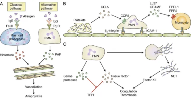
Ohnmacht et al., 2010). Jönsson et al. (2011) identified neu- trophils as important players of active and IgG-induced passive systemic anaphylaxis in experimental mice (Fig. 3 A).
Passive systemic anaphylaxis triggered by IgG antibodies was blocked by neutrophil depletion using the RB6-8C5 anti- Gr1 antibody. Active systemic anaphylaxis was partially reduced by anti-Gr1 antibodies alone, and it was nearly completely blocked by simultaneously depleting both neu- trophils and basophils (Jönsson et al., 2011). Interestingly, mice lacking all activating Fc- and Fc-receptors except FcRIV were able to mount normal active anaphylactic reactions which were, however, completely blocked by anti-Gr1 anti- bodies. Adoptive transfer of wild-type neutrophils rescued anaphylaxis development in FcR -chain-deficient animals (which lack all activating Fc- and Fc-receptors including FcRIV, and fail to mount active systemic anaphylaxis). Those results suggest that neutrophils are responsible for the FcRIV- mediated component of active systemic anaphylaxis in mice
on November 24, 2015jem.rupress.orgDownloaded from
perforin-mediated translocation of granzymes), it is unclear how this would happen in case of neutrophil elastase and how elastase would selectively cleave IRS-1 without wreaking havoc inside the target cell. The problem of protease-mediated pro- tein degradation during lysis of neutrophil-containing bio- logical samples is also well known in the scientific community;
therefore more careful studies to exclude the possibility of IRS-1 degradation during sample preparation are warranted.
Another study mentioned earlier suggests a role for neu- trophils in the pathogenesis of T1D, an autoimmune disease caused by degradation of pancreatic -cells (Diana et al., 2013). In that study, neutrophils were shown to participate in the activation of type I cytokine production by pDCs and in subsequent activation of the adaptive immune response (Diana et al., 2013). That study places neutrophils high in the hierarchy of autoimmune disease pathogenesis, participating in the shaping of the adaptive immune response.
Collectively, although the studies on the role of neutro- phils in diabetes and related metabolic diseases may open up new directions of research that may significantly improve our understanding of the inflammatory origin of metabolic dis- eases, they also raise several additional questions and concerns that have yet to be addressed.
The role of neutrophils in atherosclerosis
Cardiovascular diseases are among the most prevalent human diseases and are the leading causes of death in developed countries. Several recent studies suggest that neutrophils play an important role in those diseases, in particular the develop- ment and progression of atherosclerosis and the pathogenesis of thrombosis.
The role of neutrophils in metabolic diseases
Metabolic diseases such as diabetes and obesity are among the largest health challenges of the 21st century. Several recent stud- ies suggest unexpected roles for neutrophils in those diseases.
Obesity and the resulting insulin resistance is one of the major causes of metabolic syndrome and type 2 diabetes. It has long been known that obesity has an important inflammatory component and that this contributes to the resulting insulin re- sistance (Gregor and Hotamisligil, 2011). The primary cell type responsible for inflammation in obesity has been considered to be the inflammatory macrophage in adipose tissues (Olefsky and Glass, 2010). However, neutrophils have also been shown to be recruited to adipose tissues early during the course of diet-induced obesity in mice (Elgazar-Carmon et al., 2008) and to be present in increased numbers in the fatty liver of obese human patients (Rensen et al., 2009). A recent report has confirmed the dramatic accumulation of adipose tissue neutrophils, as well as the increased presence and activity of the neutrophil-specific protease neutrophil elastase, during high-fat diet in experimental mice (Talukdar et al., 2012).
Importantly, the pharmacological inhibition or genetic deletion of neutrophil elastase led to improved in vivo glucose tolerance and insulin sensitivity, likely due to improved responsiveness of liver and adipose tissue cells to insulin. The authors proposed that neutrophil elastase gains access to the intracellular space (endocytic compartments) of hepatocytes and degrades insulin receptor substrate 1 (IRS-1), leading to attenuation of insulin signal transduction pathways (Talukdar et al., 2012; similar to a mechanism previously proposed to occur in lung cancer cells; Houghton et al., 2010). Although there is precedent for translocation of proteases through cell membranes (such as
Figure 3. Novel functions of neutrophils in vascular diseases. (A) Neutrophils (PMN) mediate an alternative pathway of anaphylactic reaction which is distinct from the classical pathway mediated by mast cells or basophils. (B) Sequential recruitment of platelets, neutrophils, and monocytes (Mono) participates in the pathogenesis of atherosclerosis. (C) Neutrophils contribute to coagulation and thrombosis by releasing tissue factor and inhibi- tors of the tissue factor pathway inhibitor (TFPI), as well as the procoagulant NETs.
on November 24, 2015jem.rupress.orgDownloaded from
adhesion is likely mediated by interaction between 2-integrins and ICAM-1.
It should be mentioned that another recent study on vas- cular healing after wire injury, a mouse model of the vascular injury triggered by insertion of an arterial stent, placed the role of neutrophils in a different perspective (Soehnlein et al., 2011). Ly6G antibody-mediated depletion of neutrophils or genetic deficiency of CRAMP inhibited endothelial re- generation and promoted neointima formation (the major cause of arterial restenosis) after vessel injury, whereas LL37 promoted endothelial recovery in an in vitro wound-healing assay (Soehnlein et al., 2011). Most importantly, covalent coating of arterial stents with LL37 significantly reduced in- stent stenosis (Soehnlein et al., 2011). Those results suggest that neutrophils and neutrophil-derived cathelicidins may play a protective role during vascular healing after stent-mediated vascular damage and raise the possibility that LL37 treatment may reduce in-stent stenosis after stent insertion.
The aforementioned studies suggest potentially important roles for neutrophils in atherosclerosis and related vascular diseases. However, additional work will be required to delin- eate the exact mechanism of how and in which phase and spatiotemporal order neutrophils participate in plaque forma- tion. For example, do neutrophils get activated inside or outside the vasculature? How do intravascular events promote plaque formation under the endothelium? Are the roles of neutro- phils in initial monocyte adhesion and eventual plaque de- stabilization related or independent events? Although plaque formation and stent restenosis likely occur through different mechanisms, the opposing effect of neutrophils and CRAMP/
LL37 on those two endothelial damage-associated processes may also require additional mechanistic explanation. The role of CCR5 in neutrophil activation also raises questions, as this receptor is not expressed on neutrophils at high levels (although increased expression was shown upon apoptosis and at the site of inflammation; Ariel et al., 2006; Hartl et al., 2008).
Neutrophils in thrombus formation
Arterial or venous thrombosis is another major complication of vascular diseases, and causes significant morbidity and mortality worldwide. Several recent studies suggest that neu- trophils may play a major role in triggering thrombosis in experimental mice.
Neutrophils were found to be the first cell type to adhere to the site of endothelial injury in both arterial and venous thrombosis models (Darbousset et al., 2012; von Brühl et al., 2012), and their accumulation at the site of vessel injury (along with that of monocytes) preceded the activation of platelets (Darbousset et al., 2012). Importantly, thrombus formation was dramatically reduced upon depletion of neu- trophils by anti-Ly6G antibodies in a flow restriction–induced venous thrombosis model (von Brühl et al., 2012) or by anti-Gr1 antibodies in laser-induced microvascular injury (Darbousset et al., 2012). Whereas contact of plasma proteins with extra- vascular tissue factor is the primary trigger of thrombus for- mation upon vessel injury, the source of prothrombotic agents It has long been appreciated that atherosclerosis results
from chronic inflammation of the vascular wall (Hansson, 2005). Recent reports indicate that neutrophils are present in unique areas of atherosclerotic plaques in both humans and mice (Naruko et al., 2002; Ionita et al., 2010; Drechsler et al., 2010; Rotzius et al., 2010) and that their presence correlates with poor prognosis and signs of plaque destabilization in human patients (Naruko et al., 2002; Ionita et al., 2010).
Importantly, depletion of neutrophils by antibodies against Ly6G strongly reduced the area of atherosclerotic lesions in apolipoprotein E-deficient (ApoE/) mice fed a high-fat diet for 4 wk, suggesting an important role for neutrophils in atherosclerotic plaque formation (Drechsler et al., 2010).
A recent study provided additional support for the role of neutrophils in atherosclerosis, showing that the genetic defi- ciency of CRAMP, a member of the cathelicidin family of antimicrobial peptides expressed in neutrophils, leads to re- duced atherosclerotic lesion size in ApoE/ mice fed a high- fat diet (Döring et al., 2012a). CRAMP and the homologous human cathelicidin peptide LL37 (the C-terminal portion of its precursor hCAP-18) promoted the adhesion of classical (inflammatory) monocytes to the vessel wall in mice, and this was inhibited by antagonists of formyl-peptide receptors, the receptors thought to be responsible for cathelicidin-mediated leukocyte migration (Döring et al., 2012a). In addition, acti- vation of pDCs by complexes of cathelicidins with extracel- lular DNA from neutrophils or other dying cells may also contribute to atherosclerosis development in mice (Döring et al., 2012b) in a manner similar to that proposed for the pathogenesis of SLE and T1D (Lande et al., 2011; Garcia- Romo et al., 2011; Talukdar et al., 2012). The recruitment of neutrophils to the inflamed mouse arterial wall could also be reduced by platelet depletion or by the genetic deletion of CCR5, the receptor for the platelet-derived CCL5 (RANTES) chemokine (Drechsler et al., 2010) which is also required for atherosclerotic plaque formation (Braunersreuther et al., 2007). The release and deposition of CCL5 appears to be de- pendent on P-selectin on platelets (Huo et al., 2003; Drechsler et al., 2010). Platelets were previously proposed to participate in atherosclerotic plaque formation (Massberg et al., 2002;
Huo et al., 2003) and platelet-derived CCL5 was shown to promote monocyte adhesion to the luminal surface of early atherosclerotic lesions in the inflamed arterial wall in mice (von Hundelshausen et al., 2001; Huo et al., 2003). Mice lack- ing P-selectin, CD18, or ICAM-1 were previously shown to have reduced atherosclerotic plaque formation (Johnson et al., 1997; Nageh et al., 1997; Collins et al., 2000). Collectively, atherosclerotic plaque formation likely involves the sequential recruitment of platelets, neutrophils, and monocytes, whereby platelet-derived CCL5, acting on CCR5, is at least partially responsible for neutrophil recruitment and neutrophil pro- motion of monocyte recruitment by cathelicidin-induced activation of formyl-peptide receptors (Fig. 3 B). Platelet P-selectin is likely involved in platelet deposition (although an additional role of endothelial P-selectin in adhesion of leu- kocyte rolling cannot be excluded) and neutrophil/monocyte
on November 24, 2015jem.rupress.orgDownloaded from
Additional subjects not discussed in detail
Several topics of major importance could not be covered in detail in this review. A brief discussion below points to some of those topics, with key references for further reading.
The role of neutrophils in tumor biology has long been appreciated (Pekarek et al., 1995). There has been several re- cent developments in the field such as the polarization of tumor-associated neutrophils by TGF- (Fridlender et al., 2009), a proposed role of neutrophil elastase in tumor growth (Houghton et al., 2010), the inhibition of tumor metastasis by neutrophils (Granot et al., 2011), neutrophils in cancer- induced thrombosis (Demers et al., 2012), the role of neu- trophil-derived factors in maintaining B cell lymphoma (Schwaller et al., 2007), the potential role of neutrophils in tumor angiogenesis (Jablonska et al., 2010), and the role of histamine in preventing inflammation-induced carcinogenesis (Yang et al., 2011). Neutrophil-related immature myeloid cells called myeloid-derived suppressor cells also appear to play an immunosuppressive role in cancer (Gabrilovich and Nagaraj, 2009). The readers are referred to recent reviews for further information on this very interesting but diverse field of study (Gregory and Houghton, 2011; Fridlender and Albelda, 2012).
Besides the mostly positive roles of neutrophils in various physiological and pathological processes, neutrophils also play inhibitory or regulatory roles under certain conditions.
Apoptosis of neutrophils triggers an active process promoting resolution of inflammation (Serhan, 2007; Serhan et al., 2008;
Soehnlein and Lindbom, 2010). A subset of neutrophil- related immature myeloid cells called myeloid-derived sup- pressor cells play a critical role in regulation of inflammation in cancer and potentially other conditions (Gabrilovich and Nagaraj, 2009). Neutrophils may also be able to produce the antiinflammatory cytokine IL-10 (Zhang et al., 2009;
De Santo et al., 2010), though this is disputed in case of human neutrophils (Davey et al., 2011). Further information can be found in recent studies (Serhan et al., 2008; Gabrilovich and Nagaraj, 2009; Soehnlein and Lindbom, 2010; Mantovani et al., 2011).
Whereas many other leukocyte lineages have been subdi- vided into various subsets, neutrophils have so far been mostly considered as a homogeneous population. That view is be- ginning to change with novel functions ascribed to subpopu- lations of neutrophils (Fridlender et al., 2009; Puga et al., 2011; Pillay et al., 2012).
This review has focused on what neutrophils do rather than how they do it; therefore, most information regarding the mechanism of neutrophil function has been omitted.
There are, however, exciting new discoveries in those fields, as well, such as various new mechanisms and cues guiding the migration of neutrophils (McDonald et al., 2010; Woodfin et al., 2011; Proebstl et al., 2012; Stark et al., 2013). Two brand new and possibly related studies also propose exciting and provocative ideas about how neutrophils may fight mi- crobes: Timár et al. (2013) have recently proposed an antimi- crobial function of neutrophil-derived microvesicles, whereas during arterial or venous thrombosis with an intact endothe-
lium has been poorly understood. After some debate about the presence of tissue factor in neutrophils (Maugeri et al., 2005) it now appears that activated (but not resting) neutro- phils express and release a significant amount of tissue factor (Maugeri et al., 2006; Fig. 3 C). Importantly, neutrophil- mediated thrombus formation required tissue factor in a my- eloid compartment (von Brühl et al., 2012; Darbousset et al., 2012), and adoptive transfer studies suggested that neutro- phils themselves may be an important source of tissue fac- tor in a microvascular thrombosis model in mice (Darbousset et al., 2012).
The coagulation system is an important host defense sys- tem in lower organisms, and there is also significant inter- action between innate immunity and coagulation in higher vertebrates (Esmon, 2004). A recent study suggested that neu- trophil serine proteases play an important role in the recipro- cal coupling between innate immunity and coagulation in mice (Massberg et al., 2010; Fig. 3 C). The authors showed that the neutrophil-derived proteases neutrophil elastase and cathepsin G were required for thrombus formation after chemically induced vessel injury, as well as for activation of factor X upon co-incubation of neutrophils and platelets. The two enzymes inactivated the tissue factor pathway inhibitor protein, thereby promoting the coagulation cascade. Impor- tantly, triggering of coagulation also prevented systemic dis- semination of infectious Escherichia coli bacteria, suggesting the existence of a poorly appreciated level of innate host de- fense in mammals.
The aforementioned study also suggested that neutrophil- derived externalized nucleosomal structures (likely corre- sponding to NETs) also play a procoagulant role in thrombosis (Massberg et al., 2010; Fig. 3 C). Indeed, NETs were shown to be able to trigger platelet activation and thrombus forma- tion in vitro (Fuchs et al., 2010). A significant amount of extracellular DNA trap formation inside the thrombus could be observed upon flow restriction–induced venous thrombo- sis in mice (Brill et al., 2012; von Brühl et al., 2012) and obstruction-induced venous thrombosis in baboons (Fuchs et al., 2010). Importantly, blocking NET formation by DNase treatment strongly reduced venous thrombus formation (Brill et al., 2012; von Brühl et al., 2012). Interestingly, various malignant states, including chronic myelogenous leukemia and various solid tumors, promote NET formation, which may be an important pathogenic mechanism of cancer- associated thrombosis (Demers et al., 2012). The proposed mechanism of NET-induced thrombus formation may in- volve the activation of Factor XII by the negatively charged NET surface, with a direct effect on the stability of the fibrin network (i.e., independent of the activation of Factor XI;
von Brühl et al., 2012).
Collectively, neutrophils may contribute to the thrombo- sis and coagulation process by releasing tissue factor, even in the absence of vascular injury, by releasing serine proteases that inactivate the tissue factor pathway inhibitor, and/or by the formation of pro-coagulant NETs (Fig. 3 C).
on November 24, 2015jem.rupress.orgDownloaded from
humans appears to be similar in many sense, there are also significant differences, such as the different expression of Fc- receptors (Bruhns, 2012) the apparent lack of IL-10 production by human but not murine neutrophils (Davey et al., 2011), as well as more widespread differences in the overall immune response (Mestas and Hughes, 2004; Seok et al., 2013). There- fore, care should be taken when extrapolating mouse studies to human physiology and pathophysiology.
Concluding remarks and future directions
Neutrophils are the most abundant circulating leukocytes, yet their full spectrum of biological functions in immunity and beyond is just beginning to emerge, likely because of the dif- ficulties in manipulating this terminally differentiated, short- lived cell type. Studies published in the last few years propose that neutrophils play a much more diverse role than previ- ously appreciated. Their putative novel roles described in this review include functions in viral infections and infections with intracellular pathogens; intricate interaction with various other components of the immune system; and roles in diseases such as autoimmune diseases, anaphylaxis, diabetes, athero- sclerosis, and thrombosis. Although those studies have yet to be closely examined, scrutinized, and, hopefully, confirmed by the scientific community, they propose exciting novel avenues for the understanding of how our immune system contributes to health and disease.
The function of neutrophils in immune defense and their role in various immune-mediated (e.g., autoimmune;
Németh and Mócsai, 2012) diseases result in significant inter- est in this cell type in clinical medicine. The emerging infor- mation on what neutrophils do and how they do it may hopefully be translated to important novel approaches of the diagnosis and treatment of diseases with a substantial neutro- phil component.
I apologize for the omission of many seminal papers due to space limitation. The author thanks Erzsébet Ligeti, Marco Cassatella, Oliver Söhnlein, Zoltán Jakus, and Tamás Németh for helpful suggestions and careful reading of the manuscript.
This work was supported by the European Research Council (Starting Independent Investigator Award no. 206283) and the Wellcome Trust (International Senior Research Fellowship No. 087782).
Submitted: 1 October 2012 Accepted: 23 May 2013 REFERENCES
Abadie, V., E. Badell, P. Douillard, D. Ensergueix, P.J. Leenen, M. Tanguy, L.
Fiette, S. Saeland, B. Gicquel, and N. Winter. 2005. Neutrophils rapidly migrate via lymphatics after Mycobacterium bovis BCG intradermal vaccination and shuttle live bacilli to the draining lymph nodes. Blood.
106:1843–1850. http://dx.doi.org/10.1182/blood-2005-03-1281 Abi Abdallah, D.S., C.E. Egan, B.A. Butcher, and E.Y. Denkers. 2011. Mouse
neutrophils are professional antigen-presenting cells programmed to instruct Th1 and Th17 T-cell differentiation. Int. Immunol. 23:317–326.
http://dx.doi.org/10.1093/intimm/dxr007
Amulic, B., C. Cazalet, G.L. Hayes, K.D. Metzler, and A. Zychlinsky. 2012. Neutrophil function: from mechanisms to disease. Annu. Rev. Immunol. 30:459–489.
http://dx.doi.org/10.1146/annurev-immunol-020711-074942 Appelberg, R. 2007. Neutrophils and intracellular pathogens: beyond
phagocytosis and killing. Trends Microbiol. 15:87–92. http://dx.doi.org/
10.1016/j.tim.2006.11.009
Yipp et al. (2012) suggested that anuclear neutrophils (formed after releasing the nuclear material to NETs) preserve at least part of their antimicrobial activities. In addition, the role of neutrophils in host defense and tissue damage is mediated by diverse intracellular signal transduction processes (Futosi et al., 2013).
Potential caveats
After several years of being somewhat overlooked by immu- nologists, the last few years have witnessed an explosion of major studies on the diverse roles of neutrophils in health and disease. However, as with any trendy and fashionable subjects, it is reasonable to exercise healthy skepticism toward novel results until they are confirmed and widely accepted by the scientific community. As the aim of this review was to discuss novel findings that have not yet been subjected to detailed examination and scrutiny, this particularly applies to the re- ports discussed in this review.
Indeed, one has the feeling that some of the closely related papers discussed here followed each other at a pace that did not allow detailed reexamination of the original concepts and methodological approaches. As an example, the study propos- ing cleavage of intracellular IRS-1 by neutrophil elastase dur- ing high fat diet-induced insulin resistance (Talukdar et al., 2012) apparently relied on a previous similar report on lung cancer growth (Houghton et al., 2010) without carefully reexamining the approaches used to distinguish true intracel- lular cleavage from degradation during sample processing. That has left room for skepticism about the conclusion drawn, es- pecially because the very large amount of proteolytic activity in neutrophil lysates.
A large number of studies also propose interesting novel roles for NETs in autoimmunity, inflammation, and vascular diseases. However, there is ongoing debate about when and how NETs are formed in vivo, and whether they can indeed be generated in sufficient quantities under sterile conditions to support the proposed processes.
We should also admit that we do not have perfect tools to study the role of neutrophils in any given disease process.
Typical examples are the use of various antibodies to deplete neutrophils, of which antibodies against Gr1 (RB6-8C5 and, likely, NIMP-R14) may also deplete leukocyte lineages other than neutrophils. We also miss appropriate genetic tools to specifically delete neutrophils; indeed, none of the various approaches (Liu et al., 1996; Karsunky et al., 2002; Hock et al., 2003; Jonsson et al., 2005; Dzhagalov et al., 2007;
Ordoñez-Rueda et al., 2012) have been tested in sufficient detail to allow exclusion of an effect on other leukocyte lin- eages. Several proposed functions of neutrophils are also based on evidence gained from knockout mice lacking gran- ule proteins (e.g., elastase or CRAMP) expressed in neutro- phils. However, those proteins are also expressed in other myeloid cells such as macrophages, casting doubt about the lineage specificity of those experiments.
Finally, most of the studies discussed were performed on experimental mice. Although the physiology of mice and
on November 24, 2015jem.rupress.orgDownloaded from