A cta DV A cta DV A
dvances in dermatology and venereologyA
ctaD
ermato-V
enereologicaThis is an open access article under the CC BY-NC license. www.medicaljournals.se/acta doi: 10.2340/00015555-3707
SIGNIFICANCE
The control of cutaneous microbiota-induced immune ac- tivation in keratinocytes is key to the maintenance of epi- dermal homeostasis. Tumour necrosis factor alpha-induced protein 3, a regulator of nuclear factor kappa B signalling pathways, has important roles in these processes. It forms a negative regulatory feedback loop, controls the activity of toll-like receptor-induced signalling pathways, but is also regulated by different toll-like receptor ligands and by Cuti- bacterium acnes, a member of our skin microbiota. The levels of tumour necrosis factor alpha-induced protein 3 greatly influence levels of bacterium-induced inflammatory mediators in keratinocytes. This study found elevated ex- pression of tumour necrosis factor alpha-induced protein 3 mRNA in acne lesions. These findings suggest a general role of tumour necrosis factor alpha-induced protein 3, not only in the regulation of toll-like receptor-induced immune activation, but also in cutaneous immune homeostasis.
Human epidermal keratinocytes sense the presence of human skin microbiota through pathogen recogni- tion receptors, such as toll-like receptors, and induce innate immune and inflammatory events. In healthy epidermis there is an absence of inflammation despite the continuous presence of cutaneous microbes, which is evidence of an effective immune regulatory mecha- nism. The aim of this study was to investigate tumour necrosis factor alpha-induced protein 3 (TNFAIP3), a negative regulator of toll-like receptor and nuclear factor kappa B signalling pathways, and its role in these regulatory events. A broad spectrum of toll- like receptor ligands induced TNFAIP3 expression, as did live Cutibacterium acnes, which is involved in the pathogenesis of acne. Changes in bacterium-induced, dose-dependent TNFAIP3 expression were Jun kinase- and nuclear factor kappa B-dependent, and resulted in altered cytokine and chemokine levels in in vitro cul- tured human keratinocytes. In acne lesions, TNFAIP3 mRNA expression was elevated compared with non- lesional skin samples from the same individuals. These results suggest that TNFAIP3 may have a general role in fine regulation of microbiota-induced cutaneous im- mune homeostasis.
Key words: TNFAIP3; microbiota; Cutibacterium acnes; acne vulgaris; innate immunity.
Accepted Nov 19, 2020; Epub ahead of print Nov 26, 2020 Acta Derm Venereol 2021; 101: adv00369.
Corr: Kornélia Szabó, Department of Dermatology and Allergology, Uni- versity of Szeged, HU-6720 Szeged, Hungary. E-mail: szabo.kornelia@
med.u-szeged.hu
T
he human skin separates the body from the external environment and plays a role in the formation of a complex physical, chemical and immunological barrier (1). The skin also provides a natural habitat for the cuta- neous microbiota, a collection of microbial species living in close contact with human cells (2). Human epidermal keratinocytes are the major cellular components of skin and are in direct contact with these microbes.Keratinocytes are immunocompetent cells that can sense different external danger signals, known as pattern- and danger-associated molecular patterns (PAMPs and DAMPs), through their pattern recognition receptors
(PRRs), such as toll-like receptors (TLRs). PAMPs and DAMPs originate from human and microbial cells and induce TLR activation, as well as subsequent activation of innate immune and inflammation responses in kerati- nocytes (3, 4). In past decades, research efforts identified endogenous negative regulators of these signalling pat- hways that are able to control these processes. One such molecule is tumour necrosis factor alpha-induced protein 3 (TNFAIP3), also known as A20, which is a ubiquitin- editing enzyme involved in the inhibition of nuclear factor kappa B (NF-κB) activation and tumour necrosis factor (TNF)-α-induced apoptosis (5).
In healthy skin, the presence of the cutaneous microbiota does not lead to excessive immune activation, even though the microbes express various PAMP molecules and are continuously present on the skin. Meanwhile, human cells must retain their capacity to respond to pathogenic micro- organisms, necessitating complex regulation to maintain healthy body functions and homeostasis. Currently, little is known about the exact mechanisms by which keratinocytes are able to distinguish between pathogens and harmless members of the microbiota, tolerate the presence of resi- dential microbes, or control the immune and inflammatory events that they are able to induce.
Tumour Necrosis Factor Alpha-induced Protein 3 Negatively
Regulates Cutibacterium acnes-induced Innate Immune Events in Epidermal Keratinocytes
Lilla ERDEI1,2, Beáta Szilvia BOLLA1,2, Renáta BOZÓ2, Gábor TAX2, Edit URBÁN3, Katalin BURIÁN4, Lajos KEMÉNY1,2,5# and Kornélia SZABÓ2,5#
1Hungarian Centre of Excellence for Molecular Medicine - University of Szeged Skin Research Group (HCEMM-USZ Skin Research Group),
2Department of Dermatology and Allergology, 3Department of Public Health, 4Institute of Clinical Microbiology, University of Szeged, and
5MTA-SZTE Dermatological Research Group, Szeged, Hungary
#These authors contributed equally to this work.
A cta DV A cta DV A
dvances in dermatology and venereologyA
ctaD
ermato-V
enereologicaCutibacterium acnes (formerly known as Propionibac- terium acnes) is a commensal bacterium that colonizes the upper part of the epidermis and the pilosebaceous units, where it becomes a dominant species during puberty (6, 7).
Human epidermal keratinocytes recognize this bacterium via TLR2 and TLR4 (8, 9). Receptor activation leads to the induction of multiple signalling paths, including the NF-κB and mitogen-activated protein kinase (MAPK) pat- hways, resulting in upregulation of several inflammatory mediators, which, in turn, promote local inflammation (10–12). Although immune events induced by commensal microbes are well characterized, especially for C. acnes, much less is known about the factors or signalling path- ways that contribute to the negative regulation of these processes. Such processes are necessary to protect the host from the deleterious consequences of unnecessary inflammation. In a search for such factors, a recent study showed the potential role of TNFAIP3 in immune events induced by Staphylococcus epidermidis in keratinocytes (13). Based on these data, the aim of the current study was to determine whether the described negative regulatory function of TNFAIP3 is specific for processes induced by S. epidermidis, or whether TNFAIP3 has a more general role in controlling different TLR ligand-induced signal- ling pathways in keratinocytes. This study also examined whether TNFAIP3 contributes to the control of other cutaneous microbes, by assessing its role in C. acnes- induced inflammation.
MATERIALS AND METHODS Cell culture and models
The human immortalized keratinocyte cell line HPV-KER was used for this study. To generate the HPV-KER cell line, normal human adult keratinocytes (NHEK) were obtained from a healthy individual undergoing routine plastic surgery at the Department of Dermatology and Allergology, University of Szeged and transfec- ted with the pCMV vector containing the HPV16/E6 oncogene. A stable cell line was established by continuous culturing over 70 passages before the start of the experiments. Previously, we have shown, that HPV-KER cells respond to TLR stimuli, ( C. acnes, poly (I:C), poly (dA:dT)) similarly to NHEK cells (14–17).
Cells were cultured in keratinocyte serum-free medium (KSFM, Life Technologies, Carlsbad, CA, USA) containing 1% antibiotic/
antimycotic (AB/AM) solution (containing penicillin, streptomy- cin, amphotericin B) (Sigma Aldrich, St Louis, MO, USA) and supplemented with epidermal growth factor and brain pituitary extract under standard laboratory conditions (37°C in a humidified atmosphere containing 5% v/v CO2).
Ex vivo skin biopsies were taken from skin specimens obtained from the plastic surgery unit of our department. Written informed consent was obtained from investigated individuals. The study was approved by the Human Investigation Review Board of the University of Szeged (PSO-EDAFN-002, 34/2015, 3517, 23 February 2015, Szeged, Hungary) and the Medical Research Council Ethics Committee of Hungary (ETT-TUKEB 39361). The studies complied with the ethical standards of research and were in accordance with the principles of the Declaration of Helsinki.
For ex vivo organotypic skin (OS) cultures, 1×1 cm biopsies were washed first with normal saline solution (NSS) containing 2% AB/
AM, followed by a wash with AB/AM-free NSS. Subsequently, the
biopsies were placed onto the upper chamber of Transwell® Inserts (Corning, New York, USA) and kept at the air–liquid interphase.
The dermal part of the biopsies was in contact with DMEM F12 liquid culture medium (Lonza, Basel, Switzerland) supplemented with 10% FBS (EuroClone, Milan, Italy) lacking AB/AM.
Data analyses
TNFAIP3 gene expression was analysed using publicly available microarray data from the GEO Profile Database (GDS2478 da- tasets, ID: 33444972), which compares the total RNA content of healthy skin (n = 6) with lesional (n = 6) and non-lesional (n = 6) samples from patients with acne (18).
Treatments
TLR1/2 (Pam3CSK4, 5 μg/ml), TLR2 (peptidoglycan, PGN, 10 μg/ml), TLR3 (poly I:C, 20 μg/ml), TLR4 (lipopolysaccharide, LPS, 2.5 μg/ml), TLR5 (flagellin, FLA, 0.5 μg/ml) and TLR2/6 (FSL-1, 1 μg/ml) ligands were applied to HPV-KER cells for 3, 6, 12 and 24 h (all reagents from InvivoGen, Toulouse, France).
For bacterial treatment, HPV-KER cells were plated in AB/
AM-free KSFM culture medium and co-cultured with live C.
acnes strains belonging to different phylogenetic groups within the species (889, I/A, ATCC 11828, II, 6609, I/B) at various multiplicity of infection (MOI). Ex vivo skin models were treated with the C. acnes 889 strain at a density of 3×10⁷ colony forming units per cm2 for 24 h. C. acnes strains were cultured and stored, as described previously (19).
The selective inhibitors of JNK (sp 600125, 10 μM), NF-κB (Bay 11-7085, 10 μM), p38 (sb 203580, 10 μM), ERK1/2 (PD 098059, 20 μM), or, as a control, DMSO were applied to the cells for 1 h (all reagents from Sigma Aldrich, St Louis, MO, USA).
Transfection, plasmids and siRNA-mediated gene silencing For transient siRNA-mediated gene silencing, siRNA was de- livered by the Santa Cruz siRNA Transfection Reagent (Santa Cruz Biotechnology, TX, USA) according to the manufacturer’s instructions. ON-TARGETplus SMARTpool TNFAIP3 siRNA or ON-TARGETplus Non-targeting Pool (Dharmacon, Lafayette, CO, USA) constructs were used at 25 nM concentration.
NF-κB luciferase reporter assay was performed using the Path- Detect pNF-κB-Luc Cis-Reporter Plasmid (Stratagene, San Diego, CA, USA), pGL4.75[hRluc/CMV] vector, which was transfected with X-tremeGENE 9 DNA Transfection Reagent (Roche Diag- nostics, Rotkreuz, Switzerland). For analysis, the Firefly & Renilla Dual Luciferase Assay Kit (Biotium Inc, Fremont, CA, USA) was used according to the manufacturer’s instructions.
Enzyme-linked immunosorbent assay
C. acnes-treated and control HPV-KER cell culture supernatants were collected and levels of secreted IL-8, IL-6 and CCL5 were mea- sured by enzyme-linked immunosorbent assay (ELISA) (PeproTech EC Ltd, London, UK) according to the manufacturer’s instructions.
RNA isolation, cDNA synthesis and real-time RT-PCR
Total RNA from HPV-KER cells or the epidermal part of OS cultures were isolated using TRI-Reagent (Molecular Research Center; Cincinnati, USA). cDNA synthesis was performed using 1 μg RNA with the iScript TM cDNA Synthesis kit (Bio-Rad, Hercules, CA, USA). Changes in mRNA expression were detected by real-time RT-PCR using the Universal Probe Library (Roche Diagnostics) or the TaqMan Gene Expression Assay (Thermo Scientific, Rockford, IL, USA). Table SI1 lists the PCR protocols and primer sequences used. All data were normalized to the 18S
A cta DV A cta DV A
dvances in dermatology and venereologyA
ctaD
ermato-V
enereologicarRNA using the ΔΔCt method and compared with the time-matched untreated control samples.
Protein isolation and western blot analysis
For the preparation of whole cell lysates, samples were collected and lysed in lysis buffer containing 20 mM 4-(2-hydroxyethyl)- 1-piperazineethanesulphonic acid, 150 mM KCl, 1 mM MgCl2, 1 mM DTT, 5% Triton X-100, 10% glycerol, 0.1% NP-40, 1%
Protease Inhibitor Cocktail, phenylmethylsulphonyl fluoride and 0.5% SDS (all from Sigma Aldrich). Protein concentrations were measured with the BCA Protein Assay Kit (Thermo Scientific).
Samples (30 μg) were separated on a 7.5% SDS-PAGE gel and transferred to nitrocellulose membrane (Bio-Rad), blocked in Tris- buffered saline containing 5% non-fat dried milk. Membranes were incubated overnight at 4°C with primary anti-TNFAIP3 antibody diluted 1:1000 (Abcam, Cambridge, UK) and anti-actin (Sigma Aldrich) diluted 1:1000. Subsequently, membranes were incubated for 1 h at room temperature with a horseradish peroxidase-conjuga- ted anti-rabbit IgG secondary antibody (Santa Cruz Biotechnology diluted 1:2000. Proteins were visualized with luminol (Bio-Rad and Advansta Inc., CA, USA) using an Omega Lum™ G Imaging System (Gel Company, San Francisco, CA, USA) or C-digit blot scanner (LI-COR Biosciences, Lincoln, NE, USA).
Fluorescence microscopic analysis
Frozen sections of ex vivo skin models were pre-incubated with PBS for 5 min, fixed with 4% paraformaldehyde for 5 min and permeabi- lized with 0.25% TritonX-100 (Thermo Scientific, Rockford, USA) containing Tris-buffered saline (TBS) for 10 min. For blocking, TBS containing 5% foetal bovine serum (EuroClone, Milan, Italy) and 5% normal goat serum (Sigma Aldrich, St Louis, MO, USA) was used for 1 h at room temperature. Cells were stained for 1 h with anti-human TNFAIP3 antibody diluted 1:300 or rabbit IgG (Santa Cruz Biotechnology) for isotype control. As a secondary antibody, Alexa Fluor 546 conjugated anti-rabbit IgG (Thermo Scientific) was used for 1 h at room temperature. Nuclei were stained for 5 min with 4′,6-diamidino-2-phenylindole (DAPI) diluted 1:100. TNFAIP3 was visualized using a ZEISS LSM 880 Confocal Laser Scanning Microscope (Zeiss, Oberkochen, Germany), magnification × 63.
Statistical analysis
Unless otherwise noted, all data are presented as mean ± standard error of the mean (SEM) of 3 independent experiments. For real- time RT-PCR analyses and ELISA, each treatment was performed at least in triplicate; for western blot and fluorescence microscopic analysis, each treatment was performed once in every independent experiment. Data were compared using paired, 2-sample t-test with Holm–Bonferroni correction using R software, version 3.6.2 (R-Studio, Boston, MA, USA). A p-value < 0.05 was considered significant.
RESULTS
TNFAIP3 expression changes upon TLR activation in keratinocytes
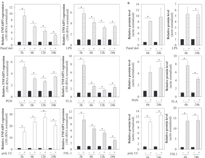
Keratinocytes are able to sense different PAMPs and DAMPs with their TLRs. This study investigated whether the activation of different TLRs using selected ligands influences TNFAIP3 expression of an in vitro cultured
human keratinocyte cell line. HPV-KER cells were trea- ted with Pam3CSK4 (TLR1/2), PGN (TLR2), poly I:C (TLR3), LPS (TLR4), FSL-1 (TLR5) and FLA (TLR2/6,) and TNFAIP3 mRNA and protein expression analysed with real-time RT-PCR and western blot analysis.
TNFAIP3 mRNA expression increased rapidly in re- sponse to all the investigated TLR ligands. The intensity and kinetics of these changes varied and depended on the ligand used (Fig. 1A). TNFAIP3 protein expression levels also increased rapidly: elevated levels were detected 6 h after ligand treatment, and these levels remained high after 24 h (Fig. 1B). These results suggest that TNFAIP3 is a generally induced molecule upon TLR activation in keratinocytes.
Cutibacterium acnes increases TNFAIP3 expression in keratinocytes
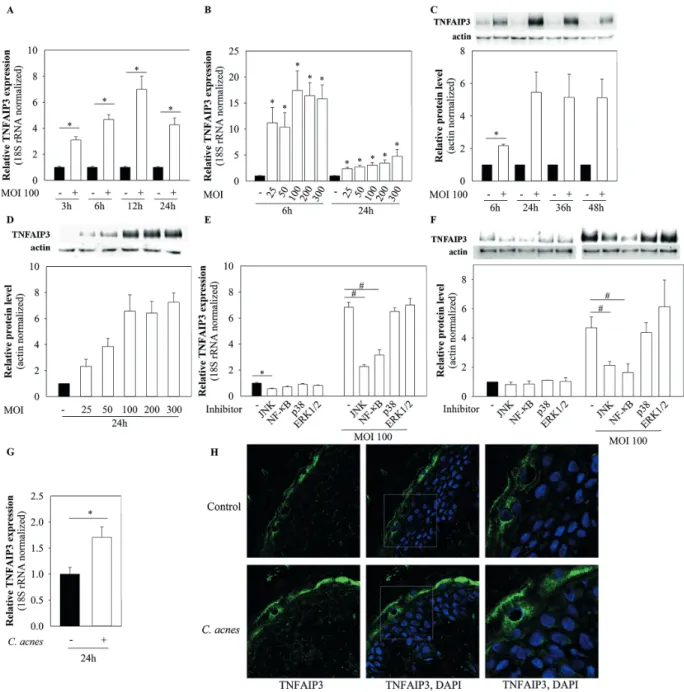
To analyse whether C. acnes affects TNFAIP3 expression levels in keratinocytes, HPV-KER cells were co-cultured with the C. acnes 889 bacterium strain (MOI 100) and the mRNA and protein expression changes analysed with real- time RT-PCR and western blot analysis. TNFAIP3 mRNA levels rapidly and transiently increased in the presence of the bacterium, reaching a maximum 12 h after bacterial treatment (Fig. 2A).
Various C. acnes strains may differentially affect the cellular and molecular properties of human keratinocytes (16, 19, 20). To analyse possible strain-specific regulation, the HPV-KER cells were co-cultured with different C.
acnes strains (MOI=100) belonging to various phyloge- netic groups within the species ( C. acnes 889 - IA, 6609 - IB, ATCC 11828 - II) and compared their effects. All of the C. acnes strains used were able to induce TNFAIP3 expression changes in HPV-KER cells. Slight differences were detected in the induction rates, but the kinetics of the expression changes appeared similar (Fig. S11). Based on these observations, the C. acnes 889 strain was used in subsequent experiments.
TNFAIP3 protein expression levels also increased ra- pidly in response to the bacterium (MOI=100) (Fig. 2C) and remained high during the time-course of the study.
This study also examined whether changes in C. acnes- induced TNFAIP3 expression were dose-dependent, by co- culturing HPV-KER cells with different MOIs. It was found that the extent of TNFAIP3 mRNA and protein expression changes depended on the bacterial dose (Fig. 2B, D).
To confirm that the observed changes were not specific properties of the immortalized keratinocyte cell line used, the experiments were also performed with NHEK cells.
Similar TNFAIP3 expression changes were found upon C.
acnes treatment as those described in HPV-KER cells. C.
acnes treatment resulted in elevated TNFAIP3 mRNA levels and dose-dependent protein expression changes (Fig. S21).
To further test TNFAIP3 inducibility in keratinocytes in the presence of C. acnes, ex vivo organotypic skin (OS) cultures were tested. In this model, normal epithelial tissue architecture is present and, as a result, skin physiology
1https://www.medicaljournals.se/acta/content/abstract/10.2340/00015555-3707
A cta DV A cta DV A
dvances in dermatology and venereologyA
ctaD
ermato-V
enereologicais better preserved. OS cultures were treated with live C. acnes bacterium and changes in the mRNA expres- sion were analysed using real-time RT-PCR. Levels of TNFAIP3 mRNA expression increased in the epidermis part of the OS cultures upon C. acnes treatment, similar to that observed in monolayer cultures (Fig. 2G).
Earlier studies have shown that TNFAIP3 protein is expressed in the human epidermis (21). The current study confirmed this finding by immunofluorescence staining of OS cultures, in which the TNFAIP3 protein was present throughout the entire epidermis, especially in the most differentiated keratinocyte layers. Immunofluorescence microscopy analysis also revealed elevated TNFAIP3 protein levels in response to C. acnes treatment (Fig. 2H).
Changes in C. acnes-induced TNFAIP3 expression are dependent on Jun kinase- and NF-κB signalling pathways This study also investigated the signalling mechanisms playing important roles in TNFAIP3 regulation. The di-
merization partner, RB-like, E2F and multi-vulval class B (DREAM) transcription complex is known to repress its transcription when bound to the DRE3 and DRE4 elements in the TNFAIP3 promoter region in human lung microvessel endothelial cells (HLMVECs). The TNFAIP3 promoter also harbours functional NF-κB binding sites (22, 23). To identify the possible signalling pathways involved in C. acnes-induced upregulation of TNFAIP3 in keratinocytes, the current study investigated the contri- bution of selected pathways playing important roles in the bacterium-induced signalling events (10–12). We inhibited the NF-κB and MAPK (JNK, p38 and ERK1/2) signalling pathways using specific inhibitors and analysed changes in TNFAIP3 mRNA and protein expression in HPV-KER cells after co-culturing the cells for 12 and 24 h with the C. acnes 889 strain.
This study found that inhibition of the JNK signalling pat- hway decreased basal TNFAIP3 mRNA expression, where- as C. acnes-induced mRNA levels were affected by JNK and NF-κB inhibition (Fig. 2E). Dual inhibition of these
Fig. 1. mRNA and protein expression of tumour necrosis factor alpha-induced protein 3 (TNFAIP3) increases in response to toll-like receptor (TLR) activation. Human immortalized keratinocyte cell line (HPV-KER) cells were treated with different TLR ligands (Pam3CSK4, PGN, poly I:C, FLA, FSL-1). TNFAIP3 mRNA and protein levels were analysed at the indicated time-points. (A) Changes in TNFAIP3 mRNA expression analysed by real-time RT-PCR; data were normalized to 18S rRNA using the ΔΔCt method and compared with the time-match untreated control values. Error bars:
standard error of the mean (SEM). (B) Changes in TNFAIP3 protein expression were analysed by western blot analysis and quantitated using Image Pro Plus (Media Cybernetics, Inc., Rockville, MD, USA); all data were normalized to actin. Statistical analyses: paired, 2-sample t-test with Holm–Bonferroni correction: *p < 0.05.
A cta DV A cta DV A
dvances in dermatology and venereologyA
ctaD
ermato-V
enereologica2 signalling pathways almost completely prevented the bacterium-induced changes in mRNA expression (Fig. S31), suggesting that these 2 pathways play major roles in the transcriptional regulation of the TNFAIP3 locus. We also detected decreased protein levels upon JNK and NF-κB path way inhibition in the C. acnes-treated samples (Fig. 2F).
TNFAIP3 affects basal and C. acnes-induced levels of inflammatory mediators in keratinocytes
To analyse the role of TNFAIP3 in the regulation of key inflammatory mediators, siRNA-mediated silencing was performed. Silencing resulted in decreased TNFAIP3
Fig. 2. Cutibacterium acnes upregulates the mRNA and protein expression of tumour necrosis factor alpha-induced protein 3 (TNFAIP3).
(A–F) Human immortalized keratinocyte cell line (HPV-KER) cells and (G, H) organotypic skin (OS) were treated with C. acnes, and changes in TNFAIP3 mRNA and protein levels were analysed. (A, B) mRNA expression was analysed by real-time RT-PCR; data were normalized to 18S rRNA and compared with the time-match untreated control values. Error bars: standard error of the mean (SEM). (C, D) TNFAIP3 protein expression was analysed by western blot and quantified using Image Pro Plus (Media Cybernetics, Inc., Rockville, MD, USA); all data were normalized to actin and compared with time-matched untreated control values. Statistical analyses: paired, 2-sample t-test with Holm–Bonferroni correction: *p < 0.05. (E, F) Cells were plated and pretreated with selective inhibitors for Jun kinase (JNK), nuclear factor kappa B (NF-κB), p38 and ERK1/2 or, as a control, Dimethyl sulphoxide (DMSO) for 1 h.
For mRNA expression studies, C. acnes challenge was performed for 12 h and analysed by real-time RT-PCR. All data were normalized to the 18S rRNA and compared with DMSO-treated control samples. For protein expression analyses, cells were treated with C. acnes for 24 h, subjected to western blot analysis and quantified with Image Pro Plus; all data were normalized to actin and compared with time-matched untreated control values. Statistical analyses: paired, 2-sample t-test with Holm–Bonferroni correction, in which basal TNFAIP3 expression, *p < 0.05, or C. acnes-induced TNFAIP3 expression,
#p < 0.05, were compared. (G, H) OS cultures were treated with 3×10⁷ bacterium/cm2 for 24 h. The epidermis was separated by dispase digestion and the mRNA expression was analysed only in epidermal cells by real-time RT-PCR. Protein expression was visualized using immunofluorescence staining for TNFAIP3 (green) and DAPI (blue) in the full-thickness skin biopsy.
A cta DV A cta DV A
dvances in dermatology and venereologyA
ctaD
ermato-V
enereologicaprotein levels (Fig. 3A). The effect of TNFAIP3 on NF-κB activity was then analysed using a luciferase reporter as- say. In these samples, the basal NF-κB promoter activity increased significantly, compared with the scrambled- siRNA (scr-siRNA) transfected controls (Fig. 3B).
The consequences of these events was also monitored, and it was found that TNFAIP3 silencing significantly increased the basal mRNA expression of TNFα, inter- leukin (IL)-1α, IL-6, IL-8 and C-C motif chemokine ligand 5 (CCL5) and the secreted levels of IL-6 and IL8, compared with the scr-siRNA control samples. These data suggest that TNFAIP3 may have important roles in the
establishment and maintenance of keratinocyte cellular homeostasis by controlling the constitutive expression of different key inflammatory mediators.
The effect of TNFAIP3 knockdown on C. acnes-induced levels of IL-6, IL8 and CCL5 were also investigated, and it was found that silencing also significantly increased the levels of mRNA and secreted protein (Fig. 3C, D).
TNFAIP3 expression differs in acne lesions
C. acnes may play important roles in the regulation and maintenance of epidermal homeostasis, as well as in
Fig. 3. Tumour necrosis factor alpha-induced protein 3 (TNFAIP3) negatively regulates downstream targets of toll-like receptor (TLR) signalling pathways. Transient siRNA-mediated silencing was performed for 48 h in human immortalized keratinocyte cell line (HPV-KER) cells. (A) TNFAIP3 protein levels were analysed by western blot analysis and quantified using Image Pro Plus (Media Cybernetics, Inc., Rockville, MD, USA). All data were normalized to actin. Error bars: standard deviation (SD). (B) Nuclear factor kappa B (NF-κB) promoter activity was measured with luciferase reporter assay, for which all data were normalized to Renilla and compared with src-siRNA containing samples. Error bars: standard error of the mean (SEM). Statistical analysis paired, 2-sample t-test: *p < 0.05. (C) After silencing, cells were treated with C. acnes for 6 h and basal and bacterium-induced mRNA expression of tumour necrosis factor alpha (TNFα), interleukin (IL)-1α, IL-6, IL-8 and C-C motif chemokine ligand 5 (CCL5) were monitored by real time RT-PCR. All data were normalized to 18S rRNA and compared with untreated scr-siRNA containing samples. Error bars: SEM. Statistical analyses: paired Student’s t-test with Holm–Bonferroni correction, *p < 0.05. (D) After silencing, cells were treated with C. acnes for 24 h and secreted IL-6, IL-8 and CCL5 proteins were measured by enzyme-linked immunoassay (ELISA). Error bars: SEM. Statistical analysis: paired Student’s t-test with Holm–Bonferroni correction, *p < 0.05.
A cta DV A cta DV A
dvances in dermatology and venereologyA
ctaD
ermato-V
enereologicaacne vulgaris pathogenesis as an opportunistic pathogen because of bacterial dysbiosis. To determine whether levels of tissue TNFAIP3 differ in healthy and lesional skin samples, data from a publicly available GEO Profile dataset were analysed, comparing the total RNA content of healthy individuals with lesional and non-lesional skin samples of patients with acne (18). Significantly increased levels of TNFAIP3 mRNA expression were found in lesion al skin samples compared with non-lesional skin of patients with acne (Fig. 4).
DISCUSSION
The cutaneous microbiota plays important roles in the maintenance of epidermal homeostasis. It protects against pathogenic microbes, promotes cell differentiation in the host, and keeps the immune system in an alert state, ready to mount a rapid response to repel pathogenic attacks (2, 7).
These functions require balanced interactions among the human cells and the microbes. When this balance shifts, microbial dysbiosis may occur, resulting in pathogenesis that manifests as skin disease, such as Malassezia spp in se- borrhoeic dermatitis, S. aureus and S. epidermidis in atopic dermatitis (AD) and C. acnes in acne vulgaris (24, 25).
Research efforts in the last few decades clearly indicate that epidermal keratinocytes are immunocompetent cells (4, 26, 27). They are equipped to recognize external micro- bes, mount immediate and efficient responses to maintain the integrity of the organism, and alert the immune system to harm. Interestingly, both pathogens and members of the skin microbiota are able to initiate this internal danger pro- gramme in keratinocytes, and induce innate immune and inflammatory events in these cells through the expression of PRRs, such as TLRs (28). However, these microbes are constantly present in the skin, and, therefore, microbiota- induced immune activations must be tightly regulated to avoid unnecessary inflammation and to maintain skin
homeostasis. In past decades, several negative regulators of the TLR and NF-κB signalling pathways have been identified, including TNFAIP3 (5).
TNFAIP3, also known as A20, is a ubiquitin editing enzyme. Through its zinc finger domains, ubiquitin ligase and deubiquitinase activity, TNFAIP3 controls TNF receptors (TNFR) and TLR pathways at different levels (29). TNFAIP3 acts directly on receptor interac- ting protein (RIP) 1 and NF-kappa-B essential modifier (NEMO), which dampen downstream signalling cascades.
TNFAIP3 also controls ubiquitin-conjugating enzyme E2 N (UBE2N) and cellular inhibitor of apoptosis proteins 1 and 2 (cIAP1/2) enzymes complexes by destabilizing their interaction. This protein also accelerates the dissociation of linear ubiquitin chain assembly complex (LUBAC) and inhibitory kappa B kinases (IKK)/NEMO, leading to the termination of NF-κB activation (30).
Tight regulation of TNFAIP3 is important for the maintenance of cellular homeostasis. In a murine model, cell-specific deletion of the TNFAIP3 gene in various cells with important innate and adaptive immune functions re- sulted in spontaneous development of inflammation (30, 31). In corroboration with these results, polymorphisms within and near the TNFAIP3 locus have been shown to be associated with different autoimmune and chronic in- flammatory diseases, including psoriasis (PsO), systemic lupus erythematosus, multiple sclerosis and rheumatoid arthritis (32–35).
In agreement with other studies (21), the current study found that TNFAIP3 is expressed through the entire epi- dermis, exhibiting higher levels in the more differentiated keratinocyte layers. This gradient may be formed in pa- rallel with natural differentiation processes, as one of the major regulators of keratinocyte differentiation is calcium (Ca2+) and, together with Ca2+-induced differentiation, TNFAIP3 expression also increases in in vitro cultured normal human keratinocytes (21, 36). The molecular details of these events are currently unknown, but it may be mediated through the DREAM transcription factor, which is Ca2+ regulated and also plays important roles in the regulation of TNFAIP3 transcription (22, 37).
To analyse the role of TNFAIP3 in the regulation of innate immune and inflammatory events in keratinocytes, different TLRs (1/2, 2, 3, 4, 5 and 2/6) were activated by challenging HPV-KER cells with well-known PAMPs (Pam3CSK4, PGN, poly I:C, LPS, FLA and FSL-1) and changes in TNFAIP3 expression were analysed. All of the investigated TLR ligands increased both the mRNA and protein expression levels of TNFAIP3 in HPV-KER cells;
however, the extent of the changes and the time-kinetics were ligand-dependent. These data are in agreement with previous findings with different cell types, suggesting that TNFAIP3 is a generally expressed, universal regulator of the TLR signalling pathways.
Based on these results, the current study aimed to in- vestigate whether TNFAIP3 also plays a role in C. acnes- induced immune and inflammatory events in human
Fig. 4. mRNA expression profile of tumour necrosis factor alpha- induced protein 3 (TNFAIP3) in healthy skin and skin from patients with acne. TNFAIP3 expression profile from GEO Profile data were analysed and compared with paired Student’s t-test with Holm–Bonferroni correction, *p < 0.05.
A cta DV A cta DV A
dvances in dermatology and venereologyA
ctaD
ermato-V
enereologicakeratinocytes. First, it was checked whether the bacterium itself influences TNFAIP3 expression in vitro and ex vivo.
The results showed that both the mRNA and protein ex- pression increased rapidly in a dose-dependent manner in response to C. acnes in cultured HPV-KER cells as well as in the epidermal part of an ex vivo skin model. A similar effect was described recently for another member of the skin microbiota, S. epidermidis (13). These results, taken together, indicate that the TNFAIP3 protein may play important roles in the regulation of immune and inflammation activation in response to various members of the cutaneous microbiota.
C. acnes is one of the most prominent cutaneous microb es in the sebum-rich skin regions from adoles- cence, and plays a role in the pathogenesis of acne vul- garis as a result of microbial dysbiosis (24). In order to determine whether TNFAIP3 levels differ in control indi- viduals and lesional and non-lesional skin samples from patients with acne, the current study compared publicly available microarray data in the GEO Profile database (ID:
33444972) and found that, in patients, mRNA expression of TNFAIP3 increases significantly in papules compared with non-lesional skin samples from the same patients.
These results suggest that, in the lesional skin, apart from the production of different pro-inflammatory mediators, expression of anti-inflammatory molecules also increases.
These latter molecules may play important roles in control of inflammation, to avoid excessive, potentially tissue- damaging reactions. Further research is necessary to determine whether increased TNFAIP3 mRNA levels in the lesional skin correspond to elevated protein levels. If mRNA and protein levels correspond, the accumulation may be an important step during the healing of lesions and the restoration of epidermal homeostasis.
Interestingly, acne is not the only inflammatory skin disease in which TNFAIP3 levels change. In PsO and AD, studies have found lower TNFAIP3 mRNA levels in lesional skin samples, compared with skin from healthy individuals. Moreover, in patients with PsO, TNFAIP3 mRNA and protein levels were lower in lesional skin than in non-lesional samples of the same patients. In contrast, TNFAIP3 mRNA expression levels in patients with atopic dermatitis (AD) did not differ in the involved and non-involved samples (21, 38). It is not clear whether this observation represents a true difference in the pat- hogenesis of these chronic inflammatory skin conditions.
One possible explanation is that the levels of negative regulators may change rapidly throughout the different phases of disease pathogenesis, and the above observa- tions represent only temporal variations. It is interesting to note, however, that inability to control innate immune activation due to decreased TNFAIP3 levels may lead to persisting, chronic inflammation, and the net outcome and the severity of inflammation possibly depends on the balance of pro- and anti-inflammatory factors. As a result, this molecule may serve as a drug target for treatment in the future.
Previous studies have found differences in the compo- sition of C. acnes strains in healthy and acne skin (39), and various strains may differently affect the cellular and molecular biology functions as well as keratinocyte viabi- lity (16, 20, 40, 41). Slight differences were found in the TNFAIP3 mRNA expression levels when the HPV-KER cells were co-cultured with different C. acnes strains (889, ATCC11828, 6609), but the expression kinetics were very similar. The C. acnes strains used differ, among others, in their growth rate, amount of secreted metabolites (e.g.
various short-chain fatty acids (SCFAs)), and pathogenic factors (e.g. Christie-Atkins-Munch-Peterson (CAMP) factors). These properties may be responsible for the ob- served differences (16, 42). This is similar to the results reported previously, in our analysis of a TNFAIP3 inte- raction partner, TNFAIP3 interacting protein 1 (TNIP1), another negative regulator, under similar experimental conditions (43). These findings suggest that strain-specific biological effects of C. acnes are probably not due to the differential induction of various negative regulators upon immune and inflammation induction in keratinocytes.
Multiple signalling pathways and transcriptional factors are involved in the transcriptional regulation of TNFAIP3.
In LPS-activated bone-marrow-derived macrophages from mice, NF-κB and p38 regulate TNFAIP3 expression through its NF-κB and CCAAT-enhancer-binding protein (C/EBPβ) binding sites (23). However, in Pam3CSK4- activated THP1 cells, NF-κB inhibition has no effect:
GSK3-related pathways regulate changes in TNFAIP3 expression, possibly through the cAMP response element binding protein (CREB) (44). Others also showed that the DREAM complex represses its transcriptional acti- vation through downstream-responsive elements (DRE3 and DRE4) binding sites in mice and HLMVECs (22).
These findings suggest that transcriptional regulation of TNFAIP3 is possibly dependent on the type of cell and stimuli. We found that the basal mRNA expression of TNFAIP3 is regulated by JNK, whereas JNK and NF- κB signalling pathways are involved in the control of C. acnes-induced changes in TNFAIP3 mRNA and protein expression, for which p38 and ERK1/2 inhibition had no effect. Dual inhibition of JNK and NF-κB signalling pathways almost completely inhibited C. acnes-induced changes in mRNA expression, which suggest that these 2 signalling pathways have a major impact on the bac- terium-induced regulation of TNFAIP3 mRNA levels in HPV-KER cells.
A recent study suggested that TNFAIP3 may help S. epi- dermidis to persist as a commensal on the human skin, th- rough the control of bacterium-induced NF-κB activation, IL-1β and human beta-defensin-2 (HBD-2) production in keratinocytes (13). Based on these findings, the current study aimed to determine whether this negative regulatory function also occurs in C. acnes-induced inflammatory processes. In these experiments, silencing TNFAIP3 significantly increased basal NF-κB promoter activities, as well as the mRNA expression of pro-inflammatory
A cta DV A cta DV A
dvances in dermatology and venereologyA
ctaD
ermato-V
enereologicacytokines and chemokines (TNFα, IL-1α, IL-6, IL-8 and CCL5) in HPV-KER cells. Co-culturing of silenced cells with the C. acnes 889 strain also leads to increased TNFα, IL-1α, IL-6, IL-8 and CCL5 mRNA expression, as well as elevated IL-8, IL-6 and CCL5 protein secretions.
These data argue that TNFAIP3 plays important roles in the regulation of C. acnes-driven molecular events in keratinocytes, as well as in the maintenance of cellular homeostasis, by regulating the cutaneous inflammatory programme under homeostatic conditions (45).
TNFAIP3 possibly operates in a negative regula- tory cycle in keratinocytes (Fig. 5). In this control loop, TNFAIP3 regulates the signalling consequences of a broad spectrum of TLR ligands, which, in turn, also affects TNFAIP3 levels. This is in agreement with the currently available research data, suggesting TNFAIP3 functions in, for example, poly-I:C-induced inflammatory responses in human keratinocytes, as well as in imiquimod- and acetone-induced PsO and dermatitis in mouse models,
respectively (21, 38). TNFAIP3 also plays important roles in the induction of Pam3CSK4-tolerance in monocytic THP-1 cells, arguing for a generalized role for this mole- cule in different TLR-mediated molecular processes (44).
In summary, TNFAIP3 plays important roles in the re- gulation of TLR-induced signalling pathways in keratino- cytes, and, through these roles, contributes to the control of C. acnes-induced innate immune and inflammatory events. By fine-tuning the microbiota-induced signalling processes, TNFAIP3 may help to maintain a homeostatic condition between the skin and the cutaneous microflora.
Currently, several different therapeutic modalities are available for the treatment of acne vulgaris. They may be clinically effective, although all of them only treat an existing condition, and most of them have substantial side-effects (e.g. antibiotics, retinoids) (46, 47). Targeting negative regulators of the C. acnes-induced inflammatory pathways themselves may help to develop novel, well- tolerated therapeutic modalities for the treatment of acne vulgaris. Further studies should evaluate the potential use and efficacy of TNFAIP3 as such a target.
ACKNOWLEDGEMENTS
The authors thank Tanácsné Bajkán Andrea for her excellent technical assistance and Shannon Frances for language editing.
This work was supported by the GINOP-2.3.2-15-2016-00015 and OTKA NK105369 research grants. The project has received funding from the EU’s Horizon 2020 research and innovation programme under grant agreement (number 739593).
KS is a recipient of the János Bolyai Research Scholarship of the Hungarian Academy of Sciences. KS was supported by the UNKP-18-4. EL was supported by the UNKP-18-3 of New Na- tional Excellence Program of the Ministry of Human Capacities.
KS was supported by the ÚNKP-19-4. EL was supported by the ÚNKP-19-3 of New National Excellence Program of the Ministry for Innovation and Technology. The funders had no role in study design, data collection and analysis, decision to publish or prepa- ration of the manuscript.
The authors have no conflicts of interest to declare.
REFERENCES
1. Eyerich S, Eyerich K, Traidl-Hoffmann C, Biedermann T.
Cutaneous barriers and skin immunity: differentiating a connected network. Trends Immunol 2018; 39: 315–327.
2. Gallo RL, Nakatsuji T. Microbial symbiosis with the innate immune defense system of the skin. J Invest Dermatol 2011;
131: 1974–1980.
3. de Koning HD, Rodijk-Olthuis D, van Vlijmen-Willems IM, Joosten LA, Netea MG, Schalkwijk J, et al. A comprehensive analysis of pattern recognition receptors in normal and infla- med human epidermis: upregulation of dectin-1 in psoriasis.
J Invest Dermatol 2010; 130: 2611–2620.
4. Pivarcsi A, Kemeny L, Dobozy A. Innate immune functions of the keratinocytes. A review. Acta Microbiol Immunol Hung 2004; 51: 303–310.
5. Liew FY, Xu D, Brint EK, O’Neill LA. Negative regulation of toll-like receptor-mediated immune responses. Nat Rev Im- munol 2005; 5: 446–458.
6. Oh J, Conlan S, Polley EC, Segre JA, Kong HH. Shifts in human skin and nares microbiota of healthy children and adults. Genome Med 2012; 4: 77.
Fig. 5. Negative regulation of Cutibacterium acnes-induced toll- like receptor (TLR) signalling events by tumour necrosis factor alpha-induced protein 3 (TNFAIP3) in keratinocytes. C. acnes induce TLR 2 and 4 activation in keratinocytes, which leads to the induction of canonical nuclear factor kappa B (NF-κB) and mitogen-activated protein kinase (MAPK)-dependent signalling cascades. When NF-κB is activated, it can regulate the expression changes of TNFAIP3 through binding to special NF-κB binding sites in its promoter region. The generated TNFAIP3 protein, in turn, can act back and inhibit signaling pathways, among others NF-kB activation, forming a negative regulatory feedback loop. In addition to NF-κB, Jun kinase (JNK) is also able to regulate changes in TNFAIP3 expression. Created with BioRender.com.
A cta DV A cta DV A
dvances in dermatology and venereologyA
ctaD
ermato-V
enereologica7. Grice EA, Segre JA. The skin microbiome. Nat Rev Microbiol 2011; 9: 244–253.
8. Kim J, Ochoa MT, Krutzik SR, Takeuchi O, Uematsu S, Legaspi AJ, et al. Activation of toll-like receptor 2 in acne triggers inflammatory cytokine responses. J Immunol 2002; 169:
1535–1541.
9. Pivarcsi A, Bodai L, Rethi B, Kenderessy-Szabo A, Koreck A, Szell M, et al. Expression and function of Toll-like receptors 2 and 4 in human keratinocytes. Int Immunol 2003; 15:
721–730.
10. Nguyen CT, Sah SK, Zouboulis CC, Kim TY. Inhibitory effects of superoxide dismutase 3 on Propionibacterium acnes- induced skin inflammation. Sci Rep 2018; 8: 4024.
11. Wang YY, Ryu AR, Jin S, Jeon YM, Lee MY. Chlorin e6-mediated photodynamic therapy suppresses P. acnes-induced inflam- matory response via NFkappaB and MAPKs signaling pathway.
PLoS One 2017; 12: e0170599.
12. Pretsch A, Nagl M, Schwendinger K, Kreiseder B, Wiederstein M, Pretsch D, et al. Antimicrobial and anti-inflammatory ac- tivities of endophytic fungi Talaromyces wortmannii extracts against acne-inducing bacteria. PLoS One 2014; 9: e97929.
13. Simanski M, Erkens AS, Rademacher F, Harder J. Staphylo- coccus epidermidis-induced interleukin-1 beta and human beta-defensin-2 expression in human keratinocytes is re- gulated by the host molecule A20 (TNFAIP3). Acta Derm Venereol 2019; 99: 181–187.
14. Danis J, Janovak L, Guban B, Goblos A, Szabo K, Kemeny L, et al. Differential inflammatory-response kinetics of human keratinocytes upon cytosolic RNA- and DNA-fragment induc- tion. Int J Mol Sci 2018; 19: 774.
15. Megyeri K, Orosz L, Bolla S, Erdei L, Razga Z, Seprenyi G, et al. Propionibacterium acnes induces autophagy in kera- tinocytes: involvement of multiple mechanisms. J Invest Dermatol 2018; 138: 750–759.
16. Tax G, Urban E, Palotas Z, Puskas R, Konya Z, Biro T, et al.
Propionic acid produced by propionibacterium acnes strains contributes to their pathogenicity. Acta Derm Venereol 2016;
96: 43–49.
17. Bolla BS, Erdei L, Urban E, Burian K, Kemeny L, Szabo K.
Cutibacterium acnes regulates the epidermal barrier proper- ties of HPV-KER human immortalized keratinocyte cultures.
Sci Rep 2020; 10: 12815.
18. Trivedi NR, Gilliland KL, Zhao W, Liu W, Thiboutot DM. Gene array expression profiling in acne lesions reveals marked upregulation of genes involved in inflammation and matrix remodeling. J Invest Dermatol 2006; 126: 1071–1079.
19. Nagy I, Pivarcsi A, Koreck A, Szell M, Urban E, Kemeny L.
Distinct strains of propionibacterium acnes induce selective human beta-defensin-2 and interleukin-8 expression in human keratinocytes through toll-like receptors. J Invest Dermatol 2005; 124: 931–938.
20. Lheure C, Grange PA, Ollagnier G, Morand P, Desire N, Sayon S, et al. TLR-2 Recognizes propionibacterium acnes CAMP factor 1 from highly inflammatory strains. PLoS One 2016;
11: e0167237.
21. Sohn KC, Back SJ, Choi DK, Shin JM, Kim SJ, Im M, et al. The inhibitory effect of A20 on the inflammatory reaction of epi- dermal keratinocytes. Int J Mol Med 2016; 37: 1099–1104.
22. Tiruppathi C, Soni D, Wang DM, Xue J, Singh V, Thippegowda PB, et al. The transcription factor DREAM represses the deu- biquitinase A20 and mediates inflammation. Nat Immunol 2014; 15: 239–247.
23. Lai TY, Wu SD, Tsai MH, Chuang EY, Chuang LL, Hsu LC, et al. Transcription of Tnfaip3 is regulated by NF-kappaB and p38 via C/EBPbeta in activated macrophages. PLoS One 2013; 8: e73153.
24. Szabo K, Erdei L, Bolla BS, Tax G, Biro T, Kemeny L. Factors shaping the composition of the cutaneous microbiota. Br J Dermatol 2017; 176: 344–351.
25. Szegedi A, Dajnoki Z, Biro T, Kemeny L, Torocsik D. Acne:
transient arrest in the homeostatic host-microbiota dialog?
Trends Immunol 2019; 40: 873–876.
26. Kollisch G, Kalali BN, Voelcker V, Wallich R, Behrendt H, Ring J, et al. Various members of the Toll-like receptor family con- tribute to the innate immune response of human epidermal
keratinocytes. Immunology 2005; 114: 531–541.
27. Steinhoff M, Brzoska T, Luger TA. Keratinocytes in epidermal immune responses. Curr Opin Allergy Clin Immunol 2001;
1: 469–476.
28. Sanford JA, Gallo RL. Functions of the skin microbiota in health and disease. Semin Immunol 2013; 25: 370–377.
29. Malynn BA, Ma A. A20: A multifunctional tool for regulating immunity and preventing disease. Cell Immunol 2019; 340:
103914.
30. Das T, Chen Z, Hendriks RW, Kool M. A20/tumor necrosis factor alpha-induced protein 3 in immune cells controls de- velopment of autoinflammation and autoimmunity: lessons from mouse models. Front Immunol 2018; 9: 104.
31. Catrysse L, Vereecke L, Beyaert R, van Loo G. A20 in inflam- mation and autoimmunity. Trends Immunol 2014; 35: 22–31.
32. Stuart PE, Nair RP, Tsoi LC, Tejasvi T, Das S, Kang HM, et al. Genome-wide association analysis of psoriatic arthritis and cutaneous psoriasis reveals differences in their genetic architecture. Am J Hum Genet 2015; 97: 816–836.
33. Dieguez-Gonzalez R, Calaza M, Perez-Pampin E, Balsa A, Blanco FJ, Canete JD, et al. Analysis of TNFAIP3, a feedback inhibitor of nuclear factor-kappaB and the neighbor intergenic 6q23 region in rheumatoid arthritis susceptibility. Arthritis Res Ther 2009; 11: R42.
34. Kawasaki A, Ito S, Furukawa H, Hayashi T, Goto D, Matsu- moto I, et al. Association of TNFAIP3 interacting protein 1, TNIP1 with systemic lupus erythematosus in a Japanese population: a case-control association study. Arthritis Res Ther 2010; 12: R174.
35. Perga S, Martire S, Montarolo F, Navone ND, Calvo A, Fuda G, et al. A20 in multiple sclerosis and Parkinson’s disease: clue to a common dysregulation of anti-inflammatory pathways?
Neurotox Res 2017; 32: 1–7.
36. Celli A, Sanchez S, Behne M, Hazlett T, Gratton E, Mauro T. The epidermal Ca(2+) gradient: measurement using the phasor representation of fluorescent lifetime imaging. Bio- phys J 2010; 98: 911–921.
37. Sours-Brothers S, Ma R, Koulen P. Ca2+-sensitive transcrip- tional regulation: direct DNA interaction by DREAM. Front Biosci (Landmark Ed) 2009; 14: 1851–1856.
38. Devos M, Mogilenko DA, Fleury S, Gilbert B, Becquart C, Quemener S, et al. Keratinocyte expression of A20/TNFAIP3 controls skin inflammation associated with atopic dermatitis and psoriasis. J Invest Dermatol 2019; 139: 135–145.
39. Fitz-Gibbon S, Tomida S, Chiu BH, Nguyen L, Du C, Liu M, et al. Propionibacterium acnes strain populations in the human skin microbiome associated with acne. J Invest Dermatol 2013; 133: 2152–2160.
40. Dagnelie MA, Corvec S, Saint-Jean M, Nguyen JM, Khammari A, Dreno B. Cutibacterium acnes phylotypes diversity loss: a trigger for skin inflammatory process. J Eur Acad Dermatol Venereol 2019; 33: 2340–2348.
41. Jasson F, Nagy I, Knol AC, Zuliani T, Khammari A, Dreno B. Different strains of propionibacterium acnes modulate differently the cutaneous innate immunity. Exp Dermatol 2013; 22: 587–592.
42. Dreno B, Pecastaings S, Corvec S, Veraldi S, Khammari A, Roques C. Cutibacterium acnes (Propionibacterium acnes) and acne vulgaris: a brief look at the latest updates. J Eur Acad Dermatol Venereol 2018; 32 Suppl 2: 5–14.
43. Erdei L, Bolla BS, Bozo R, Tax G, Urban E, Kemeny L, et al.
TNIP1 regulates cutibacterium acnes-induced innate im- mune functions in epidermal keratinocytes. Front Immunol 2018; 9: 2155.
44. Hu J, Wang G, Liu X, Zhou L, Jiang M, Yang L. A20 is critical for the induction of Pam3CSK4-tolerance in monocytic THP-1 cells. PLoS One 2014; 9: e87528.
45. Lippens S, Lefebvre S, Gilbert B, Sze M, Devos M, Verhelst K, et al. Keratinocyte-specific ablation of the NF-kappaB regulatory protein A20 (TNFAIP3) reveals a role in the control of epider- mal homeostasis. Cell Death Differ 2011; 18: 1845–1853.
46. Dreno B. Bacteriological resistance in acne: a call to action.
Eur J Dermatol 2016; 26: 127–132.
47. Orfanos CE, Zouboulis CC. Oral retinoids in the treatment of seborrhoea and acne. Dermatology 1998; 196: 140–147.