BRIEF ARTICLE
Immune phenotype in children with therapy-naïve remitted and relapsed Crohn’s disease
Aron Cseh, Barna Vasarhelyi, Kriszta Molnar, Balazs Szalay, Peter Svec, Andras Treszl, Antal Dezsofi, Peter Laszlo Lakatos, Andras Arato, Tivadar Tulassay, Gabor Veres
Aron Cseh, Barna Vasarhelyi, Kriszta Molnar, Balazs Sza- lay, Peter Svec, Andras Treszl, Research Group for Pediatrics and Nephrology, Semmelweis University and Hungarian Acad- emy of Sciences, H-1083, Budapest, Hungary
Barna Vasarhelyi, Department of Laboratory Medicine, Sem- melweis University, H-1083, Budapest, Hungary
Antal Dezsofi, Andras Arato, Tivadar Tulassay, Gabor Veres, First Department of Pediatrics, Semmelweis University, H-1083, Budapest, Hungary
Peter Laszlo Lakatos, First Department of Medicine, Semmel- weis University, H-1083, Budapest, Hungary
Author contributions: Veres G and Cseh A designed the re- search; Dezsofi A, Lakatos PL, Arato A and Veres G included the patients; Cseh A, Molnar K, Szalay B and Svec P performed the analyses; Treszl A analyzed the data; Cseh A, Vasarhelyi B and Veres G wrote the paper; Tulassay T critically reviewed the paper.
Supported by Grants TÁMOP-4.2.2-08/1/KMR-2008-0004, OTKA-76316, OTKA-K81117 and ETT-028-02
Correspondence to: Aron Cseh, MD, Research Group for Pe- diatrics and Nephrology, Semmelweis University and Hungar- ian Academy of Sciences, H-1083, Budapest,
Hungary. cseharon@gmail.com
Telephone: +36-1-4591500 Fax: +36-1-3036077 Received: March 1, 2010 Revised: June 1, 2010 Accepted: June 8, 2010
Published online: December 21, 2010
Abstract
AIM: To characterize the prevalence of subpopulations of CD4+ cells along with that of major inhibitor or stimu- lator cell types in therapy-naïve childhood Crohn’s dis- ease (CD) and to test whether abnormalities of immune phenotype are normalized with the improvement of clini- cal signs and symptoms of disease.
METHODS: We enrolled 26 pediatric patients with CD.
14 therapy-naïve CD children; of those, 10 children remitted on conventional therapy and formed the re- mission group. We also tested another group of 12 chil-
dren who relapsed with conventional therapy and were given infliximab; and 15 healthy children who served as controls. The prevalence of Th1 and Th2, naïve and memory, activated and regulatory T cells, along with the members of innate immunity such as natural killer (NK), NK-T, myeloid and plasmocytoid dendritic cells (DCs), monocytes and Toll-like receptor (TLR)-2 and TLR-4 ex- pression were determined in peripheral blood samples.
RESULTS: Children with therapy-naïve CD and those in relapse showed a decrease in Th1 cell prevalence.
Simultaneously, an increased prevalence of memory and activated lymphocytes along with that of DCs and monocytes was observed. In addition, the ratio of my- eloid /plasmocytoid DCs and the prevalence of TLR-2 or TLR-4 positive DCs and monocytes were also higher in therapy-naïve CD than in controls. The majority of alter- ations diminished in remitted CD irrespective of whether remission was obtained by conventional or biological therapy.
CONCLUSION: The finding that immune phenotype is normalized in remission suggests a link between immune phenotype and disease activity in childhood CD. Our ob- servations support the involvement of members of the adaptive and innate immune systems in childhood CD.
© 2010 Baishideng. All rights reserved.
Key words: Crohn’s disease; Dendritic cell; Infliximab;
Lymphocyte; Monocyte; Regulatory T cell; Relapse;
Remission; Therapy-naïve; Toll-like receptor
Peer reviewers: Dr. Stefan Wirth, Professor, Children’s Hos- pital, Heusnerstt. 40, Wuppertal 42349, Germany; Masahiro Iizuka, MD, PhD, Director, Akita Health Care Center, Akita Red Cross Hospital, 3-4-23, Nakadori, Akita 010-0001, Japan Cseh A, Vasarhelyi B, Molnar K, Szalay B, Svec P, Treszl A, Dezsofi A, Lakatos PL, Arato A, Tulassay T, Veres G. Immune
© 2010 Baishideng. All rights reserved.
doi:10.3748/wjg.v16.i47.6001
phenotype in children with therapy-naïve remitted and relapsed Crohn’s disease. World J Gastroenterol 2010; 16(47): 6001-6009 Available from: URL: http://www.wjgnet.com/1007-9327/full/
v16/i47/6001.htm DOI: http://dx.doi.org/10.3748/wjg.v16.
i47.6001
INTRODUCTION
Crohn’s disease (CD) is a chronic gastrointestinal disease characterized by segmental inflammation of the intestinal mucosa associated with a dysregulated action of the muco- sal immune system to the otherwise innocuous luminal an- tigens in a genetically susceptible host. About 10%-15% of patients with CD are diagnosed before 18 years of age[1]. Certain features are unique to pediatric CD in comparison to adult onset disease such as different disease location, altered response to immunosuppressive therapy and dif- ferent genetic and immune phenotype[2,3]. Different char- acteristics may suggest differences in the pathomechanism of CD in children compared to that in adults. Theories regarding CD pathomechanism include malfunctioning of the immune system. Indeed, several studies carried out in adult CD indicate the involvement of either adaptive or in- nate immunity[4-7].
In adult CD, peripheral Treg prevalence is diminished in therapy-naïve patients and in the active state of disease or relapses and is increased with therapy or in remis-
sion[8-10], while intestinal Treg prevalence is increased in
active CD[9-11]. Simultaneously, the prevalence of effector T cells[12-14] and activated T cells[15-17] is increased in the pe- riphery in active CD. The shift of T lymphocytes toward Th1 commitment in peripheral blood and biopsy speci- mens is a widely observed phenomenon in CD[18].
In addition, the prevalence of antigen presenting cells (APCs) including dendritic cells (DCs) decrease in remis- sion, and even more so in relapses at the periphery, but is increased in tissues, simultaneously with an increased expression of Toll-like receptor (TLR)-2 and 4 in therapy- naïve and treated CD adults[19-21]. Monocytes/macro- phages, another group of APCs, are comparable at the pe- riphery but increased in biopsies and showed upregulation of TLR-2 and TLR-4 in the active state and in remission of CD[22-25]. Other cell types of innate immunity such as natural killer (NK) and natural killer T (NKT) cells are less prevalent in active CD[17,26-28].
There have been a limited number of studies specifi- cally performed to investigate alterations of the immune system in children with CD. In addition, the majority of available data are on treated CD children. While in un- treated CD children some alterations in adaptive immunity have been reported, such as the skewness of the Th1/Th2 ratio to Th2 (this finding is in contrast with that observed in adult CD)[29-32], recent data suggest that the disturbance of innate immune functions is also a major factor contrib- uting to CD in children. Indeed, in untreated children the central macrophage prevalence is increased[33], and TLR-2 and 4 receptor expression is also enhanced[34]. The role
of innate immune system in the pathomechanism of CD may be inversely associated with age at disease onset[2].
The immune dysregulation in CD is affected by ongo- ing therapy. Aminosalicylates and steroids, as well as im- munosuppressive drugs used as first-line therapy in CD have strong immunomodulatory effects[35]. In addition, biological therapy including the tumor necrosis factor α inhibitor infliximab (IFX)[36] also has substantial effects on immune cell functions [many of the data are from pa- tients with rheumatoid arthritis (RA)] as it may increase the prevalence of peripheral and central Tregs[8,37], effec- tor and activated[38,39] and Th1 committed T cells[39,40]. IFX also decreases the prevalence of NK cells[38], DCs[41,42] and monocytes[43,44], as well as TLR-2 and TLR-4 expression in peripheral cells[45]. The immunological impact of conven- tional or IFX therapy on peripheral immune phenotype is, however, known exclusively for adult CD patients and has not been explored fully in CD children.
In this prospective study we aimed to explore the major cell prevalence of the adaptive and innate immune systems in therapy-naïve CD children and its alteration with the improvement of CD obtained by conventional therapy or IFX treatment.
MATERIALS AND METHODS
We enrolled the following patient groups into our study: (1) 14 therapy-naïve CD children. No drug was prescribed for these patients at the time of CD diagnosis. The diagnosis of CD was established by means of “The Porto criteria”[46]; disease activity was determined according to the Pediat- ric Crohn’s disease activity index (PCDAI)[47]; (2) During conventional treatment [steroid, azathioprine (AZT) and 5-aminosalicylate (5-ASA)], 10 children responded form- ing the remission group. Clinical remission was defined as a PCDAI < 10; (3) IFX therapy (5 mg/kg IFX at weeks 0, 2, and 6) was started in 12 CD children who failed to respond to conventional therapy forming the “relapsed group”. Non-responsiveness was defined as moder- ately increased PCDAI (PCDAI > 30) in patients under conventional therapy; and (4) Fifteen age- and gender- matched children with functional abdominal pain served as controls. All patients and controls were diagnosed, treated and followed up in the Outpatient Clinic of the First Department of Pediatrics, Semmelweis University between September 2007 and August 2009. The Insti- tutional Ethical Committee approved our study; written parental informed consent was obtained.
The patients’ clinical characteristics are shown in Table 1.
Small and large bowel was involved in 11 of 14 treatment naïve CD patients, according to the literature (L3 localiza- tion, Montreal criteria[48]). Therapy-naïve CD patients and CD patients with relapse had lower body mass index than controls. Lower body weight and body mass index is a common presenting sign in pediatric patients with CD[49]. Reduced food intake, postprandial abdominal cramps, systemic release of cytokines and malabsorptive diarrhea were listed as factors responsible for this phenomenon[50].
Together with other routine blood sampling 6 mL of lithium-heparin anticoagulated blood was taken from therapy-naïve patients at the time of diagnosis, at the time of first remission in the remission group and at the initia- tion of IFX therapy, and then 2 and 6 wk later in the re- lapsed groups. From peripheral blood mononuclear cells (PBMCs), the identification of markers (6B11, CCR4, CD3, CD4, CD8, CD11c, CD14, CD25, CD45RA, CD45RO, CD123; CD161, CXCR3, HLA-DR and Lin-1 BD Biosciences Pharmingen, San Diego, CA, USA;
TLR-2 and TLR-4 eBioscience, San Diego, CA, USA) and FoxP3 assay (eBioscience, San Diego, CA, USA) were per- formed with a BD FACS Aria (BD Biosciences Pharmin- gen, San Diego, CA, USA)[51]. Briefly, from whole blood, PBMCs were separated with gradient centrifugation using Ficoll-Paque (GE Healthcare Life Sciences, Pittsburgh, PA, USA). The isolated PBMCs were washed twice with Phosphate Buffered Saline pH 7.4 (PBS, Central Phar- macy of Semmelweis University, Budapest, Hungary) and were stained with the appropriate fluorescent anti- bodies according to the manufacturers’ instructions. For intracellular staining, cells were incubated first with Fixa- tion/Permeabilization Buffer (eBioscience, San Diego, CA, USA), then washed twice with Permeabilization Buffer (eBioscience, San Diego, CA, USA) and stained by FoxP3 and isotype control. At the end of staining, cells were washed twice with PBS and with Permeabiliza- tion Buffer for the detection of cell surface markers and FoxP3, respectively. Samples were resuspended in PBS and were measured within 1 h recording at least 50 000 events in the lymphocyte gate.
Our data did not follow normal distribution, therefore non-parametric statistical tests and median and interquar- tile ranges were used. Mann-Whitney and Friedman tests with Dunn’s post hoc comparison and Spearman’s corre- lation were used for statistical analysis, the level of signifi- cance was 5% (P < 0.05). All data are expressed as median
(interquartile range). Statistical analysis was performed with Statistica 8 (Statsoft, Tulsa, OK, USA).
RESULTS
Major clinical characteristics and laboratory data are sum- marized in Table 1. The investigated cell prevalence values and cell ratios of the adaptive and innate immune systems are summarized in Tables 2 and 3, respectively. We also highlighted the most important alterations in Figures 1 and 2.
First, we compared the immune phenotype in ther- apy-naïve CD patients with that of healthy controls.
In CD children, the prevalence of activated T cells (i.e.
CD4+CD25+ cells) increased. At the same time, the preva- lence of T cells with Th1 commitment (i.e. CD4+CXCR3+ cells) decreased resulting in a skewness of Th1/Th2 to Th2. The prevalence of memory (i.e. CD4+CD45RO+) cells increased and, therefore, a shift in the naïve/memory ratio toward memory cells was observed. The prevalence of regulatory T (i.e. CD4+CD25hiFoxP3+) cells was com- parable between the two groups.
Striking differences in cell prevalence values of innate immunity were obtained between therapy-naïve CD and healthy children. The occurrence of NK and NKT cells (marked as CD3-CD161+ and CD3+6b11+, respectively) was lower in CD children. Interestingly, the prevalence of the APCs investigated differed largely between CD and healthy children. DCs (i.e. those with Lin1-HLADR+ expression) were more prevalent and, within DC cells, the myeloid DCs (mDCs, i.e. CD11c+ cells) were more preva- lent, while plasmocytoid DCs (pDCs, i.e. CD123+ cells) were less frequent in CD than in healthy children. This leads to skewness of mDCs in the mDC/pDC ratio. The prevalence of peripheral monocytes (i.e. CD14+ cells) also increased in therapy-naïve CD patients. In addition, the prevalence of DC cells and monocytes expressing TLR-2 and TLR-4 receptors was also increased in CD. Of note,
Table 1 Clinical data and patient characteristics
Control Therapy-naïve (before conventional
therapy)
First remission (with conventional
therapy)
Relapse (before IFX therapy)
IFX therapy (before 2nd infusion)
IFX therapy (before 3rd infusion) Clinical data
n (boys/girls) 15 (6/9) 14 (6/8) 10 (4/6) 12 (5/7)
Age (yr) 12 (8-16) 10 (8.5-13) 11.5 (9.5-15.5) 14 (11-16) 14.5 (11.5-16) 14.5 (12-16)
Body mass index (kg/m2) 19.5 (16.5-22.3) 13.9 (12.5-15.8)d 18.6 (14.4-19.2)c 17.4 (14.1-19.5)a 18.8 (14.6-21.3) 19.25 (16.1-22.1) Body weight (percentile) 58 (35-79) 13 (4-23)d 24 (19-44)c 21 (7-28)a 23 (15-63) 35 (10-74) Disease duration (mo) - 10 (8.5-13) 10.5 (8.5-15.5) 11.5 (9.25-15) 12 (9.5-15) 12 (10-15)
Activity index (PCDAI) - 45 (39-58) 0 (0-5)f 45 (25-48)j 20 (7-28)g 13 (2-22)l
Localization (n, montreal criteria) - L1 (1), L2 (2), L3 (11) L1 (0), L2 (2), L3 (8) L1 (1), L2 (2), L3 (9) Laboratory data
White blood cell count (g/L) 7.9 (5.7-10.1) 12.4 (9.4-14.2)b 11.7 (8.4-14.4) 10 (8.2-11.2) 7.9 (5.5-10.6) 6.7 (4.5-10.4)e Platelet (g/L) 346 (288-375) 657 (451-746)b 442 (308-624) 479 (396-750)b 392 (293-523) 383 (308-483)e
Serum iron (mmol/L) 16 (14-21) 4 (2-12)a 8 (4-10)a 4 (2-7)d 5 (4-7)d 8 (6-8)b,e
Serum albumin (g/L) 45 (44-49) 37 (34-39)d 42 (42-45)c 40 (36-41)d 41 (38-45)b 42 (38-45)b C-reactive protein (mg/L) 0 (0-1) 21 (5-65)d 7 (2-11)b,c 27 (9-55)h, d 9 (3-11)d 5 (1-14)d,e
aP < 0.05, bP < 0.01; dP < 0.01 vs control; cP < 0.05, fP < 0.01 vs therapy-naïve; hP < 0.01, jP < 0.01 vs first remission; eP < 0.05, gP < 0.05, lP < 0.001 vs relapse.
IFX: Infliximab; PCDAI: Pediatric Crohn's disease activity index; L1: Small bowel; L2: Large bowel; L3: Small bowel and large bowel localization according to Montreal criteria[48].
4 of the 14 therapy-naïve CD children did not respond to conventional therapy. Their immune phenotype at the therapy-naïve phase did not differ from those children who responded to conventional therapy (data not shown).
In the second phase of our study, we prospectively test- ed the alteration in cell prevalence values during therapy.
At the time of first remission with conventional therapy, the Th1/Th2 ratio shifted to Th1 and normalized along with activated T cell prevalence. Memory T cells remained elevated, while all the other cell types of adaptive immu- nity were comparable to that measured before therapy. For innate immune cells, NK and NK-T remained lower than normal and total DC prevalence remained higher than the control. However, mDC and pDC ratios, total monocyte prevalence and cells expressing TLR-2/TLR-4 receptor values (including monocytes and DCs) were normal.
In children who relapsed with conventional therapy, immune phenotype again became comparable to that in therapy-naïve CD. Therefore, we measured lower Th1, increased activated T, higher DC and higher macrophage prevalence as well as higher TLR-2 and TLR-4 expression in comparison to controls. In addition, the prevalence of mDCs, simultaneously with TLR-2 and TLR-4 expressing
DCs was higher in relapsed than in remitted CD. During IFX therapy, immune cell prevalence was measured at two time points (i.e. 2 and 6 wk after the initiation of therapy).
Th1, activated T and Treg prevalence increased significantly by week 6 of therapy. Total DC, mDC, pDC, total mono- cyte, along with TLR-2 and TLR-4 expressing DC and macrophage prevalence were normal at this time. Of note, although no significant alteration was observed at week 2, some tendencies were already present (Figures 1 and 2).
DISCUSSION
In our study we investigated the major components of adaptive and innate immunity in a simultaneous manner in CD children. While CD4 numbers in therapy-naïve CD children were normal as in early studies with adult pa- tients[52,53], we noticed a shift to the Th2 direction in Th1/
Th2 committed T lymphocytes. This finding is in line with other reports on blood[29,30,32] or biopsy specimens of ther- apy-naïve CD children[31]. Similar to adults, we also found a higher than normal prevalence of activated CD4+ cells and effector memory cells and a decrease in effector cell/
naïve CD4 cell ratios[12-17]. We tested the idea that this was
Table 3 Prevalence and ratios of cellular members of innate immunity Cell prevalence in
parent population
Control Therapy-naïve (before conventional therapy)
First remission (with conventional therapy)
Relapse (before IFX therapy)
IFX therapy (before 2nd infusion)
IFX therapy (before 3rd infusion) NKT in PBMC 1.39 (0.81-2.42) 0.7 (0.11-1.33)a 0.74 (0.37-1.34) 0.72 (0.45-1.23) 0.73 (0.37-1.20) 0.83 (0.31-2.05) NK in PBMC 3.17 (1.71-4.76) 1.73 (0.72-3.49)a 1.98 (1.65-2.97) 1.95 (0.72-4.81) 2.14 (0.66-4.78) 2.45 (1.26-4.85) DC in PBMC 0.61 (0.10-2.15) 1.05 (0.54-3.32)a 1.16 (0.58-2.98)a 1.45 (1.09-3.46)a 0.86 (0.30-2.80) 0.82 (0.52-2.31) mDC in DC 46.04 (37.14-52.30) 61.66 (45.37-72.74)b 45.19 (37.83-56.68)c 69.01 (40.11-74.79)a,e 60.01 (48.03-68.50)a 57.39 (37.19-64.14)a,g pDC in DC 27.38 (20.65-33.94) 19.26 (15.08-24.58)a 21.61 (14.76-32.61)a 18.33 (16.59-26.42)a 19.03 (17.26-32.38) 27.39 (19.68-36.70)g mDC/pDC ratio 1.85 (0.44-1.46) 3.05 (2.32-6.11)d 2.35 (1.80-9.10)b,c 3.5 (1.74-7.79)d 3.04 (2.03-4.65)d 2.20 (1.66-5.54)b,g TLR-2 in DC 8.8 (4.14-17.35) 55.08 (37.27-57.94)d 17.32 (6.32-32.54)c 38.7 (19.31-54.59)b,e 25.89 (5.69-60.43) 16.70 (2.29-42.61)g TLR-4 in DC 2.24 (0.90-2.78) 14.36 (6.26-19.20)b 2.73 (1.35-7.21)f 10.11 (2.29-19.55)a,e 5.31 (2.81-13.42) 3.32 (0.10-6.05)g Monocyte in PBMC 2.01 (1.38-3.82) 8.57 (3.82-16.72)b 5.96 (2.49-21.22)a,c 7.3 (2.65-15.44)a 5.58 (2.78-17.52)a 5.31 (3.54-6.94)a TLR-2 in monocyte 16.44 (10.31-19.69) 29.65 (17.83-39.77)a 20.92 (7.87-29.15)c 20.63 (7.05-29.01) 19.89 (13.13-29.32) 18.52 (10.29-28.96) TLR-4 in monocyte 7.17 (0.45-14.73) 14.59 (3.01-35.23)a 4.43 (1.82-8.31)c 10.39 (2.10-26.54) 8.26 (2.71-17.56) 6.30 (2.48-18.94)
aP < 0.05, bP < 0.01, dP < 0.001 vs control; cP < 0.05, fP < 0.01 vs therapy-naïve; eP < 0.05 vs first remission; gP < 0.05 vs relapse. Data are expressed as median (interquartile range). IFX: Infliximab; PBMC: Peripheral blood mononuclear cell; NK: Natural killer, CD3-CD161+; NKT: Natural killer T, CD3+6b11+; DC:
Dendritic cell, Lin1-HLA-DR+; mDC: Myeloid dendritic cell, CD11c+; pDC: Plasmocytoid dendritic cell, CD123+; Monocyte (CD14+); TLR: Toll-like receptor.
Table 2 Prevalence and ratios of cellular members of adaptive immunity Cell prevalence in
parent population Control Therapy-naïve (before
conventional therapy) First remission (with
conventional therapy)Relapse (before IFX
therapy) IFX therapy (before
2nd infusion) IFX therapy (before 3rd infusion) CD4+ in PBMC 35.16 (24.81-47.79) 36.52 (18.61-44.06) 37.42 (22.27-44.26) 39.12 (24.15-51.18) 39.77 (27.71-50.30) 35.65 (24.31-48.81) Activated in CD4+ 5.92 (2.39-6.51) 10.61 (3.93-19.04)a 5.03 (3.37-7.93)g 7.84 (5.30-11.67)a 7.24 (5.67-12.25)a 9.98 (5.45-16.19)b Naïve in CD4+ 64.57 (59.86-73.03) 63.36 (41.19-74.86) 61.31 (39.09-73.05) 66.31 (60.44-71.79) 64.47 (53.90-77.87) 58.11 (53.51-64.96) Effector in CD4+ 21.91 (13.94-33.96) 31.29 (22.80-55.78)a 33.97 (28.54-39.44)a 29.47 (26.54-36.19)a 35.23 (27.74-40.36)a 37.62 (35.74-45.24)b,i Naïve/effector ratio 2.73 (1.60-4.97) 1.57 (1.02-2.54)a 1.43 (1.14-2.21)a 2.34 (1.66-2.68) 1.79 (1.47-2.36) 1.52 (1.19-1.74)a,e Th1 in CD4+ 16.19 (9.91-25.26) 6.49 (4.50-7.67)d 12.89 (5.12-19.03)c 10.9 (4.03-14.38)a 12.73 (8.12-15.81) 18.87 (12.51-38.11)e Th2 in CD4+ 4.59 (2.78-5.51) 4.7 (1.98-6.03) 5.06 (1.42-8.48) 3.97 (2.27-7.19) 4.61 (2.55-5.65) 4.46 (2.11-8.16) Th1/Th2 ratio 3.98 (3.06-5.12) 1.60 (0.50-2.11)d 1.95 (0.43-3.62)b 2.68 (0.85-5.07) 2.68 (1.42-6.14) 3.25 (1.37-4.80)e Treg in CD4+ 1.25 (1.10-2.37) 1.36 (1.09-2.48) 1.41 (1.07-3.50) 1.31 (1.16-2.63) 1.33 (1.07-2.99) 1.96 (1.47-3.77)b,e
aP < 0.05, bP < 0.01, dP < 0.01 vs control; cP < 0.05, gP < 0.01 vs therapy-naïve; eP < 0.05, iP < 0.01 vs relapse. Data are expressed as median (interquartile range). IFX: Infliximab; PBMC: Peripheral blood mononuclear cell; Activated (CD25+); Naïve (CD45RA+); Effector (CD45RO+); Th1: T helper 1 committed, CXCR3+; Th2: T helper 2 committed, CCR4+; Treg: Regulatory T lymphocytes, CD25hiFoxP3+.
due to low Treg numbers, but our data showing a normal prevalence of FoxP3 expressing CD4 cells do not support this notion. This finding is in contrast with that suggesting a diminution of Treg cells in untreated CD adults. How- ever, in an earlier study Tregs were identified according to CD4+CD25high positivity[8] currently not regarded as a sensitive marker for this cell type.
Our study provides novel information on the possible contribution of the innate immune system to a Th2 shift in therapy-naïve CD children. While some authors sug- gested that altered NK and NKT function may be a com- ponent in adult CD[17,26-28], our data are the first to show lower than normal NK and NKT prevalence in therapy- naïve CD children. In addition, we also observed a marked increase in monocyte and DC prevalence with an increase in the mDC/pDC ratio. These cell populations are major triggers of immune response and may be linked with the increase in memory T cell prevalence. As recent studies emphasized, mDC and pDC have distinct regulatory prop- erties as mDC may shift the immune response not only toward Th1, but also in the Th2 direction[54], while pDC can induce Tregs[55]. Therefore, an increase in the mDC/
pDC ratio in our patients may contribute to a lower Th1/
Th2 ratio. Furthermore, we also measured an increased prevalence of TLR-2 and TLR-4 expressing monocytes and DCs that may also play a role in the activation of im- mune cells. This finding is in accordance with our previous observation of high TLR-2 and TLR-4 expression in the colonic mucosa of therapy-naïve CD children[34].
We tested prospectively the link between immune phenotype and disease activity index in our patients. The majority of immune system alterations in therapy-naïve CD are normalized with the normalization of PCDAI.
While Th1 prevalence significantly increased compared to the therapy-naïve state and almost normalized during therapy, Th1/Th2 was still in the normal range at first remission suggesting a difference in CD immune pheno- type between adults and children[4-7].
In remitted patients, NK and NKT prevalence increased and the difference between therapy-naïve CD and healthy controls disappeared. This finding does not support previ- ous reports on decreased NK and NKT numbers in treat- ed CD adults[17,26-28]. We also observed that the mDC/pDC ratio, prevalence of monocytes and that of TLR-2 and TLR-4 expressing DCs and monocytes were also normal- ized. This may suggest that normalization of immune phe-
30
20
10
+Activated (CD25) T helper lymphocytes 0
Control Therapy-naïve
First r
emission Relapse IFX ther
apy 1 IFX ther
apy 2 a b
a a f
80
60
40
20
+Effector/memory (CD45RO) T helpers 0
Control Therapy-naïve
First r
emission Relapse IFX ther
apy 1 IFX ther
apy 2 a b
a a
a h
50 40 30 20 10
0 T helper 1 committed (CXCR3+) lymphocytes
Control Therapy-naïve
First r
emission Relapse IFX ther
apy 1 IFX ther
apy 2
d e
a c
6
4
2
0 Regulatory (FoxP3+) T lymphocytes
Control Therapy-naïve
First r
emission Relapse IFX ther
apy 1 IFX ther
apy 2
b e
A B
C D
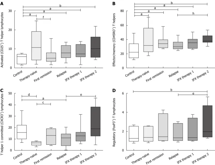
Figure 1 Prevalence of cellular members of adaptive immunity. The prevalence of activated (i.e. CD4+CD25+) (A), effector or memory (i.e. CD4+CXCR3+) (B), Th1 committed (i.e. CD4+CXCR3+) (C) and regulatory T (i.e. CD4+CD25hiFoxP3+) cells (D). aP < 0.05, bP < 0.01, dP < 0.001 vs control; cP < 0.05, fP < 0.01 vs therapy- naïve; eP < 0.05, hP < 0.01 vs relapse.
notype is linked to an improvement in PCDAI. This may be a specific feature in childhood CD as a number of stud- ies reported a higher than normal prevalence of activated and effector T lymphocytes even in treated CD adults[12-17]. Tregs probably do not play a role in this normalization as their prevalence was not altered in remission.
We performed another prospective study in 12 CD children who relapsed with conventional therapy and were
treated with IFX. In these CD children, the immune phe- notype was similar to that observed in therapy-naïve CD children (with the only exception of normal prevalence of NK and NKT cells). During IFX therapy, however, marked changes occurred in the 6th wk of IFX treatment and the prevalence of Th1 cells and the APCs investigated were normalized. Interestingly, the prevalence of activated T cells, memory cells and Treg cells were elevated further.
8
6
4
2
0 Natural killer (CD3-CD161+) cells
Control Therapy-naïve
First r
emission Relapse IFX ther
apy 1 IFX ther
apy 2 a
C D
A 4 B
3
2
1
0 Natural killer T (CD3+6b11+) cells
Control Therapy-naïve
First r
emission Relapse IFX ther
apy 1 IFX ther
apy 2 a
25 20 15 10 5 0 Peripheral monocytes (CD14+)
Control Therapy-naïve
First remission Relapse IFX ther
apy 1 IFX ther
apy 2 a a
a a
b c
6
4
2
0 Dendritic (Lin1-HLADR+) cells
Control Therapy-naïve
First remission Relapse IFX ther
apy 1 IFX ther
apy 2 a a
a
E 80 F
60
40
20
0
TLR-2 expressing dendritic cells
Control Therapy-naïve
First remission Relapse IFX ther
apy 1 IFX ther
apy 2 b
d c e g 40
30
20
10
0
TLR-4 expressing dendritic cells
Control Therapy-naïve
First remission Relapse IFX ther
apy 1 IFX ther
apy 2 a
b f e g
Figure 2 Prevalence of cellular members of innate immunity. The prevalence of natural killer T (i.e. CD3+6b11+) (A), natural killer (i.e. CD3-CD161+) (B), dendritic cell (i.e. Lin1-HLA-DR+) (C), peripheral monocyte (i.e. CD14+) (D), Toll-like receptor 2 expressing dendritic cell (E) and Toll-like receptor 4 expressing dendritic cell (F).
aP < 0.05, bP < 0.01, dP < 0.001 vs control; cP < 0.05, fP < 0.01 vs therapy-naïve; eP < 0.05 vs first remission; gP < 0.05 vs relapse.
Recently, increased numbers of Th1 cells and high blood levels of Th1-type cytokines were found in adult patients with rheumatoid arthritis (RA) after IFX treat-
ment[39,40]. Others also found an increase in effector and
activated T cells with IFX[38,39]. While the explanation for this is still unclear, an attractive hypothesis may be that IFX inhibits the homing of Th1 and activated T cells to the inflammation site and transiently increases their pe- ripheral occurrence[38,40].
Theoretically, this immune phenotype may refer to an increased risk for infections. However, our patients did not exhibit major clinical signs and symptoms of infec- tion during IFX therapy. Interestingly, simultaneously with these changes, IFX therapy also increased the prevalence of peripheral Treg cells. This phenomenon - which may be due to a resistance of Tregs to IFX-induced apoptosis - was also demonstrated previously in the peripheral blood of adult CD patients[8], and in colonic samples from CD children[37].
In our patients, the prevalence of monocytes, mDCs, mDC/pDC ratio and TLR-2 and TLR-4 expressing DCs normalized during IFX therapy indicating the possible impact of biological therapy on innate immunity. This is in line with the observation of others investigating RA and CD patients[41,43-45].
Our study has three major limitations. First, although we did correct for multiplicity when performing pairwise comparisons, the study was not powered for the multitude of statistical tests we performed, thus some significances could occur by chance alone. Second, peripheral cell prevalence values do not necessarily reflect the intestinal phenotype. Third, our results may have been affected by ageing of the patients during the follow-up period. The short duration of our prospective study (i.e. about 10 mo until the first remission or 6 wk from the beginning of IFX therapy), however, makes this bias less likely.
CD exhibits several abnormalities in adaptive im- munity (such as a decrease in Th1 cell and an increase in memory and activated T cell prevalence) and innate im- munity (such as an increase in DC, monocyte and TLR-2 and TLR-4 exhibiting APC prevalence). The majority of the observed alterations of the innate immune system are normalized with the improvement of clinical signs and symptoms of CD, irrespective of whether this is ob- tained by conventional therapy or add-on IFX therapy.
This finding suggests a link between immune phenotype and disease activity in childhood CD.
If these results are reinforced by other groups, the observations may raise the possibility that immune pheno- type is a potential biomarker for clinical response in CD children.
COMMENTS
Background
About 10%-15% of patients with Crohn’s disease (CD) are diagnosed before 18 years of age. Pediatric CD is a unique subtype of CD due to different loca- tion, altered responsiveness to therapy and different susceptibility factors com- pared with those in adult CD.
Research frontiers
Several studies indicate both the malfunction of adaptive and innate immunity in adulthood CD. Fewer data are available for CD children. The alteration of im- mune phenotype with treatment is also unclear in this population.
Innovations and breakthroughs
In this prospective study, the authors demonstrated marked alterations in both adaptive and innate immunity in childhood CD. These abnormalities were re- solved in infants responding to conventional therapy but not in non-responding children. Immune phenotype depends on disease activity.
Applications
These observations support the involvement of members of the adaptive and innate immune systems in childhood CD. They also identify immune phenotype as a possible biomarker for the follow-up of therapeutic success.
Peer review
In this study, the authors prospectively investigated the major components of adaptive and innate immunity in CD children. The authors showed several ab- normalities in adaptive and innate immunity in CD children. They also showed that the majority of observed alterations were normalized in remission stage and suggest a link between immune phenotype and disease activity in child- hood CD. Although the number of CD patients enrolled to this study was small, this prospective study demonstrated clinically interesting results in CD children.
REFERENCES
1 Dubinsky M. Special issues in pediatric inflammatory bowel disease. World J Gastroenterol 2008; 14: 413-420
2 Nieuwenhuis EE, Escher JC. Early onset IBD: what's the dif- ference? Dig Liver Dis 2008; 40: 12-15
3 Shaoul R, Karban A, Reif S, Weiss B, Shamir R, Tamir A, Da- vidovich O, Halevi J, Silver EL, Levine A. Disease behavior in children with Crohn's disease: the effect of disease dura- tion, ethnicity, genotype, and phenotype. Dig Dis Sci 2009; 54:
142-150
4 Zenewicz LA, Antov A, Flavell RA. CD4 T-cell differentia- tion and inflammatory bowel disease. Trends Mol Med 2009;
15: 199-207
5 Himmel ME, Hardenberg G, Piccirillo CA, Steiner TS, Lev- ings MK. The role of T-regulatory cells and Toll-like receptors in the pathogenesis of human inflammatory bowel disease.
Immunology 2008; 125: 145-153
6 van Lierop PP, Samsom JN, Escher JC, Nieuwenhuis EE. Role of the innate immune system in the pathogenesis of inflam- matory bowel disease. J Pediatr Gastroenterol Nutr 2009; 48:
142-151
7 Sartor RB. Mechanisms of disease: pathogenesis of Crohn's disease and ulcerative colitis. Nat Clin Pract Gastroenterol Hepatol 2006; 3: 390-407
8 Chamouard P, Monneaux F, Richert Z, Voegeli AC, Lavaux T, Gaub MP, Baumann R, Oudet P, Muller S. Diminution of Circulating CD4+CD25 high T cells in naïve Crohn's disease.
Dig Dis Sci 2009; 54: 2084-2093
9 Maul J, Loddenkemper C, Mundt P, Berg E, Giese T, Stall- mach A, Zeitz M, Duchmann R. Peripheral and intestinal regulatory CD4+ CD25(high) T cells in inflammatory bowel disease. Gastroenterology 2005; 128: 1868-1878
10 Saruta M, Yu QT, Fleshner PR, Mantel PY, Schmidt-Weber CB, Banham AH, Papadakis KA. Characterization of FOXP3+CD4+ regulatory T cells in Crohn's disease. Clin Im- munol 2007; 125: 281-290
11 Makita S, Kanai T, Oshima S, Uraushihara K, Totsuka T, Sawada T, Nakamura T, Koganei K, Fukushima T, Watanabe M. CD4+CD25bright T cells in human intestinal lamina pro- pria as regulatory cells. J Immunol 2004; 173: 3119-3130 12 Roman LI, Manzano L, De La Hera A, Abreu L, Rossi I, Al-
varez-Mon M. Expanded CD4+CD45RO+ phenotype and de- fective proliferative response in T lymphocytes from patients with Crohn's disease. Gastroenterology 1996; 110: 1008-1019 13 Alkim C, Balci M, Alkim H, Dağli U, Parlak E, Tezel A, Ulker
COMMENTS
A. The importance of peripheral immune cells in inflamma- tory bowel disease. Turk J Gastroenterol 2007; 18: 82-88 14 García de Tena J, Manzano L, Leal JC, San Antonio E, Sualdea V,
Alvarez-Mon M. Active Crohn's disease patients show a distinc- tive expansion of circulating memory CD4+CD45RO+CD28null T cells. J Clin Immunol 2004; 24: 185-196
15 Rose M, Hildebrandt M, Fliege H, Seibold S, Mönnikes H, Klapp BF. T-cell immune parameters and depression in pa- tients with Crohn's disease. J Clin Gastroenterol 2002; 34: 40-48 16 Liu ZX, Hiwatashi N, Noguchi M, Toyota T. Increased ex-
pression of costimulatory molecules on peripheral blood monocytes in patients with Crohn's disease. Scand J Gastroen- terol 1997; 32: 1241-1246
17 Senju M, Hulstaert F, Lowder J, Jewell DP. Flow cytometric analysis of peripheral blood lymphocytes in ulcerative colitis and Crohn's disease. Gut 1991; 32: 779-783
18 Zenewicz LA, Antov A, Flavell RA. CD4 T-cell differentiation and inflammatory bowel disease. Trends Mol Med 2009; 15:
199-207
19 Baumgart DC, Metzke D, Schmitz J, Scheffold A, Sturm A, Wiedenmann B, Dignass AU. Patients with active inflamma- tory bowel disease lack immature peripheral blood plasma- cytoid and myeloid dendritic cells. Gut 2005; 54: 228-236 20 Baumgart DC, Thomas S, Przesdzing I, Metzke D, Bielecki C,
Lehmann SM, Lehnardt S, Dörffel Y, Sturm A, Scheffold A, Schmitz J, Radbruch A. Exaggerated inflammatory response of primary human myeloid dendritic cells to lipopolysaccha- ride in patients with inflammatory bowel disease. Clin Exp Immunol 2009; 157: 423-436
21 Hart AL, Al-Hassi HO, Rigby RJ, Bell SJ, Emmanuel AV, Knight SC, Kamm MA, Stagg AJ. Characteristics of intestinal dendritic cells in inflammatory bowel diseases. Gastroenterol- ogy 2005; 129: 50-65
22 Frolova L, Drastich P, Rossmann P, Klimesova K, Tlaskalova- Hogenova H. Expression of Toll-like receptor 2 (TLR2), TLR4, and CD14 in biopsy samples of patients with inflammatory bowel diseases: upregulated expression of TLR2 in terminal ileum of patients with ulcerative colitis. J Histochem Cytochem 2008; 56: 267-274
23 Kamada N, Hisamatsu T, Honda H, Kobayashi T, Chinen H, Kitazume MT, Takayama T, Okamoto S, Koganei K, Sugita A, Kanai T, Hibi T. Human CD14+ macrophages in intestinal lamina propria exhibit potent antigen-presenting ability. J Im- munol 2009; 183: 1724-1731
24 Cantó E, Ricart E, Monfort D, González-Juan D, Balanzó J, Rodríguez-Sánchez JL, Vidal S. TNF alpha production to TLR2 ligands in active IBD patients. Clin Immunol 2006; 119: 156-165 25 Grimm MC, Pavli P, Van de Pol E, Doe WF. Evidence for
a CD14+ population of monocytes in inflammatory bowel disease mucosa--implications for pathogenesis. Clin Exp Im- munol 1995; 100: 291-297
26 van Ierssel GJ, van der Sluys Veer A, Verspaget HW, Griffioen G, van Hogezand RA, Lamers CB. Contribution of plasma cortisol to corticosteroid-suppressed peripheral blood natural killer cell activity in Crohn's disease. Immunopharmacology 1995;
29: 11-17
27 Grose RH, Thompson FM, Baxter AG, Pellicci DG, Cummins AG. Deficiency of invariant NK T cells in Crohn's disease and ulcerative colitis. Dig Dis Sci 2007; 52: 1415-1422
28 Kontiainen S, Scheinin T, Halme L. Number of activated T-helper cells and NK cells in peripheral blood is decreased in severe Crohn's disease. APMIS 1996; 104: 355-361
29 Mack DR, Beedle S, Warren J, Davis J, Gross T. Peripheral blood intracellular cytokine analysis in children newly diag- nosed with inflammatory bowel disease. Pediatr Res 2002; 51:
328-332
30 Holland N, Dong J, Garnett E, Shaikh N, Huen K, Harmatz P, Olive A, Winter HS, Gold BD, Cohen SA, Baldassano RN, Kirschner BS, Heyman MB. Reduced intracellular T-helper 1
interferon-gamma in blood of newly diagnosed children with Crohn's disease and age-related changes in Th1/Th2 cytokine profiles. Pediatr Res 2008; 63: 257-262
31 Desreumaux P, Brandt E, Gambiez L, Emilie D, Geboes K, Klein O, Ectors N, Cortot A, Capron M, Colombel JF. Distinct cytokine patterns in early and chronic ileal lesions of Crohn's disease. Gastroenterology 1997; 113: 118-126
32 Jo Y, Matsumoto T, Yada S, Fujisawa K, Esaki M, Onai N, Matsushima K, Iida M. CCR4 is an up-regulated chemokine receptor of peripheral blood memory CD4+ T cells in Crohn's disease. Clin Exp Immunol 2003; 132: 332-338
33 Perminow G, Reikvam DH, Lyckander LG, Brandtzaeg P, Vatn MH, Carlsen HS. Increased number and activation of colonic macrophages in pediatric patients with untreated Crohn's disease. Inflamm Bowel Dis 2009; 15: 1368-1378 34 Szebeni B, Veres G, Dezsõfi A, Rusai K, Vannay A, Mraz M,
Majorova E, Arató A. Increased expression of Toll-like recep- tor (TLR) 2 and TLR4 in the colonic mucosa of children with inflammatory bowel disease. Clin Exp Immunol 2008; 151: 34-41 35 Diefenbach KA, Breuer CK. Pediatric inflammatory bowel
disease. World J Gastroenterol 2006; 12: 3204-3212
36 Veres G, Baldassano RN, Mamula P. Infliximab therapy in children and adolescents with inflammatory bowel disease.
Drugs 2007; 67: 1703-1723
37 Ricciardelli I, Lindley KJ, Londei M, Quaratino S. Anti tu- mour necrosis-alpha therapy increases the number of FOXP3 regulatory T cells in children affected by Crohn's disease. Im- munology 2008; 125: 178-183
38 Ferkolj I, Ihan A, Markovic S, Veceric Z, Pohar M. Infliximab reduces the number of activated mucosal lymphocytes in pa- tients with Crohn's disease. J Gastrointestin Liver Dis 2006; 15:
231-235
39 Aeberli D, Seitz M, Jüni P, Villiger PM. Increase of peripheral CXCR3 positive T lymphocytes upon treatment of RA pa- tients with TNF-alpha inhibitors. Rheumatology (Oxford) 2005;
44: 172-175
40 Maurice MM, van der Graaff WL, Leow A, Breedveld FC, van Lier RA, Verweij CL. Treatment with monoclonal anti- tumor necrosis factor alpha antibody results in an accumula- tion of Th1 CD4+ T cells in the peripheral blood of patients with rheumatoid arthritis. Arthritis Rheum 1999; 42: 2166-2173 41 Richez C, Schaeverbeke T, Dumoulin C, Dehais J, Moreau JF,
Blanco P. Myeloid dendritic cells correlate with clinical re- sponse whereas plasmacytoid dendritic cells impact autoan- tibody development in rheumatoid arthritis patients treated with infliximab. Arthritis Res Ther 2009; 11: R100
42 Balanescu A, Radu E, Nat R, Regalia T, Bojinca V, Ionescu R, Balanescu S, Savu C, Predeteanu D. Early and late effect of in- fliximab on circulating dendritic cells phenotype in rheuma- toid arthritis patients. Int J Clin Pharmacol Res 2005; 25: 9-18 43 Lügering A, Schmidt M, Lügering N, Pauels HG, Domschke W,
Kucharzik T. Infliximab induces apoptosis in monocytes from patients with chronic active Crohn's disease by using a cas- pase-dependent pathway. Gastroenterology 2001; 121: 1145-1157 44 Baert FJ, D'Haens GR, Peeters M, Hiele MI, Schaible TF, Shealy
D, Geboes K, Rutgeerts PJ. Tumor necrosis factor alpha an- tibody (infliximab) therapy profoundly down-regulates the inflammation in Crohn's ileocolitis. Gastroenterology 1999; 116:
22-28
45 De Rycke L, Vandooren B, Kruithof E, De Keyser F, Veys EM, Baeten D. Tumor necrosis factor alpha blockade treat- ment down-modulates the increased systemic and local expression of Toll-like receptor 2 and Toll-like receptor 4 in spondylarthropathy. Arthritis Rheum 2005; 52: 2146-2158 46 Inflammatory bowel disease in children and adolescents:
recommendations for diagnosis--the Porto criteria. J Pediatr Gastroenterol Nutr 2005; 41: 1-7
47 Hyams JS, Ferry GD, Mandel FS, Gryboski JD, Kibort PM, Kirschner BS, Griffiths AM, Katz AJ, Grand RJ, Boyle JT.
Development and validation of a pediatric Crohn's disease activity index. J Pediatr Gastroenterol Nutr 1991; 12: 439-447 48 Satsangi J, Silverberg MS, Vermeire S, Colombel JF. The
Montreal classification of inflammatory bowel disease: con- troversies, consensus, and implications. Gut 2006; 55: 749-753 49 Mamula P, Markowitz JE, Baldassano RN. Inflammatory
bowel disease in early childhood and adolescence: special con- siderations. Gastroenterol Clin North Am 2003; 32: 967-995, viii 50 Diefenbach KA, Breuer CK. Pediatric inflammatory bowel
disease. World J Gastroenterol 2006; 12: 3204-3212
51 Svec P, Vásárhelyi B, Pászthy B, Körner A, Kovács L, Tulas- say T, Treszl A. Do regulatory T cells contribute to Th1 skew-
ness in obesity? Exp Clin Endocrinol Diabetes 2007; 115: 439-443 52 Selby WS, Jewell DP. T lymphocyte subsets in inflammatory
bowel disease: peripheral blood. Gut 1983; 24: 99-105 53 Yuan SZ, Hanauer SB, Kluskens LF, Kraft SC. Circulating
lymphocyte subpopulations in Crohn's disease. Gastroenterol- ogy 1983; 85: 1313-1318
54 Kool M, Lambrecht BN. Dendritic cells in asthma and COPD:
opportunities for drug development. Curr Opin Immunol 2007; 19: 701-710
55 de Heer HJ, Hammad H, Kool M, Lambrecht BN. Dendritic cell subsets and immune regulation in the lung. Semin Immu- nol 2005; 17: 295-303
S- Editor Tian L L- Editor Webster JR E- Editor Ma WH