Interactions between Plant Viruses
and Host Cells
K. W. MUNDRY
Max Planck Institut für Biologie, Tübingen, Germany
Introduction
All viruses have two principal features in common, genetic autonomy and the absolute inability to multiply without the help of energy- and metabolite-delivering systems of a host cell. T h e multiplication of viruses is therefore the result of a chain of close interactions between two genomes, that of the virus and that of the cell.
This chain of interactions is initiated by the infectivity of the virus, which itself must be a function of the intrinsic structure of the viral nucleic acid in connection with particular features of the whole virus particle.
It is evident that the smaller a virus particle is and the simpler its structure, the easier is the elaboration of characteristic features in connec- tion with its function. Many plant viruses are such simple viruses. In addition to having a simple structure they can be easily purified in rather large amounts and exhibit a high degree of variation. Their main dis- advantages are failure to show genetic recombination and extremely low plating efficiency. This is why the use of plant viruses for the investigation of problems in molecular biology is restricted mainly to the relation of virus structure to virus infectivity and to some aspects of virus mutation and replication.
The infectivity of virus particles cannot be investigated without the use of an infectible system, and the infectibility of a cell cannot be studied without the infectious virus particle. Although at present some character- istics of virus infectivity and of its genetics may be attributed to certain details of virus structure, the infectibility of the host cell is far from being understood. This is particularly true in the case of the plant cell. The greater part of this review will therefore deal with problems of virus structure in relation to virus infectivity and the genetic determination of
65
66 K. W. MUNDRY
virus strains. Some problems concerning the infectibility of plant cells and the multiplication of viruses will be discussed later.
Structure and Infectivity of Plant Viruses
A. Minimal Infectious Length of Elongated Plant Virus Particles A characteristic feature of many plant viruses (and also of certain bac- teriophage found by Marvin and Hoffmann-Berling, 1963, and Zinder et al., 1963) is their overall rod-shaped or threadlike structure. An early observation that the length of most such particles approximates a virus species-specific "most frequent length" or "normal length," immediately raises the question concerning the minimal length necessary for infectivity.
For example (Fig. 1), the most frequent length for particles of tobacco mosaic virus (TMV) is about 300 ιημ (Williams and Steere, 1951); for wheat streak mosaic virus the length was found to be about 750 ιημ (Brakke and Staples, 1958); and for sugar beet yellows virus, about 1250 ιημ (Mundry, 1958). The particles of tobacco rattle virus are shorter than those just mentioned (about 180 ιημ; Paul and Bode, 1955) and are always accompanied by a second component measuring about 70 ιημ. Although the existence of a particular and characteristic length is very striking in all preparations of elongated virus particles, few attempts have been made to correlate particle length with infectivity. This is particularly sur- prising since for many years the question remained open whether particles deviating in length from the normal length represented common products of virus replication or merely breakdown products resulting from the procedures employed in isolating viruses. In all cases studied, the most frequent length was found—within the limits of estimation—to be identical with the minimal length required for virus infectivity. In- vestigations on sugar beet yellows virus have yielded evidence that devia- tion from the minimal length required for infectivity by 100 Â renders the particles noninfectious (Fig. 2) (Mundry, 1958).
According to high resolution electron micrographs obtained by H o m e et al. (1959) and Russell and Bell (1963), the structure of sugar beet yellows virus particles is fundamentally similar to that of TMV particles.
The particles may consist, as in TMV, mainly or even solely of nucleic
FIG. 1. Frequency of particles of different length in preparations of four elongated plant viruses. Redrawn with data on tobacco rattle virus from Paul and Bode (1955);
tobacco mosaic virus from Williams and Steere (1951); wheat streak mosaic virus from Brakke and Staples (1958); and sugar beet yellows virus from Mundry (1958); n, number of particles; πΐμ, length of particle.
INTERACTIONS BETWEEN PLANT VIRUSES AND HOST CELLS 67
500
t
n 250
tobacco rattle virus
T — r i i l r i 9 400 800 πημ,-
10
tobacco mosaic virus
400 800 1200 1600 mp,—*►
68 K. W. MUNDRY 1.0
0.0
ΊΊ'
y\
=1050θΧ
E
ϊ/
=10 900 % ^sr
B
m
= 11300 X
FIG. 2. Relative concentration of particles of sugar beet yellows virus of a given minimal length m (solid line) and relative infectivity of the samples (dotted line).
Particle concentrations were estimated by electron microscopy via the frequency distribu- tion of particles of different length. Sets of curves for particles of 12,700 Â length and longer superimpose, indicating that the particles of 12,700 Â and longer are the infec- tious ones. From Mundry (1958).
INTERACTIONS BETWEEN P L A N T VIRUSES AND HOST CELLS 69
acid and protein. The nucleic acid of TMV is RNA which is helically twisted and surrounded by subunits of the protein coat; these subunits stack between the turn of the RNA helix. Thus the RNA strand is efficiently protected from all sides, particularly since the protein subunits of the coat extend beyond the RNA strand into the central hollow core of the particle. This hollow core has also been observed in the sugar beet yellows virus particle. T h e removal of some part of the protein coat of TMV, at the ends or elsewhere, renders the infectivity RNase-sensitive (Hart, 1955; Corbett, 1964; see also Commoner, 1959). This observation was followed by the discovery of the infectivity of isolated TMV RNA (Gierer and Schramm, 1956; Fraenkel-Conrat, 1956). Neither the whole protein coat of TMV (and probably of other mechanically transmitted plant viruses) nor some (e.g., terminal) parts of it seem to play a funda- mental role in plant virus infectivity.
B. The Role of the Integrity of Viral RNA in Infectivity
The loss of infectivity of elongated virus particles observed on reduc- tion of their original length must therefore be attributed to the loss of some part of the RNA core of the virus. T h e effect of this loss indicates either the loss of certain genetic information essential for the production of an important component, e.g., a protein intimately related to the replication process, or the loss of some specific terminal structure of the RNA molecule. Such a terminal structure might be related to the initia- tion or termination of RNA replication without necessarily containing genetic information to be translated into a peptide or protein molecule.
Data from different authors are contradictory in this respect. Today it is well established that cleavage of internucleotide bonds in TMV RNA, either by the action of RNase (Gierer, 1958), or by heat treatment (Ginoza, 1958), always renders the particle noninfectious. Both types of inactivation exhibit first-order kinetics. Therefore, it is concluded that a single break is sufficient to cause loss of infectivity in the RNA strand.
Although the results indicate that one out of about 6500 possible breaks is sufficient, the possible effects of internucleotide bond cleavage near an end of the polynucleotide strand cannot be judged from these experi- ments. T h e number of nonlethal hits will be too small to cause a detectable shoulder on the inactivation curve.
Fraenkel-Conrat and his collaborators have recently tried to charac- terize the terminal configuration of infectious TMV RNA by hydrolysis with highly purified exonucleases (Fig. 3). T h e results are interesting in several respects: (1) Neither end of the polynucleotide chain carries a
70 K. W . MUNDRY
phosphate residue (Sugiyama and Fraenkel-Conrat, 1961); (2) the terminal nucleoside at both ends is adenosine in all four strains investigated (Sugiyama and Fraenkel-Conrat, 1963); (3) removal of several nucleotides from the 3'-hydroxyl end of the chain permits certain conclusions to be drawn regarding the nucleotide sequence near this end; also, loss of these nucleotides does not interfere with the infectivity of the RNA strand nor does it influence the primary structure of the virus coat protein subunits.
Furthermore, and this observation is most curious, TMV RNA lacking about three nucleotide residues at the 3'-hydroxyl end of the chain and which, according to claims, is still infectious, produces on replication polynucleotide strands of the common structure including the sequence of terminal nucleotides removed from the molecules of the preparation prior to inoculation (Singer and Fraenkel-Conrat, 1963).
HO "P^ I^R I OH
n n = 6500
FIG. 3. T h e terminal nucleotide sequence of the ribonucleic acid of tobacco mosaic virus. Data from Sugiyama and Fraenkel-Conrat (1961, 1963), and Singer and Fraenkel- Conrat (1963).
No sensible explanation is possible at the present time unless it is assumed that the original integrity of the whole nucleotide sequence was preserved in some strands of the preparation and that these strands were the infectious ones that produced the common progeny. It would be worthwhile to study this system again. Only an accurate quantitative and kinetic comparison between the effects of enzymatic attack and the in- activation of infectivity of the RNA, however, could be accepted as proof for the statement that some terminal nucleotides are meaningless in terms of infectivity of the chain. Even then they would not necessarily be meaningless: Their removal might affect a cistron which is nonessential for the infectivity and which then might mutate. A very particular class of mutants should then be found and a wide spectrum of many different types should not result from such treatment.
Some calculations have been made concerning the number of nucleo- tides missing in slightly shortened noninfectious virus particles. For TMV, it has been reported that 50-130 missing nucleotides render the particles noninfectious (Commoner et al., 1958). Density gradient centrifugation of
INTERACTIONS B E T W E E N P L A N T VIRUSES AND HOST CELLS 71
several spherical viruses often demonstrates the existence of particle classes differing in density. In the case of broad bean mottle virus the most dense particles contain the largest amount of RNA, and only these were found to be infectious. Particles less dense seem to lack 130-360 nucleo- tides, and are noninfectious (Aronson and Bancroft, 1962).
The very loose structure of the particles of sugar beet yellows virus suggests that single turns of the nucleoprotein particle, and therefore an equivalent amount of its nucleic acid, might be lost upon isolation of the virus. It has already been mentioned that a deviation of 100 Â from the minimal length required for infectivity renders these particles non- infectious (Fig. 2). This was the smallest deviation checked in these experiments, and equals the loss of about four turns of the nucleoprotein particle, according to estimates based on Home's electron micrographs.
Unfortunately, the size of the nucleic acid molecule of this virus has not yet been determined. At the moment we can only speculate that if the RNA is similar in size to many other viruses and has a molecular weight of about 2 X 10⁶ (equaling about 6500 nucleotide residues per chain), the observed inactivation would be due to the removal of about 50 nucleotide residues. Loss of a single turn would then amount to loss of about a dozen nucleotides. These experiments should therefore be repeated with the greatest accuracy possible today.
The structure of polynucleotide chains exhibits a distinct polarity because the phosphate residues form a bridge connecting the 3'-hydroxyl group of one nucleoside with the 5'-hydroxyl group of its neighbor (Fig. 3). A given exonuclease attacks a polynucleotide chain either from its 3'-hydroxyl end or from its 5'-hydroxyl end, according to the specificity of the enzyme. The message contained in a viral RNA strand is read from one end to the other, probably in the same direction in all cases. Removal of terminal nucleotides from the 3'-hydroxyl end, as in Fraenkel-Conrat's experiments, must therefore have an effect fundamentally different from chain curtailment at the 5'-hydroxyl end. In the latter case the first
"letters" (so to speak) of the message are lost, and in the former case the last ones are missing. If we are not mistaken in believing that the loss of a few nucleotides in the 3'-hydroxyl terminal sequence does not interfere with the infectivity of viral RNA, then the removal of nucleotides from the 5'-hydroxyl end has a greater probability of being lethal in terms of infectivity.
Practically all the observations (except removal of three 3'-hydroxyl terminal nucleotides) support the view that the entire RNA chain is required for infectivity. Hence the question remains whether missing
72 K. W. MUNDRY
information can be complemented by a phenomenon similar to multi- plicity reactivation observed with phages.
Very recently, Hulett and Loring (1965) made an interesting observa- tion: According to their data the infectivity of TMV preparations is not dependent solely on the concentration of typical 300 ιημ particles.
Preparations with a higher concentration of shorter particles than con- trols with an equal concentration of ordinary particles were more infectious than was expected on the basis of concentration of 300 ιημ particles alone. That the effect might be due to a multiplicity phe- nomenon seems to be a reasonable explanation. There are, however, difficulties in this interpretation. The probability of introducing more than one virus particle into the same epidermal cell during the inocula- tion procedure is very low since the maximum number of infectible sites per leaf is much smaller than the number of epidermal cells. As will be explained later, the concentration of infectious virus particles in the inoculum must be about 1 mg/ml in order to saturate all infectible sites.
To introduce several particles (each smaller than the typical TMV particle) into the same cell, the concentration of such particles must be as high or even higher. The concentration of inoculum used by Hulett and Loring was much lower. Aggregation of particles is therefore required to ensure the introduction of more than one particle at a time into a cell, otherwise this phenomenon remains obscure.
In summary, we can say that the integrity of practically the whole nucleotide sequence of viral RNA is generally needed for its infectivity.
The loss of a few percent (or even less than 1%) of the nucleotides from one end interferes with infectivity in the cases studied so far. Perhaps the removal of a very few nucleotides from the 3'-hydroxyl end of the chain may not produce the same results. The possible mutagenicity of this effect has not been checked. The fundamental difference between the two ends of a viral polynucleotide chain as deduced from the polarity of the chain also remains to be demonstrated.
Virus Mutation
Even when the integrity of the whole nucleotide chain of plant viruses is required for the infectivity of these viruses, certain single nucleotides whose replacement by another nucleotide would result in mutation are not necessary for infectivity. Others might not participate in demonstrable genetic information at all. These problems are discussed in the following section.
INTERACTIONS BETWEEN P L A N T VIRUSES AND HOST CELLS 73
A. Virus Mutation in Vivo: Replication Errors and Growth Tempera- ture
In living cells for which genetic information is mainly conserved in chromosomes, mutations may arise for many different reasons including disturbances of cell division and chromosome partition, and disturbances of chromosome structure and replication. So far, the simple plant viruses have not yielded clear evidence for recombination events (see, however, Aach, 1961; Best, 1961) or multiplicity reactivation, and their infectivity is lost on the occurrence of single breaks in the nucleic acid strand. The mutations of plant viruses which do occur are therefore probably not the result of mere breakage and reunion of RNA fragments. Most if not all mutations of simple plant viruses occurring during virus multiplication must be considered replicating errors, possibly resulting from mispairing of bases and having the effect of base replacements. We might expect that the frequency of mispairing, if it occurs at all, is temperature-dependent.
Thus, the mutation rate should be temperature-dependent. In bacterio- phage, spontaneous mutation was not affected within a more or less physiological range of temperature (Wittmann, 1957).
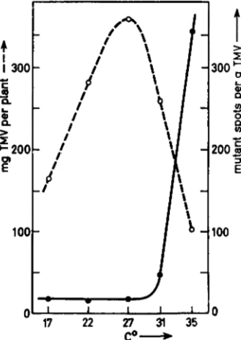
We have investigated the effect of temperature on the appearance of variant strains of TMV while the virus was multiplying in tobacco plants (Mundry, 1957b). At temperatures exceeding 30°C a sharp increase was found in the number of tissue areas containing mutants ("mutant spots") produced per gram virus finally isolated (Fig. 4). From 17° to 30°C the number of spots per gram virus was virtually constant. Lower tempera- tures were not tested. Although selection phenomena are difficult to judge, they do not seem to be responsible, for the following reasons:
(1) The types appearing with increased frequency at increased tempera- tures do not reveal evidence for positive selection; they remain im- prisoned in small areas of tissue which are surrounded by tissue infested with the wild-type strain; (2) they do not "overgrow" the wild-type strain even under conditions in which the probability of becoming predominant in the plants is great (that is, when the progeny virus moves out of the inoculated leaves and spreads into noninfected parts of the plant, par- ticularly into the upper noninoculated leaves); (3) short-time exposure of infected plants to a temperature of 35°C for periods of 24 hours produces the most pronounced effect if such treatment is applied immediately after inoculation and before the onset of massive invasion by progeny virus from noninoculated parts of the plant (Fig. 5). This indicates that the very high selection pressure resulting from the rapid multiplication of
74 K. W . MUNDRY
the wild-type strain must be maintained at a low level, otherwise, the mutants would not be able to invade enough cells to become detectable with the naked eye.
A reasonable explanation for the discrepancy between these results and the observations on bacteriophages cannot be given. It is possible that errors are more easily produced during replication of RNA than during
♦
1 1 300*-· c
Φ
>
* 2 0 0
?
100
n
- / i
/ / _/
4
A \\
1 * / ' * / Ί
/ \ / J
/ \ 1 1 w J
Λ 1
A I / * 1/ H
—1 1 1 I l-J
H 300'
>
200 2 o
CL (0
Έ S
"5
E
100
17 22 27 C°-
31 35
FIG. 4. Mutation of tobacco mosaic virus, strain "vulgäre," and the amount of virin produced per plant as a function of temperature. Plants were inoculated and then kept under continuous light at the temperatures indicated. So called "yellow-spots"
(small leaf areas infested with mutants, varying in appearance from light green to bright yellow) were counted 15 days after inoculation; dotted line, milligrams TMV produced per plant; solid line, mutant spots per gram virus produced. Redrawn with data from Mundry (1957b).
replication of DNA. The problem concerning the variability in the trans- lation of RNA-contained messages into amino acid sequences (Friedman and Weinstein, 1964), indicated by observations of the temperature de- pendence of the "meaning" of a given nucleotide triplet, might be con- sidered as evidence for temperature dependency of mispairing among ribonucleotides. Experiments with RNA phage might perhaps clarify the situation.
INTERACTIONS BETWEEN P L A N T VIRUSES AND HOST CELLS 75
B. Conversion of One Nucleotide into Another: Chemomutagenesis with Nitrous Acid
While irradiation of isolated TMV with ultraviolet light of X rays did not reveal any indication of mutagenic effects (Mundry, 1957a; 1960b), chemical modification of RNA did. Schuster and Schramm (1958) found
300
200
100
0 -
-
L
^
L-
\ 3 5 °
\22°
I I L
-]
-^
-J
\ A
, I
I 24 48 72 96 120
hours p.i.—*
FIG. 5. Influence on the mutation of TMV of the timing of 24-hour exposure to 35°C, of TMV-inoculated tobacco plants. Time 0 = hour of inoculation. Sets of 21 plants were kept in the greenhouse (average temperature 22° ± 2°C) and were ex- posed to 35°C and continuous light in a climatron for one of the first five days after inoculation. Another series of sets received similar climatron treatment at 22°C T h e dotted line represents the average number of " m u t a n t spots" (for explanation see Fig. 4) on two control sets kept in the greenhouse continuously. Data from Mundry (1957b); ordinate, mutant spots per 21 plants; abscissa, hours post-inoculation.
that nitrous acid inactivates TMV RNA without splitting internucleotide bonds. Such treatment results in deamination of the amino bases adenine, guanine, and cytosine, and yields hypoxanthine, xanthine, and uracil, while the integrity of the RNA molecule is maintained. Schuster and Schramm found no pronounced difference in the deamination rates for the three bases when isolated RNA was incubated. Inactivation of
76 K. W. MUNDRY
infectivity followed first-order kinetics. Single deaminations thus seem to be efficient in causing noninfectivity of the RNA strand. From the data they calculated that there were 3000-3300 possible lethal hits. This figure is significantly lower than the 4500 deaminations possible per TMV
TMV-infections on Java tobacco
A B C
local necrotic strain either mixed with systemic strain (e.g. means) impurities or mutants (e.g. vulgäre)
UV, HN02
secondary symptoms (sSS, on noninoculated leaves)
primary symptoms ('P.S., on inoculated leaves)
FIG. 6. Mutation from non-necrotic strains of tobacco mosaic virus to necrotic strains, and reverse mutation. Test plants are Java tobacco. Only treatment of a non- necrotic strain with nitrous acid yields localized necrotic lesions among faint chlorotic lesions of the wild type on the inoculated leaves; the reverse mutation cannot be induced; ultraviolet (and also X-ray) irradiation of purified virus is without mutagenic effect in both directions.
FIG. 7. Dependence of the concentration of mutants (number of necrotic lesions on Java tobacco relative to the maximum value) on the duration of incubation with nitrous acid, ί/τ, units of time required for inactivation to e — l, e — 2, e — 3 . . . e — 1 sur- vivors; under the conditions of these experiments τ is about 18 minutes; A> data for TMV nucleoprotein particles, all others for isolated TMV RNA. Data from Gierer and Mundry (1958).
INTERACTIONS BETWEEN P L A N T VIRUSES AND HOST CELLS 77
RNA strand. The remaining 1200-1500 amino bases either do not con- tribute to the genetic information of the virus, or deamination of at least some of them should be mutagenic.
This possibility was investigated and mutants were in fact found (Gierer and Mundry, 1958; Mundry and Gierer, 1958). We checked for the appearance of localized necrotic strains in preparations of RNA from a non-necrotic wild-type strain as a function of duration of treatment with nitrous acid (Figs. 6 and 7). First-order kinetics for inactivation and also for the appearance of necrotic mutants was observed. These results indi- cate that a single deamination per polynucleotide of 6500 nucleotide residues is mutagenic. Earlier experiments had investigated selection processes and showed that they could be well controlled (Mundry, 1957a).
These experiments were the first demonstration of the mutagenicity of single base alterations on a genetic element in vitro. Soon after publi- cation the interpretation of the results was criticized, first by Bawden (1959), and later by Markham (1963). Bawden argued that selection of strains with reduced sensitivity toward the inactivating effect of nitrous acid might have been the cause of the results observed. Such selection processes were calculated (Fig. 8) and were found not to fit the experi- mental data at all, neither quantitatively, nor qualitatively (Mundry,
1959). Even when entire resistance to nitrous acid was ascribed to all (0.4%) mutant type RNA molecules pre-existing in the incubation mix- ture, 75% of all mutants found after treatment would still have resulted from the mutagenic action of nitrous acid. This argument was rejected by Bawden (1961) without the addition of any other viewpoint.
Markham's criticism (1963) was based on the phenomenon of mutual exclusion among related strains of a plant virus: One strain will not multiply in a cell where a second strain is already multiplying. This leads to the following effects: T h e lower the concentration of infectious particles is in an inoculum, the further apart are the infections on the leaves, and the lower is the probability that pre-existing mutants or impurities consisting of other strains will be obscured by the bulk of the material. The only reply to arguments involving mutual exclusion is that all our conclusions are based on comparisons of treated samples with con- trols diluted to give approximately identical spacing of infections on the leaves. Details of this procedure were worked out before the nitrous acid experiments (Mundry, 1957a). They were presented again in connection with a critique of Bawden's argument (Mundry, 1959). Finally, mathe- matical treatment of the data included an additional correction that expressed and compared the results on the basis of concentrations of
78 K. W. MUNDRY
infectious particles. Any selection would have caused a deviation from the theoretically calculated curve, but none was observed (see Fig. 7).
Treatment of TMV RNA with nitrous acid yields many different types of mutants. For the appearance of localized necrotic lesions from a non- necrotic wild-type strain, about 180-200 single deaminations were found to be effective. In a screening experiment where as many different mutants as possible were classified, the "total mutability" was found to be several times higher than that towards necrotic strains only. It exceeded the rate
0 18 36 5V 72 90 t [min] **
FIG. 8. Production of mutants of TMV RNA by deamination with nitrous acid and theoretical course of selection of more slowly inactivated strains. A,B,C, and D:
0.4% of the infectious material assumed to be entirely resistant (A); twenty times (B);
five times (C); and three times (D) less sensitive than the bulk of the material to the inactivating effect of H N 02; solid line, experimental curve. From Mundry (1959).
toward necrotic mutants by about a factor of 8. T o the 3000-3300 "lethal hits" calculated by Schuster and Schramm, we can now add a number of about 1400-1600 mutagenic hits. Hence deaminable nucleotides in TMV RNA not participating in demonstrable genetic information could not be detected. On the basis of several observations (Mundry, 1960c, 1963;
Siegel, 1960), we might argue that such nucleotides do not exist. How- ever, according to the degeneracy of the genetic code, nucleotide triplets should exist which would not reveal any biological effect after the deami- nation of a single base (see Fig. 9). It seems reasonable to assume that the number of such possibilities in TMV RNA is small and has escaped detection.
INTERACTIONS B E T W E E N P L A N T VIRUSES AND HOST CELLS 79
C. Other Studies on Chemomutagenesis in Vitro
Induction of mutation on viral RNA by chemically defined reactions is of particular interest for investigations of the relationship between the structure of a messenger and the structure of a specific protein. For this reason, chemomutagenesis of viral RNA has attracted particular interest and the experiments with nitrous acid were followed by several other attempts to alter the genetic specificity of viral RNA (and DNA) by chemical means.
Schuster and Mundry (1959) carried out some experiments on the pos- sible mutagenic effect of bromination on TMV RNA with inconclusive results (unpublished data). We have also checked the behavior of TMV RNA toward dimethyl sulfate. From the kinetics of the inactivation (first- order kinetics) and the observation that the proportion of mutant types among the survivors remained virtually unchanged, we have concluded that treatment of TMV RNA with dimethyl sulfate is not mutagenic (Table I) (Schuster and Mundry, 1958).
A contrasting conclusion based only on the isolation of variant strains from leaves inoculated with dimethyl sulfate-treated T M V RNA was drawn by Fraenkel-Conrat. Kinetic data were not published, and the possible selection resulting from decreasing mutual exclusion generally leading to an increase in the number of mutant-type lesions has obviously not been taken into account (Fraenkel-Conrat, 1961). For this reason we regard our data as the more conclusive.
The effect of bromination of TMV RNA with bromosuccinimide was followed under similar conditions by Tsugita and Fraenkel-Conrat (1962).
It was claimed to be slightly but definitely mutagenic.
The observation that hydroxylamine treatment of bacteriophage evokes mutations (Freese et ai, 1961) has been reproduced also with TMV RNA (Schuster and Wittmann, 1963). A slight increase in the proportion of variant strains after treatment of TMV RNA with hydroxylamine has been found among the survivors. The reaction is specific for the alteration of uridylic acid residues under appropriate pH conditions.
T h e Genetic Code
A. General Features of the Genetic Code. Support of Current Hy- potheses by Studies on TMV Mutants
We have referred to problems concerning the genetic code several times.
I shall not present a detailed discussion, but a short extract will be given of what has been achieved in experiments with plant viruses (particularly
Οθ
TABLE I
REACTION OF TMV RNA WITH DIMETHYL SULFATEO
Relative virus concentration
(survivors) 1 0.1 0.01 0.001
1&
0.186 0.01 &
0.0034&
0.00 lö
Duration of treatment (minutes)
0 0 0 0 0 3 6 11 16
Total infectivity (Local lesions
on 15 leaves of xanthi n.c.
tobacco) 6221 1568 120 21 5982 2220 1365 513 116
Mutant types (Necrotic lesions
on 32 leaves of Java tobacco)
19 6 0 0 16 5 3 3 0
Proportion / Mutant lesions \ y total infectivity /
3.06 X 1 0 - 3 3.82 X 1 0 - 3
~~"
2.68 X 1 0 - 3 2.26 X 1 0 - 3 2.20 X 1 0 - 3 5.84 X 1 0 - 3
—
>
,
Average proportion
3.44 X 1 0 - 3
3.26 X 1 0 - 3
* 3 S c
Ö
*:
a From Schuster and Mundry (1958).
& Calculated via the infectivity-dilution curve.
INTERACTIONS BETWEEN P L A N T VIRUSES AND HOST CELLS 81
with mutants of TMV) on some general features of the genetic code. This seems justified because these studies are also related to some problems of plant virus replication.
The general features of the genetic code could be defined if answers were found to the following questions: (1) How many nucleotide residues are required for coding one amino acid residue (problem of code word size)? (2) How many code words can stand for one particular amino acid (problem of degeneracy)? (3) Does a single nucleotide at its location within the sequence of its neighbors participate in coding for only one or for more than one amino acid residue in the gene product (problem of over- lap of code words)? (4) Is the code universal?
The observations of Nirenberg and Matthaei (1961) revealed that in an in vitro system prepared from E. coli a messenger containing only uridylic acid residues provides sufficient information to code for the incorporation of phenylalanine into peptides synthesized by this system; similarly a chain of cytidylic acid residues codes for proline. We have found that treatment of TMV RNA with nitrous acid is mutagenic. One of the events responsible for this mutagenicity is the alteration of cytidylic acid residues into uridylic acid residues. Therefore, it should eventually be possible to induce the replacement of a proline residue in the coat protein of TMV by a phenylalanine residue. Wittmann (1959, 1962), Tsugita and Fraenkel- Conrat (1961), and Tsugita (1962a,b) have performed experiments of this kind. The proteins of many nitrous acid-induced mutants have been analyzed. A total of 49 amino acid replacements were distributed over 42 mutant lines among the 154 mutant strains checked, while 112 mutants did not exhibit any change in the amino acid composition of the coat protein (Wittmann, 1964). Among the exchanges were the following:
proline to serine (observed four times), proline to leucine (observed four times), leucine to phenylalanine (observed once); two cases of the replace- ment of serine by leucine were also found. Changes in the opposite direc- tion did not occur. This observation has two main consequences: First, it lends substantial support to the claim that cytidylic acid residues do participate in coding for proline in a natural messenger, and uridylic residues for phenylalanine. The second conclusion to be drawn from this observation is at least as important: T h e TMV RNA and not a replica of it with a reciprocal base composition is itself the messenger.
The results also support the triplet hypothesis, at least partly. Proline can be replaced by phenylalanine either by two subsequent exchanges (via serine or leucine) or by a chain of three events, to serine first, then via leucine to phenylalanine. In the latter case, provision must be made for
82 K. W. MUNDRY
three possibilities of deamination in constructing the code word for proline. Since homologous sequences of cytidylic acid residues are suffi- cient to code for proline, three such residues would allow the change of the proline code word to a phenylalanine code word via three subsequent deaminations.
It was confusing that the same final result, the substitution of proline by phenylalanine, can be achieved in two ways, either via serine or via leucine alone. The current hypotheses therefore take into account the possibility of degeneracy of the code. Degeneration means that different code words exist for one particular amino acid. The scheme in Fig. 9 combines some of the experimental findings with the triplet hypothesis and the obvious degeneracy of the code for cases of proline-phenylalanine
UpUpUp phe FIG. 9. T h e deamination of a cytidylic acid triplet to an uridylic acid triplet via
three single steps and the change of the meaning of the code words. Dotted arrows represent possibilities for deaminations that do not affect the meaning of a code word and therefore have no biological effect.
substitutions in nitrous acid-induced mutants of TMV. The assignments are paralleled by the results from the E. coli in vitro system. Similar suggestions and comparisons could be made for many of the 64 possible nucleotide triplets. Thus, the substitution of serine by leucine can be explained by a change of a triplet UCX to UUX, when X stands for either A or G.
While the triplet hypothesis is reasonable, and the degeneracy of the code well established in the experimental systems so far investigated, the problem of overlap of code words is solved by the data on amino acid replacements in the coat protein of nitrous acid-induced TMV mutants.
Simultaneous replacements of two adjacent amino acids have never been observed. This indicates that the deamination of a single nucleotide generally causes a change in meaning of only one code word while the neighboring code words remain unaffected. If code words overlap on the polynucleotide strand which represents the messenger the opposite result should have been found. However, among 49 exchanges determined by
INTERACTIONS BETWEEN P L A N T VIRUSES AND HOST CELLS 83
Wittmann (1964), not a single exchange of two adjacent amino acid residues could be demonstrated.
Finally it should be mentioned that agreement between the results from two systems so widely unrelated as the TMV-multiplying tobacco cell and the in vitro system prepared from E. coli cells strongly suggests the possi- bility that more than the main features of the genetic code is universal. At present there are no reasonable ideas concerning the possible develop- ment during evolution of different systems for selection among the code words for a single amino acid. Most of the possible triplets seem to have a meaning in terms of amino acid specification, but it has yet to be shown whether all these are used with equal frequency in all organisms.
B. The Problem of Functional Separation of Cistrons
Changes of single nucleotides not affecting the genetic information con- tained in TMV-RNA can be deduced from details of the triplet code (dotted arrows in Fig. 9), but have escaped experimental detection so far.
The question whether there are any should therefore be discussed, and also whether the existence of nucleotide sequences not dealing with the information to be translated into protein is required for the proper func- tioning of a polycistronic messenger.
TMV RNA, like other viral RNA, is probably a polycistronic messenger.
A cistron is defined as a portion of nucleic acid molecule which contains the information for a single protein. A polycistronic messenger hence con- tains information for several proteins. This information is based on specific sequences of amino acid code words. It is thus clear that the in- formation for different proteins must be kept functionally separated from one another, or the products of adjacent cistra might become intercon- nected so as to form a super-polypeptide. So far, the general occurrence of a mechanism which splits such a super-polypeptide into its proper poly- peptide units, does not seem to be indicated. Cistrons should therefore be functionally separated while they are structurally linked in a poly- cistronic messenger. These links might be nucleotide sequences carrying
"nonsense" in terms of amino acid code words, or they might be links of a non-nucleotide nature.
Several attempts have been made to demonstrate the occurrence in viral (particularly TMV) RNA of non-nucleotide material covalently linked to nucleotides somewhere in the chain, and constituting a part of the backbone of the molecule. No convincing results have been obtained so far. On the contrary, evidence is accumulating that neither amino acids nor metal ions, generally found in traces in even the purest preparations
84 K. W. MUNDRY
of viral RNA, constitute genuine components of the RNA backbone.
Whether they constitute genuine compounds of TMV or other viral RNA at all, also remains to be demonstrated (see Mundry, 1963).
Recently published data (Brenner et al., 1965) indicate the existence of
"nonsense" triplets, to which none of the common amino acids could be related (UAA and UAG). Such sequences would suit very well the purpose of functional separation of cistrons, but their existence in polycistronic messengers can hardly be demonstrated conclusively unless a mechanism has been discovered which permits the selection of triplets according to the natural reading frame.
In spite of this situation, degradation of TMV RNA via a few high- molecular distinct parts, has been repeatedly demonstrated (see Mundry,
1963; Miura et al., 1963). Similarly, the RNA of brome grass mosaic virus seems to occur within the cell as three components (probably not as a result of degradation due to the isolation procedures): The infectious RNA and two pieces resembling in size one-third and two-thirds of the infectious one, characterized by ultracentrifugation analysis (Bockstahler and Kaesberg, 1965). The nature of the weak bonds in viral RNA is still obscure and remains to be investigated, as well as the problem whether the observed distinct pieces of viral RNA are subunits in a functional sense or are mere breakdown products due to intracistronic cleavage.
T h e Size of Cistrons and the Concept of Minimal Infectious Genetic Information
Based on a triplet code, the size of the cistron which determines the primary structure of the TMV coat protein would be 3 nucleotides for each of 158 amino acids (474 nucleotides).
We have already mentioned that in our nitrous acid-induced TMV mutants, 180-200 deaminations were found, each of which could alter a non-necrotic strain of TMV into a necrotic mutant when the tests were performed on a particular variety of tobacco. The mechanism of lesion production is a hypersensitivity reaction of the host tissue which prevents the spread of virus over large areas and into noninoculated parts of the plant. This mechanism depends on the presence of a single gene in the host cells. It is conceivable that wild-type TMV contains information which counteracts the effect of this host gene, thus enabling the virus to invade the inoculated or otherwise infected plant, and to spread via the vascular system into other parts of the plant. The 180-200 amino bases found to be involved in the induction of necrotic strains by deamination
INTERACTIONS BETWEEN P L A N T VIRUSES AND HOST CELLS 85
might very well belong to a particular cistron on the RNA of TMV. This view is supported by the observation that reverse mutations from necrotic strains, including those of spontaneous origin, could not be induced to revert to non-necrotic ones by treatment with nitrous acid (Mundry, 1960a). If allowance is made for the (nondeaminable) uridylic acid resi- dues, the size of this cistron could very well be 250-300 nucleotides.
Investigations of the multiplication of several RNA viruses, including plant viruses (Ralph et al., 1965), has revealed that a specific enzyme not present in the uninfected cells is required to replicate the RNA via a double-stranded RNA. It is assumed that this enzyme is virus-specific, and that information necessary for its production is a part of the viral genome. The smallest RNA virus should therefore possess a cistron for the production of this enzyme and one for the virus' coat protein. Thus an RNA equivalent to at least two cistrons would be required to con- stitute infectious viral information.
The RNA of the smallest plant viruses so far isolated is much larger than would be anticipated from the above argument. The RNA of broad bean mottle virus contains about 3400 nucleotides (Yamazaki et al., 1961), and the RNA of brome grass mosaic virus has about 3000 nucleotides (Bockstahler and Kaesberg, 1961). The smallest viral RNA is that of a small bacteriophage in which the nucleic acid molecule has only 1600 nucleotides (Loeb and Zinder, 1961, quoted in Caspar and Klug, 1962).
This figure is probably close to the lower limit of that necessary to con- stitute infectious genetic information.
There is, however, another RNA virus with only 1160 nucleotides, apparently below this limit. It will not multiply in healthy cells of its natural host, but it will reproduce if another virus is present in the cells.
Multiplication of the tobacco necrosis satellite virus (Kassanis, 1960;
Reichmann et al., 1962; Reichmann, 1964) seems to be dependent on the presence of additional information, carried neither by itself nor by the genome of the host cell. It is reasonable to assume that the information missing in the RNA of the satellite virus is the cistron for the RNA replicating enzyme, and that the larger tobacco necrosis virus provides this information upon infection. We can speculate that the minimum number of nucleotides required to constitute fully infectious genetic in- formation is of the order of a thousand.
It should be mentioned that the two species of particles generally found in preparations of tobacco rattle virus (Fig. 1), of which only the larger is infectious (Harrison and Nixon, 1959), are also of interest in connec- tion with the concept of incomplete infectious genetic information.
86 K. W. MUNDRY
Plant Virus Replication and Some of Its Cellular Events
In the preceding sections we have tried to define some of the features characteristic of the construction of infectious genetic information, by describing the events which cause the loss of infectivity and change of the original genetic information of a virus. We cannot, however, answer the question why a certain sequence of nucleotides, like the one in T M V RNA, is infectious. It is still unknown how host cells, for example those of tobacco leaves which contain about 2 X 10⁹ base pairs of DNA per nucleus, are forced to reproduce a virus simply by the addition of an RNA strand of only 6500 nucleotides to this mass of information. Obser- vations on the fundamental processes of reproduction of viruses in the plant cell are few and scattered, and are far from fitting into a reasonably established picture. Only a few details of more general interest will be presented here.
T h e mechanism of virus uptake by plant cells is still obscure. While pinocytosis in animal cells is gaining particular interest in this respect (Smith, 1963), observations on pinocytosis-like phenomena in plant cells are few and lack details. If pinocytosis is the mechanism for the uptake of virus, the saturation of all infectible sites on a plant leaf should take place when the virus concentration in the inoculum is enough for there to be one infectious particle for each microdrop of inoculum within a single pinocytosis vesicle. I have calculated the size of a drop that would con- tain one infectious particle at a concentration of virus in the inoculum which would assure a maximum number of lesions on the leaves. A drop- let size of about 1 μ3 was found for inoculations with the nucleoprotein particles or independently with T M V R N A (Mundry, 1963). This calcu- lation agrees with the hypothesis of virus uptake into plant cells by pinocytosis.
Although the uptake of viruses by plant cells probably depends upon their physiological activity (particularly when pinocytosis may be in- volved), it also seems to be dependent upon the nature of the virus coat protein. T h e specific infectivity of mixed reconstitutions of T M V (that is, T M V rebuilt from isolated R N A of one strain and isolated coat protein of another) parallels the specific infectivity of the protein donor strain, and not that of the strain from which the R N A has been isolated (Holoubek, 1962). T h e coat protein might therefore have something to do with stimulating virus uptake. It would be interesting to know whether it also stimulates pinocytosis.
Earlier we mentioned the phenomenon of strain interference by mutual
INTERACTIONS BETWEEN P L A N T VIRUSES AND HOST CELLS 87
exclusion. Nothing is known about the time of onset of mutual exclusion.
If it is based on competition among strains for adsorption sites or entry points for virus uptake into the cell it should occur early in the infection process. This possibility was tested by experiments based on the observa- tion that rubbing leaves with buffer produces wounds or exposes to the environment attachment sites which remain susceptible to infection for a while. These sites can be brought into contact with virus by dipping the prerubbed leaves into the inoculum. In our experiments such sites were subsequently exposed to two different strains. One was a non-necrotic strain at a high concentration which had previously been shown to guarantee saturation of practically all previously produced infectible sites. The other was a local lesion-producing strain, at a concentration which would give proper lesion counts. No difference was found between the results depending upon whether prerubbed leaves were first dipped in strain A and then into strain B containing inoculum or vice versa. The lesions deriving from this treatment were always identical in number to those appearing on the controls in which the dip into the non-necrotic, highly concentrated inoculum was replaced by a dip into buffer contain- ing no virus. If time was allowed to elapse between the first virus dip and the second one no other effect could be observed than a general decrease in the tendency of the leaves to produce infections through preformed wounds. No indication of mutual exclusion was found within 10 minutes after the first dip. Within this time the leaves lost their infectibility.
Among the possible explanations of these findings are the following hypotheses: (1) There is no strain-specific adsorption of virus to receptive sites; a certain number of sites are produced, lose their infectibility, and virus adsorbed to them may be replaced by the virus applied during the second dip; (2) there are absolute strain-specific sites, and therefore no competition for infectible sites; again, a number of sites are produced upon rubbing and these lose infectibility with time; (3) the leaf is stimulated by the rubbing procedure and the stimulus leads to the con- tinuous production of very short-lived sites which are nonspecific toward strains; as the stimulus dies, fewer and fewer sites appear, and after 10 minutes, no more. In this case, the sites present during the time of the first dip are not identical with those present at the time of the second dip. These hypotheses could be tested but have not been so far (Mundry,
1958b).
Little is known and little can be said about the fate of the virus particle after it has entered the plant cell. From the following observations we can deduce that the coat protein is removed from the nucleic acid mole-
88 K. W. MUNDRY
cule of the virus particle. There is a change in the sensitivity of infective centers on inoculated leaves to the inactivating effect of ultraviolet light (Siegel and Wildman, 1960; Mundry, 1963). Infective centers become sensitive to the action of pancreatic ribonuclease from soon after the infection up to two hours post infectionem (p.i.) (Hamers-Casterman and Jeener, 1957). In addition, photoreactivation of potato virus X is only possible within 30-120 minutes p.i. (nucleoprotein particles are probably not reactivated by light) (Bawden and Kleczkowski, 1955). Hence the protein may have been removed within the first half hour p.i. This time may not be the same for different plant viruses. Details of the stripping procedure have been recently reviewed (Mundry, 1963).
Recent experiments on the detection of double-stranded RNA as the replicating form of viral RNA in bacteria have lead to similar efforts in the case of virus infected plant cells (Burdon et al., 1964; Shipp and Haselkorn, 1964; Ralph et al., 1965). The characterization of an enzyme involved in the production of double-stranded viral RNA during the course of infection has been attempted (Weissmann et al., 1964). Although details of the replication mechanism are still unknown it is remarkable that an RNA-dependent RNA replicating enzyme is not ordinarily present in normal cells, but is produced upon infection following the introduction of genetic information with the viral RNA. This hypothesis is supported by elegant experiments on a RNA phage (Spiegelman et al.,
1965). It provides a key to the problem of virus infectivity.
Finally some data related to the biochemical events of virus multiplica- tion in plant cells will be discussed. Direct observation of metabolic changes within the infected cell was performed by ultraviolet micro- spectrophotometry (Zech and Voigt-Koehne, 1956; Zech, 1963, as cited in Mundry, 1963). Zech and his collaborators studied the change in the optical density at 265 and 280 ιημ at different locations in the cell after the introduction of TMV. The measurements cover the first 24-hour period after the inoculation. The studies were performed on the cells of tobacco leaf hairs. The terminal cell was inoculated and the changes in RNA concentration or nucleolar mass determined in the cell adjacent to the inoculated one. Careful control and standardization measurements have been published separately (Zech, 1961). The data are summarized in Fig. 10.
About 30 minutes p.i. the RNA concentration in the nucleus begins to rise. At about 2 hours p.i. it has increased up to 2.3 times its original value. This concentration remains constant until 5 hours p.i. then it starts decreasing gradually until 9 hours p.i., when it reaches its original
INTERACTIONS BETWEEN P L A N T VIRUSES AND HOST CELLS 89
0 2 » 6 8 10 12 /« 16 18 20 22 2H
i I I I i
11.0+23* zero time RNA content of the nucleus 23* maintained
123+1.0* LQx maintained
1.0+03*zero time value
25'*maintained
8*maintained
RNA content of nucleus-surrounding cytoplasmic zone
8-*"* 1.0* zero time value
RNA content of cytoplasm more distant from the nucleus
8-*· 2* zero time value
zero time value ofnuc/eo/armass
bss5S5oss5ssi!S!3Ksssssss5B 10 to L3x zero time value
8 10 12 It hours post inoculation
FIG. 10. Ultraviolet-microspectrophotometric measurements on TMV-infected to- bacco leaf hair cells. Data from Zech (1963); reproduced from Mundry (1963). T h e terminal cell of a leaf hair was inoculated at time 0, and changes in RNA content or nucleolus mass in the cell adjacent to the inoculated one were followed. T h e widths of the bars in each sequence are proportional to the data obtained; each bar repre- sents an average of the data obtained from many (occasionally more than 100) cells;
data represented by hatched bars are less reliable due to technical difficulties; below the wavy line numbering begins with the cell adjacent to the inoculated one and proceeds toward the base of the hair.
90 K. W . MUNDRY
value again. There it remains constant for another 8 hours. During the eighteenth hour p.i. it continues to decrease slowly, revealing only 30%
of its original value at the end of the first day of infection. The data suggest strongly that RNA is produced within the nucleus after about 30 minutes. The nucleolus was excluded from the data and the micro- beam was focused so as not to touch the nucleolus for these measurements.
This is possibly the time required to separate the RNA from the coat protein of the virus particles. RNA production continues for about 4-5 hours. The measurements in other zones adjacent to the nucleus, or more distant from it, seem to establish the transport of RNA out of the nucleus into the cytoplasm surrounding the nucleus, and then into parts of the cell further removed from the nucleus. Electron microscopic in- vestigations (Zech, 1960; von Wettstein and Zech, 1962) paralleled these observations. Large amounts of material were found filling the endo- plasmatic reticulum. Later this material becomes tightly packed there, while the membranes of the reticulum are densely occupied with ribo- somes. The increase in RNA content in the cytoplasm surrounding the nucleus is immense; it rises within half an hour to 25 times its original value. The zone of high optical density obviously spreads radially, and the RNA content of the cytoplasm more distant from the nucleus in- creases, while that of the zone surrounding the nucleus decreases slowly.
The material deposited in vesicular enlargements of the endoplasmatic reticulum in this region is finally condensed into bodies which are almost certainly virus crystals.
The observations indicate that TMV RNA is produced in the nucleus while the bulk of viral proteins are manufactured in the cytoplasm. The absence of TMV antigens in the nucleus of infected cells (Nagaraj, 1965) supports this view. The extraction of complete TMV nucleoprotein rods from tobacco cell nuclei (Reddi, 1964) is a claim for the contrary.
Quantitative measurements in all organelles and zones within the cell were impossible for technical reasons. Thus the behavior of the nucleolus could not be followed accurately because at first it was surrounded by the highly absorbing nucleus, and later by the very dense plasma zone sur- rounding the nucleus. Only after the RNA was widely distributed, and the optical density in the cell sufficiently decreased, was it possible to establish that there was a strong increase in the optical density of the nucleolus. Because of the very low water content of the nucleolus, a 30-50% rise in its extinction value could indicate a much higher RNA accumulation or production than would a 200-300% increase in the relatively water-rich, non-nucleolar portion of the nucleus. In spite of these difficulties, the conclusion seems justified that the onset of (non-
INTERACTIONS BETWEEN PLANT VIRUSES AND HOST CELLS 91
nucleolar) nuclear RNA synthesis precedes that of the nucleolus. Viral RNA synthesis in plant cells may therefore begin with some still unknown interactions between this RNA and the chromatin in the cell nucleus. T h e nature of this interaction is obscure and may be of fundamental im- portance for regulation phenomena.
ACKNOWLEDGMENT
T h e author is greatly indebted to Dr. L. E. Bockstahler for assistance in translating the manuscript.
REFERENCES
AACH, H. G. (1961). Interferenz zweier nahe verwandter Staemme des Tabakmosaik- virus. Ber. Deut. Botan. Ges. 74, 433-435.
ARONSON, A. I., AND BANCROFT, J. B. (1962). Density heterogeneity in purified prepara- tions of broad bean mottle virus. Virology 18, 570-575.
BAWDEN, F. C. (1959). Effect of nitrous acid on tobacco mosaic virus: mutation or selection? Nature 184, B.A.27-B.A.29.
BAWDEN, F. C. (1961). Some effects of changing environment on the behaviour of plant viruses. Symp. Soc. Gen. Microbiol. 11, 296-316.
BAWDEN, F. C , AND KLECZKOWSKI, A. (1955). Studies on the ability of light to counteract the inactivating action of ultraviolet radiation on plant viruses. / . Gen. Microbiol.
13, 370-382.
BEST, R. J. (1961). Recombination experiments with strains A and E of tomato spotted wilt virus. Virology 15, 327-339.
BOCKSTAHLER, L. E., AND KAESBERG, P. (1961). Bromegrass mosaic virus: a virus contain- ing an unusual small ribonucleic acid. Nature 190, 192-193.
BOCKSTAHLER, L. E., AND KAESBERG, P. (1965). Isolation and properties of RNA from bromegrass mosaic virus. / . Mol. Biol. 13, 127-137.
BRAKKE, M. K., AND STAPLES, R. (1958). Correlation of rod length with infectivity of wheat streak mosaic virus. Virology 6, 14-26.
BRENNER, S., STRETTON, A. O. W., AND KAPLAN, S. (1965). Genetic code: T h e "nonsense"
triplets for chain termination and their suppression. Nature 182, 994-998.
BURDON, R. H., BILLETER, M. A., WEISSMANN, C , WARNER, R. C , OCHOA, S., AND KNIGHT,
C. A. (1964). Replication of viral RNA, V.Presence of a virus-specific double- stranded RNA in leaves infected with tobacco mosaic virus. Proc. Natl. Acad. Sei.
U.S. 52, 768-775.
CASPAR, D. L. D., AND KLUG, A. (1962). Physical principles in the construction of regular viruses. Cold Spring Harbor Symp. Quant. Biol. 27, 1-24.
COMMONER, B. (1959). T h e biochemistry of the synthesis and biological activity of tobacco mosaic virus. Plant Pathol. Probl. Progr., 1908-1958 pp. 483-492.
COMMONER, B., SHEARER, G. B., AND STRODE, C. (1958). Linear analysis of tobacco mosaic virus. Proc. Natl. Acad. Sei. U.S 44, 1117-1122.
CORBETT, M. V. (1964). Electron microscopy of partially degraded tobacco mosaic virus.
Virology 22, 539-543.
FRAENKEL-CONRAT, H. (1956). T h e role of the nucleic acid in the reconstitution of active tobacco mosaic virus. / . Am. Chem. Soc. 78, 882-883.
FRAENKEL-CONRAT, H. (1961). Chemical modification of viral nucleic acids. I. Alkylating agents. Biochim. Biophys. Ada 49, 169-180.
92 K. W. MUNDRY
FREESE, E., BAUTZ-FREESE, E., AND BAUTZ, E. (1961). Hydroxylamine as a mutagenic and inactivating agent. / . Mol. Biol. 3, 133-143.
FRIEDMAN, S. M., AND WEINSTEIN, I. B. (1964). Lack of fidelity in the translation of synthetic polyribonucleotides. Proc. Natl. Acad. Sei. U.S. 52, 988-996.
GIERER, A. (1958). Die Groesse der biologisch aktiven Einheit der Ribonukleinsaeure des Tabakmosaik virus. Z. Naturforsch. 13b, 485-488.
GIERER, A., AND MUNDRY, K. W. (1958). Production of mutants of tobacco mosaic virus by chemical alteration of its ribonucleic acid in vitro. Nature 182, 1457.
GIERER, A., AND SCHRAMM, G. (1956). Infectivity of ribonucleic acid from tobacco mosaic virus. Nature 177, 702.
GINOZA, W. (1958). Kinetics of heat inactivation of ribonucleic acid of tobacco mosaic virus. Nature 181, 958-961.
HAMERS-CASTERMAN, C., AND JEENER, R. (1957). An initial ribonuclease-sensitive phase in the multiplication of tobacco mosaic virus. Virology 3, 197-206.
HARRISON, B. D., AND NIXON, H. L. (1959). Separation and properties of particles of tobacco rattle virus with different lengths. / . Gen. Microbiol. 21, 569-581.
HART, R. G. (1955). Infectivity measurements of partially degraded tobacco mosaic virus. Virology 1, 402-407.
HOLOUBEK, V. (1962). Mixed reconstitution between protein from common tobacco mosaic virus and ribonucleic acid from other strains. Virology 18, 401-404.
HORNE, R. W., RUSSELL, G. E., AND T R I M , A. R. (1959). High resolution electron microscopy of beet yellows virus filaments. / . Mol. Biol. 1, 234-236.
HULETT, H. R., AND LORING, H . S. (1965). Effect of particle length distribution on in- fectivity of tobacco mosaic virus. Virology 25, 418-430.
KASSANIS, B. (1960). Properties and behaviour of a virus depending for its multiplication on another. / . Gen. Microbiol. 27, 477-488.
LOEB, T., AND ZINDER, N. D. (1961). A bacteriophage containing RNA. Proc. Natl.
Acad. Sei. U.S. 47, 282-289.
MARKHAM, R. (1963). Plant virus nucleic acids. Progr. Nucleic Acid Res. 2, 61-81.
MARVIN, D., AND HOFFMANN-BERLING, H . (1963). Physical and chemical properties of two new small bacteriophages. Nature 197, 517-518.
MIURA, K.-I., MIURA, T., HIRUKI, C., HIDAKA, Z., AND WATANABE, I. (1963). Fractionation
of infectious ribonucleic acid isolated from tobacco mosaic virus. Virology 19, 140- 146.
MUNDRY, K. W. (1957a). Zur Frage des Einflusses von Roentgen- und UV-Strahlen auf die Mutationsrate des Tabakmosaikvirus nach Bestrahlung reiner Praeparate.
Z. Vererbungslehre 88, 115-127.
MUNDRY, K. W. (1957b). Die Abhaengigkeit des Auftretens neuer Virusstaemme von der Kulturtemperatur der Wirtspflanzen Z. Vererbungslehre 88, 407-426.
MUNDRY, K. W. (1958). Ueber die Korrelation zwischen Partikellaenge und Infektiositaet beim Vergilbungsvirus der Rueben. Z. Naturforsch. 13b, 19-27.
MUNDRY, K. W. (1958b). Unpublished data.
MUNDRY, K. W. (1959). T h e effect of nitrous acid on tobacco mosaic virus: mutation, not selection. Virology 9, 722-726.
MUNDRY, K. W. (1960a). Mutationsuntersuchungen am Tabakmosaikvirus in vitro. I.
Die Abhaengigkeit des Erfolges der Mutagenese mit H N 02 von der Mutations- richtung. Z. Vererbungslehre 91, 81-86.
MUNDRY, K. W. (1960b). Mutationsuntersuchungen am Tabakmosaikvirus in vitro. II.