https://doi.org/10.1007/s00239-019-09913-4 REVIEW
Virus–Host Coevolution with a Focus on Animal and Human DNA Viruses
Győző L. Kaján1 · Andor Doszpoly1 · Zoltán László Tarján1 · Márton Z. Vidovszky1 · Tibor Papp1
Received: 6 May 2019 / Accepted: 23 September 2019 / Published online: 10 October 2019
© The Author(s) 2019
Abstract
Viruses have been infecting their host cells since the dawn of life, and this extremely long-term coevolution gave rise to some surprising consequences for the entire tree of life. It is hypothesised that viruses might have contributed to the forma- tion of the first cellular life form, or that even the eukaryotic cell nucleus originates from an infection by a coated virus. The continuous struggle between viruses and their hosts to maintain at least a constant fitness level led to the development of an unceasing arms race, where weapons are often shuttled between the participants. In this literature review we try to give a short insight into some general consequences or traits of virus–host coevolution, and after this we zoom in to the viral clades of adenoviruses, herpesviruses, nucleo-cytoplasmic large DNA viruses, polyomaviruses and, finally, circoviruses.
Keywords Virus · Coevolution · Antiviral defence · Endogenous viral elements
Coevolution in General
Viruses are obligatory cellular parasites, developing with their hosts since the dawn of life. Coevolution, when the virus and the host reciprocally affect each other’s evolution, is often detected. According to the Red Queen Hypothesis, both the parasite and the host are perpetually struggling to maintain a constant fitness level (McLaughlin and Malik 2017). This long-term evolutionary pressure gave rise to some surprising consequences for the entire tree of life.
The Origin of Viruses—Which Came First:
The Chicken or the Egg?
What can be the origin of an obligatory cellular parasite?
Three concurring hypotheses describe the origin of viruses:
(i) the primordial virus world or the virus first hypothesis
claims that the ancestors of viruses existed already in the pre-cellular world; (ii) the escaped genes theory describes viruses as mobile genetic elements, which became independ- ent of their host cells; (iii) whereas according to the cellular regression theory, viruses are regressed intracellular para- sites (Holmes 2013).
The Primordial Virus World
According to the primordial virus world hypothesis, in the primordial soup multiple pre-cellular and pre-viral Ur- organisms competed with each other. The last universal common ancestor—the first cellular life form—emerged from these primordial replicators, and at least a fraction of current viruses might originate from the remaining ones (Moelling and Broecker 2019). Ancient viruses might have even contributed to the formation of the last universal com- mon ancestor of all living organisms.
It is generally accepted that an RNA-based world pre- ceded today’s DNA- and protein-based one. RNA viruses and especially the capsidless viroids might resemble this ancient world (Elena et al. 1991). However, the evolu- tion of viruses—which has been especially rapid for RNA viruses—destroyed the possible signal of genetic related- ness a long time ago, making phylogenetic reconstruction already impossible. Luckily, some sort of evidence is still
Handling editor: Konstantinos Voskarides.
* Győző L. Kaján
kajan.gyozo@agrar.mta.hu
1 Institute for Veterinary Medical Research, Centre
for Agricultural Research, Hungarian Academy of Sciences, Hungária krt. 21, Budapest 1143, Hungary
within reach: the existence of viral protein fold superfami- lies without cellular counterparts. Proteins without any primary sequence homology are still grouped based on their conserved tertiary structure, their folding: e.g. the major capsid proteins of the PRD1 bacteriophage, adeno- viruses and some archaeal viruses all have a double jelly- roll fold structure (Benson et al. 1999; Nasir and Caetano- Anollés 2015, 2017), or there are structural homologies in the RNA- and DNA-dependent polymerases too (Gor- balenya et al. 2002). There are fold superfamilies con- taining both viral and cellular proteins (we will return to this phenomenon by the escaped genes theory), but there are also numerous examples without cellular homologues found in phylogenetically very diverse viruses: e.g. viral RNA-dependent RNA polymerases are not homologous to their cellular counterparts (Iyer et al. 2003). According to the most parsimonious assumption, these proteins have a common origin in the primordial virus world, and phy- logenetic tree reconstructions of these fold superfamilies confirm this as well (Nasir and Caetano-Anollés 2015).
Viruses became obligatory parasites either only after the emergence of the last universal common ancestor, or it is equally possible that they were already parasitizing on other ancient replicators. According to the viral eukar- yogenesis hypothesis, even the eukaryotic cell nucleus originates from an infection by a coated virus: the virus became an endosymbiont usurping the role of the original archaean nucleus (Bell 2009). The endosymbiont pox-like virus had no possibility for a lytic viral cycle, and under this evolutionary pressure started to replicate by means of cell-to-cell fusion using its fusion proteins. When two such cells—infected by related viruses—fused to each other, the viral DNA was copied, and homologous viral chro- mosomes formed tetrads. Cell division resulted in four daughter cells with one copy of the endosymbiont virus in each. This might be the origin of meiotic cell division (Bell 2006). After its development, sexual reproduction offered a significant fitness advantage in the fight against any parasite (Hamilton et al. 1990), including viruses.
Escaped Genes
This theory states that mobile genetic elements escaped the cell, acquired a protein capsid and started to replicate autonomously (Moreira and López-García 2005, 2009).
Numerous homologies can be observed among viral and cellular genes. On the other hand, the root position, and thus the certain direction of evolution, might be challeng- ing to determine. For example, cellular and viral DNA polymerases are related (Filée et al. 2002), but the exact direction of the lateral gene transfer is unknown (Shack- elton and Holmes 2004).
The Cellular Regression Theory
Perhaps the least favoured theory currently claims that viruses are the extremely reduced descendants of obligatory intracellular parasites, incapable of autonomous extracel- lular life (Bândea 1983). According to this theory, the viral genome is the remnant of a heavily reduced cellular genome and the capsid is that of a cell membrane. The nucleo-cyto- plasmic large DNA viruses (NCLDVs) are good candidates to represent the end result of this mechanism: they prolifer- ate in the cytoplasm, and have particle and genome sizes comparable to those of the smallest prokaryotes (Arslan et al. 2011; Colson et al. 2012, 2011). The modern version of this theory was mentioned when discussing the primordial virus world: viruses might be the reduced descendants of pre-cellular Ur-organisms, converting into obligatory cel- lular parasites (Claverie 2006; Forterre 1991).
Viral Genetic Elements in Host Genomes
PolintonsThe theories of viral origin are a topic of heated debate, but most possibly none of them is mutually exclusive, and the suspected processes of two or more could have occurred parallel and/or sequentially as well. A small proportion of mobile genetic elements might have its root in the primordial virus world, but numerous regression or escape occasions must have occurred too. The latter were perhaps facilitated by the cellular genomic insertion of viral genetic elements.
Polintons, also known as Mavericks, are mobile genetic ele- ments with multiple homologues in both cellular and viral genomes. They have escaped several times and gave rise to different viruses and mobile genetic elements such as adenoviruses or mitochondrial linear plasmids (Krupovic and Koonin 2015, 2017). It must be stressed again that these genetic elements are homologous and related, but the direc- tion of the genetic exchange is unknown in most cases. Such transfers are common from host to virus and vice versa too.
Endogenous Viral Elements
Endogenous viral elements (EVEs) are viral genes or sometimes complete genomes inserted into host genomes.
As genome integration is a compulsory step in retroviral replication, most of these elements are of retroviral origin, but further virus families were detected as well: hepadna- viruses, adeno-associated viruses, herpesviruses and oth- ers (Aiewsakun and Katzourakis 2015; Bill and Summers 2004; Broecker and Moelling 2019; Morissette and Flamand
2010; Young and Samulski 2001). If the insertion occurs in the germ cell line, the inserted sequence stretch might get inherited. And if it provides a selective advantage, e.g.
protection from a viral infection, the genomic change will be fixed in the population. In case this advantage disappears, the inserted stretch might mutate over time and lose its pro- tective nature (Broecker et al. 2016).
EVEs are important sources for virus–host coevolution- ary research, as they provide a map for viral host range.
The history of coevolution can be traced by the detection of homologous endogenous viral sequences in different host species. This provides information about the historical host range of a virus clade, and even the time frame of coevolu- tion can be inferred based on the known diversification times of the hosts, or on phylogenetic analyses of exo- and endog- enous viral sequences. E.g. based on endogenous lentiviral sequences in lemurs, it was hypothesised that these viruses have been coevolving with primates for several millions of years (Katzourakis et al. 2007).
Viruses represent a major force in evolutionary pressure (Emerman and Malik 2010; Villarreal and Witzany 2010).
Retroviruses, for example, are so effective in genome inte- gration and have been coevolving with vertebrates so long, that over 50% of the human genome was estimated to consist of endogenous retroviral elements (de Koning et al. 2011).
Some of these regions provide important and fundamental attributes to their integrator hosts, like the syncytin genes, which have an important role in placental development not only in various mammals but in viviparous lizards as well (Cornelis et al. 2017; Imakawa and Nakagawa 2017). Other transposable elements contribute to embryonic development, stem cell pluripotency or cell differentiation in eukaryotes (Chuong et al. 2017). In prokaryotes, important functions were attributed to the integrated viruses as well. E.g. in E.
coli, deleting all prophages has resulted in increased sus- ceptibility to environmental factors and slower cell growth (Wang et al. 2010).
Effects of the Bottleneck Phenomenon
Essentially, the viral mutation rate and copy number deter- mine the variability within the host and in fine the stock of viral evolution (Duffy et al. 2008; Peck and Lauring 2018;
Sanjuán and Domingo-Calap 2016). Generally, viral path- ogens exist in diverse populations in vivo (McCrone and Lauring 2018). As a substantial evolutionary effect on the population dynamics of viruses, genetic bottleneck (reduc- tion of effective population size due to environmental events, for instance new habitat colonisation) affects the virus–host coevolution: it decreases both the genetic variation and the fitness of the virus, and furthermore it is responsible for the founder effect (Novella et al. 1995). Changes of genotype
frequencies by stochastic population size reduction are referred to as genetic drift (Bergstrom et al. 1999; Den- nehy et al. 2006; Elena et al. 2001; McCrone and Lauring 2018; Zwart and Elena 2015). Furthermore, it is important to stress that the phenomenon of genetic bottleneck is inter- pretable from both virus and host perspective (Voskarides et al. 2018).
In the case of the viruses, the newly colonised region might be a new host (host switch) or organ (Voskarides et al.
2018; Zwart and Elena 2015). Bottleneck events occur dur- ing both intra- and inter-species steps of the viral life cycle (Gutiérrez et al. 2012). An interesting aspect of virus evo- lution and the bottleneck effect is the case of multipartite viruses (e.g. plant-infecting nanoviruses or animal-infecting bidnaviruses, alphatetraviruses, nodaviruses and picobirna- viruses). These package their genetic material as indepen- dently encapsidated separate segments (Lucía-Sanz and Manrubia 2017). Self-evidently, bottleneck events have a crucial role for these viruses, as all genomic segments are needed for a successful infection. Furthermore, the different genomic segments drift at different rates (Gallet et al. 2018).
Antiviral Defence Mechanisms
Under the constant threat of an infection, cellular organisms have developed multiple layers of different defence mecha- nisms (not solely) against these genetic parasites to protect themselves and their genomic integrity.
Shuttled Weapons
Most strikingly, antiviral defence mechanisms often have viral origins too (Broecker and Moelling 2019; tenOever 2016; Villarreal 2009). The simplest mechanism is the superinfection exclusion, where an integrated and expressed endogenous viral protein provides protection from exoge- nous infection. This was first described in the tobacco plant, but it is applied by prokaryotes, animals or other plants as well (Moelling et al. 2017). The development of such a defence mechanism was observed in sheep and koalas for example. Retroviral elements were endogenised even as recently as 200 and 100 years ago in the genomes of sheep and koala, respectively, providing protection against certain exogenous retroviral infections (Armezzani et al. 2014; Tar- linton et al. 2006).
But such basic integrations are not the only examples.
The recently described bacterial antiphage system, CRISPR- Cas (Charpentier and Doudna 2013), has its roots in at least five different classes of mobile genetic elements (Koonin and Makarova 2017). The Argonaute proteins show struc- tural and functional homologies to the retroviral replication machinery and provide protection against invading nucleic
acids in prokaryotes (Moelling et al. 2006). The RNA inter- ference system of eukaryotes shares the same homologies, and it is available in each of the five eukaryotic superking- doms, suggesting a very ancient evolutionary origin (Cerutti and Casas-Mollano 2006). Though present, this system does not provide antiviral effects in chordates. But also here, endogenous retroviruses provide transcription factor bind- ing sites for interferon-stimulated genes (Chuong et al. 2016;
Ito et al. 2017), whereas the Rag recombinases—needed for the diversification of antibodies—originate from transposons found in the genomes of starfish, oysters and sea urchins (Kapitonov and Koonin 2015).
Other Mechanisms
The most ancient defence mechanism might be the use of antisense RNAs, where the gene translation of the invad- ing pathogen is interfered by small complementary RNAs (Gottesman and Storz 2011). Against DNA phages, bacte- ria often use restriction endonucleases, cleaving the viral genome (Kobayashi 2001). Piwi-interacting RNAs associ- ate with nucleases and provide defence against transpos- able elements. The latter are available in all metazoans, and they may have a common origin with the prokaryotic Argonaute system (Iwasaki et al. 2015). As already men- tioned, the RNA interference does not provide an antiviral effect in chordates; instead, pattern recognition receptors were developed. Engagement of these by pathogen-associ- ated molecular patterns (i) induces antiviral cytokines: the tumour necrosis factor, available in most eukaryotic line- ages, but in chordates rather the interleukins and interferons (Levy et al. 2011), and (ii) activates the natural killer cells too (Esteso et al. 2017). The APOBEC3 protein is part of the innate immune system in humans, it targets specifically retroviruses and interferes with their reverse transcription (Sheehy et al. 2002). However, HIV counteracts this effect by the viral infectivity factor, which triggers the degrada- tion of APOBEC3 (Donahue et al. 2008). Furthermore, it was also observed that human leucocyte antigen I and II loci are genetically more diversified in pathogen- (most pre- dominantly virus-) rich environments (Prugnolle et al. 2005;
Sanchez-Mazas et al. 2012).
After this short general introduction on the highlights of the topic, we focus on the coevolutionary processes of selected DNA viruses: adeno-, herpes-, NCLD-, polyoma- and circoviruses.
Adenoviruses
Adenoviruses are DNA viruses, the major capsid proteins of the non-enveloped, icosahedral capsid are the hexon, the penton base and the protruding fibre, responsible for receptor
binding. Their non-segmented, double-stranded, linear genome varies between 26 and 45 kbp in size, and the DNA is covalently bound to the terminal protein (Harrach 2014).
Adenoviruses infect vertebrate hosts, and are clustered into five accepted and one proposed genera. Members of the gen- era Mastadenovirus and Aviadenovirus infect mammals and birds, respectively. Atadenoviruses were detected in reptiles, birds, ruminants and a marsupial possum; siadenoviruses in birds, a frog and a tortoise. The single member of the genus Ichtadenovirus is the white sturgeon adenovirus. The sixth genus was proposed recently: testadenoviruses were detected in testudinoid turtles only until now (Doszpoly et al. 2013).
As these viruses were described from five major classes of the vertebrates, the hypothesis was formed that adeno- viruses had started to coevolve with the vertebrates 450 million years ago, before the divergence of fish from other vertebrates (Kovács et al. 2003). Mast-, avi-, at-, si- and ichtadenoviruses had been thought to coevolve with mam- mals, birds, reptiles, amphibians and fish, respectively (Benkő and Harrach 2003). This hypothesis was partially questioned later, as the white sturgeon and the frog adeno- virus—the latter belonging to genus Siadenovirus—are the single fish or amphibian adenoviruses, respectively, discov- ered until now (Davison et al. 2000; Doszpoly et al. 2019).
Furthermore, several avian adenoviruses clustered into the genus Siadenovirus (Ballmann and Harrach 2016; Kovács and Benko 2011; Kovács et al. 2010; Lee et al. 2014). Yet, it is equally possible that several further amphibian and fish adenoviruses are awaiting discovery.
Adenoviruses are generally thought to be host specific with usually one or very few host species for a specific viral serotype. Still, during the hundreds of millions of years, host changes did happen, e.g. there were ten predicted host switches in the evolution of human adenoviruses in 4.5 mil- lion years (Hoppe et al. 2015). The most striking assumed host switch happened for some atadenoviruses. These are thought to be the lineage coevolving with squamatid reptiles originally (Wellehan et al. 2004), but presumably a virus strain had jumped to an ancient ruminant during the evolu- tionary history. Atadenoviruses were isolated from cattle, sheep and mule deer, suggesting some level of coevolution already. However, the high genomic A + T content—hence the name atadenovirus—contradicts long cospeciation of these viruses (Benkő and Harrach 1998).
Non-reptile atadenovirus genomes are characterised by 57.0–66.3% A + T content, whereas reptile atadenoviruses are not affected by such bias (Farkas et al. 2002; Harrach 2008; Papp et al. 2009; Wellehan et al. 2004). Examples from other virus families are known: the influenzaviral nucleotide composition changes following a host jump from bird to mammal (Greenbaum et al. 2008), or similar differ- ences were observed between flaviviruses or herpesviruses of different hosts (Jenkins et al. 2001; McGeoch et al. 2006).
Obviously, this composition has an effect on codon usage, thus the complete genome sequence might be under evo- lutionary pressure (Shackelton et al. 2006). Still, the exact cause of this bias in atadenoviruses is unknown, but it is hypothesised that longer coevolutionary times—adaptation of the virus to the host—result in a balanced nucleotide composition (Farkas et al. 2002; Papp et al. 2009; Wellehan et al. 2004).
Similar observations were made about pathology. Adeno- viruses generally cause a mild disease, and there are apa- thogenic strains at least in healthy individuals. E.g. recent research shows that only specific and not all serotypes of the species Fowl aviadenovirus D and E cause inclusion body hepatitis in chicken (Schachner et al. 2018; Zadravec et al. 2011). But recent host switches might result in an elevated pathogenicity (Benkő and Harrach 2003; Jánoska et al. 2011; Kohl et al. 2012; Vidovszky et al. 2015). For instance, duck adenovirus 1 is apathogenic in ducks and geese, but causes the egg drop syndrome in chickens (Hess et al. 1997). Another atadenovirus causes a haemorrhagic epizooty among mule deer (Lehmkuhl et al. 2001; Woods et al. 1996). Or bovine adenovirus 10—though a mastad- enovirus—is only distantly related to other bovine mastad- enoviruses, and it causes a severe acute fibrinous enterocol- itis (Horner et al. 1989). The A + T content of the known genome part is over 59%, and from serologically identical strains several fibre size variants were described (Ursu et al.
2004). These mutations might be the consequences of an ongoing evolution, and represent the adaptation to a rela- tively new host, cattle (Benkő and Harrach 2003).
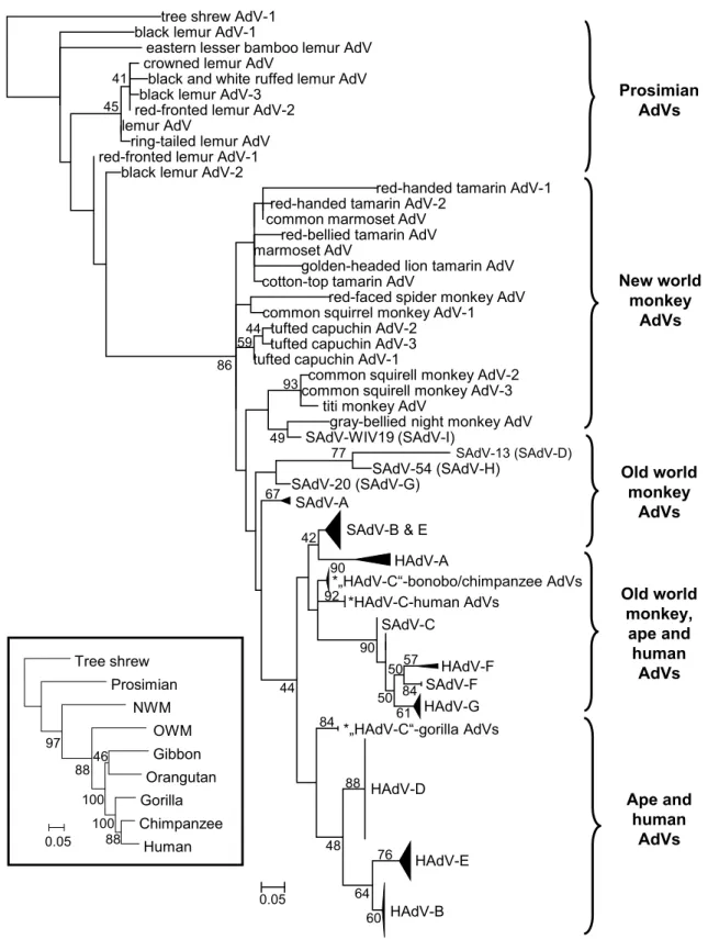
The most investigated adenoviruses are evidently human and other primate adenoviruses. Codivergence is not obvious here at the first look as host switches blur the picture (Hoppe et al. 2015; Purkayastha et al. 2005; Roy et al. 2009; Wevers et al. 2011), but it is still observable. The most ancient line- ages are the prosimian and New World monkey adenoviruses (Podgorski et al. 2018). On the next branches we can find the Old World monkey adenoviruses including members of Human mastadenovirus (HAdV) A, F and G (Gilson et al.
2016; Pantó et al. 2015; Podgorski et al. 2016) (Fig. 1). Evo- lutionarily speaking, strains of HAdV-B and -E are of gorilla or chimpanzee origin, respectively; and strains of HAdV-B jumped to the common ancestor of humans and chimpan- zees two times most probably (Hoppe et al. 2015). HAdV-C seemingly codiverged with gorillas, chimpanzees, bonobos and humans, whereas HAdV-D viruses infect only humans (Hoppe et al. 2015). The long-term coevolution of HAdV-D and humans is supported by the high variety of types within the species (Ismail et al. 2018b; Kaján et al. 2018), and their usually facultative pathogen nature.
Exactly in HAdV-D strains it is often observed that homologous recombination has a driving force in adenovirus evolution (Crawford-Miksza and Schnurr 1996; Gonzalez
et al. 2015; Ismail et al. 2018a; Kaján et al. 2017; Robinson et al. 2013; Walsh et al. 2009). Predominantly, intraspecies recombination events are common, but intergenus recom- binants have also been reported already: the head domain of the porcine adenovirus 5 (genus Mastadenovirus) fibre seems to be of atadenoviral origin (Nagy et al. 2002), though a porcine atadenovirus has not been discovered yet.
Recombination enables an accelerated, modular evolution of viruses, ‘and has been associated with such features as the evasion of host immunity (Malim and Emerman 2001), the development of antiviral resistance (Nora et al. 2007), the ability to infect new hosts (Hon et al. 2008), increases in virulence (Khatchikian et al. 1989) and even the creation of new viruses (Weaver 2006)’ (Holmes 2013).
Herpesviruses
Herpesviruses (HVs) are large (150–200 nm) icosahedral, enveloped viruses having a so-called tegument layer between the capsid and the envelope. HVs possess a linear, non- segmented, double-stranded DNA genome (135–295 kbp) (Pellett et al. 2011). They are known to infect all classes of vertebrates, from fish to humans; moreover, HVs were discovered in some mollusc species as well (Davison et al.
2009). Chronologically, HVs of mammals and birds were known first causing different diseases in humans, livestock and poultry. Later, several decades ago, HVs of reptiles, fishes and amphibians were also reported (Fawcett 1956;
Rebell et al. 1975; Wolf and Darlington 1971). These novel viruses were tentatively classified into the family Herpesvir- idae because of their morphological features. The first non- vertebrate HV was discovered in a mollusc species in the 1970s (Farley et al. 1972). As more sequence data became available, molecular analysis provided insight into the evolu- tion of HVs, and these newly discovered viruses were also officially classified into the family Herpesviridae (Davison 1998; Davison et al. 2006, 2005; Davison and Davison 1995;
McGeoch and Gatherer 2005). In 2009, the taxonomy of HVs changed radically, as the family Herpesviridae was split into three families. The novel family Herpesviridae contains only the HVs of higher vertebrates (Amniotes), while the family Alloherpesviridae contains the HVs of amphibians and fish (Anamnia), and the family Malacoherpesviridae contains the HVs of molluscs. The three families were clus- tered under the novel order Herpesvirales (Davison et al.
2009). These three groups of HVs are related only tenu- ously to each other. There are only a few genes showing homology in all known HVs, the most conserved gene is the putative ATPase subunit of terminase, its gene product being responsible for the packaging of the viral DNA into the capsid (Davison et al. 2009). Interestingly, a homologous gene was found in T4 bacteriophages (Myoviridae) implying
tree shrew AdV-1 black lemur AdV-1
eastern lesser bamboo lemur AdV crowned lemur AdV
black and white ruffed lemur AdV black lemur AdV-3
red-fronted lemur AdV-2 lemur AdV
ring-tailed lemur AdV red-fronted lemur AdV-1
black lemur AdV-2
red-handed tamarin AdV-1 red-handed tamarin AdV-2
common marmoset AdV red-bellied tamarin AdV marmoset AdV
golden-headed lion tamarin AdV cotton-top tamarin AdV
red-faced spider monkey AdV common squirrel monkey AdV-1
tufted capuchin AdV-2 tufted capuchin AdV-3 tufted capuchin AdV-1
common squirell monkey AdV-2 common squirell monkey AdV-3
titi monkey AdV
gray-bellied night monkey AdV SAdV-WIV19 (SAdV-I)
SAdV-54 (SAdV-H) SAdV-20 (SAdV-G)
SAdV-A
SAdV-B & E HAdV-A
*„HAdV-C“-bonobo/chimpanzee AdVs
*HAdV-C-human AdVs SAdV-C
HAdV-F SAdV-F HAdV-G
*„HAdV-C“-gorilla AdVs
HAdV-D
HAdV-E
HAdV-B 84
88
76
60 64 48
77 93
49 41
45
86 5944
67
44 90 42
92
90
84 5057
5061
0.05
SAdV-13 (SAdV-D)
Prosimian AdVs
New world monkey
AdVs
Old world monkey
AdVs
Old world monkey,
ape and human
AdVs
Ape and human
AdVs Tree shrew
Prosimian NWM
OWM Gibbon Orangutan Gorilla Chimpanzee
Human 97
88
10088 46
100
0.05
Fig. 1 Phylogeny reconstruction (maximum likelihood analysis) based on partial sequences of the IVa2 protein of primate adenovi- ruses (AdVs). A phylogenetic tree of primates is shown in the bottom left corner to demonstrate the parallel evolution of AdVs and hosts.
SAdV-A, Simian mastadenovirus A; HAdV-B, Human mastadenovi-
rus B, etc. Hosts of AdVs presumably belonging to species HAdV- C are marked with asterisks due to their separation. [From Podgorski et al.: Adenoviruses of the most ancient primate lineages support the theory on virus–host coevolution (2018) Acta Vet Hung 66:474, with permission from Akadémiai Kiadó.]
the common origin of HVs and tailed bacteriophages (Baker et al. 2005).
The phylogenetic relationship of HVs is well studied and their evolution was found to be largely synchronous with host lineages (McGeoch et al. 2000, 2006). Exceptions have been noted among the mammalian (McGeoch et al. 2006) and fish HVs (Kelley et al. 2005). The major sublineages within the subfamilies (Alpha-, Beta-, Gammaherpesvirinae) of Herpesviridae emerged probably before the mammalian radiation, 60 to 80 million years ago, while the diversifica- tion time point of the three subfamilies had been around 200 million years ago (McGeoch et al. 1995, 2000). The most common ancestor of all known HVs (order Herpesvi- rales) existed around 400 million years ago based on similar phylogenetic branching patterns of HVs and host lineages (McGeoch et al. 2006).
As for mammalian HVs, several studies were conducted on their evolutionary route spanning shorter time periods.
For example, Bovine herpesvirus 4 (BoHV-4) has been iso- lated from cattle worldwide and this species was thought to be the original host of the virus. However, BoHV-4 has been reported from wild buffalo and other ruminants as well (Ros- siter et al. 1989; Todd and Storz 1983). Later, serological studies implied that the original host of the virus might be the African buffalo (Syncerus caffer) (Dewals et al. 2005).
This hypothesis was confirmed by phylogenetic calculations, suggesting that BoHV-4 has been coevolving with the Afri- can buffalo for the last 1.5 million years, and the virus was passed to cattle on at least three independent occasions much more recently, sometimes via an intermediate species (Dew- als et al. 2006). In another study, the phylogenetic trees of primate cytomegaloviruses and their hosts’ were compared, and primate cytomegalovirus genomes were also analysed.
The obtained results demonstrated the coevolution of these viruses with their hosts (Russell et al. 2016). Furthermore, the avian and reptilian HVs were clustered into the subfam- ily Alphaherpesvirinae, relating to the development of birds from reptilian progenitors (McGeoch and Gatherer 2005).
As for the members of the family Alloherpesviridae, phy- logenetic inferences strongly support the monophyly of fish and amphibian HVs within the order Herpesvirales (Waltzek et al. 2009). The comparison of the phylogenetic trees of viruses and hosts implied that closely related HVs in the family may have coevolved with their hosts, whereas codi- versification was not supported at deeper nodes of the tree (Waltzek et al. 2009). In other words, the separation of the main alloherpesvirus lineages does not fully resemble that of the host taxa. One explanation can be that the ancestors of the different viruses diverged before the separation of the various fish lineages, and thus the different virus lineages evolved independently. The strong similarity among viruses of distantly related fish species could be explained by host switches (Doszpoly et al. 2011).
Integrated herpesviral genomes in host chromosomes were also discovered in the last years, giving insight into the virus–host coevolution and providing interesting data about the genome organisation and content of ancient virus species (Aswad and Katzourakis 2017; Inoue et al. 2017; Savin et al.
2010). The discovery of a new lineage of alloherpesviruses associated with at least fifteen different fish species was reported. One of them seems to represent a full-length viral genome in salmon (Salmo salar) (Aswad and Katzourakis 2017). An interesting discovery of a HV genome integrated into the genome of an invertebrate chordate provides links to the ancient ancestors of mollusc and vertebrate HVs:
the genome of Branchiostoma floridae (Florida lancelet or amphioxus) contains several genes showing homology to herpesviral genes, implying the existence of a HV associated with this invertebrate chordate. The terminase and polymer- ase gene sequences from the putative amphioxus HV show higher similarity to those of the mollusc HVs than to any vertebrate HVs (Savin et al. 2010). The discovery of this virus gave data about HVs dating back to the separation of vertebrates and invertebrates.
Nucleo‑cytoplasmic Large DNA Viruses
Nucleo-cytoplasmic large DNA viruses (NCLDV) are a group of DNA viruses including the families Ascoviridae, Asfarviridae, Iridoviridae, Marseilleviridae, Mimiviridae, Pithoviridae, Phycodnaviridae and Poxviridae (Koonin and Yutin 2010, 2019). A new virus order, the Megavirales was proposed [but not yet accepted by the International Commit- tee on Taxonomy of Viruses (ICTV)] to collect the above- mentioned virus families (Colson et al. 2013). Their host range spans from unicellular eukaryotes via arthropods to vertebrates (even mammals). NCLDVs replicate within the cytoplasm of the infected cells, yet in some families (e.g. iri- doviruses) a nuclear stage is also present. Hence, NCLDVs encode many genes required for their own successful repli- cation, but still use the translational apparatus of the host.
Although the genome size (between 100 and 2500 kb) and host range of NCLDVs vary greatly, they appeared to form a monophyletic group with a common ancestor, based on a subset of about 30 conserved genes (Filée et al. 2008). For instance, iridoviral homologues of ATPase, the A1L/VLTF2 transcription factor, the major capsid protein and the DNA polymerase proteins show considerable sequence similarity with asfar-, asco-, mimi-, pycodna- and poxvirus counter- parts (Boyer et al. 2009).
In 2003, the discovery of mimivirus, the first giant amoeba virus (La Scola et al. 2003) with its large virion (700 nm) in the size range of bacteria and archaea, altered a century-long vision about the sizes and genome structures in the virosphere. Its genome was also huge
(1200 kb) (Raoult et al. 2004), comparable to the smallest free-living prokaryote genomes (Koonin 2009), and con- tained several genes of distant origins (eukaryotic, bacte- rial or viral) acquired by lateral/horizontal gene transfer (Filée 2009). This led to the concept of ‘giant viruses are giant chimeras’ (Moreira and Brochier-Armanet 2008).
The mimivirus was found to contain nearly all NCLDV core genes and clustered phylogenetically with phycodna- viruses based on these (Claverie et al. 2009, 2006). This suggested that it was an oversized NCLDV. Nonetheless, the translation system component genes seemed to illus- trate a divergent evolutionary past. On the corresponding phylogenetic trees, mimiviruses clustered on a distinct branch, separated from the three established domains of cellular life (bacteria, archaea and eukaryotes). This strik- ing novel observation has triggered the ‘fourth domain hypothesis’, according to which giant viruses evolved from cellular ancestors, most likely of an extinct fourth domain, via the reductive evolution route (Colson et al.
2012, 2011). However, the most recent evolutionary recon- struction favoured a less peculiar alternative hypothesis, the multiple origin of viral gigantism (Koonin and Yutin 2018). This latter analysis, based on well-conserved genes, defined three major branches on the NCLDV tree, suggest- ing at least three independent emergences from smaller viruses. For the translation-related genes, phylogenetic analysis showed kinship between viral and different eukar- yotic lineages, suggesting lateral gene transfers at different time points of the virus evolution. Further evolutionary reconstructions revealed a connection between NCLDVs and smaller eukaryotic viruses, e.g. adenoviruses, and ultimately, derived all these viruses from tailless bacte- riophages (Koonin and Yutin 2019). The reconstructed phylogeny was as follows. Branch #1 included most of the real giants (>500 kb): the extended family Mimiviri- dae and pandoraviruses. Branch #2 gathered the families Asco-, Irido-, Marseille- and Pithoviridae. This branch contained the widest variation of genome sizes (100 kb to 1500 kb). Branch #3 included only non-giants in two distinct clades: Asfarviridae (African swine fever virus and its protist-infecting relatives) and Poxviridae. Accord- ing to the present knowledge, a major host switch took place two times along this branch. Once during the early history of poxviruses, which infect both arthropods and vertebrates, two animal phyla that radiated from the com- mon ancestor more than 500 million years ago (Oliveira et al. 2017). Thereafter an apparent coevolution with the hosts also took place within the two subfamilies resulting in what we see today: several genera and poxvirus ‘types’
infecting a wide range of arthropod and vertebrate species.
The second host switch happened at a later evolutionary stage in the sensu lato asfarviruses. Within this group a few apparently related viruses infect protists and pigs too
(Alonso et al. 2018; Andreani et al. 2017; Bajrai et al.
2016).
Shifting the perspective to branch #2 families: we can see that using a concatenated set of 9 genes common to these, a close relationship has been confirmed, suggesting that asco- viruses emerged recently and share a common ancestor with invertebrate iridoviruses (Piégu et al. 2015). However, the replication strategies and morphologies are markedly dif- ferent in these two virus groups replicating in invertebrate hosts (Federici et al. 2009). The evolutionary steps leading to these significant alterations still remain obscure.
This phylogenetic reconstruction of the NCLDVs dem- onstrated a ‘turbulent evolution’, which was dominated by gene gain, while on other branches substantial gene loss was apparent. The branches that include giant (protist) viruses are the most prominent gene gainers, whereas NCLDVs infecting animal hosts have undergone considerable gene losses, a sort of ‘genome contraction’ during their evolu- tion from ancestral protist viruses. It was hypothesised that in animals, the selective pressure for virus genome size is stronger than in protists, yet its mechanism needs to be ana- lysed (Koonin and Yutin 2018).
Polyomaviruses
Polyomaviruses (PyVs) are non-enveloped icosahedral DNA tumour viruses with a double-stranded, cca. 5000-bp-long circular DNA genome. Approximately 80 accepted PyV species form the family Polyomaviridae; the species are clustered into four genera (Alpha-, Beta-, Gamma- and Del- tapolyomavirus) according to the ICTV. The taxonomical classification of PyVs is based on the genetic distance of the large tumour antigen. Still, some PyVs cannot be categorised into any accepted genus, so these require additional genera in the future (Moens et al. 2017a).
The organisation of the circular PyV genome is highly conserved: the 5–7 genes are located on both strands, and distributed into early and late regions. There is a non-coding control region as well. The early coding region contains reg- ulatory proteins: the small and the large tumour antigen. The late coding region contains the genes of the structural pro- teins, like the major (VP1) and two minor capsid proteins.
The approximately 500-bp-long non-coding control region contains regulatory elements and transcription promoters, the origin of DNA replication (Moens et al. 2017b).
PyVs are known to infect mammals and birds, but using viral metagenomics, PyVs were identified and characterised also in fish and arthropods (Buck et al. 2016; Peretti et al.
2015). Analysing the available divergent sequence data of PyVs, results indicate that PyVs have been progressively coevolving with their hosts for about half billion years. Still, the current taxonomic classification system does not reflect
this coevolution. This might be explained using phyloge- netic analyses of PyV genes: some modern PyV species arose after ancient recombination events involving distantly related PyV lineages (Buck et al. 2016). The mammalian PyVs are known to be highly host specific and coevolving with their hosts. PyVs are generally apathogenic in humans, causing symptoms mainly in immunosuppressed individu- als. However, e.g. Merkel cell polyomavirus (species Human polyomavirus 5) is oncogenic like several other mammalian PyVs.
Intra-host molecular evolution is also a feature of some PyV infections in humans. Mutations in the early region can lead to the expression of a truncated form of the large tumour antigen protein. Rearrangements in the non-coding control region and point mutations in VP1 have been described in two important human PyVs: JCPyV and BKPyV (Helle et al.
2017). In the case of JCPyV, mutations of these loci lead to the development of progressive multifocal leukoencephalop- athy. Non-coding control region rearrangements in BKPyV are proposed to play a direct role in the development of PyV- associated nephropathy. Therefore, intra-host viral evolution appears to be an essential component of the disease process (McIlroy et al. 2019).
As discussed already, a recent host switch or a broader host range is usually associated with elevated pathogenic- ity. This is true for PyVs too: closely related PyVs can be detected in healthy bats of the genus Rhinolophus (Carr et al. 2017), whereas the goose haemorrhagic polyomavirus infects geese, Muscovy ducks and mulards, and the budg- erigar fledgling disease virus (species Aves polyomavirus 1) infects birds of diverse families (Johne and Müller 2007).
Circoviruses
Circoviruses (family Circoviridae) are vertebrate-infecting, single-stranded DNA (ssDNA) viruses with one of the smallest (~2 kb) virus genomes (Parrish 2011). The cir- cular genome contains two genes (rep and cap) encoding the replication-associated (Rep) and the capsid (Cap) pro- teins, respectively (Biagini et al. 2011). The first recognised circoviruses, namely porcine circovirus 1 infecting swine and wild boar (Tischer et al. 1982, 1974) and the beak and feather disease virus of parrot species (Pass and Perry 1984) have been known for a long time. Still, the diversity of cir- coviruses and circular replication-associated protein encod- ing ssDNA (CRESS DNA) viruses was not revealed until recently. The continuous improvement of molecular methods has facilitated the discovery of numerous novel circo- and CRESS DNA viruses over the past few years (Delwart and Li 2012; Rosario et al. 2017; Shulman and Davidson 2017;
Zhao et al. 2019).
Unfortunately, as most of this knowledge originates from metagenomic surveys of environmental samples, the exact host–virus linkage is often hard to decipher. In such cases, different comparative in silico approaches, like cophylogeny analysis of host and virus or a nucleotide composition analy- sis might help find a solution (Harrach 2000; Kapoor et al.
2010; Kemenesi et al. 2017; Shackelton et al. 2006). The results of some phylogenetic tree reconstructions suggested the potential long-term adaptation of some circovirus groups to birds, mammals, reptiles and fish (Altan et al. 2019; Del- wart and Li 2012; Dennis et al. 2018; Fehér et al. 2013).
In relation to circoviruses and their hosts, the research of EVEs and their potential role in host cells has deepened recently. It was revealed that the complete or partial circo- virus genome might be integrated into the host genome as an EVE. To date, primarily rep- but also cap-homologues have been detected in different animal genomes using bio- informatics approaches (Aiewsakun and Katzourakis 2015;
Aswad and Katzourakis 2012; Belyi et al. 2010; Dennis et al. 2019, 2018; Fehér et al. 2013; Gibbs et al. 2006; Gil- bert et al. 2014; Holmes 2011; Horie and Tomonaga 2011;
Katzourakis and Gifford 2010; Krupovic and Forterre 2015;
Liu et al. 2011). There are various possible functions—e.g.
antiviral protection—proposed for different circoviral EVEs depending on the location and type of integration, but the exact impact of these is poorly known (Aswad and Katzou- rakis 2012; Dennis et al. 2018; Honda and Tomonaga 2016;
Horie and Tomonaga 2011; Katzourakis and Gifford 2010;
Liu et al. 2011). In the authors’ opinion, the quick evolution of these viruses via recombination (Lefeuvre et al. 2009;
Martin et al. 2011; Rosario et al. 2012) and the RNA virus- like high mutation rates (Duffy et al. 2008; Firth et al. 2009;
Rosario et al. 2012) can facilitate genomic integration, adap- tation to the host cell or host switches. This hypothesis is supported by studies of related viruses (Gibbs and Weiller 1999).
By comparative analyses of the Rep-encoding gene super- family, some authors propose the plant-infecting geminivi- ruses as the ancestors of both nanoviruses and circoviruses (Londoño et al. 2010; Mankertz et al. 1997; Meehan et al.
1997; Rosario et al. 2012). In the opinion of Gibbs and Weiller (1999), a plant-infecting nanovirus had switched to a herbivorous vertebrate first, and then recombined with a picorna-like virus (supposedly a calicivirus) via a retro- transposable element or a retrovirus. If we dig even deeper, the homologues of the ssDNA virus Rep were described in plasmids of eubacteria and algae, suggesting an evolution- ary relationship (Gibbs et al. 2006; Liu et al. 2011; Oshima et al. 2001). In some authors’ opinion, the close phylogenetic relatedness of several circoviruses replicating in various host species may be the sign of cross-species transmissions (Del- wart and Li 2012; Gibbs et al. 2006; Li et al. 2011; Liu et al.
2011). The common presence of the highly conserved Rep
and the mechanism of the rolling circle replication (Faurez et al. 2009) are the most obvious evidences for the common ancestry of the ssDNA virus taxa and ssDNA molecules (Martin et al. 2011). Members of the family Circoviridae and related viruses have been existing for 40 to 50 million years. Some authors hypothesise even 100 million years of coevolution with the vertebrates based on EVEs (Belyi et al. 2010; Delwart and Li 2012; Katzourakis and Gifford 2010). By that period—from the middle Cretaceous to the late Eocene epoch—the main vertebrate groups had evolved already (Ravi and Venkatesh 2008). Others estimate the age of bird and mammalian circoviruses to be a mere 500 years, questioning long-term coevolution with the hosts (Firth et al.
2009). The evolutionary route of these viruses is not clear yet, and further research is needed both for circoviruses and their hosts.
Concluding remarks
As seen from the example of this latest mentioned dispute concerning the appearance of circoviruses, there are a num- ber of unsettled questions in connection with the coevolu- tionary processes observed in DNA viruses and their hosts.
In the paragraphs above, we have listed these major pro- cesses. (1) Apparent speciation and coevolution of adenovi- rus lineages (genera) with the different vertebrate families.
Here, host switches to new taxa were marked both with shift in the genome content (codon usage, A + T content) and more severe pathology in the non-coevolved hosts. (2) In connection with herpesviruses (HVs) a longer coevolu- tionary past was supposed, largely synchronous with the more divergent (vertebrate and invertebrate) host lineages.
Integrated HV genomes in host chromosomes were dem- onstrated to provide new data about the genome content of ancient HVs and a new HVs lineage. Moreover, the discov- ery of a HV core gene homologue in tailed bacteriophages established the phylogenetic relatedness between two seem- ingly unrelated virus orders (Herpesvirales, Caudovirales) and broadened the perspective for the coevolutionary stud- ies. (3) Concerning NCLDVs, we have discussed that many earlier unrelated virus families with large/giant capsid and genome sizes and even more divergent host range (from protists to vertebrates) can be phylogenetically linked based on a subset of core genes. This phylogenetic reconstruction demonstrated a ‘turbulent evolution’, which was dominated by gene gain and gene loss via horizontal gene transfer both from other viruses and from host genomes. (4) In connection with polyomaviruses (PyV) the recombination among PyV lineages generating novel virus species was also mentioned, and the importance of the intra-host molecular evolution was discussed in more detail. It was demonstrated that point mutations and rearrangements in non-coding control regions
both can critically alter the pathogenic potential, immuno- genicity and target organs of related PyVs of a single host species. (5) In the case of circoviruses (or more broadly CRESS DNA viruses), higher mutation rates and rolling cir- cle type replication of their ssDNA, alongside with recom- bination events and integration into host genomes as well as presumed cross-species transmissions of distant hosts were simultaneously forming the coevolutionary processes.
The above list is extensive, yet not exhaustive. The pre- sented examples cover a large portion of the animal DNA viruses, but many known and yet unknown families with potentially diverse coevolutionary strategies were not cov- ered. One of these could be the recently discovered ‘adoma- viruses’, which are apparently products of rampant gene exchange between adeno-, papilloma- and polyomaviruses and their ancient and more recent (fish) hosts (Welch et al.
2018). As mentioned in the introduction, protein struc- ture based homology searches can reveal such similarities between poorly sequence-conserved proteins and unravel the otherwise hidden evolutionary connections of viruses and their hosts.
Viruses have always been around. Their parasitic lifestyle had an enormous driving effect on the evolution of all organ- isms, and thus life would not be the same without them.
Future research will hopefully shed light on the exact origin and coevolutionary history of all viruses.
Acknowledgements Open access funding provided by MTA Centre for Agricultural Research (MTA ATK). The research of Győző Kaján is supported by the OMA Foundation (Grant 101öu6), that of Andor Doszpoly and Márton Z. Vidovszky by the National Research, Devel- opment and Innovation Office (K127916 and NN128309, respectively).
Győző L. Kaján and Tibor Papp are recipients of the Bolyai Researcher Grant of the Hungarian Academy of Sciences. All authors would like to thank Prof. Dr. Mária Benkő and Prof. Dr. Balázs Harrach in gen- eral for establishing and maintaining the Comparative and Molecular Virology Research Group, and specifically for their thought-provoking comments concerning the topic of this paper.
Compliance with Ethical Standards
Conflict of interest The authors declare that they have no conflicts of interest.
Open Access This article is distributed under the terms of the Crea- tive Commons Attribution 4.0 International License (http://creat iveco mmons .org/licen ses/by/4.0/), which permits unrestricted use, distribu- tion, and reproduction in any medium, provided you give appropriate credit to the original author(s) and the source, provide a link to the Creative Commons license, and indicate if changes were made.
References
Aiewsakun P, Katzourakis A (2015) Endogenous viruses: connecting recent and ancient viral evolution. Virology 479–480:26
Alonso C, Borca M, Dixon L, Revilla Y, Rodriguez F, Escribano JM, ICTV Report Consortium (2018) ICTV virus taxonomy profile:
asfarviridae. J Gen Virol 99:613
Altan E, Kubiski SV, Burchell J, Bicknese E, Deng X, Delwart E (2019) The first reptilian circovirus identified infects gut and liver tissues of black-headed pythons. Vet Res 50:35
Andreani J, Khalil JYB, Sevvana M, Benamar S, Di Pinto F, Bitam I, Colson P, Klose T, Rossmann MG, Raoult D, La Scola B (2017) Pacmanvirus, a new giant icosahedral virus at the crossroads between asfarviridae and faustoviruses. J Virol 91:e00212
Armezzani A, Varela M, Spencer TE, Palmarini M, Arnaud F (2014)
"Ménage à Trois": the evolutionary interplay between JSRV, enJSRVs and domestic sheep. Viruses 6:4926
Arslan D, Legendre M, Seltzer V, Abergel C, Claverie JM (2011) Distant mimivirus relative with a larger genome highlights the fundamental features of Megaviridae. Proc Natl Acad Sci USA 108:17486
Aswad A, Katzourakis A (2012) Paleovirology and virally derived immunity. Trends Ecol Evol 27:627
Aswad A, Katzourakis A (2017) A novel viral lineage distantly related to herpesviruses discovered within fish genome sequence data.
Virus Evol 3:vex016
Bajrai LH, Benamar S, Azhar EI, Robert C, Levasseur A, Raoult D, La Scola B (2016) Kaumoebavirus, a new virus that clusters with faustoviruses and asfarviridae. Viruses 8:278
Baker ML, Jiang W, Rixon FJ, Chiu W (2005) Common ancestry of herpesviruses and tailed DNA bacteriophages. J Virol 79:14967 Ballmann MZ, Harrach B (2016) Detection and partial genetic char- acterisation of novel avi- and siadenoviruses in racing and fancy pigeons (Columba livia domestica). Acta Vet Hung 64:514 Bândea CI (1983) A new theory on the origin and the nature of viruses.
J Theor Biol 105:591
Bell PJ (2006) Sex and the eukaryotic cell cycle is consistent with a viral ancestry for the eukaryotic nucleus. J Theor Biol 243:54 Bell PJ (2009) The viral eukaryogenesis hypothesis: a key role for
viruses in the emergence of eukaryotes from a prokaryotic world environment. Ann N Y Acad Sci 1178:91
Belyi VA, Levine AJ, Skalka AM (2010) Sequences from ancestral single-stranded DNA viruses in vertebrate genomes: the parvo- viridae and circoviridae are more than 40 to 50 million years old. J Virol 84:12458
Benkő M, Harrach B (1998) A proposal for a new (third) genus within the family Adenoviridae. Arch Virol 143:829
Benkő M, Harrach B (2003) Molecular evolution of adenoviruses. Curr Top Microbiol Immunol 272:3
Benson SD, Bamford JK, Bamford DH, Burnett RM (1999) Viral evo- lution revealed by bacteriophage PRD1 and human adenovirus coat protein structures. Cell 98:825
Bergstrom CT, McElhany P, Real LA (1999) Transmission bottlenecks as determinants of virulence in rapidly evolving pathogens. Proc Natl Acad Sci USA 96:5095
Biagini P, Bendinelli M, Hino S, Kakkola L, Mankertz A, Niel C, Okamoto H, Raidal S, Teo C, Todd D (2011) Circoviridae. In:
King AMQ, Lefkowitz E, Adams MJ, Carstens EB (eds) Virus taxonomy: IXth report of the international committee on tax- onomy of viruses. Elsevier, San Diego, pp 343–349
Bill CA, Summers J (2004) Genomic DNA double-strand breaks are targets for hepadnaviral DNA integration. Proc Natl Acad Sci USA 101:11135
Boyer M, Yutin N, Pagnier I, Barrassi L, Fournous G, Espinosa L, Rob- ert C, Azza S, Sun S, Rossmann MG, Suzan-Monti M, La Scola B, Koonin EV, Raoult D (2009) Giant Marseillevirus highlights the role of amoebae as a melting pot in emergence of chimeric microorganisms. Proc Natl Acad Sci USA 106:21848
Broecker F, Moelling K (2019) Evolution of immune systems from viruses and transposable elements. Front Microbiol 10:51 Broecker F, Horton R, Heinrich J, Franz A, Schweiger MR, Lehrach
H, Moelling K (2016) The intron-enriched HERV-K(HML-10) family suppresses apoptosis, an indicator of malignant transfor- mation. Mob DNA 7:25
Buck CB, Van Doorslaer K, Peretti A, Geoghegan EM, Tisza MJ, An P, Katz JP, Pipas JM, McBride AA, Camus AC, McDermott AJ, Dill JA, Delwart E, Ng TF, Farkas K, Austin C, Kraberger S, Davison W, Pastrana DV, Varsani A (2016) The ancient evolu- tionary history of polyomaviruses. PLoS Pathog 12:e1005574 Carr M, Gonzalez G, Sasaki M, Dool SE, Ito K, Ishii A, Hang’ombe
BM, Mweene AS, Teeling EC, Hall WW, Orba Y, Sawa H (2017) Identification of the same polyomavirus species in different African horseshoe bat species is indicative of short-range host- switching events. J Gen Virol 98:2771
Cerutti H, Casas-Mollano JA (2006) On the origin and functions of RNA-mediated silencing: from protists to man. Curr Genet 50:81 Charpentier E, Doudna JA (2013) Biotechnology: rewriting a genome.
Nature 495:50
Chuong EB, Elde NC, Feschotte C (2016) Regulatory evolution of innate immunity through co-option of endogenous retroviruses.
Science 351:1083
Chuong EB, Elde NC, Feschotte C (2017) Regulatory activities of transposable elements: from conflicts to benefits. Nat Rev Genet 18:71
Claverie JM (2006) Viruses take center stage in cellular evolution.
Genome Biol 7:110
Claverie JM, Ogata H, Audic S, Abergel C, Suhre K, Fournier PE (2006) Mimivirus and the emerging concept of "giant" virus.
Virus Res 117:133
Claverie JM, Abergel C, Ogata H (2009) Mimivirus. Curr Top Micro- biol Immunol 328:89
Colson P, Gimenez G, Boyer M, Fournous G, Raoult D (2011) The giant Cafeteria roenbergensis virus that infects a widespread marine phagocytic protist is a new member of the fourth domain of Life. PLoS ONE 6:e18935
Colson P, de Lamballerie X, Fournous G, Raoult D (2012) Reclassifi- cation of giant viruses composing a fourth domain of life in the new order Megavirales. Intervirology 55:321
Colson P, De Lamballerie X, Yutin N, Asgari S, Bigot Y, Bideshi DK, Cheng XW, Federici BA, Van Etten JL, Koonin EV, La Scola B, Raoult D (2013) "Megavirales", a proposed new order for eukaryotic nucleocytoplasmic large DNA viruses. Arch Virol 158:2517
Cornelis G, Funk M, Vernochet C, Leal F, Tarazona OA, Meurice G, Heidmann O, Dupressoir A, Miralles A, Ramirez-Pinilla MP, Heidmann T (2017) An endogenous retroviral envelope syncytin and its cognate receptor identified in the viviparous placental.
Proc Natl Acad Sci USA 114:E10991
Crawford-Miksza L, Schnurr DP (1996) Analysis of 15 adenovirus hexon proteins reveals the location and structure of seven hyper- variable regions containing serotype-specific residues. J Virol 70:1836
Davison AJ (1998) The genome of salmonid herpesvirus 1. J Virol 72:1974
Davison AJ, Davison MD (1995) Identification of structural proteins of channel catfish virus by mass spectrometry. Virology 206:1035 Davison A, Wright K, Harrach B (2000) DNA sequence of frog adeno-
virus. J Gen Virol 81:2431
Davison AJ, Trus BL, Cheng N, Steven AC, Watson MS, Cunningham C, Le Deuff RM, Renault T (2005) A novel class of herpesvirus with bivalve hosts. J Gen Virol 86:41
Davison AJ, Cunningham C, Sauerbier W, McKinnell RG (2006) Genome sequences of two frog herpesviruses. J Gen Virol 87:3509
Davison AJ, Eberle R, Ehlers B, Hayward GS, McGeoch DJ, Minson AC, Pellett PE, Roizman B, Studdert MJ, Thiry E (2009) The order Herpesvirales. Arch Virol 154:171
de Koning AP, Gu W, Castoe TA, Batzer MA, Pollock DD (2011) Repetitive elements may comprise over two-thirds of the human genome. PLoS Genet 7:e1002384
Delwart E, Li L (2012) Rapidly expanding genetic diversity and host range of the Circoviridae viral family and other Rep encoding small circular ssDNA genomes. Virus Res 164:114
Dennehy JJ, Friedenberg NA, Holt RD, Turner PE (2006) Viral ecol- ogy and the maintenance of novel host use. Am Nat 167:429 Dennis TPW, Flynn PJ, de Souza WM, Singer JB, Moreau CS, Wil-
son SJ, Gifford RJ (2018) Insights into circovirus host range from the genomic fossil record. J Virol 92:JVI00145
Dennis TPW, de Souza WM, Marsile-Medun S, Singer JB, Wilson SJ, Gifford RJ (2019) The evolution, distribution and diver- sity of endogenous circoviral elements in vertebrate genomes.
Virus Res 262:15
Dewals B, Gillet L, Gerdes T, Taracha EL, Thiry E, Vanderplasschen A (2005) Antibodies against bovine herpesvirus 4 are highly prevalent in wild African buffaloes throughout eastern and southern Africa. Vet Microbiol 110:209
Dewals B, Thirion M, Markine-Goriaynoff N, Gillet L, de Fays K, Minner F, Daix V, Sharp PM, Vanderplasschen A (2006) Evo- lution of Bovine herpesvirus 4: recombination and transmis- sion between African buffalo and cattle. J Gen Virol 87:1509 Donahue JP, Vetter ML, Mukhtar NA, D’Aquila RT (2008) The
HIV-1 Vif PPLP motif is necessary for human APOBEC3G binding and degradation. Virology 377:49
Doszpoly A, Somogyi V, LaPatra SE, Benko M (2011) Partial genome characterization of acipenserid herpesvirus 2: taxo- nomical proposal for the demarcation of three subfamilies in Alloherpesviridae. Arch Virol 156:2291
Doszpoly A, Wellehan JF, Childress AL, Tarján ZL, Kovács ER, Harrach B, Benkő M (2013) Partial characterization of a new adenovirus lineage discovered in testudinoid turtles. Infect Genet Evol 17:106
Doszpoly A, Harrach B, LaPatra S, Benkő M (2019) Unconventional gene arrangement and content revealed by full genome analysis of the white sturgeon adenovirus, the single member of the genus Ichtadenovirus. Infect Genet Evol 75:103976
Duffy S, Shackelton LA, Holmes EC (2008) Rates of evolutionary change in viruses: patterns and determinants. Nat Rev Genet 9:267
Elena SF, Dopazo J, Flores R, Diener TO, Moya A (1991) Phylogeny of viroids, viroidlike satellite RNAs, and the viroidlike domain of hepatitis delta virus RNA. Proc Natl Acad Sci USA 88:5631 Elena SF, Sanjuán R, Bordería AV, Turner PE (2001) Transmission
bottlenecks and the evolution of fitness in rapidly evolving RNA viruses. Infect Genet Evol 1:41
Emerman M, Malik HS (2010) Paleovirology–modern consequences of ancient viruses. PLoS Biol 8:e1000301
Esteso G, Guerra S, Valés-Gómez M, Reyburn HT (2017) Innate immune recognition of double-stranded RNA triggers increased expression of NKG2D ligands after virus infection.
J Biol Chem 292:20472
Farkas S, Benkő M, Elő P, Ursu K, Dán A, Ahne W, Harrach B (2002) Genomic and phylogenetic analyses of an adenovirus isolated from a corn snake (Elaphe guttata) imply a common origin with members of the proposed new genus Atadenovirus.
J Gen Virol 83:2403
Farley CA, Banfield WG, Kasnic G, Foster WS (1972) Oyster herpes- type virus. Science 178:759
Faurez F, Dory D, Grasland B, Jestin A (2009) Replication of porcine circoviruses. Virol J 6:60
Fawcett D (1956) Electron microscope observations on intra-cellular virus-like particles associated with the cells of the Lucke renal adenocarcinoma. J Biophys Biochem Cytol 2:725
Federici BA, Bideshi DK, Tan Y, Spears T, Bigot Y (2009) Asco- viruses: superb manipulators of apoptosis for viral replication and transmission. Curr Top Microbiol Immunol 328:171 Fehér E, Székely C, Lőrincz M, Cech G, Tuboly T, Singh HS, Bányai
K, Farkas SL (2013) Integrated circoviral rep-like sequences in the genome of cyprinid fish. Virus Genes 47:374
Filée J (2009) Lateral gene transfer, lineage-specific gene expansion and the evolution of nucleocytoplasmic large DNA viruses. J Invertebr Pathol 101:169
Filée J, Forterre P, Sen-Lin T, Laurent J (2002) Evolution of DNA pol- ymerase families: evidences for multiple gene exchange between cellular and viral proteins. J Mol Evol 54:763
Filée J, Pouget N, Chandler M (2008) Phylogenetic evidence for exten- sive lateral acquisition of cellular genes by nucleocytoplasmic large DNA viruses. BMC Evol Biol 8:320
Firth C, Charleston MA, Duffy S, Shapiro B, Holmes EC (2009) Insights into the evolutionary history of an emerging livestock pathogen: porcine circovirus 2. J Virol 83:12813
Forterre P (1991) New hypotheses on the origin of prokaryotes, eukary- otes and viruses. In: Trân Thanh Vân J, Mounolou J, Schneider J, McKay C (eds) Frontiers of life. Editions Frontières, Gif sur Yvette, pp 221–233
Gallet R, Fabre F, Thébaud G, Sofonea MT, Sicard A, Blanc S, Micha- lakis Y (2018) Small bottleneck size in a highly multipartite virus during a complete infection cycle. J Virol 92:e00139
Gibbs MJ, Weiller GF (1999) Evidence that a plant virus switched hosts to infect a vertebrate and then recombined with a verte- brate-infecting virus. Proc Natl Acad Sci USA 96:8022 Gibbs MJ, Smeianov VV, Steele JL, Upcroft P, Efimov BA (2006) Two
families of rep-like genes that probably originated by interspecies recombination are represented in viral, plasmid, bacterial, and parasitic protozoan genomes. Mol Biol Evol 23:1097
Gilbert C, Meik JM, Dashevsky D, Card DC, Castoe TA, Schaack S (2014) Endogenous hepadnaviruses, bornaviruses and circovi- ruses in snakes. Proc Biol Sci 281:20141122
Gilson T, Blanchette P, Ballmann MZ, Papp T, Pénzes JJ, Benkő M, Harrach B, Branton PE (2016) Using the E4orf6-based E3 ubiq- uitin ligase as a tool to analyze the evolution of adenoviruses. J Virol 90:7350
Gonzalez G, Koyanagi KO, Aoki K, Watanabe H (2015) Interregional coevolution analysis revealing functional and structural inter- relatedness between different genomic regions in human mas- tadenovirus D. J Virol 89:6209
Gorbalenya AE, Pringle FM, Zeddam JL, Luke BT, Cameron CE, Kalmakoff J, Hanzlik TN, Gordon KH, Ward VK (2002) The palm subdomain-based active site is internally permuted in viral RNA-dependent RNA polymerases of an ancient lineage. J Mol Biol 324:47
Gottesman S, Storz G (2011) Bacterial small RNA regulators: versatile roles and rapidly evolving variations. Cold Spring Harb Perspect Biol 3:a003798
Greenbaum BD, Levine AJ, Bhanot G, Rabadan R (2008) Patterns of evolution and host gene mimicry in influenza and other RNA viruses. PLoS Pathog 4:e1000079
Gutiérrez S, Michalakis Y, Blanc S (2012) Virus population bottle- necks during within-host progression and host-to-host transmis- sion. Curr Opin Virol 2:546
Hamilton WD, Axelrod R, Tanese R (1990) Sexual reproduction as an adaptation to resist parasites (a review). Proc Natl Acad Sci USA 87:3566
Harrach B (2000) Reptile adenoviruses in cattle? Acta Vet Hung 48:485