THESIS
DR. ROLAND PÓSA
KAPOSVÁR UNIVERSITY
FACULTY OF AGRICULTURAL AND ENVIRONMENTAL SCIENCES
2014
KAPOSVÁR UNIVERSITY
FACULTY OF AGRICULTURAL AND ENVIRONMENTAL SCIENCES DEPARTMENT OF ANIMAL PHYSIOLOGY AND HYGIENE
The Head of Doctoral (Ph.D.) School:
DR. MELINDA KOVÁCS correspondent member of the HAS
Supervisors:
DR. MELINDA KOVÁCS correspondent member of the HAS
DR. TIBOR MAGYAR DSc, honorary professor Institute for Veterinary Medical Research
Centre for Agricultural Research Hungarian Academy of Sciences
Budapest
COMPUTED TOMOGRAPHY BASED EXAMINATION OF THE COMPLEX RESPIRATORY DISEASES OF SWINE
Written by:
DR. ROLAND PÓSA
Kaposvár 2014
TABLE OF CONTENTS
ABBREVIATIONS ... 3
1. REVIEW OF THE LITERATURE ... 5
1.1. The porcine respiratory disease complex (PRDC) ... 5
1.2. The most important respiratory viruses ... 6
1.2.1. Porcine reproductive and respiratory syndrome virus (PRRSV) ... 6
1.2.2. Swine influenza virus (SIV) ... 7
1.2.3. Porcine circovirus (PCV) ... 8
1.2.4. Aujeszky’s disease virus ... 9
1.2.5. Porcine respiratory coronavirus (PRCV) ... 9
1.3. The most important respiratory bacteria ... 10
1.3.1. Mycoplasma hyopneumoniae ... 10
1.3.2. Bordetella bronchiseptica ... 11
1.3.3. Pasteurella multocida ... 13
1.3.4. Actinobacillus pleuropneumoniae ... 14
1.3.5. Haemophilus parasuis ... 15
1.3.6. Streptococcus suis... 15
1.4. Other pathogens of minor importance ... 15
1.5. Prevalence of different pathogens in PRDC... 18
1.6. Mycotoxins as environmental predisposing factors ... 20
1.7. Use of computed tomography (CT) for examination of the lungs in humans and animals 22 1.7.1. Development of CT imaging ... 22
1.7.2. The lung CT imaging ... 26
1.7.3. Interpretation of lung disorders ... 27
2. CONCLUSIONS DRAWN FROM THE DATA OF THE LITERATURE ... 29
3. ANTECEDENTS AND OBJECTIVES OF THE DISSERTATION ... 31
4. METHODOLOGICAL SUMMARY OF THE DISSERTATION ... 32
4.1. Artificial rearing of piglets ... 32
4.2. Experimental design ... 33
4.3. Methods applied during the study ... 35
4.4. Statistical analysis ... 38
4.5. Approval of the experiments ... 39
5.1. CHAPTER ... 40
Non Invasive (CT) Investigation of the Lung in Bordetella bronchiseptica Infected Pigs ... 40
5.2. CHAPTER ... 44
Interaction of Bordetella bronchiseptica, Pasteurella multocida and fumonisin B1 in the porcine respiratory tract as studied by computed tomography ... 44
5.3. CHAPTER ... 52
Use of Computed Tomography and Histopathologic Review for Lung Lesions Produced by the Interaction Between Mycoplasma hyopneumoniae and Fumonisin Mycotoxins in Pigs ... 52
6. GENERAL DISCUSSION ... 62
7. CONCLUSIONS ... 67
8. NEW SCIENTIFIC RESULTS ... 69
9. SUMMARY ... 70
10. ACKNOWLEDGEMENTS ... 74
11. REFERENCES ... 75
12. PUBLICATIONS & PRESENTATIONS ... 98
13. CURRICULUM VITAE ... 102
ABBREVIATIONS
ANOVA analysis of variance
A. pleuropneumoniae (APP) Actinobacillus pleuropneumoniae T. pyogenes Trueperella pyogenes
A. suis Actinobacillus suis
B. bronchiseptica (Bb) Bordetella bronchiseptica
CF complement fixation
CT computed tomography
ELISA enzyme-linked immunosorbent assay
EM electron microscopy
FA direct fluorescent antibody assay
FAT fluorescent antibody test
FB1 fumonisin B1 toxin
GLM general linear model
H. parasuis Haemophilus parasuis
HI hemagglutination inhibition
HRCT high resolution CT
HU Hounsfield Unit
IFA indirect fluorescent antibody assay
IHA indirect hemagglutination
IHC immunohistochemistry
IPMA indirect immunoperoxidase monolayer assay
ISH in-situ hybridization
IT intratracheal infection through an endotracheal tube LSD least significant difference
M. hyopneumoniae (Mh) Mycoplasma hyopneumoniae M. hyorhinis Mycoplasma hyorhinis
MIP Medical Image Processing software
MRI magnetic resonance imaging
NOAEL no observed adverse effect level P. multocida (Pm) Pasteurella multocida
PCR polymerase chain reaction
PCV porcine circovirus
PDNS porcine dermatitis and nephropathy syndrome
PMWS postweaning multisystemic wasting syndrome
PMT Pasteurella multocida toxin
PPE porcine pulmonary oedema
PRCV porcine respiratory coronavirus
PRDC porcine respiratory disease complex
PRRSV porcine reproductive and respiratory syndrome virus RFLP restriction fragment length polymorphism S. suis Streptococcus suis
SAS Statistical Analysis System
SIV swine influenza virus
SPF specific pathogen free
SVN serum-virus neutralization
TGEV transmissible gastroenteritis coronavirus
VI virus isolation
1. REVIEW OF THE LITERATURE 1.1. The porcine respiratory disease complex (PRDC)
The overwhelming majority of porcine respiratory diseases are disease entities developing in the simultaneous presence of multiple pathogens. In the case of these syndromes, the appearance of clinical signs and the magnitude of economic losses are decisively influenced by the predisposing factors (primarily the management, care and feeding conditions). Today, the multifactorial diseases belonging to this category are referred to in the special literature as porcine respiratory disease complex (PRDC) (Dee, 1996, Halbur, 1998, Thacker, 2001a). In connection with such disease entities, we can speak about obligate and opportunistic pathogens.
Obligate pathogens can overcome the host’s natural resistance and cause disease also on their own. However, opportunistic pathogens require predisposing factors, which may be adverse environmental effects or lesions caused by an obligate pathogen. The obligate pathogens include respiratory viruses (PRRS virus, influenza viruses, porcine circovirus type 2, Aujeszky’s disease virus, porcine respiratory coronavirus) and bacteria (Mycoplasma hyopneumoniae, Actinobacillus pleuropneumoniae, Bordetella bronchiseptica). The opportunistic pathogens primarily include different bacteria (Pasteurella multocida, Haemophilus parasuis, Streptococcus suis, Actinobacillus suis, Trueperella pyogenes). Depending on the circumstances, certain opportunistic bacteria can behave like obligate pathogens (Brockmeier et al., 2002a).
In most pig herds affected by PRDC, the respiratory disease is due to a combination of one or two viruses, M. hyopneumoniae and several bacteria causing secondary infection. M.
hyopneumoniaehas long been regarded by experts as one of the most prevalent and economically most harmful pathogens responsible for porcine respiratory diseases. However, in the past two decades numerous new pathogens appeared and became known, which have acquired decisive importance in the aetiology of PRDC. At the end of the 1980s, the appearance and worldwide spread of PRRS virus brought major changes in the health status of pig herds. Subsequently, further pathogens playing an important role in the aetiology of PRDC became known, such as porcine circovirus type 2, porcine coronavirus and new strains of swine influenza viruses (e.g.
H3N2), which also became widespread all over the world in the past 10–20 years and cause serious losses in countries having a well-developed pig production industry (Brockmeier et al., 2002a).
The past few decades have seen a rapid increase of productivity also in the pig production sector, accompanied by the size increase and concentration of pig herds and the spread of intensive management technologies. These changes, however, have brought along a drastic increase in the prevalence of respiratory diseases (Brockmeier et al., 2002a).
The so-called non-infectious predisposing factors resulting from the management technology and the adverse environmental effects substantially contribute to the development of respiratory diseases, either by increasing the chance of pathogen transmission or by raising the animals’ stress level and damaging the respiratory organs. The most important environmental predisposing factors are the different nutritional anomalies, overcrowding, inadequate ventilation, extreme temperature conditions, commingling of pigs of different origin, insufficient downtime and disinfection between consecutive production groups, and inadequate animal health management. Overcrowding and inadequate ventilation lead to overheating or cooling as well as enhanced stress. Elevated dust and ammonia levels of the air in pig houses adversely affect the natural resistance of the respiratory organs. The commingling of pigs originating from different breeding herds and the continuous use of pig houses without proper disinfection and downtime between consecutive groups highly facilitate the spread of respiratory diseases (Brockmeier et al., 2002a).
Although intensive pig production implies the possibility of efficient infectious disease control, in large pig herds it sometimes occurs that a subpopulation of infected pigs develops and persists, transmitting pathogens to unprotected piglets in the period around weaning, when the piglets’ passive protection derived from the colostrum wanes. Many respiratory pathogens are present in almost all pig herds, and on many pig farms a multitude of respiratory pathogens are circulating. It occurs even in the best managed pig herds that diseases are introduced with latently infected animals, causing severe disease outbreaks in naive animals, whose immune system has not yet been exposed to a given pathogen (Brockmeier et al., 2002a).
1.2. The most important respiratory viruses
1.2.1. Porcine reproductive and respiratory syndrome virus (PRRSV)
PRRS virus is a widely prevalent pathogen of pigs, which can cause not only respiratory but also reproductive disease. The disease was first observed and described at the end of the 1980s in the United States (Keffaber, 1989), while its first European occurrence was reported in Northwestern Germany (Lindhaus and Lindhaus, 1991).
Its presence in Hungary has been known since 1995 based upon the serological test results of Hornyák et al. (1996). PRRS virus belonging to the European genotype was first isolated in Hungary in 1999 (Medveczky et al., 2001).
According to experts, about 10% of the Hungarian pig herds can be considered infected with PRRSV. This ratio is much more favourable than that reported in countries west of Hungary, in some of which the prevalence of PRRSV exceeds 50% (Balka, 2009).
The results of clinical studies demonstrate that PRRS is often associated with disease outbreaks caused by other pathogens, and PRRS virus is nowadays one of the viruses most frequently isolated from cases of PRDC. Concomitant infection with B. bronchiseptica and PRRS virus highly predisposes pigs to secondary bacterial infections, especially to colonisation by P. multocida (Brockmeier et al., 2001). Concomitant infection by PRRS virus and M.
hyopneumoniae was reported to cause pneumonia of increased severity (Thacker et al., 1999).
There is evidence that PRRS virus interacts with other viruses causing respiratory disease, such as porcine coronavirus and swine influenza virus, and that it changes the typical course of disease caused by certain pathogens such as H. parasuis (van Reeth and Pensaert, 1994; van Reeth et al., 1996; Solano et al., 1997). In contrast, certain infection experiments using a combination of PRRS virus and other pathogens (P. multocida, M. hyopneumoniae and TGEV) did not show an increase in the occurrence and severity of clinical signs (Cooper et al., 1995;
Carvalho et al., 1997; Wesley et al., 1998; Thacker et al., 1999; Brockmeier et al., 2001).
1.2.2. Swine influenza virus (SIV)
Swine influenza virus has been known for almost one hundred years. The disease was first observed in the United States in 1918 and became known as ‘hog flu’; however, the influenza virus causing it was not detected until 1931. Up to the 1970s, it had occurred almost exclusively in the United States, but subsequently it became widespread in the pig herds of Europe (Nardelli et al., 1978) as well as Asia (Scholtissek et al., 1998; Varga, 1999). The subtype H1N1 became highly prevalent throughout North America, Europe and Asia. Before 1998, swine influenza was caused almost exclusively by the H1N1 subtype in America (Nfon et al., 2011). However, from the middle of 1998 the H3N2 subtype spread throughout the United States within a very short time (Zhou et al., 1999). Between 1998 and 2000, the H3N2 subtype was identified in more than 50% of the swine influenza cases diagnosed by the Iowa State University (Schneider and Yoon, 2001). Both subtypes are present in the European pig herds and cause acute outbreaks in the endemically infected countries from time to time. From the 1970s, with the spread of swine
influenza virus the acute respiratory disease outbreaks were most frequently due to swine influenza in the Netherlands and Belgium (van Reeth and Nauwynck, 2000; de Jong et al., 2001).
The third most prevalent subtype is H1N2, which was first isolated in the United Kingdom (Brown et al., 1995) but has since been described in the United States as well (Karasin et al., 2000).
Although swine influenza virus is often isolated from PRDC cases, its role in the pathogenesis of PRDC has not been clarified yet. The increased susceptibility of pigs to concomitant infections is perhaps due to the damage of the mucociliary apparatus and the impaired macrophage function (Brockmeier et al., 2002a). When acting in combination with other respiratory viruses, SIV can produce more severe disease. Simultaneous infection with PRRSV or M. hyopneumoniae resulted in respiratory disease of higher severity and longer duration (van Reeth et al., 1996; Thacker et al., 2001b). A case study reported that simultaneous infection with SIV and PCV-2 resulted in clinical signs and morbidity rates typical of acute SIV infection, but combined infection resulted in a disease of longer duration and associated with higher mortality in the herd (Harms et al., 2002).
1.2.3. Porcine circovirus (PCV)
Porcine circovirus type 2 (PCV2) is currently one of the pathogens causing the highest economic losses to the pig industry. One type of the diseases caused by PCV2, the circovirus- induced postweaning multisystemic wasting syndrome (PMWS) was first described in Canada in 1991 (Harding and Clark, 1996). By now the pathogen has become widespread all over the world. Since the appearance of PMWS, the number of diseases appearing to be related to PCV2 has increased considerably; therefore, today the term ‘PCV-related diseases’ is already more commonly used (Cságola, 2009). So far, PCV2 has been brought into connection with the following diseases, in addition to PMWS: PRDC (Halbur, 1998; Thacker, 2001a; Kim et al., 2003a), porcine dermatitis and nephropathy syndrome (PDNS) (Segalés et al., 1998a; Rosell et al., 2000), fetal myocarditis and reproductive disturbances (West et al., 1999; O’Connor et al., 2001), necrotic pneumonia (Pesch et al., 2000), necrotic tracheitis (Candotti et al., 2001), exudative epidermitis (Wattrang et al., 2002; Kim and Chae, 2004), and congenital tremor (Stevenson et al., 2001; Choi et al., 2002). With the exception of PMWS, PRDC, reproductive disorders and certain cases of PDNS, the causative role of PCV2 has not been proved in all cases. However, the presence of the virus indicates that it may participate in inducing the given disease entity, if not on its own, then at least as a concomitant infection.
1.2.4. Aujeszky’s disease virus
Earlier, Aujeszky’s disease virus was one of the most widespread pathogens in the pig herds. Because of the huge economic losses caused by the disease, after the appearance of vaccines of adequate efficacy several countries started eradication programmes to stamp out the disease. By now, most countries of the European Union as well as the domesticated pig population of the United States, Canada and New Zealand have already attained the disease-free status. In several countries (including Hungary) the procedure aimed at the official recognition of Aujeszky’s disease free status is currently in progress. In countries where eradication of Aujeszky’s disease has not been started yet, the disease causes a very severe problem to the pig producers even at present (OIE, 2012).
It is experimentally proven that Aujeszky’s disease virus has synergistic effects with numerous bacteria (A. pleuropneumoniae, P. multocida, S. suis, M. hyopneumoniae, H. parasuis) and also with other viruses (PRRS virus), giving rise to diseases of increased severity (Fuentes and Pijoan, 1987; Iglesias et al., 1992b; Sakano et al., 1993; Narita et al., 1994; Shibata et al., 1998; de Bruin et al., 2000).
1.2.5. Porcine respiratory coronavirus (PRCV)
Porcine respiratory coronavirus is a deletion mutant variant of TGE virus and, thus, does not have the S gene present in TGE virus (Rasschaert et al., 1990; Laude et al., 1993). PRCV was first described in Europe in 1984 (Pensaert et al., 1986) and then in the United States in 1989 (Wesley et al., 1990). The spread of PRCV was much faster in Europe where it became enzootic in several countries within a short time (Laval et al., 1991; Groschup et al., 1993; van Reeth and Pensaert, 1994).
The role of this virus in the aetiology of PRDC is still being studied; however, this virus can commonly be isolated together with PRRS virus and/or swine influenza virus, enhancing the host’s susceptibility to secondary bacterial infections (Lanza et al., 1992; van Reeth and Pensaert, 1994; van Reeth et al., 1996).
1.3. The most important respiratory bacteria 1.3.1.Mycoplasma hyopneumoniae
The structure of mycoplasmas is in several aspects markedly different from that typical of other bacteria. Among others, they contain only a small amount of genetic material and do not have a cell wall, being surrounded by a thin cytoplasmic membrane only. They are characterised by diversity in shape, and can have coccoid, lemon, pear or branching filamentous shape. They are Gram-negative and do not readily take up stains. They are microorganisms difficult to culture (Varga et al., 1999; Thacker, 2006).
Mycoplasma pneumonia of pigs is a disease that often takes a chronic course and causes heavy economic losses. Its causative agent is M. hyopneumoniae that causes infection only in the pig. It was isolated from pigs suffering from respiratory disease in the 1960s, then the disease caused by it, which was to become known later as enzootic pneumonia of pigs, was successfully reproduced experimentally (Goodwin et al., 1965; Mare and Switzer, 1965). Subsequently, the outstanding pathological role of M. hyopneumoniae was demonstrated by numerous studies all over the world. This agent is present mainly in growing and fattening pigs, and gives rise to a health problem reducing the profitability of pig fattening (Brockmeier et al., 2002a; Thacker, 2006; Sibila et al., 2009). The route of infection is predominantly airborne, after which the mycoplasmas adhere to the epithelial cells of the airways with the help of their adhesin proteins.
First they colonise the upper airways, then spreading deeper and deeper, they eventually permanently colonise the lower airways. The toxic metabolic products of mycoplasmas damage the epithelial cells of the airways, the cells become degenerated and denuded, losing their cilia, then they die and become detached from the surface of the mucous membrane. This severely impairs the functioning of the mucociliary apparatus and weakens the local immune defence of the respiratory tract mucosa (DeBey and Ross, 1994; Brockmeier et al., 2002a; Thacker, 2006).
The inflammatory process developing around the site of infection is first characterised by cellular infiltration (with neutrophil granulocytes, monocytes, and lymphocytes), followed by increased epithelial cell proliferation. The cranioventral lung areas are the most severely affected: these are often atelectatic, of reddish and then of greyish colour, and compact and meaty to the touch. The mycoplasmas also damage the macrophages participating in the local immunity of the respiratory tract, resulting in local and generalised immunosuppression. As a consequence of this and the damage of the mucociliary apparatus, complicated disease forms accompanied by the proliferation of other pathogens can easily develop (Caruso and Ross, 1990;
Varga et al., 1999; Brockmeier et al., 2002a; Thacker, 2004).
M. hyopneumoniae exerts a nonspecific stimulating (mitogenic) effect on lymphocytes (Messier and Ross, 1991). In addition, the production of proinflammatory cytokines plays an important role in pneumonia caused by M. hyopneumoniae, and it exerts an effect also on other pathogens present in the respiratory tract of piglets (Asai et al., 1993, 1994; Thacker et al., 2000).
Direct spread from animal to animal is the most important route of transmission involved in the epidemiology of mycoplasma pneumonia. Infection is most often spread by infected animals introduced into herds or groups of susceptible animals previously not exposed to the pathogen. The disease usually occurs in a massive form in the autumn and winter months, when the ab ovo not perfect environmental conditions tend to worsen further. Young animals are more susceptible to infection, and the disease spreads rather slowly, usually not manifesting itself in clinical signs before a few months of age. The disease often takes an asymptomatic course and its presence can be inferred from the poor performance parameters and the slaughterhouse findings only. The clinical signs of the above-mentioned different complications may be diverse, but most often dyspnoea and dry cough occur (Varga et al., 1999; Brockmeier et al., 2002a).
Concomitant infection occurring in the presence of other pathogens is common; in this case, the spread of infection accelerates and the disease assumes a more severe form. Thus, the dead animals rarely show gross pathological changes typical of a pure mycoplasma pneumonia; deaths occur and necropsies are performed mainly in the complicated cases, which are therefore seen only during examination at the slaughterhouse in most cases (Brockmeier et al., 2002a; Choi et al., 2003; Hansen et al., 2010; Fablet et al., 2012).
M. hyopneumoniae has been experimentally shown to predispose pigs to infection by A.
pleuropneumoniae and P. multocida infection (Yagiashi et al., 1984; Ciprian et al., 1988; Amass et al., 1994) and to increase the severity and duration of PRRSV-induced pneumonia (Thacker et al., 1999).
1.3.2.Bordetella bronchiseptica
B. bronchiseptica is a short rod-shaped, Gram-negative bacterium capable of active motility. The virulent strains produce numerous virulence factors, the most important of which are adhesins (filamentous haemagglutinin, pertactin, fimbriae) and toxins (dermonecrotoxin, adenylate cyclase-haemolysin toxin, tracheal cytotoxin) (Magyar, 1999; Brockmeier et al., 2002a; Brockmeier et al., 2002b; Magyar et al., 2002).
Also when acting alone, B. bronchiseptica can produce pathological changes in the respiratory tract of numerous animal species and also of humans. In pigs, its aetiological role
played in the development of atrophic rhinitis was described first (Switzer, 1956). According to the present status of our knowledge, B. bronchiseptica acting alone produces a milder form of atrophic rhinitis. However, in combination with the toxic strains of P. multocida it plays a role in giving rise to a more severe and progressive form of that disease (Rutter and Rojas, 1982;
Chanter et al., 1989). In the case of PRDC, B. bronchiseptica acts as a obligate pathogen facilitating the colonisation and enhancing the pathogenic effect of other bacteria and viruses, thus contributing to the production of also other, more severe diseases (Brockmeier et al., 2002a).
Colonisation by B. bronchiseptica occurs early, already in a few days old piglets.
Adherence to the mucous membranes of the airways is facilitated by adhesins produced by the bacterium. The dermonecrotoxin (Roop et al., 1987; Magyar, 1999) and the adenylate cyclase- haemolysin toxin (Brockmeier et al., 2002a) produced by the bacterium have a decisive role in the production of the characteristic clinical signs (sneezing, serous nasal discharge) and pathological changes (inflammation of the nasal mucosa, atrophy of the turbinate cartilages) as well as in facilitating colonisation by other pathogens (P. multocida in the case of atrophic rhinitis). However, colonisation by B. bronchiseptica and the severity of changes caused by it depend partly on the immune status of the dam (older and vaccinated sows and those having undergone a B. bronchiseptica infection previously transfer more antibodies to their piglets), and partly on the age of piglets exposed to the pathogen (the severity of changes markedly decreases with age) (Varga et al., 1999).
As a result of the pathological processes taking place in the upper and lower airways the mucous membrane of the airways will become hyperaemic, while the epithelial cells become damaged and lose their cilia. In the lungs, small haemorrhages due to injury of the walls of alveoli can be seen, together with leukocytic infiltration in the perialveolar and peribronchiolar tissues. Later on this is replaced by the proliferation and fibrosis of the epithelial cells (Brockmeier et al., 2002a).
B. bronchiseptica belongs to the pathogens frequently isolated from disease entities of the PRDC (Schöss and Alt, 1995; Runge et al., 1996; von Altrock, 1998; Palzer et al., 2007). Earlier experiments demonstrated that in young piglets B. bronchiseptica can produce pneumonia also when acting alone (Meyer and Beamer, 1973; Janetschke et al., 1977; Underdahl et al., 1982).
The disease entity occurring in the simultaneous presence of B. bronchiseptica and other respiratory pathogens is more severe than that induced by B. bronchiseptica alone (Brockmeier et al., 2000; Brockmeier et al., 2004; Brockmeier et al., 2008).
B. bronchiseptica has been demonstrated to enhance colonisation by, and/or exacerbation of the disease caused by P. multocida, S. suis and H. parasuis (Cowart et al., 1989; Vecht et al., 1992; Wesley et al., 1998; Brockmeier et al., 2001). Furthermore, while PRRSV or B.
bronchiseptica alone does not increase susceptibility to pneumonia caused by P. multocida, the combination of the two already does (Brockmeier et al., 2001).
1.3.3.Pasteurella multocida
P. multocida is a Gram-negative bacterium of coccoid or short rod shape. It sometimes occurs that freshly isolated strains stain only at their two ends (bipolar staining). Most strains form a capsule and their colonies are viscous and mucinous. Serotyping is based on the capsular and cell wall antigens. Based on the capsular antigens, so far five serotypes (A, B, D, E and F), while according to the cell wall antigens 11 serotypes (by agglutination test) and 16 serotypes (by precipitation test) have been distinguished. Classification based upon the capsular antigens is more commonly used, and this form of classification is mentioned more often in the literature as well (Varga et al., 1999).
From mastitis cases P. multocida A strains (Pijoan et al., 1984; Cowart et al., 1989; Vena et al., 1991; Djordjevic et al., 1998; Davies et al., 2003; Ross, 2006; Palzer et al., 2008), while from cases of atrophic rhinitis serotype D strains are isolated more often, although both types have been recovered from both diseases (Pijoan et al., 1983; Kielstein, 1986). According to reports published in the literature, pure P. multocida infections usually cause respiratory diseases of mild course. However, in the previous presence of other pathogens and following the damage of the respiratory tract, P. multocida may cause respiratory organ changes of varying severity, with the corresponding clinical signs (Brockmeier et al., 2002a).
The toxin of protein nature, produced by certain strains of P. multocida (P. multocida toxin, PMT) is the primary virulence factor of this bacterium in atrophic rhinitis, when atrophy of the turbinates, deviation of the nasal septum and distortion of the nasofacial part of the head are attributable to the effects of this toxin. This was demonstrated in experiments in which the above-mentioned changes could be produced by administering the purified toxin into the airways, without the presence of the bacterium itself (Dominick and Riemler, 1986).
Prior colonisation by B. bronchiseptica is known to be a very important predisposing factor (van Diemen et al, 1994). The role played by PMT in the aetiology of pneumonia is not clarified yet. From the majority of pneumonia cases non-toxigenic type A strains can be isolated (Varga et al., 1999; Brockmeier et al., 2002a).
Especially in the case of type A strains, the capsule may be an important virulence factor involved in the avoidance of phagocytosis (Fuentes and Pijoan, 1987). It is difficult to infect piglets experimentally with a pure P. multocida culture. Monoinfection is very rare in the practice; even if it occurs, it causes only very mild clinical signs and discrete changes in the respiratory apparatus (Bentley and Farrington, 1980; Hall et al., 1990; Ono et al., 2003).
Numerous viruses and bacteria have been demonstrated to predispose to secondary infection by P. multocida. The clinical signs and pathological lesions occurring in such cases are usually more severe than what the obligate agent alone would cause. Chronic intermittent coughing, dyspnoea and growth retardation have been reported (Fuentes and Pijoan, 1987; Ciprián et al., 1988;
Amass et al., 1994; Chung et al., 1994; Halloy et al., 2005).
Several studies have reported that P. multocida could be detected the most frequently from respiratory diseases due to mixed infections (Bentley and Farrington, 1980; Schöss and Alt, 1995; Runge et al., 1996; von Altrock, 1998; Palzer et al., 2007; Palzer et al., 2008).
1.3.4. Actinobacillus pleuropneumoniae
A. pleuropneumoniae is one of the most important pathogens involved in the aetiology of porcine pleuropneumonia. It occurs all over the world. The pathogen was first isolated in the 1960s in Great Britain, California and Argentina (Matthew and Pattison, 1961; Shope, 1964), and was designated as Haemophilus pleuropneumoniae (Shope, 1964). As a result of studies conducted in the 1980s, it was reclassified into the genus Actinobacillus under the name of A.
pleuropneumoniae (Pohl et al., 1983; Kilian and Biberstein, 1984).
The complex effects of A. pleuropneumoniae and other respiratory pathogens on the respiratory organs have already been demonstrated in several experiments. In combination with PRRS virus (Pol et al., 1997), Aujeszky’s disease virus (Sakano et al., 1993) and M.
hyopneumoniae (Caruso and Ross, 1990), A. pleuropneumoniae causes more severe disease than alone, and the Apx toxins predispose pigs to infection by P. multocida (Chung et al., 1994).
1.3.5.Haemophilus parasuis
H. parasuis is a Gram-negative bacterium that causes fibrinous polyserositis, polyarthritis and meningitis (Glässer’s disease) (Amano et al., 1994) or pneumonia (Little, 1970) in pigs.
Very often other pathogens can also be cultured from cases of pneumonia, and the type and severity of lesions depend on the presence of these pathogens (Rapp-Gabrielson et al., 2006).
There have been attempts to reproduce pneumonia caused by H. parasuis experimentally;
however, it appears that the presence of other pathogens, such as Aujeszky’s disease virus (Narita et al., 1994) or PRRS virus (Solano et al., 1997; Solano et al., 1998; Segales et al., 1998b;
Segales et al., 1999) is also needed for the development of pneumonia.
1.3.6.Streptococcus suis
S. suis is a Gram-positive bacterium carried by pigs in their tonsils and nasal cavity. It sporadically causes systemic and respiratory disease. Suckling piglets are infected by the sow very early; thus, the transmission cycle cannot be broken even by early weaning in the practice.
At least 35 capsular serotypes of S. suis are known to exist. Capsular type 2 is the most common serotype, which can be isolated from affected pigs.
Like H. parasuis, S. suis is also frequently isolated from PRDC cases; however, induction of pneumonia with experimental S. suis infection is not typical, indicating that mixed infections can play a role in inducing the pathological processes taking place in the lungs. Certain studies have demonstrated that other respiratory pathogens, such as Aujeszky’s disease virus (Iglesias et al., 1992b), PRRS virus (Halbur et al., 2000; Thanawongnuwech et al., 2000) and B.
bronchiseptica (Vecht et al., 1989, 1992; Griffiths et al., 1991) predispose piglets to the disease caused by S. suis.
1.4. Other pathogens of minor importance
Pathogens less frequently isolated from PRDC cases include porcine cytomegalovirus, paramyxovirus, encephalomyocarditis virus, haemagglutinating encephalomyocarditis virus, adenovirus, Salmonella choleraesuis, A. suis and T. pyogenes; these pathogens may occur sporadically and in some places also endemically.
Table 1: Respiratory diseases in swine (Iowa State University Hompage, 2014)
Disease and etiologic agent
Clinical signs Lesions gross - micro
Diagnostic aids/
comments Mycoplasmal
pneumonia Mycoplasma hyopneumoniae
Persistent, dry cough, retarded growth, high morbidity/low mortality. Endemic in many herds.
Pneumonia and atelectasis in cranioventral lobules (gray-purple). Marked lymphoid hyperplasia around airways.
History and lesions are suggestive of diagnosis.
FAT and IHC applied to lung tissue or PCR of airway swabs. Serology useful on a herd basis.
Atrophic rhinitis Bordetella
bronchiseptica and/or toxigenic Pasteurella multocida
Sneezing, snorting, nasal discharge, epistaxis. Tear staining at medial canthi.
Usually a deviation of snouts in a few pigs.
Variable turbinate atrophy. Perhaps deviation of snout. Tear staining. Secondary pneumonia often present.
Based on signs, history of reduced performance, typical lesions at necropsy or at slaughter.
Culture and identify the agent(s) from turbinates or nasal swabs. Confirm toxigenicity.
Pneumonic pasteurellosis Pasteurella multocida
Coughing, dyspnea, fever, prostration.
Firm, fibrinous pneumonia cranioventrally and extending into diaphragmatic lobes.
Variable pleuritis, perhaps
adhesions/abscesses.
Culture Pasteurella multocidafrom lung lesions, possibly also from parenchymatous organs. Gray, firm, and subacute nature of lung lesions are suggestive of pasteurellosis. Organism a frequent, secondary, respiratory pathogen.
Pleuropneumonia (APP)
Actinobacillus pleuropneumoniae
Acute: High temperatures, prostration, dyspnea, mouth breathing, perhaps bloody foam from nose. Short course, high morbidity and mortality.
Chronic: chronic cough, unthriftiness
Pneumonia is firm, necrotic and bloody with fibrinous pleuritis.
Chronic cases have pleural adhesions often with necrotic masses in lungs. Common in diaphragmatic lobes.
Signs and lesions are highly suggestive.
Isolate agent from lung lesions and identify serotype. Serologically identify carrier herds by ELISA or CF tests.
Swine influenza (SIV) Influenza type A Subtypes H1N1 and H3N2 most common;
diversity recognized.
Sudden onset, rapid spread of fever, serous nasal discharge, dyspnea, prostration.
This is followed by loud coughing, and usually rapid recovery in seven days.
Multifocal to diffuse red firmness. Usually has lobular
anteroventral
distribution. Secondary bacterial pneumonia is common.
History and signs are very suggestive.
Morbidity high, mortality low. Confirm presence of SIV by PCR or ELISA on nasal swabs or by PCR, IHC, FAT or VI from lung tissue.
Serology by IHA or ELISA. Subtype determined by PCR.
Porcine reproductive and respiratory syndrome virus (PRRSV) Arterivirus
Respiratory disease and poor reproductive performance. High pre- weaning mortality.
Respiratory dyspnea, fever and prolonged course, usually with abundant secondary agents.
In young pigs, focal or diffuse interstitial pneumonia in any or all lobes. Generally swollen lymph nodes.
Perhaps secondary bronchopneumonia.
Chronic respiratory disease in growing swine. Identify virus in lung by IHC, FAT, or PCR. Isolate virus and identify. Demonstrate rising antibody titers in herd. Bronchoalveolar lavage is good specimen for PCR and isolation.
Sequencing commonly performed by PCR.
Porcine Circovirus Type 2 (PCV2) Circovirus
These pigs have manifest ill thrift and labored respiration and often cough.
Granulomatous bronchointerstitial pneumonia with bonchiolitis and bronchiolar fibrosis, and abundant PCV2 antigen associated with the lesions is suggestive of PCV2- associated
pneumonia and PRDC.
Characteristic lesions of PCV2-associated pneumonia.
Detection of PCV2 nucleic acids (PCR, ISH).
Detection of anti- PCV2 antibodies (SVN, IPMA, IFA, ELISA).
Detection of PCV2 virus or viral antigen (VI, IHC, EM, IFA/FA, antigen- capture ELISA.
Further
characterization of PCV2 (RFLP, sequencing).
Pseudorabies (PRV) Herpesvirus
Central nervous system (CNS) signs
predominate in neonates and the very young. (Sudden death common in very young) Swine >3 weeks old: sneezing, coughing, nasal discharge, dyspnea
Gross: often no lesions.
Perhaps rhinitis, tonsillitis, tracheitis, keratoconjunctivitis.
Histology:
Nonsuppurative meningoencephalitis.
Necrotic bronchitis, bronchiolitis, alveolitis.
Isolate and identify virus. Histopath with IHC. FAT on tonsil, spleen, brain. Serologic tests reveal herd infection. PRV was eradicated from US commercial swine in 2004. PRV should now be thought of as a foreign disease and all suspicious cases reported and investigated.
1.5. Prevalence of different pathogens in PRDC
According to slaughterhouse data, the different types of pneumonia show 25–78%
occurrence in the pig herds (Osborne et al., 1981; Wilson et al., 1986; Grest et al., 1997;
Christensen and Enoe, 1999; Maes et al., 2001). The prevalence and incidence of pathogens playing a role in the aetiology of PRDC vary widely by country. The results of a large survey conducted at the Iowa State University in the United States between 1993 and 2000 support the presence of the above-mentioned most important respiratory pathogens in the respiratory diseases of pigs. According to the data reported at the end of the survey, in 2000, the most frequently isolated pathogens were the following: PRRS virus (in 1587 cases), P. multocida (in 971 cases), M. hyopneumoniae (in 823 cases), S. suis (in 639 cases), swine influenza virus (in 627 cases), porcine circovirus type 2 (in 456 cases), A. pleuropneumoniae (in 224 cases), B. bronchiseptica (in 206 cases), S. choleraesuis (in 177 cases), and A. suis (in 141 cases). The most important statement of that study was that during the 8 years of the survey there was a considerable increase in the incidence of PRRS virus (13-fold increase) and porcine circovirus type 2 (a 456-fold increase since the first isolation of the virus in 1996) in pigs affected by pneumonia (Brockmeier et al., 2002a). In 2000–2001, the lungs of a total of 2872 pigs affected by respiratory disease were examined for the presence of respiratory pathogens in the Veterinary Diagnostic Laboratory of the University of Minnesota. The results of that study were similar to those obtained during the Iowa survey with regard to the prevalence of pathogens. From the lung samples, the different pathogens were detected with the following frequencies: PRRS virus (35.4%), P. multocida (31.6%), M.
hyopneumoniae (27.0%), swine influenza virus (22.2%, of that 50.1% H3N2, 47.8% H1N1 and 1.7% H1N2), H. parasuis (22.0%), porcine circovirus type 2 (18.6%), A. pleuropneumoniae (18.2%), B. bronchiseptica (8.0%), and porcine coronavirus (1.5%). Two or more pathogens were present in the same sample in 88.2% of the samples examined. PRRS virus occurred alone in 3.3% of the cases, while in the case of concomitant infections it was detected together with P.
multocida (10.4%), M. hyopneumoniae (7.0%), A. pleuropneumoniae (6.2%), H. parasuis (6.1%), swine influenza virus (3.8%), and porcine circovirus type 2 (2.3%). Swine influenza virus alone was detected in 3.1% of the cases, but this agent also occurred more frequently together with other pathogens: with P. multocida (5.2%), M. hyopneumoniae (4.3%), PRRS virus (3.8%), and porcine circovirus type 2 (1.9%) (Choi et al., 2003).
In a Danish study, in which 148 pneumonic pigs were examined at two slaughterhouses, the most frequently detected pathogens were PCV-2, M. hyopneumoniae, Mycoplasma hyorhinis and P. multocida (Hansen et al., 2010).
In France, the incidence of pneumonia and the presence of different bacterial respiratory pathogens were studied in a survey involving 3731 pigs of 125 pig herds. The pathogens most frequently detected from pneumonic pigs were M. hyopneumoniae (69.3%), P. multocida (36.9%), A. pleuropneumoniae (20.7%), S. suis (6.4%) and H. parasuis (0.99%) (Fablet et al., 2012).
In a study involving 44 pig farms, conducted in Spain between 2000 and 2005, a 80–85%
prevalence of PRRS and swine influenza was found, while the prevalence of Aujeszky’s disease virus decreased from 41% to 30% during the period studied (López-Soria et al., 2010).
In South Korea, in a total of 105 PRDC cases PCV-2 was found in 85 cases, PRRSV in 66 cases and swine influenza virus in 14 cases; concomitant infection by more than one agent was found in 85 out of the 105 cases. Besides PCV-2, P. multocida (in 38 cases) and M.
hyopneumoniae (in 33 cases) were isolated most frequently (Kim et al., 2003b).
In Germany, Palzer et al. (2008) studied pigs suffering from pneumonia (n = 239), from the bronchoalveolar lavage fluid of which M. hyopneumoniae, M. hyorhinis, PRRS virus, PCV-2, influenza virus type A, alpha-haemolytic Streptococcus species, beta-haemolytic Streptococcus species, P. multocida, B. bronchiseptica, H. parasuis and A. pleuropneumoniae could be isolated.
In most cases, these pathogens occurred in some combination, and only rarely were they detected alone, in monoinfection. In pigs not showing clinical signs (n = 100) PCV-2 and alpha- haemolytic streptococci were detected.
In Slovakia, Holko et al. (2004) conducted a study in which respiratory pathogens were isolated from 98 pigs that had died among respiratory signs. P. multocida was isolated in 41 cases (44%), A. pleuropneumoniae in 38 cases (40.8%) and M. hyopneumoniae in 27 cases (29%). In five cases all three pathogens could be detected, but the most frequent concomitant infection was M. hyopneumoniae combined with P. multocida (Holko et al., 2004).
Monitoring studies have been conducted in several countries to determine the prevalence of the most important respiratory and other pathogens of domestic pigs in the wild boar population. The serological tests have confirmed that porcine circovirus type 2 has rather high prevalence in the wild boar population of the United States and also of some European countries:
its prevalence was 59% in some hunting areas of North Carolina (Sandfos et al., 2012), 48% in Spain (Boadella et al., 2012), 43% in the Czech Republic (Sedlak et al. 2008) and 16.0% in Germany (Sattler et al., 2012); in addition, the prevalence of Aujeszky’s disease virus, swine influenza virus and PRRS virus may also be remarkably high, although it varies by country.
1.6. Mycotoxins as environmental predisposing factors
Microscopic filamentous fungi contaminating feed and food raw materials produce numerous toxins (mycotoxins) which enter the soil–plant–animal–human food chain and pose a major public health risk. In addition to this, mycotoxins cause huge economic losses to the animal production sector. The number of toxic fungal metabolites known at present is more than one thousand, but newly discovered mycotoxins are continuously being added to the diverse array of these compounds. So far, about 100 mycotoxins have been demonstrated to exert adverse effects on living organisms, but according to our current state of knowledge only 15–20 mycotoxins have outstanding human and animal health importance (Rafai, 2003).
In the 1950s, the development of an until then unknown pulmonary oedema of pigs was regularly observed in Hungary, especially after the feeding of newly harvested maize; the disease showed massive occurrence in the affected herds. At that time the disease was described by Domán (1952) and Petrás (1952), and the entity became known as the ‘fattening or unique pulmonary oedema of pigs’.
Fumonisins (FB1, FB2, FB3, FB4) constitute a group of mycotoxins that was discovered and identified relatively recently, in 1988 (Gelderblom et al., 1988). In experiments with horses, fumonisin B1 (FB1) induced encephalitis and cerebral atrophy (encephalomalacia) (Marasas et al., 1988).
The mycotoxin FB1 causes degeneration of the liver and kidney as well as pulmonary oedema in pigs (Harrison et al., 1990). It disturbs the homeostasis of cells and induces cell death, apoptosis (Wang et al., 1991). After the horse, the pig is the second most sensitive animal species to the mycotoxin FB1.
The natural occurrence of fumonisins was first reported in the United States, in maize samples brought into connection with equine encephalomalacia (Norred et al., 1989), while that of FB1 was first detected in South Africa, in maize originating from Transkei province (Sydenham et al., 1990), where the prevalence of human oesophageal cancer was very high. In Hungary, the first survey aimed at determining the FB1 content of maize samples was started by Fazekas and his colleagues at the Veterinary Institute of Debrecen, in the middle of the 1990s.
They determined the FB1 content of mouldy maize samples collected from arable land, from
‘average’ maize samples collected at harvest, from pig and poultry diets, as well as from maize flour and maize meal samples prepared for human consumption. According to the results obtained, between 1993 and 1996 FB1 occurred in about 70% of the mouldy samples, in an average concentration of 2.60–8.65 mg/kg, depending on the vegetation year (minimum value 0.05 – maximum value 75.10 mg/kg).
The mean FB1 content of average maize samples was 0.1–1.6 mg/kg (minimum value 0.05 – maximum value 7.0 mg/kg). The mean FB1 concentration measured in pig and poultry diets was 0.3–0.6 mg/kg. In maize products intended for human consumption, FB1 was found only in trace amounts (0.016–0.058 mg/kg) (Fazekas et al., 1998).
FB1 contamination was equally high in samples collected during the storage and the harvesting of maize, indicating that a substantial proportion of FB1 is produced during the vegetation period of maize (Fazekas et al., 1998). In addition, it should be emphasised that the highest risk of FB1 contamination is posed by maize stored in an obsolete manner (in a shed or on the cob) as well as by the maize tailings separated during sieving after combine harvesting (Fazekas et al., 1998).
Research aimed at obtaining a more comprehensive understanding of these pathological processes were started after American authors had first described the disease entity termed
‘porcine pulmonary oedema’ (PPE) of pigs, proven to be induced by the mycotoxin FB1, together with the most important lesions related to it (pulmonary oedema, liver and kidney degeneration) (Harrison et al., 1990).
In Hungary, the first report on the results of FB1 feeding experiments in pigs was published in 1997 (Fazekas et al, 1998).
The Animal Breeding and Animal Hygiene Research Group of the Hungarian Academy of Sciences and Kaposvár University conducted several experiments to study the changes caused by FB1 toxin in pigs depending on the dose and the exposure time, to determine the still tolerable FB1 limits and the ‘no observed effect level’ (NOAEL) (Zomborszky-Kovács et al., 2000, 2002).
They have demonstrated pathological changes of acute pulmonary oedema not manifested in clinical signs that FB1 toxin causes already at a low dose (5–10 mg/kg of feed); in addition, they demonstrated the chronic pulmonary fibrosis causing effect of low-dose (5–10 mg/kg of feed) FB1 toxicosis and determined the pathogenesis of the disease which was accompanied by liver, kidney and myocardial damage in some cases. Based on these results, the maximum allowable limit of FB1 was determined and declared as 5 mg/kg first in the Hungarian Feed Code (Codex Pabularis Hungaricus) and subsequently in the recommendation of the European Union (Commission Recommendation, 2006).
So far, few studies have been conducted to determine the predisposing effect of fumonisin B1 toxin for diseases caused by the most important porcine respiratory pathogens. Halloy et al.
(2005) demonstrated that in the presence of FB1 toxin P. multocida serotype A induced a more severe and more extensive pneumonia than it did alone, unassisted by exposure to FB1.
The immunosuppressive effect of FB1 toxin might be related to the accumulation of free sphingoid bases which inhibits lymphocyte proliferation (Taranu et al., 2005).
Oswald et al. (2003) have shown that FB1 taken up with the feed reduced natural resistance to Escherichia coli, resulting in enhanced colonisation of the small intestine by these bacteria.
Stoev et al. (2012) found reduced antibody production in response to vaccination against Aujeszky’s disease in pigs treated with the FB1 mycotoxin.
Similarly, Halloy et al. (2005) assumed that the effect of FB1 predisposing pigs for P.
multocida infection was based on the impaired immune response and reduced phagocytosing ability of the pulmonary macrophages. Increased sensitivity of the pulmonary capillaries to FB1
infection may also make the lungs more susceptible to infection (Halloy et al., 2005). Several studies have confirmed that this mycotoxin has a proinflammatory action as well (Taranu et al., 2005).
Ramos et al. (2010) could also demonstrate the predisposing effect of FB1 toxin in pigs experimentally infected with PRRS virus and treated with FB1 toxin, and also attributed it to the immunosuppressive effect of the toxin.
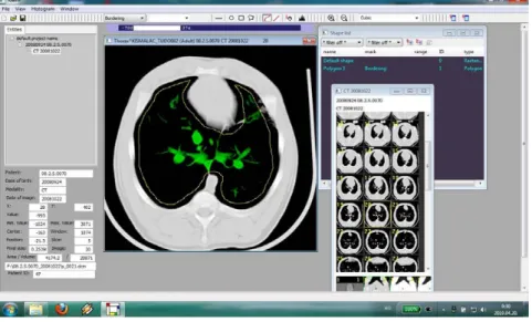
1.7. Use of computed tomography (CT) for examination of the lungs in humans and animals
1.7.1. Development of CT imaging
In 1917, the mathematician J. H. Radon gave evidence that the distribution of a material in an object layer can be calculated depending on the integral values along any number of lines passing through the same layer are known (Radon, 1917). The first application of Radon's reconstruction mathematics was carried out by Bracewell (Bracewell, 1956). The first succesful practical implementation was established by an English engineer G. N. Hounsfield in 1972, now generally known as the inventor of computed tomography. In 1979 Hounsfield and Cormack, a physicist, were awarded the Nobel Prize for medicine for their outstanding achievements. The development of CT scanners started with Hounseld’s experimental set-up, which was termed the „rst generation” of CT. The rst commercial scanners, the so-called „second generation”, differed only slightly from Hounseld’s scanning system. To speed up scanning and to utilize the available x-ray power more efciently detectors were added to entail going from a pencil beam to a small fan beam.
The translatory motion became obsolete, and the systems executed a rotatory motion only.
The rst whole-body scanners with fan beam systems were launched in 1976 providing scan times of 20 s per image. In the rst scanners of this type both the x-ray tube and the detector rotated around the patient. This concept was called „third generation” CT scanner. Only a little later scanners followed with a ring-like stationary detector fully encircling the patient so that only the x-ray tube rotated; which was termed the „fourth generation” CT. The translation- rotation systems vanished entirely. The type of rotation system of the „third generation” turned to be more effective. Thus today, the so-called third-generation CT's became standard (i.e.
conventional) (Kalender, 2006).
Figure 1: Four scanner generations were promoted in the 1970s (Kalender, 2006).
In these, the patient is scanned one slice at a time. The X-ray tube and detectors rotate for 360 degrees or less to scan one slice while the table and patient remain stationary. This slice-by- slice scanning is time-consuming, consequently efforts were made to increase the scanning speed. After the great technological progress in the 1970s, new achievment was seen in the start of 1990s, when the spiral/helical CT scanners were invented (Kalender, 1990). The novelty of this invention is that the X-ray beam traces a path around the patient (Kalender, 2006).
Figure 2: The „Spiral CT” (Kalender, 2006).
In 1998, all major CT manufacturers introduced multi-slice CT (MSCT) systems. It typically offered simultaneous acquisition of 4 slices at a rotation time of down to 0.5 s.
This was a significant development in scanning speed and longitudinal resolution and offered better utilization of the available X-ray power (Klingenbeck et al., 1999, McCollough and Zink, 1999, Ohnesorge et al., 1999, Hu, 2000). These improvements were quickly recognized as revolutionary developments that would finaly enable users to do real isotropic 3D imaging (Kalender, 2006).
Figure 3: The development from fan-beam to cone-beam CT in the early 2000s (Kalender, 2006).
Volume scanning has resulted in the routine application of such advanced techniques as CT fluoroscopy, CT angiography, three-dimensional imaging and virtual reality imaging. Its main applications are in cardiovascular studies, functional imaging, trauma and oncology. In cardiology, gated studies with the multi-slice scanners can provide clear non-invasive images of the heart and its major vessels, as well as fast coronary artery imaging including distal segments and multiple branches. The speed and precision of the multi-slice scanners has also changed the approach to the diagnosis and treatment of cancer. Instead of just studying the morphology of a tumor and monitoring changes in size, it is now possible to follow the perfusion of a contrast agent through and around the tumor, which allows early information on the response to therapy.
Other emerging applications for multi-slice scanners provide evaluation of carotid artery plaque, diagnostic of pulmonary diseases, and low-dose pediatric applications (Imhof, 2006).
Pixels in an image optained by CT scanning are displayed in terms of relative radiodensity.
Each pixel is assigned a numerical value (CT number), that is the average of all the attenuation values pertained within the corresponding voxel. This number is compared to the attenuation value of water and displayed on a scale of arbitrary units named Hounsfield units (HU) after Sir Godfrey Hounsfield. This scale assigns water as an attenuation value (HU) of zero. The range of CT numbers is 2000 HU wide although modern scanners have a greater range of HU up to 4000.
Each numeral value represents a shade of grey with +1000 (white) and –1000 (black) at either end of the spectrum. The "Partial Volume Effect" means the phenomenon that one part of the detector cannot differentiate between different tissues. This is typically done via a process of
"windowing", which maps a range (the "window") of pixel values to a greyscale ramp. The X- ray density of the object of interest determines the window used for display in order tooptimize the visible detail. The „lung windows” tipically have a window mean of approximately -600 to - 700 HU and a window width of 1000 to 1500 HU. Lung windows best demonstrate lung anatomy and pathology, contrasting soft-tissue structures with surrounding air-field lung parenchyma. For example, CT images of the lung are commonly viewed with a window extending from -1100 HU to -100 HU. Pixel values of -1100 and lower, are displayed as black;
values of -100 and higher are displayed as white; values within the window are displayed as a grey intensity proportional to position within the window. The window used for display must be matched to the X-ray density of the object of interest, in order to optimize the visible detail (Webb et al., 2014).
1.7.2. The lung CT imaging
The initial imaging tool for the lung parenchyma remains the chest radiograph. It is unsurpassed in the amount of information it yields as far as its cost, radiation dose, availability, and ease of performance. However, the chest radiograph has its limitations. In several studies, the chest radiograph has been shown to have an overall sensitivity of 80 percent and a specificity of 82 percent for detection of diffuse lung disease. Chest radiography could provide a confident diagnosis in only 23 percent of cases, and those confident diagnoses proved correct only in 77 percent of cases (Mathieson et al., 1989).
Advances in CT scanner technology have made isotropic volumetric, multiplanar high- resolution lung imaging possible in a single breath-hold, an essential advance over the incremental high-resolution CT (HRCT) technique in which noncontiguous images sampled the lung, but lacked anatomic continuity. HRCT of the lungs is an established imaging technique for the diagnosis and management of interstitial lung disease, emphysema, and small airway disease, providing a noninvasive detailed evaluation of the lung parenchyma, and providing information about the lungs as a whole and focally (Sundaram et al., 2010). HRCT, which has a sensitivity of 95 percent and a specificity approaching 100 percent, can often provide more information than either chest radiography or conventional CT scanning. A confident diagnosis is possible in roughly one-half of cases, and these are proven correct an estimated 93 percent of the time (Mathieson et al., 1989). The technique of HRCT was developed with relatively slow CT scanners, which did not make use of multi-detector (MDCT) technology. The parameters of scan duration, z-axis resolution and coverage were interdependent. To cover the chest in a reasonable time period with a conventional chest CT scan required thick sections (e.g. 10mm) to ensure contiguous coverage. As performing contiguous thin sections required unacceptably prolonged scan time, HRCT examination was performed with widely spaced sections. Because of the different scan parameters for conventional and HRCT examinations, if a patient required both, they had to be performed sequentially. Modern MDCT scanners are able to overcome this interdependence, and are capable of imaging at full resolution yet retain very fast coverage - images can then be reconstructed retrospectively from the volumetric raw data (Dodd et al., 2006).
1.7.3. Interpretation of lung disorders (Smithuis et al., 2006)
Reticular pattern
In the reticular pattern there are too many lines, either as a result of thickening of the interlobular septa or as a result of fibrosis.
Nodular pattern
In most cases small nodules can be placed into one of three categories: perilymphatic, centrilobular or random distribution.
Low Attenuation pattern
The fourth pattern includes abnormalities that result in decreased lung attenuation or air- filled lesions. These include: emphysema, lung cysts, bronchiectasia, honeycombing.
High Attenuation pattern
Moderate increased lung attenuation is called ground-glass-opacity (GGO) if there is a hazy increase in lung opacity without obscuration of underlying vessels. The more increased lung attenuation is called consolidation if the increase in lung opacity obscures the vessels. In both ground glass and consolidation the increase in lung density is the result of replacement of air in the alveoli by fluid, cells or fibrosis.
Ground-glass-opacity (GGO)
In GGO the density of the intrabronchial air appears darker as the air in the surrounding alveoli. Ground-glass opacity (GGO) represents: A) Filling of the alveolar spaces with pus, edema, hemorrhage, inflammation or tumor cells; B) Thickening of the interstitium or alveolar walls below the spatial resolution of the CT as seen in fibrosis.
So ground-glass opacification may either be the result of air space disease (filling of the alveoli) or interstitial lung disease (i.e. fibrosis).
Consolidation
Consolidation is synonymous with airspace disease. Is it pus, edema, blood or tumor cells.
Acute consolidation is seen in: pneumonias (bacterial, mycoplasmal), pulmonary edema, hemorrhage, acute eosinophilic pneumonia. Chronic consolidation is seen in: organizing pneumonia, chronic eosinophilic pneumonia, fibrosis, bronchoalveolar carcinoma or lymphoma.
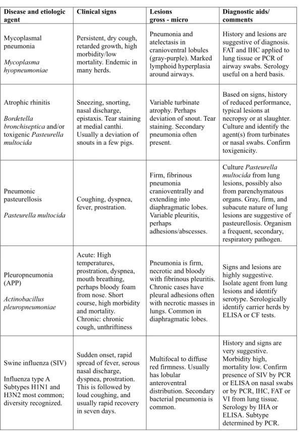
Figure 4: Broncho-alveolar cell carcinoma with ground-glass opacity and consolidation
Mosaic attenuation
The term mosaic attenuation is used to describe density differences between affected and non-affected lung areas. There are patchy areas of black and white lung. When ground glass opacity presents as mosaic attenuation consider: A) infiltrative process adjacent to normal lung;
B) normal lung appearing relatively dense adjacent to lung with air-trapping; C) Hyperperfused lung adjacent to oligemic lung due to chronic thromboembolic disease
Crazy Paving
Crazy Paving is a combination of ground glass opacity with superimposed septal thickening. It was first thought to be specific for alveolar proteinosis, but later was also seen in other diseases.
Crazy Paving can also be seen in: infection (viral, mycoplasmal, bacterial), pulmonary hemorrhage, edema.
2. CONCLUSIONS DRAWN FROM THE DATA OF THE LITERATURE
Over the past few decades, there has been a very pronounced increase in productivity also in the pig industry, brought about by the size increase and concentration of pig herds and by the spread of intensive management technologies. This productivity increase, however, was accompanied by a drastic rise in the prevalence of respiratory diseases, among other changes.
The vast majority of respiratory diseases often affecting large numbers of animals represent syndromes of multifactorial aetiology induced by the simultaneous presence of multiple pathogens, which are now collectively termed as porcine respiratory disease complex (PRDC) in the special literature.
Mycoplasma hyopneumoniae is one of the most prevalent respiratory pathogens. It has played a decisive role in the aetiology of respiratory diseases for several decades, usually in association with other pathogenic microorganisms, thus resulting in a complex respiratory syndrome. The prevalence of the other respiratory pathogens varies considerably; however, in the past two decades numerous new pathogens appeared or became known, which have assumed decisive importance in the development of PRDC. The appearance and worldwide spread of PRRS virus at the end of the 1980s brought major changes in the health status of pig herds.
Subsequently, further pathogens playing an important part in the aetiology of PRDC became known, such as porcine circovirus type 2, porcine coronavirus and new strains of swine influenza virus (e.g. H3N2), which also spread all over the world in the past 10–20 years and are now causing major economic losses in the pig-producing countries.
Atrophic rhinitis is also a long-known and widely distributed pig disease, in the aetiology of which the interaction between toxigenic strains of two pathogenic bacteria, Bordetella bronchiseptica and Pasteurella multocida, is thought to play the primary role. These two pathogens are frequently isolated from the respiratory tract of pigs and they play a major role in the development of PRDC. Based on the data of the literature, the toxigenic strains of P.
multocida are more often found in lesions restricted to the upper airways, whereas in the pathological processes taking place in the lungs mostly P. multocida strains not capable of toxin production are involved.
Microscopic filamentous fungi contaminating feed and food raw materials produce numerous toxins (mycotoxins) which, when entering the soil-plant-animal-human food chain, are a source of major public health risks. In addition, they cause substantial economic losses in the animal production sector. Fumonisins (FB1, FB2, FB3, FB4) constitute a relatively recently identified group of mycotoxins, which was discovered in 1988.
After American authors had first described the syndrome termed porcine pulmonary oedema (PPE), confirmed to be attributable to fumonisin B1 (FB1) toxin, together with the main pathological lesions associated with it (pulmonary oedema, liver and renal degeneration), research studies aimed at getting a closer insight into the pathological processes were started.
Hungarian researchers have achieved significant results in the study of disease entities caused by FB1 toxin. It was on the basis of these results that the maximum permissible limit for FB1 (5 mg/kg) was established and incorporated into the Codex Pabularis Hungaricus (the Hungarian Feed Code) and the relevant recommendations of the European Union.
To date, few studies have been done to determine the potential predisposing role of FB1
toxin in syndromes induced by the most important porcine respiratory pathogens. The interaction between this mycotoxin and the above-mentioned pathogens was studied in pigs experimentally infected with serotype A of P. multocida and treated with FB1 toxin (Halloy et al., 2005), as well as in pigs experimentally infected with PRRS virus and treated with FB1 toxin (Ramos et al., 2010).