Edited by:
Gabor Jakab, University of Pécs, Hungary
Reviewed by:
Massimiliano Morelli, Istituto per la Protezione Sostenibile delle Piante (CNR), Italy Stefanie Christine Becker, University of Veterinary Medicine Hannover, Germany
*Correspondence:
Eva Varallyay varallyay.eva@abc.naik.hu
Specialty section:
This article was submitted to Plant Microbe Interactions, a section of the journal Frontiers in Microbiology
Received:30 October 2017 Accepted:18 January 2018 Published:06 February 2018
Citation:
Czotter N, Molnar J, Szabó E, Demian E, Kontra L, Baksa I, Szittya G, Kocsis L, Deak T, Bisztray G, Tusnady GE, Burgyan J and Varallyay E (2018) NGS of Virus-Derived Small RNAs as a Diagnostic Method Used to Determine Viromes of Hungarian Vineyards.
Front. Microbiol. 9:122.
doi: 10.3389/fmicb.2018.00122
NGS of Virus-Derived Small RNAs as a Diagnostic Method Used to
Determine Viromes of Hungarian Vineyards
Nikoletta Czotter1, Janos Molnar2,3, Emese Szabó1, Emese Demian1, Levente Kontra1, Ivett Baksa1, Gyorgy Szittya1, Laszlo Kocsis4, Tamas Deak5, Gyorgy Bisztray5,
Gabor E. Tusnady2, Jozsef Burgyan1and Eva Varallyay1*
1National Agricultural Research and Innovation Center, Agricultural Biotechnology Institute, Gödöllo, Hungary,2Research Center of Natural Sciences, Institute of Enzymology, HAS, Budapest, Hungary,3Department of Biotechnology, Nanophage-therapy Center, Enviroinvest Corporation, Pécs, Hungary,4Department of Horticulture, Georgikon Faculty, University of Pannonia, Keszthely, Hungary,5Department of Viticulture, Institute of Viticulture and Oenology, Szent-Istvan University, Budapest, Hungary
As virus diseases cannot be controlled by traditional plant protection methods, the risk of their spread have to be minimized on vegetatively propagated plants, such as grapevine.
Metagenomic approaches used for virus diagnostics offer a unique opportunity to reveal the presence of all viral pathogens in the investigated plant, which is why their application can reduce the risk of using infected material for a new plantation. Here we used a special branch, deep sequencing of virus-derived small RNAs, of this high-throughput method for virus diagnostics, and determined viromes of vineyards in Hungary. With NGS of virus-derived small RNAs we could detect not only the viruses tested routinely, but also new ones, which had never been described in Hungary before. Virus presence did not correlate with the age of the plantation, moreover phylogenetic analysis of the identified virus isolates suggests that infections are mostly caused by the use of infected propagating material. Our results, validated by other molecular methods, raised further questions to be answered before this method can be introduced as a routine, reliable test for grapevine virus diagnostics.
Keywords: grapevine, vineyard, virus, virome, diagnostics, metagenomics, small RNA, NGS
INTRODUCTION
Grapevine can host more than 60 viruses and viroids (Al Rwahnih et al., 2009; Martelli, 2014), often as multiplied infection. Vegetative propagation and long lifetime of the plantation increase the risk of virus infection which cannot be controlled by traditional plant protection methods. The use of highly adaptive cultivars globalizes not only the presence of the particular cultivar but also the spread of new pathogens and their vector organisms. Safety regulations deal only with a limited number of known viruses and ignore new invading pathogens, which can lead to the use of infected propagating material and produce a new level of persistent infection risk. Traditional diagnostic methods can only answer the question whether or not the investigated virus is present in our sample, and need preliminary information about the pathogen (antigen for ELISA and sequence information of the particular variant for PCR-based methods). In striking contrast, deep sequencing
offers a unique opportunity to reveal any virus or viroid present in the sample, expected or not (Boonham et al., 2014).
Indeed, different platforms were used for the description of new grapevine viruses (Martelli, 2014) and also to create the virome of a vineyard (Coetzee et al., 2010). During virus infection small interfering (si) RNAs having the same sequence as the infecting viruses are formed (Baulcombe, 2004; Molnar et al., 2005; Donaire et al., 2009; Kreuze et al., 2009; Szittya et al., 2010) by the RNA interference (RNAi) based defense reaction of the plant. Deep sequencing of the small RNA (sRNA) population extracted directly from field plants offers a unique opportunity in virus diagnosis to identify several variants of grapevine infecting viroids (Navarro et al., 2009) or viruses (Pantaleo et al., 2010) even if they are alien on the plant or have never been described before (Zhang et al., 2011; Giampetruzzi et al., 2012; Wu et al., 2012). In our work we used this cutting-edge technique, the deep sequencing of virus-derived siRNAs to reveal the sanitary status of vineyards in our country. Analysis of the sRNA sequence dataset obtained using our bioinformatics pipeline enabled us to describe viruses never before reported from our country (Grapevine Syrah virus 1 and Grapevine Pinot Gris virus, Grapevine Satellite virus). Beside these new descriptions we analyzed our samples for the most widespread viruses with RT- PCR (using published diagnostic primers) and sRNA NGS in parallel. In most cases our results could be validated, but we also found contradictions in several cases, which is also discussed.
MATERIALS AND METHODS
Plant Material, Sample Preparation
Samples were collected from 14 vineyards representing 18 different varieties and nine wine-growing-regions of Hungary.
Shoots of 1–10 randomly chosen individual plants per plantation were collected on the field or from sprouted canes. RNA was extracted from various organs: shoot tips, young leaves (until the 3rd internode from the shoot apex), older leaves (lower than the 3rd internode), tendrils, and inflorescenses by CTAB method (Gambino and Gribaudo, 2008). RNA pools representing each plant were generated mixing equal amounts of RNA originating from the different organs. These individual plant RNA pools (library 14–18) or a plantation pool generated by the same strategy, representing all of the sampled plants from the same plantation (library 1–13) was used for small RNA library preparation (18 libraries in total) and sequenced using single index on a HiScanSQ by UD Genomed (Debrecen, Hungary) 50 bp, single end (8 samples/1 sequencing lane). Fastq files of the sequenced libraries are deposited to the GEO and can be accessed through series accession number GSE106240.
Pipeline for Data Evaluation of NGS Results (Bioinformatics)
The resulting reads were sorted according to their indexes.
Adapters of the sequenced reads were removed by the Trimmomatic program (Bolger et al., 2014), their quality was checked by the FastQC program (http://www.bioinformatics.
babraham.ac.uk/projects/fastqc) and deduplicated by the Picard MarkDuplicates tool (http://broadinstitute.github.io/picard). For
virus detection we used two different pipelines in parallel: (A) Short reads were mapped to viral reference genomes (Refseq viral database of NCBI from only plant and invertebrate hosts were used) by the BWA-aln short read aligner (Li and Durbin, 2009) with default options. Mapped reads were counted both with and without deduplication using samtools idxstats (Li, 2011). Redundant reads of the resulted hits were equalized to read/million read. Consensus viral sequences from the aligned deduplicated reads were generated using the samtools/bcftools (Li and Durbin, 2009) pipeline. Coverage of the appropriate genome was counted as % of the genome covered by nucleotide information from the mapped small RNA reads. (B) De novo assembling of the deduplicated reads was performed using Velvet with k-mer: 13, 15, 17 (Zerbino and Birney, 2008). The generated contigs were annotated by BLAST megablast (Morgulis et al., 2008) to the RefSeq of NCBI.
Sequence Comparison
To compare consensus sequences of virus variants of the different libraries or sequenced PCR products we used the CLUSTAL Omega program (Sievers et al., 2011) and neighbor-joining algorithm implemented in MEGA v.6 (Tamura et al., 2013) Bootstrap values>70% (1000 bootstrap replicates) were used.
Validation of Predicted Virus Diagnostics by RT-PCR
cDNA was synthetized from pooled RNA extracts representing each plantation using random primer and the RevertAid First Strand cDNA Synthesis Kit (Thermo Fisher Scientific, USA) (according to the manufacturer’s instructions). The cDNA generated was used as templates for PCR reactions using Phire Hot Start II DNA Polymerase (Thermo Fisher Scientific, USA) and published diagnostic primers or new ones (see Supplementary Table 5) designed according to the consensus sequence generated by mapping our small RNA reads to the reference genomes. To detect GRVFV, we used cDNA generated with a GRVFV-specific GRVFV-R/6391 primer. PCR products were analyzed by agarose gel electrophoresis. For Sanger sequencing cDNA was synthetized from pooled RNA extracts of individual plants and virus-specific PCR was done using Phusion Hot Start II High-Fidelity DNA Polymerase (Thermo Fisher Scientific, USA) or Q5 Hot Start High-Fidelity DNA Polymerase (New England Biolabs, UK) DNA polymerase. The purified products were cloned into pJET 1.2 vector (Thermo Fisher Scientific, USA) and sequenced. Sequences were deposited into GenBank (for GenBank Accession Numbers see Supplementary Figure 6).
Validation by Northern Blot
For Northern blot analyses 4–5µg of total RNA was separated on formaldehyde-1.2% agarose gel and blotted to Amersham Hybond-NX membrane (GE Healthcare, UK), by capillary method using 20xSSC. Hybridization was carried out at 65◦C in Church buffer (0.5 M Phosphate buffer, pH 7.2 containing 1% BSA, 1 mM EDTA, 7% SDS) overnight with the appropriate radioactively labeled probe, washed for 5 min in 2×SSC, 0.1%
SDS and for 15 min in 0.5×SSC, 0.1% SDS at the temperature of the hybridization and exposed to an X-ray film.
Virus-specific, P32-labeled, DNA probes were prepared by using the DecaLabel DNA Labelling Kit (Thermo Fischer Scientific, USA). As a template we used the PCR-amplified and purified product of cloned region of viral genome.
The virus piece cloned was a 1663-bp part amplified with GPGV5557F and GPGV7220R for GPGV, a 1324-bp product amplified by RBDV_RNA1F_4082 and RBDV_RNA1R_5406 for RBDV RNA1, and a 927-bp product amplified by GSVsatF72 GSVsatR999 for GSV.
RESULTS AND DISCUSSION
Sample Collection and Sequencing
As a survey to detect virus infections in Hungarian vineyards samples were collected directly from the field, in a random fashion, independently of any apparent symptom, in May 2014 or from sprouts of single bud cuttings. 14 vineyards differing in the variety grown and the age of the plantation from 9 wine- growing regions of the country were sampled (Supplementary Figure 1 and Supplementary Table 1, 2). Small RNA libraries were prepared from pooled samples representing either the plantation (libraries 1–13), or different varieties at the same plantation (library 14–18) and sequenced.
Initial Statistics
As a result of sequencing 8–14 million raw reads/library were generated (Supplementary Table 3). After trimming of the adapters, duplicates were removed and non-redundant reads (560000-1.6 million/library), without removing grapevine- specific sRNAs, were used for virus diagnostics. In different libraries 3.3–13% of the total non-redundant reads and 2.3–
11.7% of the total redundant reads were mapped to viral reference genomes, representing 2–13 different viruses and viroids.
Size Distribution of Sequenced Srnas
Size distribution of redundant sRNA sequences showed that the majority of the reads was between 21 and 24 nt, indicating that the library preparation was successful (Figure 1A). Most of the reads were 21 nt long and contained miRNA sequences in accordance with our previous report (Pantaleo et al., 2010). As for non-redundant reads the 24 nt-long size class was overrepresented (Figure 1B), likely responsible for transcriptional gene silencing (TGS) (Borges and Martienssen, 2015). sRNAs are products of different plant DICERs (DCL1,−2,−3, and 4) and each DCL enzyme activity produces a specific size class. The products of DCL1 and DCL4 are 21 nt-long, whereas DCL2 generates 22 nt-long, and DCL3, 24 nt-long sRNAs. Whereas DCL1 has a key role in miRNA biogenesis, DCL3 generates siRNAs for TGS against parasitic nucleic acids (e.g., transposons) (Parent et al., 2012).
The Vitis vinifera genome encodes four DCLs homologous to DCLs characterized inArabidopsis thaliana(Zhao et al., 2015).
Although according to that work VvDCL1 contains only one RNaseIII domain, VvDCL2 and VvDCL3 lacks a dsRB domain, moreover VvDCL4 lacks a PAZ domain, we think they must be
fully functional since all characteristic sRNA size classes (21, 22, and 24 nt) were present in our samples.Figures 1A,Bshow the size distribution of all host-derived sRNA reads.
During antiviral silencing DCL4 and DCL2 process virus- derived dsRNAs into sRNAs. Virus- and viroid-specific sRNAs in our samples were almost exclusively 21–22 nt long, supporting the idea that in grapevine DCL2 and DCL4 are the key enzymes in virus-derived sRNA biogenesis (Figure 1C).
Origin of Viral SRNAS
In order to identify viruses present in our plantations, sRNA reads were aligned and mapped to reference genomes of all known viruses of plant or insect host origin. Coverage (in %) of the whole viral reference genome was also calculated. During this analysis virus-specific contigs were assembled with different k-mers (kmer13, 15, 17), and the resulting contigs were also aligned to this set of reference genomes. In Supplementary Table 4 the results of the bioinformatics analysis for quarantine viruses: Grapevine fanleaf virus (GFLV), Arabis mosaic virus (ArMV), Grapevine leafroll-associated virus 1-3 (GLRaV1-3), Grapevine virus A (GVA), Grapevine virus B (GVB), Grapevine fleck virus (GFkV), together with Grapevine chrome mosaic virus (GCMV), Grapevine red globe virus (GRGV), Grapevine asteroid mosaic-associated virus (GAMaV), Grapevine vein feathering virus (GRVFV), Grapevine Syrah virus 1 (GSyV1), Grapevine rupestris stem pitting-associated virus (GRSPaV), Grapevine Pinot gris virus (GPGV), Raspberry bushy dwarf virus (RBDV), Grapevine satellite virus (GSV) and viroids: Hop stunt viroid (HSVd) and Grapevine yellow speckled viroid 1-2 (GYSVd- 1 and 2) are summarized. A virus or viroid was diagnosed as present if any virus/viroid specific contigs (if any with any kmer) was present, and coverage of the viral genome by small RNA reads was higher than 40% (in case of viruses) or 80% (in case of viroids). According to these results the tested plantations are free from GFLV, ArMV, and GLRaV2, but we usually found simultaneous presence of up to 13 of different viruses and viroids in the same plantation.
Validation of Deep Sequencing Results
In order to validate our deep sequencing results we synthetized cDNA from RNAs representing plantation pools and set up PCR reactions by published diagnostic primers or with primers designed according to the sequenced sRNA reads (Supplementary Table 5). Positive controls (cDNA from virus- containing samples) and negative controls were always included.
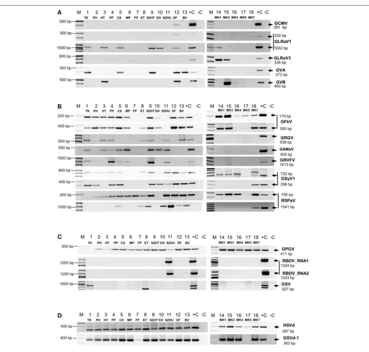
PCR products were analyzed by separation on 1.2 % agarose gels (Figure 2) and traditional Sanger sequencing. Results of the sRNA NGS virus diagnostics and its comparison with RT- PCR are summarized in Table 1. Sequences were deposited to GenBank and used for phylogenetic comparison (see Supplementary Table 6 for summary). Our results showed that the reliability of sRNA NGS as a diagnostic tool varied from virus to virus, and we discuss it for each of the identified viruses.
Nepoviruses
The only Nepovirus detected was GCMV present in 12_DF where partial CP could be amplified and sequenced (Figure 2A)
FIGURE 1 |Size distribution of(A)trimmed,(B)non-redundant,(C)viral, redundant sequenced reads of the sequenced libraries.
FIGURE 2 |RT-PCR validation of sRNA NGS for(A)Nepo-,Leafroll-, and Vitiviruses,(B)Tymoviruses,(C)viruses which presence is not routinely tested and(D) viroids. cDNA was synthetized from pooled RNA extracts representing each vineyard using random primer and used as templates for PCR reactions with published diagnostic primers or new ones designed according to the consensus sequence generated by mapping our small RNA reads to the reference genomes. To detect GRVFV we used cDNA generated with a GRVFV-specific primer. PCR products were analyzed by agarose gel electrophoresis. (M), GenRuler 100 bp+; (+C), cDNA containing the tested virus was used as positive, or (–C), water as negative control.
(accession number MF100927). Interestingly enough, all of the GCMV RNA2 sequences in GenBank originated from Hungarian accessions (Elbeaino et al., 2014), and are more closely related to each other (more than 96% identity in this CP region) than to the isolate what we found (88–89% identity compared to other GCMV RNA2 sequences in this CP region) (Supplementary Figure 2A). The geographical origins of these Hungarian GCMV accessions are unknown. According to a recombination analysis of full GCMV RNA2 sequences, GCMV suggested to be a
putative interspecies recombinant of GARSV and TBRV (Digiaro et al., 2015), which question could be further investigated incorporating HUDF isolate, however for its recombination analysis full RNA2 must be sequenced.
Grapevine Leafroll Associated Viruses
GLRaV1 was detected in nine of our samples, but validation with published diagnostic primers failed in six samples (Figure 2A, Table 1). Testing the assumption that diagnostic primers were
TABLE1|SummaryofthesmallRNANGSvirusdiagnosticsanditsRT-PCRvalidationforeachlibrary. GFLVArMVGCMVGLRaV1GLRaV2GLRaV3GVAGVBGFkVGRGVGAMaVGRVFV newGRVFVGSyVGRSPaVHSVdGYSVd-1 contig blast0001001011111101011111 read bwa0001001011111100011110 PCR000S00S0SSSSS00S1111 contig blast000000001001110100116 read bwa000000001101110100118 PCR00000000S00SSS00117 contig blast000100001111010100118 read bwa0001000111111101001111 PCR000S000SSSS1SS001S10 contig blast000000001111110100118 read bwa000000001111111100119 PCR00000000SSSSSS001S9 contig blast000100101011110100119 read bwa0001001011111111001111 PCR000S00S000SSSS00119 contig blast000000001011000100115 read bwa000000001011010100117 PCR00000000S0S1SS00118 contig blast000000000000000100102 read bwa000000000000001100103 PCR000000000000SS00114 contig blast000000001101110100117 read bwa000000000101111100117 PCR00000000SS01SS001S7 contig blast000100101101100100119 read bwa0001001011111111001111 PCR000S00S0SSSSSS001111 contig blast000100001111010100119 read bwa0001000010111111001110 PCR000S0000S0SS0S00117 contig blast000000000001011110117 read bwa000000000111011010118 PCR000000000SSSSSS0119 contig blast0011001010010111001110 read bwa0011000010111111001112 PCR00SS00S1S00SSS001111 contig blast0001001011011101011110 read bwa0001001011111111111113 PCR000S00S0SS0SSSSS1112 contig blast000101001101110100119 read bwa0001010011111101001110 PCR000S0S00S001SS00118 contig blast000001011100010100118 read bwa000001011100011100118 PCR00000S0SS001SS00118 contig blast000000000000010100114 read bwa000000000000010100114 PCR00000000,50001SS00S15 contig blast000000000000000100001 read bwa000000000000000100001 PCR0000000000000S00102 contig blast000000001000011100115 read bwa000100001000011100117 PCR00010000S001SS00117 001801531298131413221413
14 16 ? 6 4
total number of viral pathogens
Age of the plantation in years 20 7
type of analysis 0 S S S
ViroidsViruses not routinly tested S
Library code 7_FP 13 stock collection regularly virus tested
1 100< ?
? 100< 17_MK5 18_MK7
15_MK3 16_MK4
8_ET 9_SZHT 10_EH 13
GPGVRBDVsGSV 1_TK
Grapevine leafroll virusesVitivirusesTymoviruses S
Nepoviruses 13_BV
5_CS 6_MP
2_PH 3_HT 4_PP number of plantation where the virus
0 0 S 0 S 0S S 0 0
14_MK1
11_SZHU 12_DF 0 0 Resultofthebioinformaticsanalysis(contigblastandreadbwa)wasmarkedby0or1.0wasusediftherewasnohitfortheappropriatevirus,whereas1wasusedifanycontigscouldbebuilt,orthecoverageofthevirusgenomewas higherthan40%(incaseofviruses)or80%(incaseofviroids).ResultofRT-PCRvalidationwasmarkedas1,ifPCRproductwaspresentorbySifnotonlytheproductwaspresentbutitwasSangersequenced.Anyhitpresentwas highlightedwithgraycolor.Summaryofthetotalnumberofviralpathogensinthelibrary,andnumberoftheplantationswherethevirusispresent,washighlightedwithredandgreencolorrespectively.
designed to variable regions, new primers were designed to the HSP70 coding region based on the sequenced sRNAs reads.
With this new set of primers we could successfully validate the presence of GLRaV1 in five additional libraries (Figure 2A). In 13_BV we could only detect the virus in two of the individuals (Supplementary Figure 3A), which might explain the lack of the PCR product in the pool. Phylogenetic analysis of our isolates showed that they clustered into two distinct groups, the same E and A as was suggested by Kominek et al. (2005) (Supplementary Figure 2B) and are only 82–92% identical to the reference genome, supporting the operation of high-level variability, due to which GLRaV1 can be easily overlooked by traditional diagnostic methods (Esteves et al., 2013). The only Hungarian GLRaV1 isolate (CSE_6.4.1.H) in GenBank (Cseh et al., 2013) (clustered to group E) was collected at the same region of the country from where HUTK and HUHT (clustered to group A) originated, suggesting that the source of the infection is more likely the propagation material. These results show that detection of GLRaV1 by sRNA NGS seems reliable, but its validation by RT-PCR may be problematic due to the high variability of the virus.
For GLRaV3 the result of sRNA NGS and its validation by RT-PCR using published diagnostic primer pairs correlated well (Table 1), detecting its presence in 14_MK1 and 15_MK3.
Phylogenetic analysis of the sequenced part of the CP showed that these isolates (HUMK1 and HUMK3) are very closely related (98% identity on nt level) but share only 91% identity with the NY1 Reference genome from the USA (Supplementary Figure 2C). They clustered together with isolates from Brazil, Israel, and South Africa, but share only 90–91% identity with European strains. Their phylogenetical relationship based on the HSP70 region showed that they cluster into two distinct group together with isolates from different regions of the country (Cseh et al., 2013)(Supplementary Figure 2D). Samples 14_MK1 and 15_MK3 are different varieties in different rows of the same plantation. As these geographically linked variants tended to be divergent, it seems possible that the infection originated from an infected propagation material and is not the result of an onsite infection.
Vitiviruses
GVA was detected at five, whereas GVB at two plantations (Table 1,Figure 2A). In 12_DF we could clone an RT-PCR product from both of these viruses although we failed to detect GVB by sRNA NGS. Testing individuals for their presence we have found that in this sample only one plant was infected (Supplementary Figures 3B,C), which could decrease the concentration of the virus below the detection limit by sRNA NGS, in the pool. GVA isolates were only 85–90% identical to the Italian Reference genome and grouped into two distinct clades within Group I, together with other European strains (Goszczynski, 2014)(Supplementary Figure 2E). GVB isolates showed higher variation: they were only 85% identical to each other and grouped into different clades (Fonseca et al., 2016) (Supplementary Figure 2F). We can conclude that sRNA NGS based virus diagnostics worked well for vitiviruses, but our results
showed that using plantation pools containing extracts of non- infected plants can lower the virus concentration, and without further investigation slight and uneven infections can easily be overlooked.
Tymovirales
Grapevine Fleck Virus (GFKV)
GFkV was one of the most widespread viruses present in 14 samples. Virus diagnostics by sRNA NGS and RT-PCR validation for GFKV presence usually correlated well (Table 1,Figure 2B).
Although using diagnostic primers we failed to validate its presence in 8_ET, validation was successful with a new set of primers, designed according to the sequenced sRNA reads (Figure 2B). Sequence comparison of the isolates showed high variability: they were 85–95% identical to the Italian reference (Supplementary Figure 2G). Sequencing of the cloned PCR product in 5_CS showed that this is a product of GRVFV (MF461275) and not of GFKV, highlighting the high-level identity of these two viruses. However, validation of the presence of GFKV failed, it is possible that sequencing more clones would have yielded a GFKV-specific product.
Tymoviruses, involved in fleck complex, are closely related;
they can coexist in the same plant and GFKV is often presents in co-infection together with GRGV, GAMaV, GRVFV (Sabanadzovic et al., 2000; Cretazzo et al., 2017) and sometimes with GSyV1, therefore it is not surprising that we could also detect these viruses in most of our samples.
Grapevine Red Globe Virus (GRGV)
GRGV was detected in many of our samples but its presence could only be validated in seven libraries (Table 1,Figure 2B).
GRGV has been identified in different parts of Europe (Sabanadzovic et al., 2000; Beuve et al., 2015; Cretazzo et al., 2017; Voncina et al., 2017) and also in California (El Beaino et al., 2001) and in Brazil (Fajardo et al., 2017). We detected GRGV in different regions of the country; the sequenced strains have 87–95% identity with the Reference strain (NC_030693) and clustered separately (Supplementary Figure 2H). Variability of different isolates and the fact that GRGV was not found in Czech and Slovak accessions—not even with NGS (Eichmeier et al., 2016)—further supports the idea that it is not originally present in Central-Europe, and possibly originates from infected propagation material of a geographically different origin.
Grapevine Asteroid Mosaic Associated Virus (GaMaV) We could detect GAMaV in eight samples (Table 1,Figure 2B).
Although described and well-known since 1994 (Boscia et al., 1994), there is still only limited sequence information about GAMaV. The Reference genome was only uploaded to GenBank in 2016 (NC_031692), but since that time—thanks to NGS surveys - it has been reported from Canada (Xiao and Meng, 2016) and from France (Candresse et al., 2017a). Our isolates are 94–96% identical to the Reference and 93–96%
identical to each other, showing less variability than GRGV. As Hungarian GAMaV isolates clustered with isolates from different geographical origins (Supplementary Figure 2I), the use of
virus-infected propagation material is the most straightforward explanation for their presence.
Grapevine Rupestris Vein Feathering (GRVFV)
GRVFV was detected in 11 and 13 libraries using a full genome (AY706994) and a Reference Genome (NC_034205) (Reynard et al., 2017) for its detection respectively (Table 1and Supplementary Table 4). The new Reference is only 77% identical to the first, Californian full genome, which shows the very high variability of this virus and the reason why the validation of its widespread presence has usually been failed (Pantaleo et al., 2010; Reynard et al., 2017). Our RT-PCR validation using cDNA produced by random probe also failed in all cases. We tried to increase the concentration of virus specific cDNA using virus- specific primer for cDNA synthesis and could amplify GRVFV in 9 libraries, but still failed in four samples (Figure 2B). Our isolates share 79–96 and 79–87% identity with AY706994 and NC034205, respectively, further confirming the diversity of this virus. Moreover, sequencing different individuals from the same plantation (HUCS, HUPP, HUTK, HUDF) revealed the presence of distinct variants (78–92% identical to each other) at the same plantation, clustering separately (Supplementary Figure 2J). This case shows that sRNA NGS has difficulties in accurate diagnostics of viruses with high variable genomes, however the presence of a Tymovirus could be accurately detected.
Grapevine Syrah Virus 1 (GSyV1)
As we have reported previously we have found GSyV1 in Hungarian vineyards (Czotter et al., 2015 present in 15 libraries (Table 1). With DetF-DetR (Al Rwahnih et al., 2009#3) amplifying putative MP, we could validate its presence in 10 while with primers amplifying part of the CP (Sabanadzovic et al., 2009) in further six samples (Figure 2B). High prevalence of GSyV1 have also been found in Czech and Slovak grapevines which also showed high variability at the 5’ putative MP coding region (Glasa et al., 2015), resulting false negative result if the above primers were used for diagnostics. Our isolates grouped distantly both into the two major and the diverged third clades suggested by Glasa (Glasa et al., 2015) (Supplementary Figure 2K).
Moreover sequences of GSyV1 from different individuals of the same plantation (HU11TK2 and HU11TK9) showed high variability what supports the idea that GSyV1 population in Central Europe is more diverse than the North American ones.
Grapevine Rupestris Stem Pitting-Associated Virus (GRSPaV)
GRSPaV is known to be the most widespread virus infecting grapevine, but to our surprise we could detect its presence by both contig blast and read bwa in only three of our samples (Supplementary Table 4). In a striking contrast, with RT- PCR using diagnostic primers amplifying a very short part of the replicase we obtained a product in 16 of our samples (Figure 2B). GRSPaV’s viral RdRp has a very low proofreading activity and frequent recombination events, because the coexistence of different variants in the same plant led to the evolution of diverse variants (Morelli et al., 2011; Glasa et al., 2017). To test that if the number of virus-specific reads or
coverage will increase if we use a different reference during bioinformatics analysis, we made the direct sRNA BWA using 5 distinct full GRSPaV genomes (AF057136=NC_001948 _Ref_1_USA, AY881627_BS_Canada, KR054734_JF_China, AY881626_SG1_USA, AY368590_SY_USA) (Supplementary Table 7). According to this analysis we obtained higher than 40% coverage to different GRSPaV strains only in two additional libraries. GRSPaV is mostly spread by vegetative propagation;
as a result, it is particularly difficult to eliminate by sanitation techniques and coexists with grapevine for a long time (Meng et al., 2006). Their coevolution led to gene expression changes of the host with mutual advantages, resulting in slight down- regulation of stress genes in the presence of the virus (Gambino et al., 2012). Because of these advantages it is possible that during this coevolution an acceptable balance of the virus and the host defense reaction was achieved. The presence of a GRSPaV-coded silencing suppressor with an activity to block virus-derived sRNA biogenesis can also explain these results, but must be further investigated.
To be able to analyse phylogenetical relationship of GRSPaV isolates, we amplified and sequenced a longer 3’ part of their genome (Figure 2B). Hungarian variants clustered with the GRSPaV-1 and Tannat variants from the USA and Uruguay, but it is possible that sequencing more clones or full genomes would alter this phylogenetic picture (Supplementary Figure 2L).
Grapevine Pinot Gris Virus (GPGV)
According to our survey, GPGV, never described before, seems widespread in our country: it is present in 17 libraries (Table 1).
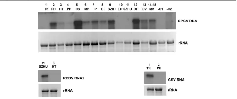
In striking contrast to the predominance of 21 nt GPGV- derived reads with both sense and antisense orientation, a 24 nt-long antisense excess was found in 1_TK and 11_SZHU (Supplementary Figure 4A). In these samples the number of virus-specific reads was very low, but we have found 2 GPGV- annotated contigs in each of them. Aligning these contigs to GPGV revealed that they were generated from the 5’ part of the genome (Supplementary Table 8). In this region (155–235) the Italian Reference (NC_015782) differs from all of the other sequenced genomes and contains a stretch with exact match toV. vinifera shotgun sequences. Similarly to GPGV’s original description (Giampetruzzi et al., 2012), our bioinformatics pipeline does not contain removal of host/grapevine-specific reads before virus diagnostics. This suggests that GPGV- identified sRNA reads in these libraries could be false positives of host origin, which what could explain their size distribution and the contradiction between contigs and the results based on direct sRNA alignment. To investigate the question why we could get a GPGV-specific RT-PCR product in 11_SZHU (Figure 2C), if the virus-derived sRNAs are false positives, extracts of individuals and different tissues were investigated by RT-PCR for the presence of the virus. The analysis showed that at this very young plantation only one plant was infected, and GPGV was present only in its young leafs and shoot tips (Supplementary Figure 3D). We proved the presence of GPGV by Northern blot in 11 vineyards (using pooled RNA for 14–18 libraries, originating from different varieties of the
same plantation) (Figure 3A). In 10_EH the low amount of loaded RNA, whereas in 11_SZHU low amount of GPGV could be the reason why we couldn’t get a signal. Since its first description (Giampetruzzi et al., 2012) GPGV has been reported from all over the world, including Slovakia (Glasa et al., 2014), Slovenia (Plesko et al., 2014), Croatia (Voncina et al., 2017), Serbia, Romania, and Ukraine (Bertazzon et al., 2016), i.e., almost all of Hungary’s neighboring countries.
According to their CP sequences our isolates showed slight variation, but grouped distantly and together with isolates of different geographical origins (Supplementary Figure 2M), which supports the possibility that GPGV spread from Eastern Europe to Italy (Bertazzon et al., 2016; Malagnini et al., 2016), and from Europe to other parts of the world (Wu and Habili, 2017). Its spread by a putative slow-moving eriophyid mite vector (Bertazzon et al., 2017) from surrounding infected plants could explain its unequal presence in our youngest (1-year-old) plantation (11_SZHU). In spite of its high prevalence, symptoms caused by GPGV are rare, and the varieties we sampled did not show symptoms connected to GPGV. Requirements for being latent or virulent strain are still elusive (Saldarelli et al., 2015); however, according to the polymorphism at the end of the MP, all isolates from our country belong to the latent group, having MPs shorter by six amino acids.
Raspberry Bushy Dwarf Virus (RBDV)
Since its first description in Slovenia (Mavric et al., 2003), RBDV has been reported rarely, but also from Hungary, to infect grapevine (Plesko et al., 2012). We found the presence of
RBDV in 11_SZHU, where presence of contigs, high number of normalized virus-specific and high coverage of both RNA1 and RNA2 by small RNA reads were present (Supplementary Table 4). In this very young, 1-year-old Furmint plantation we successfully validated the presence of both RNAs by RT- PCR (Figure 2C), and in case of RNA1 by Northern blot (Figure 3B). Phylogenetical analysis of the cloned part of RNA1 showed that it differs from the RBDV RNA1 sequences, which was only available fromRubushost (Supplementary Figure 2N upper panel). The same analysis of the MP part of RNA2 (Supplementary Figure 2N lower panel) showed that RBDV at Tokaj, in the north-eastern part of the country, is closer to the isolate originating from the Slovenian Vitis host than to Hungarian isolates from the same host, from the southwestern part of the country (JQ928628 and JQ928629), which strongly suggests its origin from the plantation material. Although we couldn’t find RBDV-specific RT-PCR product from the 13_BV plantation pool, we tested individuals at this plantation, because RBDV-specific sRNAs were present and coverage of both RNA1 and RNA2 was about 40%. An RT-PCR product with primers amplifying RNA1 at the expected size was found in BV2 plant (Supplementary Figure 3E) and proved to be RBDV1-specific by Sanger sequencing, having 98.6% identity to the RBDV RNA1 sequence from 11_SZHU (Supplementary Figure 5). BV2 is Balafánt, an ancient Hungarian variety, which has been present in the collection for a long time, raising the question how it could be infected by this virus in the north-eastern part of the country.
In addition to viruses, we also identified a viral satellite and viroids in our vineyards.
FIGURE 3 |Validation of the sRNA NGS by Northern blot hybridization for A/GPGV, B/RBDV RNA1, and C/GSV. Four to five micrograms total RNA from pooled samples was separated on 1,2% agarose gel, blotted to Nytran membrane and hybridized with radioactively labeled virus specific probes. Relative gel loadings are indicated by ethidium bromide staining of ribosomal RNAs.(A)The presence of GPGV was investigated in all vineyards. In lane 1-13 library pools, in lane 14 plantation pool prepared from library 14-18 was used. RNA from grapevine (–C1) orNicotiana benthamiana(–C2) not containing GPGV was used as negative control.(B)The presence of RBDV_RNA1 was investigated in 11_SZHU vineyard. RNA from library 3_HT, not containing this virus was used as negative control.(C)The presence of investigated in 1_TK vineyard. RNA from library 2_PH, not containing this virus was used as a negative control.