viruses from acute respiratory infections of Egyptian children
IMAN S. NAGA
1*, GAMAL ELDIN ELSAWAF
1, MAHMOUD ELZALABANY
2, MOHAMED YOUSSEF ELTALKHAWY
1, and OLA KADER
11Department of Microbiology, Medical Research Institute, University of Alexandria, Alexandria, Egypt
2Department of Pediatrics, Faculty of Medicine, University of Alexandria, Alexandria, Egypt
Received: October 15, 2019 • Accepted: December 2, 2019 Published online: March 09, 2020
ABSTRACT
Respiratory infections have a significant impact on health worldwide. Viruses are major causes of acute respiratory infections among children. Limited information regarding its prevalence in Egypt is avail- able. This study investigated prevalence of 10 respiratory viruses; Adenovirus, influenza A, B, respi- ratory syncytial virus (RSV), Parainfluenza virus (PIV)type 1-4, enterovirus, and human coronavirus OC43 (HCoV-OC43) among children in Alexandria, Egypt presenting with acute lower respiratory tract infections. The study was conducted on children <14 years of age selected from ElShatby Pediatric Hospital, Alexandria University, Egypt. One hundred children presenting during winter season with influenza-like illness were eligible for the study. Oropharyngeal swabs were collected and subjected to viral RNA and DNA extraction followed by polymerase chain reaction. Viral infections were detected in 44% of cases. Adenovirus was the most common, it was found in 19% of the patients. Prevalence of PIV (3 and 4) and enterovirus was 7% each. Prevalence of RSV and HCoV-OC43 was 5% and 3%
respectively. Two percentage were Influenza A positive and 1% positive for influenza B. Mixed viral infection was observed in 7%.To the best of our knowledge, this is the first report of the isolation of HCoV-OC43 from respiratory infections in Alexandria, Egypt.
KEYWORDS
human coronavirus OC43, acute respiratory infections, respiratory viruses, PCR
INTRODUCTION
Acute respiratory infections (ARIs) are a significant cause of acute illness and a leading cause of morbidity and mortality in children and elderly people worldwide [1]. Viruses have already been recognized as the most common cause of ARI in young children [2]. Approximately one- third of children will develop lower respiratory tract infections (LRTI) within thefirst year of life [3]. Viral ARIs present the second most common cause of morbidity and mortality in children under the age of 5 [2]. Unfortunately, more than half of this mortality is recorded among developing countries as low social level and malnutrition double the burden [4–6].
Determining ARIs etiology solely based on symptoms, clinical findings, and biochem- ical tests without adequate laboratory testing is not possible as pathogen-specific clinical symptoms are lacking. The introduction of molecular-based detection methods such as polymerase chain reaction (PCR) and real-time PCR assays that are sensitive and specific tools for virus detection has made diagnosis quicker and cheaper and increased the ability to detect more than one virus simultaneously [7] rather than the use of viral cultures or direct immunofluorescence assay (DFA) and rapid antigen test. Accurate diagnosis of viral
Acta Microbiologica et Immunologica Hungarica
67 (2020) 2, 112-119 DOI:
10.1556/030.2020.01059
© 2020 Akademiai Kiado, Budapest
ORIGINAL ARTICLE
*Corresponding author:
E-mail:Imannaga80@gmail.com;
iman.naga@alexu.edu.eg
ARIs has been shown to reduce the misuse of antibiotics and shorten the duration of hospital stay [8].
Viral etiology of ARIs is complex and diverse. In devel- oped countries, the major causes of ARI in children and adults are influenza A and B viruses, parainfluenza virus types 1-3 (PIV1-3), respiratory syncytial virus (RSV), adenovirus (ADV) and rhinovirus [9]. Mixed viral infections were also described [8].
The burden of several respiratory viruses in Middle East countries has been described in several studies [10–16]. In Egypt, few studies have tackled the viral etiology of ARIs in children [17–20]. Nevertheless, little information is known regarding the viral etiology of respiratory tract infections in pediatric patients in Egypt particularly emerging viral pathogens.
Therefore, this study aimed to detect the prevalence of different respiratory viruses and their contribution in ARIs among children in Alexandria, Egypt.
MATERIALS AND METHODS
Patients and respiratory samples
Pediatric patients less than 14 years of age presenting with influenza-like illness, including fever (more than 37.5 8C) and at least one general symptom (headache, malaise, myalgia, sweats, retrosternal pain, asthenia) and one respi- ratory symptom (cough, sore throat, nasal congestion or runny nose) selected from El-Shatby Pediatric Hospital, Alexandria University were eligible for the study. All sam- ples were collected in winter season. A written consent was taken from each child guardian prior to sample taking.
All relevant information was collected from each patient including personal and demographic data (age, sex, resi- dence) as well as medical data (onset of symptoms, use of antibiotics and presence or absence of chronic diseases or immunosuppressive diseases). Clinical examination was carried out prior to sample collection, focusing on respira- tory system examination.
Oropharyngeal samples were taken by Virocult (P
-Virocult®) swabs and placed into the supplied transport buffer. On arrival to the laboratory, samples were divided into aliquots and stored at–80 8C for subsequent DNA or RNA extraction.
Viral diagnosis
This study detected the presence of one DNA virus; Adeno- virus and nine RNA viruses, influenza A, B viruses, RSV, PIV type 1-4, enteroviruses, and human coronaviruses OC 43 (HCoV-OC43). All primers used are listed inTable 1[21–23].
For detection of Adenovirus, viral DNA was extracted using Quick-DNA™ Miniprep Kit (Zymo Research, UK) ac- cording to manufacturer’s instructions. The extracted DNA was used as a template for PCR amplification. PCR reactions were performed in 25
m
L total volume containing 12.5 (23) Hot start MyTaq PCR master mix (Thermoscientific), 10 pmol of forward and reverse primers and 10m
L of extracted DNA.Table1.SequenceofprimerpairsusedforPCRassay VirusForwardprimerReverseprimer
Annealing temperature, 8CAmplicon sizeReference Adenovirus50CCTACGCACGATGTGACCACAGACCG0350GTGTTGTAGGCAGTGCCGGAGTAGGG0365217bp21 InfluenzaAvirus50CAGAGACTTGAAGATGTCTTT GCTGG3050GCTCTGTCCATGTTATTTG3055212bp22 InfluenzaBvirus50AAAATTACACTGTTGGTTCGGTG3050AGCGTTCCTAGTTTTACTTG3055362bp22 Parainfluenzavirus150CCGGTAATTTCTCATACCTATG3050CCTTGGAGCGGAGTTGTTAAG3055317bp22 Parainfluenzavirus250AACAATCTGCTGCAGCATTT3050ATGTCAGACAATGGGCAAAT3055507bp22 Parainfluenzavirus350-CTCGAGGTTGTCAGGATATAG-3050-CTTTGGGAGTTGAACACAGTT-3055189bp22 Parainfluenzavirus450-CTGAACGGTTGCATTCAGGT-3050-TTGCATCAAGAATGAGTCCT-3055451bp22 Humancoronavirus OC43(HCoV-OC43)50-GGCTTATGTGGCCCCTTACT-3050-GGCAAATCTGCCCAAGAATA-3055335bp22 Respiratorysyncytial virus(RSV)50-GGAACAAGTTGTTGAGGTTTATGA ATATGC-3050-TTCTGCTGTCAAGTCTAGTACACT GTAGT-3055279bp22 Humanenterovirus50-CAAGCACTTCTGTTTCCCCGG-3050-ATTGTCACCATAAGCAGCCA-3050155bp23
The reaction was performed on Veriti thermal cycler (Applied Biosystems, USA).
For detection of RNA viruses, viral RNA was extracted using QIAamp viral RNA mini spin Kit (Qiagen, Germany) according to manufacturer’s instructions. The extracted RNA was reverse transcribed into cDNA using“SensiFAST™ cDNA Synthesis Kit” (Zymo Research, UK) and random hexamer primers. PCR reactions were performed in 25
m
Ltotal volume containing 12.5 (23) Hot start MyTaq PCR master mix (Thermo scientific), 10 pmol of forward and reverse primers and 3
m
L of cDNA. The reaction was per- formed on Veriti thermal cycler (Applied Biosystems, USA).RESULTS
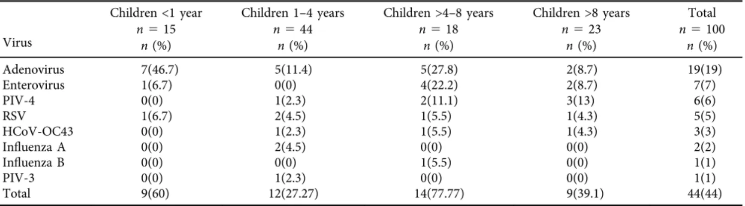
The present study included 100 pediatric patients. Males represented 41% of patients while females were 59%. Chil- dren were categorized into 4 age groups, children younger than 1 year old (60%), children between 1 and 4 years old (27.27%), children between 4 and 8 years old (77.77%) and children older than 8 years old (39.1%). Viral infections were detected in 44% out of the 100 children included in this study with the highest prevalence (77.7%) in the age group between 4 and 8 years.
Adenovirus was the most common, it was found in 19%
of the patients. Interestingly, HCoV-OC43 was detected in
3% of samples which up to our knowledge was not docu- mented previously in Egypt.
PIV 3 and 4 were detected in 7% while PIV1 and PIV 2 were not detected in all samples. Enterovirus was also detected in 7%. Prevalence of RSV was 5%. Only 2% were positive for Influenza A and 1% was positive for influenza B (Table 2).
As regards the respiratory symptoms, all children having respiratory viral infections had fever. Respiratory illness caused by Adenovirus in 6(31.6%), influenza virus in 1(50%) and PIV in 2(33.3%) had wheezes, while all 3 children with HCoV-OC43 infection had wheezes. Rhinorrhea was a major symptom (85.7–100%) in children infected with Coronavirus, Adenovirus and Enterovirus. Also, sore throat was a major symptom with most viruses. No hospitalization was needed among patients included (Table 3).
Mixed viral infection was observed in 7 (15.9%) out of the 44 infected children. PIV4 was the most common virus associated with mixed infection (71.4%). PIV4 and enterovirus were the most common viruses associated together (Fig. 1).
DISCUSSION
ARI is one of the most common human diseases, and the heaviest burden of viral respiratory illness is carried by children. Determining the etiology of LRTIs in children has long been a point of interest to researchers. Viruses have Table 2.Prevalence of respiratory viruses according to age of the patient
Virus
Children <1 year n515
Children 1–4 years n544
Children >4–8 years n518
Children >8 years n523
Total n5100
n(%) n(%) n(%) n(%) n(%)
Adenovirus 7(46.7) 5(11.4) 5(27.8) 2(8.7) 19(19)
Enterovirus 1(6.7) 0(0) 4(22.2) 2(8.7) 7(7)
PIV-4 0(0) 1(2.3) 2(11.1) 3(13) 6(6)
RSV 1(6.7) 2(4.5) 1(5.5) 1(4.3) 5(5)
HCoV-OC43 0(0) 1(2.3) 1(5.5) 1(4.3) 3(3)
Influenza A 0(0) 2(4.5) 0(0) 0(0) 2(2)
Influenza B 0(0) 0(0) 1(5.5) 0(0) 1(1)
PIV-3 0(0) 1(2.3) 0(0) 0(0) 1(1)
Total 9(60) 12(27.27) 14(77.77) 9(39.1) 44(44)
Table 3.Clinical manifestation of the patients with viral etiology
Clinical manifestations
Viruses
Adenovirus n519
Enterovirus n57
PIV-4 n56
RSV n55
HCoV-OC43 n53
Influenza A n52
Influenza B n51
PIV-3 n51
Total n5 44
n(%) n(%) n(%) n(%) n(%) n(%) n(%) n(%) n(%)
Fever 19(100) 7(100) 6(100) 5(100) 3(100) 2(100) 1(100) 1(100) 44(100)
Cough 10(52.6) 6(85.7) 4(66.6) 3(60) 2(66.6) 2(100) 1(100) 0 28(63.6)
Sore throat 17(89.5) 4(57.1) 3(50) 4(80) 2(66.6) 2(100) 1(100) 1(100) 34(77.3)
Wheezes 6(31.6) 1(14.3) 2(33.3) 0 3(100) 1(50) 0 0 13(29.5)
Rhinorrhea 17(89.5) 7(100) 1(16.7) 2(40) 3(100) 1(50) 0 0 31(70.5)
Tachypnea 10(52.6) 4(57.1) 3(50) 3(60) 1(33.3) 2(100) 1(100) 1(100) 25(56.8)
Chest pain 3(15.8) 2(28.6) 0 1(20) 0 1(50) 1(100) 0 8(18.2)
Nausea and vomiting 6(31.6) 4(57.1) 0 2(40) 0 1(50) 1(100) 0 14(31.8)
been shown to be the causative agent in 36–85% of LRTIs among children [24–28].
Several laboratory techniques ranging from enzyme immunoassay, direct fluorescent antibody staining, cell culture, and nucleic acid amplification tests are available for diagnosis of viral infections. The PCR technique is consid- ered an easy technique that provides higher sensitivity and specificity [29]. Nevertheless, different sampling methods and different geographical areas affect greatly the observed burden from each virus [19].
In this study, PCR and RT-PCR techniques were used to detect Adenovirus, a DNA virus, and 9 RNA viruses;
influenza A, B viruses, RSV, PIV type 1-4, enteroviruses, and HCoV-OC43. One hundred oropharyngeal swabs were ob- tained from Egyptian pediatric patients in Alexandria pre- senting with ARIs symptoms during winter season. Viral infection was confirmed in 44% of children.
In developing countries, viruses represent a considerable proportion of the pathogens associated with ARIs, varying from 40 to 90% across studies [30–34]. This wide differences in detection rates in the literature was accused to heteroge- neity in study populations, differences in presenting respi- ratory symptoms, number of respiratory pathogens tested, method used for detection and genetic variability between populations [30, 35].
In a study in Saudi Arabia in 2014, the frequency of respiratory virus detection in 135 children with ARIs was 80.7% [36]. A study in Egypt in 2018 reported the detection of respiratory viruses in 59.09% cases out of 132 patients [37]. A study in Turkey in 2011 reported a viral etiology in 36.7% out of 147 children [38]. In addition, a study in Amman, Jordan in 2010 reported 88% out of 728 suffered from viral infection [39].
It has been previously reported that the prevalence and severity of viral ARIs in male children is higher than females.
This was explained by the impact of sex hormones on T- helper 1/T-helper 2 cytokine balance [40]. In this study, 41%
of the children enrolled were males and 59% were females.
Viral infections were confirmed in 58.5% and 33.9% of the males and females respectively with male to female ratio of 1.4:1. Similar ratio was reported by Ljubin-Sternak et al.
[41], who detected ARI in 109 out of 134 patients with a male to female ratio of 1.5: 1. Liu et al. [42] examined 4,242 pediatric patients in southern China for respiratory viruses and reported even a higher male to female ratio of 1.92:1.
In the present study, the highest prevalence of viral infection was in the age group >4–8 years (77.77%) followed by the age group <1 year (60%). A significantly lower prevalence of 39.1% and 27.27% was observed in age group of >8 years and 1–4 years respectively. The higher preva- lence in the age groups >4–8 years and >8 years than in the age group 1–4 years could be explained by close contact with infected children in kindergarten and school respectively.
On the other hand, infection rate is still high in the age group less than 1 year old which could be attributed to a higher infection rate, lower viral clearance rate due to un- derdeveloped immune system, and higher load of the in- fectious agent associated with living conditions such as crowding. Furthermore, parents of younger children may seek healthcare earlier in the course of disease due to parental anxiety [30, 31, 35].
Thomazelli et al. [43] showed that the largest number of positive cases occurred in infants aged less than 1 year.
Moreover, Anders et al. [44] found that the incidence of ARI was higher in infants ≥6 months of age than in those <6 months. Tsai et al. [45], on the other hand reported that the Figure 1.Prevalence of respiratory viruses detected among 100 children presenting with respiratory symptoms
highest prevalence of viral infection was in children between 1 and 3 years old.
Adenovirus is the most common cause of pharyngitis and coryza in young children and causes 5–10% of all febrile illnesses in infants and young children. Of all young children who contract Adenovirus, 10% will develop pneumonia [46].
In the present study, the most commonly isolated virus was Adenovirus with a prevalence of 19%. The highest prevalence being 46.6% among the age group <1 year fol- lowed by 27.7% in the age group >4–8 years old with a higher exposure of males (29.2%) than females (11.8%).
Previous studies in Egypt showed that prevalence of Adenovirus infection ranged from 4.5% to 16.7% [19, 20, 37, 47]. Kajon et al. [46] investigated 165 respiratory Adeno- virus in Argentina, Chile and Uruguay, they reported that the highest prevalence 48.3% was in the age group 0–5 months followed by the age group 6–11 months 34.2% while the least prevalence was in the age group 2–5 years 4.7%
with a higher exposure of males 67.5.2% than females 32.5%.
Giamberardin et al. [48] reported that Adenoviruses, which are generally associated with severe infections in hospitalized patients were detected in 12.6% out of their 492 pediatric pa- tients in southern Brazil. The distribution of the frequency of the virus according to the age group showed that most of them were detected in children aged between 24 and 36 months.
It was estimated that 90 million cases of influenza occur each year in children aged <5 years around the world; of these, 20 million are associated with acute LRTI [49].
Influenza virus type A is the most virulent and is associated with seasonal (winter) epidemics in temperate regions [50].
In the present study, influenza A virus was detected in only 2% in the age group 1–4 years. On the other hand, only 1% in the age group >4–8 years was positive for influenza B.
El Baroudy et al. [37], detected influenza B virus in 2.3%
of his cases and influenza A was not detected among the samples. Shafik et al. [19]detected influenza A in 3.5% and influenza B in 1.1% cases in Cairo, Egypt. Thomazelli et al.
[43] detected influenza A virus in 5% of their 336 Brazilian samples, while influenza B virus was not detected. Choi et al.
[51] detected influenza A virus in 4.7%, and influenza B virus in 1.7% of their cases.
Type 4 PIV is very difficult to culture in-vitro and immunofluorescence assay has only been possible for a short while since the production of a first monoclonal anti-PIV-4 antibody [52]. For these reasons’PIV-4 was practically never sought in virology laboratories. But it has been shown that it can cause bronchiolitis or pneumonia in young children and its prevalence appears to be higher than originally thought [53].
In the present study, 7% of cases were positive for PIV 3 and 4.6 (85.7%) out of the 7 positive PIV cases belonged to PIV-4 while PIV-1 and PIV-2 were not detected in this study. However, Shafik et al. [19] detected PIV-1 in 23 cases, PIV-2 in only 1 case and PIV-3 in 22 cases. Also, Thomazelli et al. [43] found that the most common PIV type among their 336 samples was type 3 (8.3%). On the other hand, type 1 was detected in two samples and type 2 was not detected.
RSV is the most common respiratory agent in infants and young children worldwide. It is the most common agent that leads to acute bronchiolitis and viral pneumonia, and the second most common cause of infant deaths after the neonatal period [54]. In the present study, RSV was detected in only 5% of cases.
Similar results were reported by Tsai et al. [45] who detected RSV infection in 4.4% among their cases. However, Bonzel et al. [55] reported that RSV was the most frequently detected pathogen 44.1%. Liu et al. [42] as well reported that RSV was the most frequently isolated virus 32.5% among their 2,361 children occurring mainly among children less than 2 years old. Moreover, Shafik et al. [19] reported that RSV was the most commonly isolated virus in 23.8%.
Respiratory enteroviruses have been extensively studied during the past years and development of molecular tools has allowed better detection of enteroviruses which had remained undetected due to their inability to grow in stan- dard cell culture [2]. Ljubin-Sternak et al. [41] detected enteroviruses in 13.2% of their cases preceded only by RSV.
Liu et al. [42] using real-time polymerase chain detected enterovirus in, 13.3% out of their 2,361 children. In the present study, enterovirus was detected in 7 (7%) cases.
In a large Egyptian surveillance study that addressed the epidemiological patterns of severe ARI due to viruses and atypical bacteria in both children and adult population conducted over a 4 year-period Hatem et al. reported that no coronavirus was detected among their studied group [47].
Interestingly, in the present study, 1 male in the age group >4–8 years and 2 females in the age group 1-4 and >8 years were positive for HCoV-OC43. Al-Ayed et al. [36]
from Saudi Arabia, detected human coronavirus (hCoV NL63 and hCoV OC43) in 3.7% of their patients including 2 cases with bronchiolitis and 2 cases with pneumonia.
Jean et al. [56] in 2013 detected HCoV-OC43 in 1.8%
of their specimens. Higher prevalence was reported by Kristoffersen et al. [57] who detected HCoV-OC43 in 44 (8.2%) naspopharyngeal swabs collected from 536 episodes of RTI in 452 Norwegian children. Isaacs et al. [58] found that HCoVs were present in 30% of 108 acute respiratory infections. Liu et al. [42] also detected HCoVs in only 5.8%
of their 2,361 cases. Similarly, Ljubin-Sternak et al. [41]
detected HCoVs in 4.2% of their cases.
In September 2012, a novel human coronavirus, called the Middle East respiratory syndrome coronavirus (MERS- CoV), was first identified in Saudi Arabia [59]. Strains of MERS-CoV that are identical to human strains have been isolated from dromedaries in several countries, including Egypt, Oman, Qatar, and Saudi Arabia. The WHO reported largest outbreaks of MERS-CoV seen in Saudi Arabia, United Arab Emirates, and the Republic of Korea. The virus does not pass easily from person to person unless there is close contact, such as providing unprotected care to an infected patient [60].
To the best of our knowledge, this is the first report of the isolation of HCoV-OC43 as an etiological agent of res- piratory infections in Alexandria, Egypt. This finding urges the need to pursue the detection of all human Coronaviruses
including MERS-Cov in order to detect and report novel respiratory viral pathogens ahead of time in our country.
Mixed viral infection was observed in 7(15.9%) out of the 44 infected children, PIV-4 and enterovirus were the most common viruses associated with mixed infection (71.4%).
Dual infection and infection with more than 2 viruses was reported in Egyptian children by Shafik et al. [19] and concluded that with the development of the PCR scientists were able to detect co-infections at a level not previously possible. A study by Cui and his team mentioned that co- infection is reportedly related to the time of year when cir- culations of multiple viruses occur, and that there could be interplay between climatic, environmental and immunity level that contribute to viral co-infection [8].
Liu et al. [42] reported mixed viral infection in 21.3% of positive samples. They suggested the hypothesis that the proportion of the specific pathogen, rather than the path- ogen itself, is relevant for co-infections. The discrepancy in proportion of viral agents may be due to differences in pathogen epidemiology, study populations, and/or the time the study was conducted due to seasonal variation. Some studies [8, 61, 62] showed significant association between severity of infection and the detection of mixed viral etiol- ogies. Some studies went further to suggest that single infection resulted in more severe outcomes [63, 64]. In the present study, the respiratory virus profile differed from other studies since the most commonly isolated virus was adenovirus followed by enteroviruses, also PIV-4 was the commonly detected PIV with absence of PIV1 and 2. Res- piratory virus profiles in the other studies showed that RSV was the most commonly detected virus followed by Influ- enza A and PIV [42, 54, 55]. Additionally, HCoV-OC43 was never reported before in human respiratory samples in Egypt.
The results of the current study did not confirm viral etiology of RTIs in 56% of patients. Bacterial pathogens or viruses other than ones investigated may be involved in such finding. Nevertheless, these results are a good representative of viruses circulating in children in Alexandria, Egypt.
In summary, respiratory infections in children are mostly due to viral etiologies. Therefore, antibiotic therapy in these cases may be unnecessary. This data is helpful for guiding the antiviral therapy among children. HCoV-OC43 is pre- sent in Egypt and further studies should be conducted to detect other coronaviruses. This study provides baseline data against which to compare future levels of disease and the impact of future interventions.
Conflicts of interest: All contributing authors declare no conflicts of interest.
REFERENCES
[1] Pientong C, Ekalaksananan T, Teeratakulpisarn J, Tanu- wattanachai S, Kongyingyoes B, Limwattananon C. Atypical bacterial pathogen infection in children with acute
bronchiolitis in northeast Thailand. J Microbiol Immunol Infect 2011; 44(2): 95–100.
[2] Bariffi F, Sanduzzi A, Ponticiello A. Epidemiology of lower respiratory tract infections. J chemother 1995; 7(4): 263–76.
[3] Kusel MM, de Klerk NH, Holt PG, Kebadze T, Johnston SL, Sly PD. Role of respiratory viruses in acute upper and lower respiratory tract illness in thefirst year of life: a birth cohort study. Pediatr Infect Dis J 2006; 25(8): 680–6.
[4] Tazinya AA, Halle-Ekane GE, Mbuagbaw LT, Abanda M, Atashili J, Obama MT. Risk factors for acute respiratory in- fections in children underfive years attending the Bamenda Regional Hospital in Cameroon. BMC Pulm Med 2018; 18(1):
7.
[5] Rajatonirina S, Razanajatovo NH, Ratsima EH, Orelle A, Ratovoson R, Andrianirina ZZ, et al. Outcome risk factors during respiratory infections in a paediatric ward in Antana- narivo, Madagascar 2010-2012. PLoS One 2013; 8(9): e72839.
[6] Ho NT, Thompson C, Nhan LNT, Van HMT, Dung NT, Tran MP, et al. Retrospective analysis assessing the spatial and temporal distribution of paediatric acute respiratory tract in- fections in Ho Chi Minh City, Vietnam. BMJ Open 2018; 8(1):
e016349.
[7] Liu WK, Liu Q, Chen DH, Liang HX, Chen XK, Chen MX, et al. Epidemiology of acute respiratory infections in children in Guangzhou: a three-year study. PLoS One 2014; 9(5):
e96674.
[8] Cui B, Zhang D, Pan H, Zhang F, Farrar J, Law F, et al. Viral etiology of acute respiratory infections among children and associated meteorological factors in southern China. BMC Infect Dis 2015; 15: 124.
[9] Berman S. Epidemiology of acute respiratory infections in children of developing countries. Rev Infect Dis 1991;
13(Suppl. 6): S454–62.
[10]Naghipour M, Cuevas LE, Bakhshinejad T, Dove W, Hart CA.
Human bocavirus in Iranian children with acute respiratory infections. J Med Virol 2007; 79(5): 539–43.
[11]Al-Sonboli N, Hart CA, Al-Aghbari N, Al-Ansi A, Ashoor O, Cuevas LE. Human metapneumovirus and respiratory syncy- tial virus disease in children, Yemen. Emerg Infect Dis 2006;
12(9): 1437–9.
[12]Kaplan NM, Dove W, Abu-Zeid AF, Shamoon HE, Abd-Eld- ayem SA, Hart CA. Evidence of human metapneumovirus infection in Jordanian children. Saudi Med J 2006; 27(7):
1081–3.
[13]Khuri-Bulos N, Williams JV, Shehabi AA, Faouri S, Al Jundi E, Abushariah O, et al. Burden of respiratory syncytial virus in hospitalized infants and young children in Amman, Jordan.
Scand J Infect Dis 2010; 42(5): 368–74.
[14]El Sayed Zaki M, Raafat D, El-Metaal AA, Ismail M. Study of human metapneumovirus-associated lower respiratory tract infections in Egyptian adults. Microbiol Immunol 2009;
53(11): 603–8.
[15]Yahia S, Kandeel AY, Hammad E, El-Gilany AH. Human Metapneumovirus (hMPV) in acute respiratory infection: a clinic-based study in Egypt. Indian J Pediatr 2012; 79(10):
1323–7.
[16]Hatipoglu N, Somer A, Badur S, Unuvar E, Akcay-Ciblak M, Yekeler E, et al. Viral etiology in hospitalized children with
acute lower respiratory tract infection. Turk J Pediatr 2011;
53(5): 508–16.
[17]Imam IZ, Labib A, Hassan A, Fathy M. Etiology of viral pneumonia in Cairo 1967–1968. J Egypt Public Health Assoc 1969; 44(5): 486–97.
[18]El Sayed Zaki M, Goda T. Clinico-pathological study of atypical pathogens in community-acquired pneumonia: a prospective study. J Infect Dev Ctries 2009; 3(3): 199–205.
[19]Shafik CF, Mohareb EW, Yassin AS, Amin MA, El Kholy A, El-Karaksy H, et al. Viral etiologies of lower respiratory tract infections among Egyptian children under five years of age BMC Infect Dis 2012; 12: 350.
[20]Gad NM, Refaay D, Gad NM, Mohamed AZ. Viral infections in Egyptian hospitalized children with acute respiratory tract infections. J Clin Cell Immunol 2017; 8: 526.
[21]Osiowy C. Direct detection of respiratory syncytial virus, parainfluenza virus, and adenovirus in clinical respiratory specimens by a multiplex reverse transcription-PCR assay. J Clin Microbiol 1998; 36(11): 3149–54.
[22]Bellau-Pujol S, Vabret A, Legrand L, Dina J, Gouarin S, Petitjean-Lecherbonnier J, et al. Development of three multi- plex RT-PCR assays for the detection of 12 respiratory RNA viruses. J Virol Methods 2005; 126(1): 53–63.
[23]Zoll GJ, Melchers WJ, Kopecka H, Jambroes G, Van der Poel HJ, Galama JM. General primer-mediated polymerase chain reaction for detection of enteroviruses: application for diagnostic routine and persistent infections. J Clin Microbiol 1992; 30(1): 160–5.
[24]Drummond P, Clark J, Wheeler J, Galloway A, Freeman R, Cant A. Community acquired pneumonia–a prospective UK study. Arch Dis Child 2000; 83(5): 408–12.
[25]Juven T, Mertsola J, Waris M, Leinonen M, Meurman O, Roivainen M, et al. Etiology of community-acquired pneu- monia in 254 hospitalized children. Pediatr Infect Dis J 2000;
19(4): 293–8.
[26]Korppi M, Heiskanen-Kosma T, Jalonen E, Saikku P, Leinonen M, Halonen P, et al. Aetiology of community-acquired pneu- monia in children treated in hospital. Eur J Pediatr 1993;
152(1): 24–30.
[27]Hatipoglu N, Somer A, Badur S, Unuvar E, Akcay-Ciblak M, Yekeler E, et al. Viral etiology in hospitalized children with acute lower respiratory tract infection. Turk J Pediatr 2011;
53(5): 508–16.
[28]Sutmoller F, Ferro ZP, Asensi MD, Ferreira V, Mazzei IS, Cunha BL. Etiology of acute respiratory tract infections among children in a combined community and hospital study in Rio de Janeiro. Clin Infect Dis 1995; 20(4): 854–60.
[29]Erdman DD,Weinberg GA, Edward KM, Walker FJ, Anderson BC,Winter J, et al. GenScan reverse transcriptase-PCR assay for detection of six common respiratory viruses in young children hospitalized with acute respiratory illness. J Clin Microbiol 2003; 41: 4298–303.
[30]Van der Zalm MM, van Ewijk BE, Wilbrink B, Uiterwaal CS, Wolfs TF, van der Ent CK. Respiratory pathogens in children with and without respiratory symptoms. J Pediatr 2009; 154:
396–400.
[31]Huijskens EG, Biesmans RC, Buiting AG, Obihara CC, Rossen JW. Diagnostic value of respiratory virus detection in symp- tomatic children using real-time PCR. Virol J 2012; 9: 276.
[32]Kwofie TB, Anane YA, Nkrumah B, Annan A, Nguah SB, Owusu M. Respiratory viruses in children hospitalized for acute lower respiratory tract infection in Ghana. Virol J 2012; 9: 78.
[33]Bicer S, Giray T, Coel D, Erdag GC, Vitrinel A, G€urol Y, et al.
Virological and clinical characterizations of respiratory in- fections in hospitalized children. Ital J Pediatr 2013; 39: 22.
[34]Huang G, Yu D, Mao N, Zhu Z, Zhang H, Jiang Z, et al. Viral etiology of acute respiratory infection in Gansu Province, China, 2011. PLoS One 2013; 8: e64254.
[35]Suryadevara M, Cummings E, Bonville CA, Bartholoma N, Riddell S, Kiska D, et al. Viral etiology of acute febrile respi- ratory illnesses in hospitalized children younger than 24 months. Clin Pediatr (Phila) 2011; 50: 513–7.
[36]Al-Ayed MS, Asaad AM, Qureshi MA, Ameen MS. Viral eti- ology of respiratory infections in children in southwestern Saudi Arabia using multiplex reverse-transcriptase polymerase chain reaction. Saudi Med J 2014; 35(11): 1348–53.
[37]El Baroudy NR, El Refay AS, Abdel Hamid TA, Hassan DM, Soliman MS, Sherif L. Respiratory viruses and atypical bacteria co-infection in children with acute respiratory infection. Open Access Maced J Med Sci 2018; 6(9): 1588–93.
[38]Hatipoglu N, Somer A, Badur S, Un€uvar E, Akçay-Ciblak M, Yekeler E, et al. Viral etiology in hospitalized children with acute lower respiratory tract infection. Turk J Pediatr 2011;
53(5): 508–16.
[39]Khuri-Bulos N, Williams JV, Shehabi AA, Faouri S, Al Jundi E, Abushariah O, et al. Burden of respiratory syncytial virus in hospitalized infants and young children in Amman, Jordan.
Scand J Infect Dis 2010; 42(5): 368–74.
[40]Muenchhoff M, Goulder PJ. Sex differences in pediatric in- fectious diseases. The J Infect Dis 2014; 209(Suppl. 3): S120–6.
[41]Ljubin-Sternak S, Marijan T, Ivkovic-Jurekovic I, Cepin- Bogovic J, Gagro A, VranešJ. Etiology and clinical charac- teristics of single and multiple respiratory virus infections diagnosed in Croatian children in two respiratory seasons. J Pathog 2016; 20: 6–11.
[42]Liu WK, Liu Q, Chen DH, Liang HX, Chen XK, Chen MX, et al. Epidemiology of acute respiratory infections in children in Guangzhou: a three-year study. PLoS One 2014; 9(5):
e96674.
[43]Thomazelli LM, Vieira S, Leal AL, Sousa TS, Oliveira DB, Golono MA, et al. Surveillance of eight respiratory viruses in clinical samples of pediatric patients in southeast Brazil. J Pediatr 2007; 83(5): 422–8.
[44]Anders KL, Nguyen HL, Nguyen NM, Van Thuy NT, Van NT, Hieu NT, et al. Epidemiology and virology of acute respiratory infections during thefirst year of life: a birth cohort study in Vietnam. Pediatr Infect Dis J 2015; 34(4): 361.
[45]Tsai HP, Kuo PH, Liu CC, Wang JR. Respiratory viral in- fections among pediatric inpatients and outpatients in Taiwan from 1997 to 1999. J Clin Microbiol 2001; 39(1): 111–8.
[46]Kajon AE, Mistchenko AS, Videla C, Hortal M, Wadell G, Avenda~no LF. Molecular epidemiology of adenovirus acute lower respiratory infections of children in the south cone of South America (1991–1994). J Med Virol 1996; 48(2): 151–6.
[47]Hatem A, Mohamed S, Abu Elhassan UE, Ismael EAM, Magda S, Rizk MS, et al. Clinical characteristics and outcomes of patients with severe acute respiratory infections (SARI): results
from the Egyptian surveillance study 2010–2014. Multidiscip Respir Med 2019; 14: 11.
[48]Giamberardin HI, Homsani S, Bricks LF, Pacheco AP, Guedes M, Debur MC, et al. Clinical and epidemiological features of respiratory virus infections in preschool children over two consecutive influenza seasons in southern Brazil. J Med Virol 2016; 88(8): 1325–33.
[49]Nair H, Brooks WA, Katz M, Roca A, Berkley JA, Madhi SA, et al. Global burden of respiratory infections due to seasonal influenza in young children: a systematic review and meta- analysis. Lancet 2011; 378(9807): 1917–30.
[50]Viboud C, Alonso WJ, Simonsen L. Influenza in tropical re- gions. PLoS Med 2006; 3: e89.
[51]Choi EH, Lee HJ, Kim SJ, Eun BW, Kim NH, Lee JA, et al. The association of newly identified respiratory viruses with lower respiratory tract infections in Korean children, 2000–2005.
Clin Infect Dis. 2006; 43(5): 585–92.
[52]Downham MA, McQuillin J, Gardner PS. Diagnosis and clinical significance of parainfluenza virus infections in chil- dren. Arch Dis Child 1974; 49(1): 8–15.
[53]Lindquist SW, Darnule A, Istas A, Demmler GJ. Parainfluenza virus type 4 infections in pediatric patients. Pediatr Infect Dis J 1997; 16(1): 34–8.
[54]Lozano R, Naghavi M, Foreman K, Lim S, Shibuya K, Aboyans V, et al. Global and regional mortality from 235 causes of death for 20 age groups in 1990 and 2010: a systematic analysis for the Global Bur-den of Disease Study 2010. Lancet 2012;
380: 2095–128.
[55]Bonzel L, Tenenbaum T, Schroten H, Schildgen O, Schweitzer- Krantz S, Adams, O. Frequent detection of viral coinfection in children hospitalized with acute respiratory tract infection using a real-time polymerase chain reaction. The Pediatr Infect Dis J 2008; 27(7): 589–94.
[56]Jean A, Quach C, Yung A, Semret M. Severity and outcome associated with human coronavirus OC43 infections among children. Pediatr Infect Dis J 2013; 32(4): 325–9.
[57]Kristoffersen AW, Nordbø SA, Rognlien A-GW, Christensen A, Døllner H. Coronavirus causes lower respiratory tract in- fections less frequently than RSV in hospitalized Norwegian children. Pediatr Infect Dis J 2011; 30(4): 279–83.
[58]Isaacs D, Flowers D, Clarke JR, Valman HB, MacNaughton MR. Epidemiology of coronavirus respiratory infections. Arch D Child 1983; 58(7): 500–3.
[59]Zaki AM, van Boheemen S, Bestebroer TM, Osterhaus AD, Fouchier RA. Isolation of a novel coronavirus from a man with pneumonia in Saudi Arabia. N Engl J Med 2012; 367: 1814–20.
[60]WHO Factsheet March. Middle East respiratory syndrome coronavirus (MERS-CoV); 2019. Available from:https://www.
who.int/news-room/fact-sheets/detail/middle-east-respiratory- syndrome-coronavirus-(mers-cov).
[61]Essa S, Owayed A, Altawalah H, Khadadah M, Behbehani N, Al-Nakib W. Mixed viral infections circulating in hospitalized patients with respiratory tract infections in Kuwait. Hindawi Publishing Corporation. Adv Virol 2015: 8. 714062
[62]Harada Y, Kinoshita F, Yoshida LM, Minh le N, Suzuki M, Morimoto K. Does respiratory virus coinfection increases the clinical severity of acute respiratory infection among children infected with respiratory syncytial virus? Pediatr Infect Dis J 2013; 32: 441–5.
[63]Chorazy ML, Lebeck MG, McCarthy TA, Richter SS, Torner JC, Gray GC: Polymicrobial acute respiratory infections in a hospital- based pediatric population. Pediatr Infect Dis J 2013; 32: 460–6.
[64]Martin ET, Kuypers J, Wald A, Englund JA. Multiple versus single virus respiratory infections: viral load and clinical dis- ease severity in hospitalized children. Influenza Respi Viruses.
2012; 6: 71–7.