barbel (Barbus barbus): First molecular evidence for the presence of CyHV-1 in fish other than carp (Cyprinus carpio)
R EKA BORZ AK
1, BOGL ARKA SELLYEI
1, FERENC BASKA
2, CSABA SZ EKELY
1and ANDOR DOSZPOLY
1p1Institute for Veterinary Medical Research, Centre for Agricultural Research, P.O. Box 18, Budapest, H-1581, Hungary
2Department of Exotic Animal and Wildlife Medicine, University of Veterinary Medicine, Budapest, Hungary
Received: November 22, 2019 • Accepted: January 14, 2020
ABSTRACT
Two adult barbels (Barbus barbus) with visible skin tumours were subjected to histopathological and molecular examinations. Thefish were caught in the River Danube near Budapest. Papillomas were found around their oral cavity, at the operculum and at the pectoralfins, while epidermal hyperplasias were seen on the body surface. Cyprinid herpesvirus 1 (CyHV-1) was detected in the kidney of the specimens by polymerase chain reaction (PCR), and barbel circovirus 1 (BaCV1) was found in all internal organs and in the tissues of the tumours. The whole genome of BaCV1 and three conserved genes from the genome of CyHV-1 were sequenced. Previously, BaCV1 had been reported only once from a mass mortality event among barbel fry. The whole genome sequence of our circovirus shared 99.9% nucleotide identity with that of the formerly reported BaCV1. CyHV-1 is known to infect common carp and coloured carp (Cyprinus carpio), and has been assumed to infect other cyprinidfish species as well. We found the nucleotide sequences of the genes of CyHV-1 to be identical in 98.7% to those of the previous isolates from carp. To the best of our knowledge, this is thefirst molecular confirmation of the presence of CyHV-1 DNA in cyprinidfish species other than carp.
KEYWORDS
alloherpesviridae, cyprinid herpesvirus, fish herpesvirus, fish circovirus, epidermal tumour
INTRODUCTION
The common barbel (Barbus barbus) belongs to the family Cyprinidae. It is a native fresh- waterfish in Europe, inhabiting mostly fast-flowing rivers. The barbel is an excellentfish for sporting activities, and its importance in aquaculture is growing.
Little is known about the diseases affecting the barbel, with the exception of a few parasitic (Myxobolus spp., Aspidogaster limacoides, Pomphorhynchus laevis) (Thielen et al., 2004;Molnar et al., 2012) and viral (circovirus, rhabdovirus) infections (L}orincz et al., 2011;
Vicenova et al., 2011). However, recently a large variety of tumours with uncertain origin have been reported from barbels. For example, skin and gonadal tumours have been observed in barbels originating from the River Ohre, Czech Republic (Palikova et al., 2007), and a branchial osteogenetic neoplasm has been reported in specimens from the River Adige, North Italy (Manera and Biavati, 1999). In the River Lee in Southern England, numerous barbels with epidermal hyperplasias and papillomas have been found. Hyperplasias (1–3 mm thick) were found on the scales and papillomas (1–2 cm in diameter) at the base of thefins. They had a cauliflower-like appearance (Barnes et al., 1993). Although viral particles were not
Acta Veterinaria Hungarica
DOI:
10.1556/004.2020.00004
© 2020 Authors
ORIGINAL ARTICLE
*Corresponding author.
E-mail:doszpoly.andor@agrar.mta.hu
observed by electron microscopy, Dixon (2008) surmised that these hyperplasias were due to cyprinid herpesvirus 1 (CyHV-1). In the last few years, several adult barbels with distinct skin tumours were caught in the middle and upper reaches of the Danube close to Budapest during the autumn months. Tumour formation is often considered multifacto- rial, and viruses, chemical and biological toxins, physical effects, hormones as well as the age, sex, genetic predispo- sition and immunological competence of the hosts are among the suspected causes (Anders and Yoshimizu, 1994;
Roberts, 2012).
Alloherpesviruses (AHVs) are large, enveloped dsDNA viruses infecting fish and amphibians (Davison et al., 2009).
The family Alloherpesviridae comprises four genera (Batra- chovirus, Salmonivirus, Ictalurivirus and Cyprinivirus). The genusCypriniviruscontains four virus species accepted by the International Committee on Taxonomy of Viruses (Pellett et al., 2011), three of which (Cyprinid herpesvirus 1, Cyprinid herpesvirus 2, Cyprinid herpesvirus 3) are associated with common carp (Cyprinus carpio) or goldfish (Carassius aur- atus). The fourth one (Anguillid herpesvirus 1) originated from the European eel (Anguilla anguilla).
CyHV-1 is the causative agent of the so-called carp pox disease in common carp (Sano et al., 1985). Similar clinical signs were reported in other fish species of the family Cyprinidae, including barbel, bleak (Alburnus alburnus), freshwater bream (Abramis brama), chub (Squalius cepha- lus), Crucian carp (Carassius carassius), ide (Leuciscus idus), roach (Rutilus rutilus) and tench (Tinca tinca)(Mawdesley- Thomas and Bucke, 1967; McAllister et al., 1985; Dixon, 2008); however, it has never been confirmed by molecular methods that these viruses were actually CyHV-1, nor were any of these viruses successfully isolated.
Circoviruses (CVs) are small (12–27 nm) viruses with an icosahedral capsid containing a short single-stranded, cova- lently closed circular DNA genome (1.7–2.3 kilobases). The Circoviridaeis a relatively newly established family of viruses which is rapidly growing as a result of the recent evolution of molecular detection techniques. The family is divided into two genera,CircovirusandCyclovirus(Breitbart et al., 2017).
The very first discovery of fish CVs (barbel circoviruses, BaCV1 and 2) was during a mass mortality event in farmed barbel fry (L}orincz et al., 2011). Later the presence of CVs was reported in wels catfish (Silurus glanis), European eel (Anguilla anguilla), asp (Aspius aspius), roach and round goby (Neogobius melanostomus) as well (L}orincz et al., 2012;
Doszpoly et al., 2014;Tarjan et al., 2014;Borzak et al., 2017).
In the present paper we report the detection of CyHV-1 and BaCV1 by molecular methods in barbels displaying epidermal tumours.
MATERIALS AND METHODS
Specimen collection and pathology
In November of 2014, two specimens of barbel (70 cm, approx. 4 kg) bearing skin tumours were delivered to our
laboratory for histopathological and molecular examina- tions. The fish were caught by a sport fisherman in the River Danube near Budapest. After euthanasia, the fish were necropsied with routine tissue collection from the main organs (skin papilloma, gills, brain, liver, kidney, spleen and intestine) for histopathological, virus isolation and molecu- lar examinations. Tissues were fixed in Bouin’s fixative, washed in 80% ethanol, embedded in paraffin, sectioned (at 4–5
m
m), stained with haematoxylin and eosin, and viewed by light microscopy according to standard pro- cedures. The barbels were subjected to parasitological ex- aminations as well. In addition, swab samples were collected from the gills and tumours for general microbiological ex- amination on Tryptic Soy Agar (TSA) at 258C for 24 h.Virus isolation
Virus isolation was attempted on EPC (Epithelioma Pap- ulosum Cyprini) cell line (Fijan et al., 1983). The pooled internal organ homogenates were diluted to a 10% (w/v) suspension in MEM medium (Gibco) complemented with antibiotics (Penicillin 300 U/mL, Streptomycin 300
m
g/mL).The suspensions were centrifuged at 20003gfor 10 min at 168C. Inoculations with the supernatant (1 mL suspension perflask) were made in 25-cm2flasks of EPC monolayer at 80% confluency. The flasks were incubated at 17 8C and 20 8C and checked daily for the appearance of cytopathic effect (CPE).
PCR assays
For the molecular investigations, organ samples were homogenised using the TissueLyser high-throughput disruption instrument (Qiagen, Germany), and the DNA extraction was carried out by DNeasy®Blood and Tissue Kit (Qiagen, Germany) from 25 mg of tissue homogenates ac- cording to the manufacturer’s recommendations. The sam- ples were tested by two different polymerase chain reactions (PCRs), one developed for the detection of large dsDNA viruses (Hanson et al., 2006) and another one for the amplification of CVs and cycloviruses (Halami et al., 2008).
After ascertaining the presence of CyHV-1 and BaCV1, amplification of the whole genome of BaCV1 was attempted by an inverse PCR described previously (L}orincz et al., 2011). For amplifying longer fragments from the genes of the DNA-dependent DNA polymerase (DNA pol), helicase, and ATPase subunit of the terminase (terminase) of the putative novel CyHV-1, consensus primers designed for cypriniviruses were used (Doszpoly et al., 2015). The PCR products were excised from the agarose gel (1%), purified with the NucleoSpin Gel and PCR Clean-up kit (Macherey- Nagel, Germany), and sequenced bidirectionally with the forward and reverse primers. The sequencing PCRs were performed with the use of the BigDye Terminator v3.1 Cycle Sequencing Kit (Applied Biosystems, USA), and the elec- trophoresis was carried out by a commercial service provider (SZBK Sequencing Platform, Szeged, Hungary) on an ABI PRISM 3100 Genetic Analyzer. The sequence analysis and
contig assembly were performed by the MEGA5.2 software (Tamura et al., 2011).
RESULTS
Gross and microscopic pathology
Macroscopically, the barbels had tumours located around the mouth and the lower jaw, on the operculum, at the pectoral fins, and on the skin/scales throughout the body (Fig. 1). The number of the tumours was less than ten in each barbel. The size of the soft polypoid, pink-coloured masses was found to range between 0.5 and 3 cm in diam- eter. Additionally, the gross pathological examination revealed intensive infestation with a common acantho- cephalan worm (Pomphorhynchus laevis, M€uller), many specimens of which were present in the gut of each barbel.
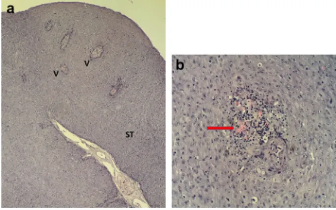
By bacteriological examination, only commensal bacteria, ubiquitous in the water environment, could be detected on the surface of gills and tumours. Histopathological ex- aminations of the excised skin tumours revealed that the stroma was well vascularised in its axis. In the benign tu- mours, the cells were polymorphic. Foci of secondary lym- phocytic inflammation were also seen (Fig. 2). The typical herpesvirus-induced nuclear changes including margination of the chromatin and the formation of inclusions (Sano et al., 1985) were not observed. These features are not always present in tissues infected with CyHV-1 (Hedrick et al., 1990). No other significant pathological/histopathological alterations were revealed.
Virus isolation
No CPE was observed during the virus isolation attempts.
After 14 days, a blind passage was carried out and then in a fortnight a second blind passage was conducted. There was no CPE in the second and third passages, and even the PCR of the tissue culture supernatants for the detection of viral DNA was negative in the consecutive passages.
PCR assays and sequence analysis
The presence of CyHV-1 was confirmed by the PCR tar- geting the DNA pol of large dsDNA viruses (Hanson et al., 2006) from the kidney. The other organs did not yield any products by this PCR. Subsequently, the PCRs for obtaining
longer sequences from the three conserved genes (Doszpoly et al., 2015) were successful. From the DNA pol gene a 787- bp-long fragment was amplified and sequenced. The PCR for the terminase gene yielded a 942-bp-long fragment, while from the helicase gene a 564-bp-long part was gained.
The sequences were deposited to the GenBank (Acc. Nos.
MK507841-507843). The nucleotide sequences of the three genes of the CyHV-1 originating from the barbels shared 98.7% nt identity with those of the CyHV-1 described pre- viously from common carp (Davison et al., 2013). Merely one amino acid discrepancy was detected in the terminase gene. The CV was detected in all examined organs of both barbels. The whole genome proved to consist of 1957 nt and shared 99.9% nt and 100 amino acid identity with BaCV1 (L}orincz et al., 2011).
DISCUSSION
Tumour formation is often considered a multifactorial dis- order, resulting from the combined influence of genetic factors, pathogens and environmental insults (Anders and Yoshimizu, 1994;Roberts, 2012). Neoplastic changes at the mouth and pectoralfins have been observed mostly infish feeding on the bottom. Epidermal injuries could increase the
Figure 1.External gross pathology. (a, b) Barbel showing papilloma around the oral cavity and at the operculum. (c) Epidermal hyperplasia on the body surface
Figure 2.Histopathologicalfindings: (a) Cross-sectional view of the papilloma. The stroma is well vascularised in its axis. ST: stroma; V:
vessels. Magnification:330. (b) Inside the benign tumour, the cells are polymorphic, and some foci of secondary lymphocytic inflammation can also be seen (arrow). Magnification: 3400.
Histological section, haematoxylin and eosin (HE)
risk of tumour development, since carcinogenic chemicals or oncogenic viruses couldfind their way easier into the body through an injury than via an intact epithelial surface (Mawdesley-Thomas and Fraser, 1972).Barnes et al. (1993) observed that tumours appeared as epidermal hyperplasias on body surfaces exposed to abrasion, while in the relatively more protected areas the tumours developed into papil- lomas. In our cases, papillomas were located not only in protected areas but also around the mouth and at the pec- toralfins (Fig. 1a) which are more exposed to rubbing, while hyperplasias were observed on the skin/scales throughout the body.
CyHV-1 is a well-known viral pathogen of common carp causing benign epidermal proliferations (Sano et al., 1985), and recently it has also been detected in cutaneous squa- mous cell carcinoma of koi carp (Sirri et al., 2018). Ac- cording to current knowledge, herpesviruses have high host specificity (Davison, 2002). Nonetheless, CyHV-1 was described as the putative causative agent of carp-pox-like disease in other cyprinid fish species based on electron microscopic investigations (Mawdesley-Thomas and Bucke, 1967; McAllister et al., 1985; Dixon, 2008). Yet, this has never been confirmed by molecular methods. Very recently, Sellyei et al. (unpublished) have found that carp-pox-like diseases in roach and asp were associated with a novel cyprinid herpesvirus (cyprinid herpesvirus 5); this finding might imply the need for the molecular examination of cyprinid fishes showing the typical signs of the disease.
To the best of our knowledge, this is the first molecular confirmation of the presence of CyHV-1 DNA in fish spe- cies other than its usual host, the carp. Without virus isolation and experimental infection, it is not possible to confirm the causal relationship between the tumour devel- opment and the presence of CyHV-1. More so, since the virus was detected only in the kidney, and there was no RNA extraction performed for checking the viral replication.
However, the seasonality and manifestation of the disease resembled those of CyHV-1 infection in common carp (Hedrick et al., 1990).
CV infection was described in European eel showing signs of the so-called cauliflower disease (benign stomato- papilloma); however, a direct connection between the pres- ence of CV and the disease could not be demonstrated (Doszpoly et al., 2014). Moreover, later the prevalence of eel CV was found to be high (35.5%) in an apparently healthy eel population in Lake Balaton, Hungary (Borzak et al., 2017). The CVs studied in more detail (porcine and avian CVs) are known to have an immunosuppressive effect (Todd, 2004), which may enhance the pathogenicity of some agents causing concomitant infections (e.g. CyHV-1). It is also possible that CVs can cause generalised infection in immunocompromised individuals. Similar conclusions were drawn from the examination of a novel small ssDNA virus in sea turtles with fibropapillomas (Ng et al., 2009). The infection with CVs might also promote the interspecies transmission of the strictly carp-pathogenic virus (CyHV-1).
However, it cannot be excluded that water pollution or other predisposing factors also play a role in the manifestation of
tumours associated with CyHV-1 and BaCV1 infection in barbels.
ACKNOWLEDGEMENTS
The excellent technical assistance of Gy€orgyi Ostoros is gratefully acknowledged. This work was supported by a grant provided by the Hungarian Scientific Research Fund (OTKA K127916).
REFERENCES
Anders, K. and Yoshimizu, M. (1994): Role of viruses in the in- duction of skin tumors and tumor-like proliferations of fish.
Dis. Aquat. Org.19, 215–232.
Barnes, A., Owen, A. G., Feist, S. W. and Bucke, D. (1993): An investigation into the occurrence of epidermal hyperplasia and papilloma in barbel (Barbus barbusL.) from a river in southern England. Bull. Eur. Assoc. Fish Pathol.13, 115–118.
Borzak, R., Sellyei, B., Szekely, C. and Doszpoly, A. (2017):
Molecular detection and genome analysis of circoviruses of European eel (Anguilla anguilla) and sichel (Pelecus cul- tratus). Acta Vet. Hung. 65, 262–277.
Breitbart, M., Delwart, E., Rosario, K., Segales, J., Varsani, A. and ICTV Report Consortium. (2017): ICTV virus taxonomy pro- file: circoviridae. J. Gen. Virol.98, 1997–1998.
Davison, A. J. (2002): Evolution of the herpesviruses. Vet. Micro- biol.86, 69–88.
Davison, A. J., Eberle, R., Ehlers, B., Hayward, G. S., McGeoch, D. J., Minson, A. C., Pellett, P. E., Roizman, B., Studdert, M. J. and Thiry, E. (2009): The order Herpesvirales. Arch. Virol. 154, 171–177.
Davison, A. J., Kurobe, T., Gatherer, D., Cunningham, C., Korf, I., Fukuda, H., Hedrick, R. P. and Waltzek, T. B. (2013):
Comparative genomics of carp herpesviruses. J. Virol. 87, 2908–2922.
Dixon, P. F. (2008): Virus diseases of cyprinids. In: Eiras, J. C. and Segner, H. (eds) Fish Diseases, Book 1. Science Publishers, Enfield, Jersey, Plymouth.
Doszpoly, A., Papp, M., Deakne, P. P., Glavits, R., Ursu, K. and Dan, A. (2015): Molecular detection of a putatively novel cyprinid herpesvirus in sichel (Pelecus cultratus) during a mass mortality event in Hungary. Arch. Virol.160, 1279–1283.
Doszpoly, A., Tarjan, Z. L., Glavits, R., M€uller, T. and Benk}o, M.
(2014): Full genome sequence of a novel circo-like virus detected in an adult European eel Anguilla anguilla showing signs of cauliflower disease. Dis. Aquat. Org.109, 107–115.
Fijan, N., Sulimanovic, D., Bearzotti, M., Muzinic, D., Zwillenberg, L., Chilmonczyk, S., Vautherot, J. and Dekinkelin, P. (1983): Some properties of the Epithelioma-papulosum-cyprini (EPC) cell-line from carpCyprinus carpio. Ann. Virol.134, 207–220.
Halami, M. Y., Nieper, H., M€uller, H. and Johne, R. (2008):
Detection of a novel circovirus in mute swans (Cygnus olor) by using nested broad-spectrum PCR. Virus Res. 132, 208–212.
Hanson, L. A., Rudis, M. R., Vasquez-Lee, M. and Montgomery, R. D.
(2006): A broadly applicable method to characterize large DNA viruses and adenoviruses based on the DNA polymerase gene.
Virol. J.3, 28.
Hedrick, R. P., Groff, J. M., Okihiro, M. S. and McDowell, T. S.
(1990): Herpesviruses detected in papillomatous skin growths of koi carp (Cyprinus carpio). J. Wildl. Dis.26, 578–581.
L}orincz, M., Csagola, A., Farkas, S. L., Szekely, C. and Tuboly, T.
(2011): First detection and analysis of afish circovirus. J. Gen.
Virol.92, 1817–1821.
L}orincz, M., Dan, A., Lang, M., Csaba, G., Toth, A. G., Szekely, C., Csagola, A. and Tuboly, T. (2012): Novel circovirus in European catfish (Silurus glanis). Arch. Virol.157, 1173–1176.
Manera, M. and Biavati, S. (1999): Branchial osteogenetic neoplasm in barbelBarbus barbus plebejus. Dis. Aquat. Org.37, 231–236.
Mawdesley-Thomas, L. E. and Fraser, W. (1972): The conservation offish. Br. Vet. J.128, 337–346.
Mawdesley-Thomas, L. E. and Bucke, D. (1967): Fish pox in the roach (Rutilus rutilusL.). Vet. Rec.81, 56–57.
McAllister, P. E., Lidgerding, B. C., Herman, R. L., Hoyer, L. C. and Hankins, J. (1985): Viral diseases offish:first report of carp pox in golden ide (Leuciscus idus) in North America. J. Wildl. Dis.
21, 199–204.
Molnar, K., Eszterbauer, E., Marton, S., Szekely, C. and Eiras, J.
(2012): Comparison of theMyxobolusfauna of common barbel from Hungary and Iberian barbel from Portugal. Dis. Aquat.
Org.100, 231–248.
Ng, T., Manire, C., Borrowman, K., Langer, T., Ehrhart, L. and Breitbart, M. (2009): Discovery of a novel single-stranded DNA virus from a sea turtle fibropapilloma by using viral meta- genomics. J. Virol.83, 2500–2509.
Palikova, M., Navratill, S., Svobodova, Z., Tichy, F., Recek, L. and Pikula, J. (2007): Skin and gonadal tumours in a barbel(Barbus barbus) – a case report. Bull. Eur. Assoc. Fish Pathol. 27, 234–238.
Pellett, P. E., Davison, A. J., Eberle, R., Ehlers, B., Hayward, G. S., Lacoste, V., Minson, A. C., Nicholas, J., Roizman, B., Studdert, M. J.
and Wang, F. (2011): FamilyHerpesviridae. In: King, A. M. Q., Adams, M. J., Carstens, E. B. and Leftkowitz, E. J. (eds) Virus Taxonomy, IXth Report of the International Committee on Tax- onomy of Viruses. Elsevier, Academic Press, London.
Roberts, R. (2012): Neoplasia of teleosts. In: Roberts, R. and Saunders, W. (eds) Fish Pathology, 4th edition. Wiley-Blackwell, Chichester, UK. pp. 167–185.
Sano, T., Fukuda, H., Furukawa, M., Hosoya, H. and Moriya, Y.
(1985): A herpesvirus isolated from carp papilloma in Japan. In:
Ellis, A. E. (ed.) Fish and Shellfish Pathology. Academic Press, London.
Sirri, R., Ciulli, S., Barbe, T., Volpe, E., Lazzari, M., Franceschini, V., Errani, F., Sarli, G. and Mandrioli, L. (2018): Detection of Cyprinid Herpesvirus 1 DNA in cutaneous squamous cell car- cinoma of koi carp (Cyprinus carpio). Vet. Dermatol.29, 60–e24.
Tamura, K., Peterson, D., Peterson, N., Stecher, G., Nei, M. and Kumar, S. (2011): MEGA5: molecular evolutionary genetics analysis using maximum likelihood, evolutionary distance, and maximum parsimony methods. Mol. Biol. Evol.28, 2731–2739.
Tarjan, Z. L., Penzes, J. J., Toth, R. P. and Benk}o, M. (2014): First detection of circovirus-like sequences in amphibians and novel putative circoviruses infishes. Acta Vet. Hung.62, 134–144.
Thielen, F., Zimmermann, S., Baska, F., Taraschewski, H. and Sures, B. (2004): The intestinal parasitePomphorhynchus laevis (Acanthocephala) from barbel as a bioindicator for metal pollution in the Danube River near Budapest, Hungary. Envi- ron. Pollut.129, 421–429.
Todd, D. (2004): Avian circovirus diseases: lessons for the study of PMWS. Vet. Microbiol.98, 169–174.
Vicenova, M., Reschova, S., Pokorova, D., Hulova, J. and Vesely, T.
(2011): First detection of pike fry-like rhabdovirus in barbel and spring viraemia of carp virus in sturgeon and pike in aqua- culture in the Czech Republic. Dis. Aquat. Org.95, 87–95.
This is an open-access article distributed under the terms of the Creative Commons Attribution 4.0 International License (https://creativecommons.org/licenses/by/4.0/), which permits unrestricted use, distribution, and reproduction in any medium, provided the original author and source are credited, a link to the CC License is provided, and changes–if any–are indicated. (SID_1)