Taxonomy and nomenclature of bacteria with clinical and scientific importance: current concepts for pharmacists and pharmaceutical scientists
MÁRIÓ GAJDÁCS
1Department of Pharmacodynamics and Biopharmacy, Faculty of Pharmacy, University of Szeged, Szeged, Hungary
*Corresponding author: Márió Gajdács Email: gajdacs.mario@pharm.u-szeged.hu Received: 17 October 2019 / Accepted: 1 January 2020
1. Introduction to (bacterial) taxonomy Taxonomy (from the greek words taxis=arran ge- ment or order, and nemein=to distribute or govern) is the science of the classification of various living organisms [1,2]. In case of bacteria, taxonomy con- sists of three independent, but interrelated disci- plines, namely classification, nomenclature and iden- tification (sometimes referred to as the ‘trinity’ of taxonomy) [2]. The most basic taxonomic group (i.e. unit) in bacterial taxonomy is the species, while groups of species are collected into genera (genus), which are then collected into families (Fa- milia), families into orders (Ordo), orders into class- es (Classis), classes into phyla (Phylum) and phyla into a domain (or Kingdom, the highest level), how- ever, there are subgroups to these main classifica-
tions (see Table I and II for examples). Groups of bacteria at each rank or level have names with endings or suffixes characteristic to that rank or level (Table I) [1-3].
Nevertheless, taxonomic units under species may still be relevant (especially in the case of medically-rel- evant bacteria), because members among specific spe- cies can be distinguished on the basis of certain biologi- cal or genetic characteristics: these members may be classified in a sub-group of members, called subspecies [1-3]. An example for this is the differentiation of Cam- plyobacter species: C. fetus subsp. veneralis is a caus- ative agent of sexually transmitted diseases and miscar- riage among cattle, while C. fetus subsp. fetus may cause intrauterine infection in humans [4]. Antigenic characteristics may be another possible way to distin- guish subgroups under the threshold of species, called Abstract
Taxonomy is the science of the classification of various living organisms consisting of three independent, but interrelated disciplines, namely classification, nomenclature and identification. With the advent of molecular biological methods and sequencing, a revolution is currently occurring with regards to the reporting of novel taxa and changes in the taxonomy of already described bacterial species.
The applications of taxonomic changes can be broad ranging: they may impact the clinical care of patients, through variations in choos- ing the appropriate antimicrobial susceptibility testing standards or data interpretation, or even their clinical relevance and epidemiol- ogy. The aim of this paper was to aid healthcare professionals and pharmaceutical scientists to navigate through the ‘maze’ of bacterial taxonomy, and to aid in finding authentic information regarding the description of taxonomic changes and to present some examples of changes in bacterial taxonomy which have proven to be clinically significant.
Keywords: bacteria, taxonomy, nomenclature, identification, molecular, microbiology, educational
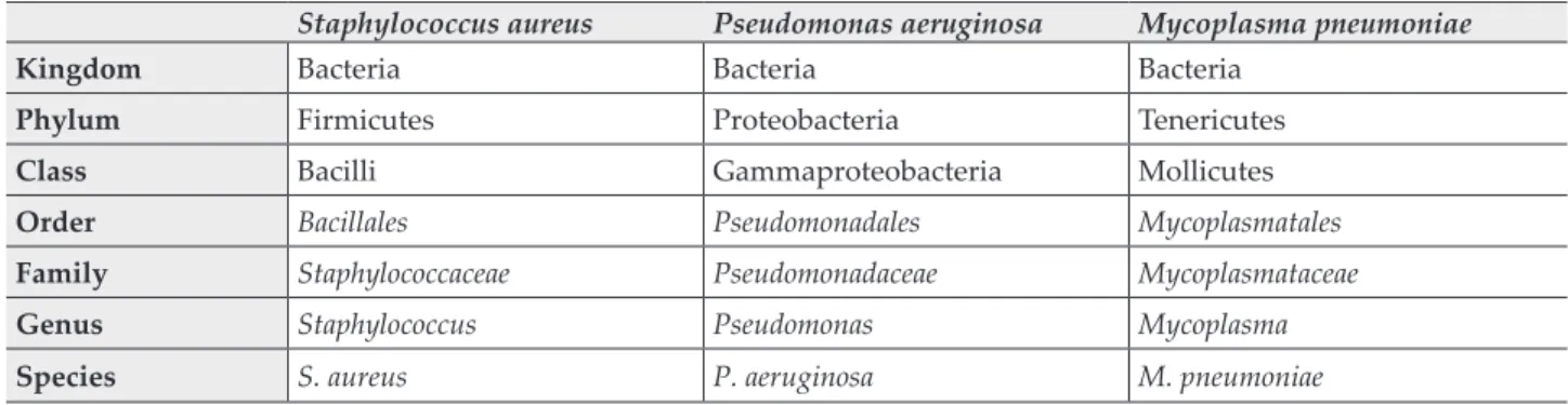
Table I Example of taxonomic classification for a common Gram-positive, Gram-negative and an atypical pathogen Staphylococcus aureus Pseudomonas aeruginosa Mycoplasma pneumoniae
Kingdom Bacteria Bacteria Bacteria
Phylum Firmicutes Proteobacteria Tenericutes
Class Bacilli Gammaproteobacteria Mollicutes
Order Bacillales Pseudomonadales Mycoplasmatales
Family Staphylococcaceae Pseudomonadaceae Mycoplasmataceae
Genus Staphylococcus Pseudomonas Mycoplasma
Species S. aureus P. aeruginosa M. pneumoniae
DOI: 10.33892/aph.2019.89.99-108
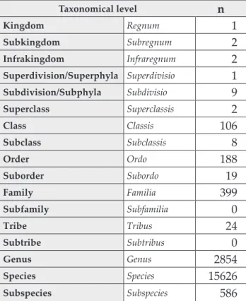
Table II Characteristics of the current bacterial classification and the number validly published names for each
classification level [22]
Taxonomical level n
Kingdom Regnum 1
Subkingdom Subregnum 2
Infrakingdom Infraregnum 2
Superdivision/Superphyla Superdivisio 1
Subdivision/Subphyla Subdivisio 9
Superclass Superclassis 2
Class Classis 106
Subclass Subclassis 8
Order Ordo 188
Suborder Subordo 19
Family Familia 399
Subfamily Subfamilia 0
Tribe Tribus 24
Subtribe Subtribus 0
Genus Genus 2854
Species Species 15626
Subspecies Subspecies 586
serogroups or serovariants [5]. In case of gut bacteria or Enterobacteriaceae (especially important for Salmo- nella species and Escherichia coli), hundreds of differ- ent serovariants may be differentiated, based on the cell wall (O; somatic antigen, based on oligosaccharides), capsule (K, from the German Kapsel or Bacterienkap- sel) and flagellar (H; from the German Hauch meaning
“breath” or “mist”) antigens [6,7]. In fact, this is the ba- sis of the Kauffman–White classification, which was frequently used for routine clinical microbiology and public health purposes for serotyping [8]. Similarly, bacteria may be further characterized based on their disease-causing capacity (pathogenicity) into pathot- ypes, e.g., extraintestinal-pathogenic E. coli (ExPEC), enteropathogenic E. coli (EPEC), enterotoxin-produc- ing E. coli (ETEC), enteroinvasive E. coli (EIEC), en- teroaggregative E. coli (EAEC), diffusely adherent E.
coli (DAEC) and so on [9,10].
However, a lot has changed since the first de- scription of taxonomy (Augustin Pyramus de Can- dolle, 1813), when the available methods for the characterization of bacterial, plant or animal spe- cies were very limited [11]. Nowadays, with the advent of molecular biological methods and se- quencing, a revolution is currently occurring with regards to the reporting of novel taxa [12]. The de- scription of new bacterial species was further fa-
cilitated by the newfound interest in the character- ization of the human microbiome [13]. One of the most important milestones was the launch of the Human Microbiome Project (HMB; the first phase of which was launched in 2007), with the aim of characterize the human gut microbial flora in healthy (physiological) and disease states; the long-term aim of this project was to find causation between human pathologies (e.g., autoimmune disorders, obesity, diabetes, neuropsychiatric dis- orders, diseases affecting the cardiovascular sys- tem) and qualitative/quantitative changes in the microbiome [14-16]. Microbial culturomics (a tech- nique allowing for the culturing of previously un- culturable bacterial species by reproducing their natural habitats using complex methods, with the aid of matrix-assisted laser desorption-ionization time-of-flight mass spectrometry [MALDI-TOF MS] and whole-genome sequencing [WGS]) has also resulted in the description of a staggering number of novel taxa [17-19]. Sequencing technol- ogies also had a significant role in the description of the prokaryotic genetic diversity.
Between 1990 and 2000, there was on average 200 novel bacterial species described per year [20].
Owing to these recent developments, the number of validly published genera and species has in- creased by approximately 50% since 2004, reduc- ing the percentage of known prokaryotes that have been implicated in animal or human clinical conditions from ~15% to ~10% [21]. Based on the records of the bacterio.net database, there are cur- rently 19,717 validly published bacterial names and 383 so-called candidate names published (as of 20th of October, 2019) [22]. However, the database of EZBioCloud.net (a freely-accesible database on prokaryotic diversity) contains 81,189 taxa (out of which, 24.89% has been validly published and 0.51% are candidate names), 64,416 16S rRNA se- quences (a highly conserved, evolutionally-con- stant region) and 146,704 qualified genomes (as of 9th of August, 2019) [23]. Nonetheless, there are re- ports estimating that the currently known/de- scribed microbiological diversity only represents around 1-5% of the global prokaryotic diversity [20-21].
Bacterial systematics is a field, which is fre- quently used synonymously with taxonomy, how- ever, the scope of systematics is much broader, in- cluding data on bacterial morphology, physiolo- gy, molecular biology and biochemistry, metabol- ic products, pathogenic potential, ecological nich- es and epidemiology to characterize, arrange and
classify bacteria [24]. Systematics became more relevant after the widespread adoption of molecu- lar biological methods, ever higher resolution characterization of bacterial species [25] (Figure 1).
Due to the rapid developments in bacterial tax- onomy, both consisting of the description of novel taxa and reclassification of existing bacterial gen- era to other taxonomical units (e.g., the history of S. maltophilia: it first described as Bacterium booker (1943), later on, it was redesignated as Pseudomo- nas maltophilia (1958) and Xanthomonas maltophilia (1981); finally, in 1993, the genus Stenotrophomonas was proposed), it is very difficult, if not impossible for researchers, officials, public health microbiolo- gists and healthcare professionals to keep in mind all the accepted or proposed changes [26,27].
However, the importance of correct taxonomy in scientific communication and the diagnostics and therapy of bacterial infections cannot be underesti- mated [3]. Even if they are not aware of all the changes and the newly introduced species, rele- vant persons should be able to quickly find them in medical literature, scientific publications or oth- er sources (Web pages or blogs kept up by taxono- mists).
There are various resources for pharmaceutical scientists and microbiologists to get informed re- garding the recognition of novel bacterial species or describing proposed reclassification of an older species. The official publication prokaryotic taxon- omy and source of data regarding these matters in the International Journal of Systematic and Evolu- tionary Microbiology (IJSEM); the main aim and role of this publication is to report the description of new taxa or the reclassification of existing spe-
cies [28]. The rules associated with the proposal of new bacterial taxa were described in the Bacterio- logical Code (1990), which was updated though the publication of the Taxonomic Outline of the Bacte- ria and Archaea (TOBA; 2006) [29,30]. Additional amendments to these rules are generally pub- lished in IJSEM. The proposed new species and species names (candidate) are to be submitted to the Editorial Office of IJSEM for evaluation, with the suffix nova (genus nova, species nova). The new taxonomy and nomenclature can only be consid- ered as official, if the Editorial Board of IJSEM and the International Committee on Systematics of Prokaryotes (ICSP) both approve the submission [21]. If approved, the proposed name receives the approved state (valid name), which is formalized by the certificate of approval awarded by the IJSEM and ICSP [21,28]. However, once these taxa are on the approved lists, they may still be subject to reclassification, based on the designation of syn- onyms or due to a transfer to another genus. The validation of a new taxa is finalized if designated type strains of the species are deposited into inter- nationally-recognized culture collections (e.g., American Type Culture Collection [ATCC], Asian Bacterial Bank [ABB], Anaerobe Reference Labora- tory, Helsinki Collection [AHN], Culture Collec- tion of Switzerland [CCOS], Collection de l’Institut Pasteur [CIP], United Kingdom National Culture Collection [UKNCC]) at least in two separate countries [31].
The Antoine van Leeuwenhoek Journal of Mi- crobiology has become the second main journal in this field in the recent years, reporting on >100 new candidate species per year, since 2014 [32]. In addition, several other journals with interests in microbiology/infectious diseases may be vehicles in reporting novel taxa, including but not limited to: Systematic and Applied Microbi- ology, Journal of Medical Microbiolo- gy, Current Microbiology, Clinical Microbiology and Infection, Diagnos- tic Microbiology and Infectious Dis- ease, Anaerobe, Infection, Genetics and Evolution, Journal of Antimicro- bial Chemotherapy, Emerging Mi- crobes and Infections, New Microbes and New Infections, Microbiology and Immunology, Frontiers in Genet- ics, Frontiers in Microbiology, Ar- chives of Microbiology, Microbiolo- gyOpen, Standards in Genomic Sci- Figure 1 Relationship of the field of taxonomy and bacterial systematics
ences, Acta Pathologica Microbiologica et Immu- nologica Scandinavica (APMIS), Zentralblatt für Bakteriologie, Research in Microbiology [33]. Nev- ertheless, it is important to note that for the novel or revised taxa to be validly published (and the study was not submitted to IJSEM), the proposi- tion must be included on an “approved” list in IJSEM. IJSEM publishes papers entitled “List of new names and new combinations previously effective- ly, but not validly, published” six-to-twelve times per year, which gives a good idea about the mo- mentum of bacterial taxonomy [21,28,33]. The pro- posed taxa that have been previously described in other journals will be footnoted in IJSEM.
2. What is in a name: nomenclature in bacteriology
The discipline of nomenclature is mainly concerned with the assignment of names to taxonomic units or groups, on the basis of specific rules [34]. Before a name could be designated for any microorgan- ism, one has to describe its biological characteris- tics (for its future identification), allowing for its classification in the subordinate system, as previ- ously described [1-3]. The origins of nomenclature date back to 1753, when Carl Linnaeus published Species Plantarum, introducing the binomial nomen- clature and the currently used classification hier- archy, based on greek-latin terms as the normal system of naming organisms [35]. Species Planta- rum (later functioning as the International Code of Botanical Nomenclature [ICBN]) was first set of rules and recommendations of its kind. Because bacteria were once classified among plants, the nomenclature of these microorganisms was sub- ject to the rules of the ICBN until the 1930s, when the bacteriological society has decided on the preparation of an independent code. The Interna- tional Code of Bacteriological Nomenclature (or Bacteriological Code for short) was first approved in Copenhangen, 1947 [29,30]. The entire frame- work of naming bacteria is too complex to be de- scribed in its entirety, as the Bacteriological Code currently had more than 500 rules and regulations regarding name proposals for novel species names (which are periodically being updated; the up- dates are published in IJSEM) [36]. As the “intel- lectual capital” (i.e. available empirical and exper- imental data) available for the scientists submit- ting the candidate names for consideration con- stantly grows, so does the scientific accuracy of the bacterial names. The etymology (the study of
the origin and history of words) of bacterial ge- nus/species name is very diverse; here are several examples on the etymology of some bacterial gen- era:− Famous microbiologists (or scientists): Rothia
(Genevieve D. Roth), Kingella and Elizabethkingia (Elizabeth O. King), Escherichia (Theodor Esche- rich), Pasterurella (Louis Pasteur), Gaffyka (Georg Theodor A. Gaffky), Burkholderia (W.H. Burk- holder), Ehrlichia (Paul Ehrlich), Serratia (Serafi- no Serrati, physicist)
− Mythological names: Cronobacter (Cronos, a ti- tan), Proteus (Proteus, a prophetic sea-god), Tel- luria (Tellus, a Roman goddess personifying the Earth)
− Morphological characteristics: Bacillus (rod), Streptococcus (spheres with grape-like organiza- tion), Helicobacter (helical-shaped), Campylobacter (curved rod), Clostridium (greek word for spin-
− Biochemical characteristics: Achromobacter (has dle) no pigment), Acinetobacter (non-motile), Chomo- bacterium (produces pigment), Anaerococcus (strict anaerobe)
− Geographical (e.g., site of first isolation): Bud- vicium (Latin name of the city Cěské Budějovice where the bacterium was first isolated), Hafnia (old name for Copenhagen), Orientia (the Orient, the area where the organisms are widely dis- tributed), Sinorhizobium (which lives in a root in China)
− Distribution: Aerococcus (air), Enterococcus (gut), Coprococcus (feces), Leptotrichia (fine hair [of rab- bits])
− Disease-causing capability: S. pneumoniae (pneumonia), Vibrio cholerae (cholera: watery di- arrhoea), N. meningitidis (meningitis), P. multoci- da (lethal to many)
− Institutions: Centers for Disease Contol and Prevention (CDC): Cedaceae, Armed Forces Insti- tute of Pathology (AFIP): Apifia
Changes at higher taxonomical levels (e.g., at orders and classes) are obviously much less likely to occur than in lower units, therefore one of the most common conventions on denominations is that the family name is based on the name of the most typical genus (i.e. a type species) in its do- main [29-36]. A typical example is the Legionellace- ae family, where the most characteristic genera is the one containing the Legionella species, namely L. pneumophila, the causative agent of Legion- airre’s disease. In contrast, for Enterobacteriaceae, E.
coli is considered the type species, but the family
is not called Escherichiaceae; instead, due to the an- atomical localization of most of these bacteria in the gut flora, they are classified in Enterobacteriace- ae. Interestingly, the family Enterobacteriaceae con- tains a genus called Enterobacter, however, if one of the members of this genus would be the type species in the family, it would need to be called Enterobacteraceae [29-37]. The correct writing of bacterial taxonomical designations are also strictly defined by this convention, e.g. names of the spe- cies, genera and family are written in italics, how- ever, at higher taxonomical designations, this is not done [29-36].
The use of abbreviations is also common in the literature and the routine clinical practice. Al- though there are official (defined in the Bacterial Code) three-letter abbreviations for a variety of bacterial genera (e.g., Acp. for Acidophilum, Rsc. for Roseococcus) to ease correspondences regarding anoxygenic phototrophic bacteria, other “real word” examples include mosaic terms derived from names of bacterial groups (e.g., GAS: Group A Streptococcus; ESKAPE: Enterococcus faecium, S.
aureus, Klebsiella spp., Acinetobacter baumannii, P.
aeruginosa, Enterobacter spp.), therapeutic recom- mendations (e.g., MRSA: methicillin-resistant S.
aureus) or public health significance (e.g., MSTM:
multidrug-resistant Stenotrophomonas maltophilia, MDRAB: multidrug-resistant A. baumannii) [27, 38-43]. It must be noted that in medicine (especial- ly as far as the clinical microbiologist-physician relationship is concerned), the use of commonly known names is preferred, which are not subject to change (irrespective of taxonomic changes), so that the doctors reading the reports, e.g., of a sus- ceptibility test can comprehend them [29-36].
3. Laboratory methods used in bacterial taxonomy and identification
The discipline of classification refers to the act of arranging bacteria into these group or taxa, based on their evolutionary relationships and similarity [44]. In the early days of bacterial taxonomy, the basis of classification was solely on the determina- tion of microscopic morphology and phenotypic characteristics, which could be observed by a light microscope or by organoleptic analysis in liquid or solid media [11]. Later on, this was comple- mented by the detection of the presence or ab- sence of various enzymes, such as coagulase (dif- ferentiates between coagulase positive S. aureus and coagulase-negative Staphylococcus species),
catalase (among other things, it differentiates be- tween Staphylococcus and Streptococcus species), oxidase (aids in the differentiation of non-ferment- ing Gram-negative bacteria, e.g., Pseudomonas and Acinetobacter), urease (among other things, it dif- ferentiates between Ureaplasma and Mycoplasma species) and the study of the use of different sug- ars (i.e. their oxidative or fermentative breakdown) [45]. For a very long period of time, these bio- chemical tests were the mainstay of identification in clinical bacteriology. Identification may be con- sidered as applied taxonomy, during which microbi- ologists determine whether a particular isolate be- longs to a recognized taxon (i.e., genus, species or subspecies) [1-3]. One of the main utilizers of bac- terial identification in medicine is the field of clini- cal microbiology (where bacterial pathogens are identified from various clinical samples to estab- lish the patient’s illness and to guide targeted an- timicrobial therapy) and public health (the follow- up of outbreaks caused by bacteria), however, companies involved in pharmaceutical research, biotechnology, forensics are all relevant stakehold- ers [21].
In the last several decades, pronounced changes were brought about in bacterial taxonomy, due to the introduction of nucleic acid-based and molec- ular techniques, thus making phenotypic methods less and less relevant [46]. These methods have demonstrated that genotypic/phylogenetic relat- edness does not necessary correlate well with phe- notypic attributes, such as a Gram-staining pat- tern, microscopic morphology, oxygen-tolerance or fastidious growth characteristics [47]. These molecular methods include comparison of DNA- denaturation or melting temperatures (Tm), char- acterization of GC (guanine and cytosine) ratios of bacterial DNA, DNA-DNA and DNA-RNA hy- bridization, pulse-field gel electrophoresis (PFGE), multi-locus sequence typing (MLST), average nu- cleotide identity (ANI), MALDI-TOF MS, 16S rRNA gene and whole genome sequencing (WGS) and next-generation sequencing (NGS) [48-50].
The use of these methods in increasingly preva- lent not only in classification, but also in identifi- cation thus, revolutionizing the field of microbiol- ogy in the process [48-50]. The prevalence of their use is mainly determined by their accuracy, ro- bustness and their price. Based on the abovemen- tioned methods, two bacteria are considered to be the same species, if their nucleotide sequences are at least 70% identical, and the difference between their Tm values is less, than 5% [47]. The analysis
of the GC ratio (G+C content) in genomic DNA is also a suitable taxonomic method: the GC ratio is the most variable in prokaryotic genomes (20- 80%); however, in strains of a specific species, the GC content is shown to be constant. It is basically defined as percentage of the G and C amino nucle- otides in the bacterial genome, which was fre- quently used for the division of various bacterial genera (e.g., staphylococci and micrococci highly resemble each other, based on phenotypic and biochemical characteristics, however, the differ- ence in their GC ratios has been shown to be pro- nounced [~30-40% vs. ~65-75%]) [51]. DNA–DNA hybridization was considered the gold standard for decades: genomic hybridization allows for the measurement of the degree of similarity between two genomes; this technique is very useful in the differentiation of closely-related bacterial species [52,53]. In contrast, DNA-RNA hybridization is useful in the genetic analysis of two phylogeneti- cally distant bacteria: this is possible, because ri- bosomal RNA (rRNA) is transfer RNA (tRNA) only represent a minor portion of bacterial genes, which evolves in a slower pace (i.e. they are more conserved), compared to other genes coding for proteins [52,53]. Currently, various nucleic acid sequencing methods (of which, WGS and NGS are one of the most modern) represent the top-tier methods for bacterial classification and the com- parison of genomic structures [48-50]. Nucleic acid (DNA and RNA) sequencing is another mo-
lecular characteristic that helps directly compare the genomic structures. The sequencing of 5S rRNA (from the 50S prokaryotic ribosomal sub- unit), 16S rRNA and 16S rDNA (from the 30S pro- karyotic ribosomal subunit) has received the most substantial attention [47-50]. In fact, current rec- ommendations state that for the submission of a novel species, the performance of MLST or se- quencing (to characterize genomic relatedness) and the submission of a preferably full-length 16S rRNA gene sequence are recommended [47].
Nevertheless, it is now well-known that the phenotypic as well as the genotypic characteristics of bacteria may be subject to change due to exoge- nous genetic material (i.e. conjugation, transfor- mation and transduction), which entails the trans- fer of plasmid DNA from one species/genus/fami- ly of bacteria to another. In reality, these proper- ties may also be useful to characterize relations between different bacterial taxa [47-50]. E. coli spe- cies conjugate well with Salmonella and Shigella species (which are more closely related taxonomi- cally), but not with members of the genera Proteus, Providencia or Enterobacter. Similar results were found in transformation studies on Rhizobium, Mi- crococcus, Bacillus and Haemophilus species, show- ing that transformation events more frequently occur with different species of the same genera (smaller genomic variation), compared to species of different genera (larger genomic variation) [47- 50].
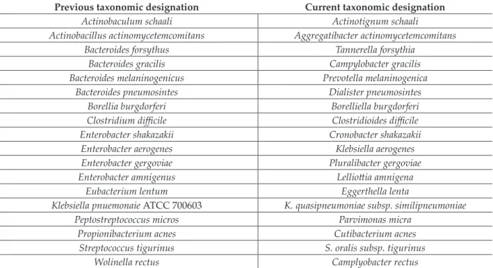
Table III Examples of bacterial species undergone taxonomic revisions in the last 20-year period
Previous taxonomic designation Current taxonomic designation
Actinobaculum schaali Actinotignum schaali
Actinobacillus actinomycetemcomitans Aggregatibacter actinomycetemcomitans
Bacteroides forsythus Tannerella forsythia
Bacteroides gracilis Campylobacter gracilis
Bacteroides melaninogenicus Prevotella melaninogenica
Bacteroides pneumosintes Dialister pneumosintes
Borellia burgdorferi Borelliella burgdorferi
Clostridium difficile Clostridioides difficile
Enterobacter shakazakii Cronobacter shakazakii
Enterobacter aerogenes Klebsiella aerogenes
Enterobacter gergoviae Pluralibacter gergoviae
Enterobacter amnigenus Lelliottia amnigena
Eubacterium lentum Eggerthella lenta
Klebsiella pnuemonaie ATCC 700603 K. quasipneumoniae subsp. similipneumoniae
Peptostreptococcus micros Parvimonas micra
Propionibacterium acnes Cutibacterium acnes
Streptococcus tigurinus S. oralis subsp. tigurinus
Wolinella rectus Camplyobacter rectus
4. Practical relevance of taxonomical changes After the official recognition and acknowledge- ment of taxonomic alterations or a revised no- menclature, significant changes may occur in the everyday practice of physicians and microbiolo- gists dealing with infectious diseases, epidemiol- ogists, university educations and other relevant stakeholders [47]. Changes in bacterial taxonomy, and nomenclature is usually greeted with con- servatism and resistance among taxonomists, mi- crobiologists, healthcare-professionals and scien- tist alike, for the simple reason that nobody likes change [1-3,21,47]. The applications of taxonomic changes can be broad ranging: they may impact the clinical care of patients, through variations in choosing the appropriate antimicrobial suscepti- bility testing standards or data interpretation, or even their clinical relevance and epidemiology (commensal/colonizer/pathogen). These changes also affect companies supplying laboratories with testing equipment and software (i.e. the lab- oratory information system or LIS) and even ad- ministrative stakeholders (e.g., accreditation ser- vices, conformity with legal documentation); of course, the clinical relevance of these changes is also relative to the isolation frequency and inva- siveness of the abovementioned bacteria [33,36,37,43,44,47]. Some recent changes of inter- est in bacterial taxonomy are discussed below and presented in Table III.
Gram-negative bacteria (especially ones repre- senting gut bacteria) have seen a plethora of taxo- nomic revisions since the beginning of the 21st century. Among other things, some Vibrio species have been reclassified into the genera Photobacte- rium (e.g., P. damselae) and Grimontia (G. hollisae), and the phylogenetically heterogenous members of the E. cloacae complex has been reassigned to the genera Kosakonia, Lelliottia, and Pluralibacter [54-56]. Another relevant change was the one af- fecting the genus Salmonella, where only Salmonel- la enterica strains remained in the species status, while other serovariants (e.g., Enteritidis, Ty- phimurium, Typhi) are no longer recognized on the species level, therefore their names should no longer be italicized [6,57]. However, one of the major taxonomical changes affecting Gram-nega- tive bacteria (and subsequently, the medical com- munity) is the recent reclassification of the family Enterobacteriaceae into the order Enterobacterales, containing seven distinct families (namely Entero- bacteriaceae, Erwiniaceae, Pectobacteriaceae, Yersinia-
ceae, Hafniaceae, Morganellaceae and Budiviciaceae) based on recent phylogenetic analyses [58]. Other suggestions include the differentiation of all Burk- holderia species into two distinct groups: the ge- nus Burkolderia would contain the human patho- genic species, while a newly designated genus Paraburkholderia would hold the non-pathogenic species to humans [59]. In contrast, it was pro- posed that the genera Chlamydia and Chlamydophi- la (containing C. pneumoniae and C. psittaci) should be fused together, eliminating the latter genus in the process [60].
Pronounced taxonomic changes have also oc- cured regarding anaerobic bacteria in the last 30- 40 years [16]. The restriction of the genus Bacte- roides to B. fragilis and related species has led to the relclassification and transfer of numerous species to the genera Prevotella and Porphyromon- as (based on pigmentation, bile-sensitivity and saccharolytic properties) and the introduction of novel genera [61-64]. Marked changes have also occured in the field of Gram-positive anaerobic cocci with the introduction of novel species, such as Finegoldia, Parvimonas and Peptinophilus, based on phylogenetic analysis [16,65-66]. Eubacterium species were also subject to taxonomic revisions, leading to the introduction of novel genera, such as Slackia, Pseudoramibacter, Mogibacterium, Egger- thella and Cryptobacterium [16,65]. Perhaps the most controversial taxonomic revision occured regarding the causative agent of antibiotic-asso- ciated diarrhoea and pseudomembranous en- terocolitis, namely Clostridium (Clostridioides) dif- ficile, which may be considered as the prime ex- ample why taxonomic changes have to be care- fully considered [67-68]. After the proposal to re- stric the genus Clostridium to C. butyricum and other related species, it was found that C. difficile was phylogenetically closest to C. mangenotii with a 94.7% similarity, however, this species was located in the family Peptostreptococcaceae [69]. This would have lead to a nomenclature re- vision of C. difficile as Peptoclostridium difficile;
however, due to the significance of this pathogen in nosocomial infection and as a public health threat, a lot of energy, time and money was put into the education of the public and healthcare professionals around the globe, regarding the dangers of “C. diff” (as it is colloquially known) and CDD/CDAD (C. difficile-associated diarrhea), with educational campaigns, fliers, books and so on [16, 67-69]. The proposed taxonomic change would have put forth issues in this educational
campaign (a sudden change of “C. diff” to “P.
diff” and CDAD to PDAD and so on); for this rea- son, the reclassification as P. difficile was rejected, instead, a novel genus Clostridioides gen. nov.
was proposed for C. difficile (now Clostridioides difficile) and C. magnerotii was also reclassified to this new genus; therefore previously used, collo- quial designations for this pathogen (C. diff, CDD/CDAD) also remained valid [70,71].
5. Conclusions
Taxonomy is concerned with the classification of living organisms, which operates in three distinct domains, namely classification, nomenclature and identification. Compared to the taxonomic trends in the 19th century, current methods and technologies allow for more detailed phylogenetic analyses, leading to the description of a tremen- dous amount of novel bacterial species and the re-classification of several already described bac- teria. This ‘explosion’ in microbial taxonomy (fur- ther aided by the developments in bacterial sys- tematics) presents an everyday challenge to medi- cal professionals (e.g., clinical pharmacists, physi- cians and nurses), pharmaceutical scientists and stakeholders in healthcare. However, the up-to- date knowledge on bacterial taxonomy is impor- tant as it may significantly impact the everyday practice of these healthcare professionals. This is especially true for scientists who use various bac- terial strains for screening of antimicrobial activi- ty of various compounds or utilizing any kind of bacterial model system during laboratory assays.
The aim of this paper was to aid the abovemen- tioned healthcare professionals to navigate through the ‘maze’ of bacterial taxonomy, to aid in finding authentic information regarding the description of taxonomic changes and to present some examples of changes in bacterial taxonomy which proven to be clinically significant.
6. Acknowledgements
M.G. was supported by the National Youth Excel- lence Scholarship [Grant Number NTP-NTFÖ- 18-C-0225] and the ESCMID Observership Pro- gramme.
7. Competing interests
The author declares no conflict of interest, mone- tary or otherwise.
8. References
1. Cowan, S.T. Sense and nonsense in bacterial tax- onomy. J. Gen. Microbiol. 1971; 67: 1-8. https://doi.
org/10.1099/00221287-67-1-1
2. Cowan, S.T. Principles and practice of bacterial tax- onomy. Microbiology 1965; 39: 148-158. https://doi.
org/10.1099/00221287-39-1-143
3. Kraft, C.S., McAdam, A.J., Carroll, K.C. A Rose by any other name: Practical updates on micro- bial nomenclature for clinical microbiology. J. Clin.
Microbiol. 2017; 55: 3-4. https://doi.org/10.1128/
JCM.02169-16
4. Huang, H., Brooks, B.W., Lowman, R., Carrillo, D.
Campylobacter species in animal, food, and envi- ronmental sources, and relevant testing programs in Canada. Can. J. Microbiol. 2015; 61: 701-721.
https://doi.org/10.1139/cjm-2014-0770
5. Johnson, J.R., Orskov, I., Orskov, F., Goullet, P., Picard, B., Moseley, S.L., Roberts, P.L., Stamm, W.E. O, K, and H antigens predict virulence fac- tors, carboxylesterase B pattern, antimicrobial resistance, and host compromise among Esch- erichia coli strains causing urosepsis. J. Infect.
Dis. 1994; 169: 119-126. https://doi.org/10.1093/
infdis/169.1.119
6. Brenner, F.W., Villar, R.G., Angulo, F.J., Tauxe, R., Swaminanthan B. Salmonella Nomenclature.
J. Clin. Microbiol. 2000; 38: 2465-2467. https://doi.
org/10.1128/JCM.38.7.2465-2467.2000
7. Fratamico, P.M., DebRoy C., Liu, Y., Neddleman, D.S., Baranzoni, G.M., Feng, P. Advances in Mo- lecular Serotyping and Subtyping of Escherichia coli. Front. Microbiol. 2016; 7: 644. https://doi.
org/10.3389/fmicb.2016.00644
8. Ryan, M.P., O’Dwyer, J, Adley, C.C. Evaluation of the Complex Nomenclature of the Clinically and Veterinary Significant Pathogen Salmonella.
Biomed. Res. Int. 2017; 2017: 3782182. https://doi.
org/10.1155/2017/3782182
9. Palaniappan, R.U.M., Zhang, Y, Chiu, D., Torres, A., DebRoy, C., Whittam, T.S., Chang, Y.F. Differen- tiation of Escherichia coli Pathotypes by Oligonu- cleotide Spotted Array. J. Clin. Microbiol. 2006; 44:
1495-1501. https://doi.org/10.1128/JCM.44.4.1495- 1501.2006
10. Browne-Robins, R.M., Holt, K.E., Ingle, D.J., Hock- ing, D.M., Yang, J., Tauschek, M. Are Escherichia coli Pathotypes Still Relevant in the Era of Whole- Genome Sequencing? Front. Cell Infect. Microbiol.
2016; 6: 141. https://doi.org/10.3389/fcimb.2016.00141 11. Cain, A.J. Deductive and inductive meth- ods in post-Linnaean taxonomy. Proc. Linn.
Soc. London 1959; 170: 185-217. https://doi.
org/10.1111/j.1095-8312.1959.tb00853.x
12. Parks, D.H., Chuvchina, M., Waite, D.W., Rinke, C., Skarshewki, A., Chaumeil, P.A., Hugenholtz, P. A standardized bacterial taxonomy based on genome phylogeny substantially revises the tree of life. Nat.
Biotechn. 2018; 36: 996-1004. https://doi.org/10.1038/
nbt.4229
13. Turnbaugh, P.J., Ley, R.E., Hamady, M., Fraser- Liggett, C.M., Knight, R., Gordon, J.I. The Human Microbiome Project: exploring the microbial part of ourselves in a changing world. Nature 2007; 449:
804-810. https://doi.org/10.1038/nature06244
14. Hahn, S.A., Altman, T., Konwar, K.M., Hanson, N.W., Kim, D., Relman, D.A., Dill, D.L., Hallam, S.J. A geographically-diverse collection of 418 hu- man gut microbiome pathway genome databases.
Sci. Data 2017; 4: 1700035. https://doi.org/10.1038/
sdata.2017.35
15. Almeida, A., Mitchell, A.L., Boland, M., Forster, S.C., Gloor, G.B., Tarkowska, A., Lawley, T.D., Finn, R.D. A new genomic blueprint of the human gut microbiota. Nature 2019; 568: 499-504. https://doi.
org/10.1038/s41586-019-0965-1
16. Gajdács, M., Spengler, G., Urbán, E. Identification and Antimicrobial Susceptibility Testing of Anaero- bic Bacteria: Rubik’s Cube of Clinical Microbiology?
Antibiotics 2017; 7: 25. https://doi.org/10.3390/anti- biotics6040025
17. Lagier, J.C., Armougom, F., Million, M., Hugon, P., Pagnier, I., Robert, C., Bittar, F., Fournous, G., Gimenez, G., Maraninchi, M., Trape, J.F., Koonin, E.V., La Scola, B., Raoult, D. Microbial culturomics:
paradigm shift in the human gut microbiome study.
Clin. Microbiol. Infect. 2012; 18: 1185-1193. https://
doi.org/10.1111/1469-0691.12023
18. Lay, J.O. MALDI-TOF mass spectrometry and bac- terial taxonomy. Trends Anal. Chem. 2000; 19: 507- 516. https://doi.org/10.1016/S0165-9936(00)00027-3 19. Garrity. G.M. A New Genomics-Driven Taxono-
my of Bacteria and Archaea: Are We There Yet? J.
Clin. Microbiol. 2016; 54: 1956-1963. https://doi.
org/10.1128/JCM.00200-16
20. Siqueira, J.F. Taxonomic changes of bacteria as- sociated with endodontic infections. J. Endodont.
2003; 29: 619-623. https://doi.org/10.1097/00004770- 200310000-00001
21. Janda, M.J. Taxonomic update on proposed nomen- clature and classification changes for bacteria of medical importance, 2016. Diagn. Microbiol. Infect.
Dis. 2017; 88: 100-105. https://doi.org/10.1016/j.diag- microbio.2017.02.003
22. Bacterio.net database http://www.bacterio.net/- number.html (Accessed on: 24th of October, 2019).
23. EZBioCloud Database of Prokaryotic Diversity https://www.ezbiocloud.net/dashboard (Accessed on: 24th of October, 2019).
24. Moore, E.R.B., Mihaylova, S.A., Vandamme, P., Krichevsky, M.I., Dijksoorn, L. Microbial systemat- ics and taxonomy: relevance for a microbial com- mons. Res. Microbiol. 2010; 161: 430-438. https://doi.
org/10.1016/j.resmic.2010.05.007
25. Vandamme, P., Pot, B., Gills, M., de Vos, P., Kersters, K., Swings, J. Polyphasic taxonomy, a consensus approach to bacterial systematics. Microbiol. Mol.
Biol. Rev. 1996; 60: 407-438. https://doi.org/10.1128/
MMBR.60.2.407-438.1996
26. de Vos, P., Trüper, H.G. Judicial Commission of the International Committee on Systematic Bacteriol- ogy. IXth International (IUMS) Congress of Bacte- riology and Applied Microbiology. Minutes of the meetings, 14, 15 and 18 August 1999, Sydney, Aus- tralia. Int. J. Syst. Evol. Microbiol. 2000; 50: 2239- 2244. https://doi.org/10.1099/00207713-50-6-2239 27. Gajdács, M., Urbán, E. Epidemiological Trends
and Resistance Associated with Stenotrophomonas maltophilia Bacteremia: A 10-Year Retrospective Cohort Study in a Tertiary-Care Hospital in Hun- gary. Diseases 2019; 31: 41. https://doi.org/10.3390/
diseases7020041
28. International Journal of Systematic and Evolution- ary Microbiology https://www.microbiologyre- search.org/content/journal/ijsem (Accessed on: 24th of October, 2019).
29. Lapage SP, Sneath PHA, Lessel EF, Skerman VBD, Seeliger HPR, Clark WA (eds.). International Code of Nomenclature of Bacteria: Bacteriological Code, 1990 Revision. Washington (DC): ASM Press; 1992.
30. Taxonomic Outline of the Bacteria and Archaea (Formerly the Taxonomic Outline of the Prokary- otes) Release 7.7 http://www.taxonomicoutline.org/
content/7/7/ (Accessed on: 24th of October, 2019).
31. Baltrus, D.A. Divorcing strain classification from species names. Trends Microbiol. 2016; 24: 431-439.
https://doi.org/10.1016/j.tim.2016.02.004
32. Antonie van Leeuwenhoek Journal of Microbiology https://www.springer.com/journal/10482/aims-and- scope (Accessed on: 24th of October, 2019).
33. Janda, M.J. Proposed nomenclature or classification changes for bacteria of medical importance: Taxo- nomic Update 4. Diagn. Microbiol. Infect. Dis. 2019;
94: 205-208. https://doi.org/10.1016/j.diagmicro- bio.2018.12.009
34. Berger, S.A., Edberg, S.C. Microbial nomenclature:
A list of names and origins. Diagn. Microbiol. Infect.
Dis. 1987; 6: 343-356. https://doi.org/10.1016/0732- 8893(87)90185-4
35. Carl Linnaeus, Species Plantarum. Sweden, 1753.
36. Munson, E., Carroll, K.C. An Update on the Novel Genera and Species and Revised Taxonomic Status of Bacterial Organisms Described in 2016 and 2017.
J. Clin. Microbiol. 2019; 57: e01181-18. https://doi.
org/10.1128/JCM.01181-18
37. Gajdács, M., Urbán, E. Resistance Trends and Epi- demiology of Citrobacter-Enterobacter-Serratia in Urinary Tract Infections of Inpatients and Out- patients (RECESUTI): A 10-Year Survey. Medicina (Kaunas) 2019; 55: 285. https://doi.org/10.3390/me- dicina55060285
38. Bacterio.net Three-letter code for abbreviations of generic names. http://www.bacterio.net/-abbrevia- tions.html (Accessed on: 24th of October, 2019).
39. Stevens, D.L. Invasive group A streptococcus infec- tions. Clin. Infect. Dis. 1992; 14: 2-11. https://doi.
org/10.1093/clinids/14.1.2
40. Gajdács, M. The Concept of an Ideal Antibiotic: Im- plications for Drug Design. Molecules 2019; 24: 892.
https://doi.org/10.3390/molecules24050892
41. Gajdács, M. The Continuing Threat of Methicillin- Resistant Staphylococcus aureus. Antibiotics 2019;
8: 52. https://doi.org/10.3390/antibiotics8020052 42. Gajdács, M. Burián, K., Terhes, G. Resistance Lev-
els and Epidemiology of Non-Fermenting Gram- Negative Bacteria in Urinary Tract Infections of Inpatients and Outpatients (RENFUTI): A 10-Year Epidemiological Snapshot. Antibiotics 2019; 8: 143.
https://doi.org/10.3390/antibiotics8030143
43. Baron, E.J., Allen, S.D. Should clinical laborato- ries adopt new taxonomic changes? If so, when?
Clin. Infect. Dis. 1993; 16: S449-S450. https://doi.
org/10.1093/clinids/16.Supplement_4.S449
44. Mandel, M. New approaches to bacterial taxonomy:
perspectives and prospects. Ann. Rev. Microbiol.
1969; 23: 239-274. https://doi.org/10.1146/annurev.
mi.23.100169.001323
45. Leber, A.L. (eds.) Clinical Microbiology Procedures Handbook, Fourth Edition. 2016; American Society
for Microbiology, Washington DC, USA. https://doi.
org/10.1128/9781555818814
46. Gajdács, M., Urbán, E. The relevance of anaerobic bacteria in brain abscesses: a ten-year retrospective analysis (2008-2017). Infect. Dis. 2019;, 51: 779-781.
https://doi.org/10.1080/23744235.2019.1648857 47. Janda, J.M. Clinical Decisions: How Relevant is
Modern Bacterial Taxonomy for Clinical Microbi- ologists? Clin. Microbiol. Newslett. 2018; 40: 51-57.
https://doi.org/10.1016/j.clinmicnews.2018.03.005 48. Wayne, B.D., Coldwell, R.R., Grimont, P.A.D., Kan-
dler, O., Krichesvsky, J.I. et al. Report of the ad hoc committee on reconciliation of approaches to bacte- rial systematics. Int J Syst Bacteriol. 1987; 166: 463- 464. https://doi.org/10.1099/00207713-37-4-463 49. Mahato, N.K., Gupta, V., Singh, P., Kumari, R.,
Verma, H., Tripathi, C., et al. Microbial taxonomy in the era of OMICS: application of DNA sequences, computational tools and techniques. Antonie Van Leeuwenhoek. 2017; 110: 1357-1371. https://doi.
org/10.1007/s10482-017-0928-1
50. Colabella, C., Corte, L., Roscini, L., Bassetti, M., Tas- cini, C., Mellor, J.C., Meyer, W., Robert, V., Vu, D., Cardinali, G. NGS barcode sequencing in taxonomy and diagnostics, an application in “Candida” patho- genic yeasts with a metagenomic perspective. IMA Fungus 2018; 9: 91-105. https://doi.org/10.5598/ima- fungus.2018.09.01.07
51. Thompson, C.C., Chimetto, L., Edwards, R.A., Swings, J., Stackerbrandt, E., Thompson, F.L. Micro- bial genomic taxonomy. BMC Genomics 2013; 14:
913. https://doi.org/10.1186/1471-2164-14-913 52. Cho, J.C., Tiedje, J.M. Bacterial Species Determi-
nation from DNA-DNA Hybridization by Using Genome Fragments and DNA Microarrays. Appl.
Environ. Microbiol. 2001; 67: 3677-3682. https://doi.
org/10.1128/AEM.67.8.3677-3682.2001
53. Dong, D., Li, J., Gao, Q., Huang, X., Xu, Y., Li, R. Uti- lizing RNA/DNA hybridization to directly quantify mRNA levels in microbial fermentation samples. J.
Microbiol. Methods 2009; 79: 205-210. https://doi.
org/10.1016/j.mimet.2009.09.002
54. Pérez-Catalunia, A., Lucena, T., Tarazona, E., Ara- hal, D.R., Macián, M.C., Pujalte, M.J. An MLSA approach for the taxonomic update of the Splen- didus clade, a lineage containing several fish and shellfish pathogenic Vibrio spp. Syst. Appl. Micro- biol. 2016; 39: 361-369. https://doi.org/10.1016/j.sy- apm.2016.03.010
55. Genus Enterobacter http://www.bacterio.net/en- terobacter.html (Accessed on: 24th of October, 2019).
56. Davin-Regli, A., Pagés, J.M. Enterobacter aero- genes and Enterobacter cloacae; versatile bacterial pathogens confronting antibiotic treatment. Front.
Microbiol. 2015; 6: 392. https://doi.org/10.3389/
fmicb.2015.00392
57. Tindall, B.J., Grimont, P.A.D., Garrity, G.M., Euzé- by, J.P. Nomenclature and taxonomy of the genus Salmonella. Int. J. Syst. Evol. Microbiol. 2005; 55:
521-524. https://doi.org/10.1099/ijs.0.63580-0
58. Genome-based phylogeny and taxonomy of the’Enterobacteriales’: proposal for Enterobactera- lesord. nov. divided into the families Enterobacte-
riaceae, Erwiniaceae fam. nov., Pectobacteriaceae fam. nov., Yersiniaceae fam.nov., Hafniaceae fam.
nov., Morganellaceae fam. nov., and Budviciaceae fam. nov. Int. J. Syst. Evol. Microbiol. 2016; 66: 5575- 5599. https://doi.org/10.1099/ijsem.0.001485
59. Coenye, T., Vandamme, P., Govan, J.R.W., LiPuma, J.J. Taxonomy and Identification of the Burkhold- eria cepacia Complex. J. Clin. Microbiol. 2001; 39:
3427-3436. https://doi.org/10.1128/JCM.39.10.3427- 3436.2001
60. Stephens, R.S., Myers, G., Eppinger, M., Bavoil, P.M.
Divergence without difference: phylogenetics and taxonomy of Chlamydia resolved. FEMS Immu- nology & Medical Microbiology. 2009; 55: 115-119.
https://doi.org/10.1111/j.1574-695X.2008.00516.x 61. Shah HN, Collins MD. Proposal to restrict the
genus Bacteroides (Castellani and Chalmers) to Bacteroides fragilis and closely related species.
Int. J. Syst. Bacteriol. 1989; 39: 85-87. https://doi.
org/10.1099/00207713-39-1-85
62. Shah HN, Collins DM. Prevotella, a new genus to include Bacteroides melaninogenicus and related species formerly classified in the genus Bacteroi- des. Int. J. Syst. Bacteriol. 1990; 40: 205-8. https://doi.
org/10.1099/00207713-40-2-205
63. Shah HN, Collins DM. Proposal for reclassification of Bacteroides asaccharolyticus, Bacteroides gingi- valis, and Bacteroides endodontalis in a new genus, Porphyromonas. Int. J. Syst. Bacteriol. 1988; 38: 128- 131. https://doi.org/10.1099/00207713-38-1-128 64. Jousimies-Somer H, Summanen P. Recent taxonom-
ic changes and terminology update of clinically sig- nificant anaerobic gram-negative bacteria (exclud- ing spirochetes). Clin. Infect. Dis. 2002; 35: S17-S21.
https://doi.org/10.1086/341915
65. Sumamanen P. Recent Taxonomic Changes for An- aerobic Gram-Positive and Selected Gram-Negative Organisms. Clin. Infect. Dis. 1993; 16: S168-S174.
https://doi.org/10.1093/clinids/16.Supplement_4.S168 66. Gajdács, M., Urbán, E., Terhes, G. Microbiological
and Clinical Aspects of Cervicofacial Actinomyces Infections: An Overview. Dent. J. 2019; 7: 85. https://
doi.org/10.3390/dj7030085
67. Knight, D.R., Elliott, B., Chang, B.J., Perkins, T.T., Ri- ley, T.V. Diversity and Evolution in the Genome of Clostridium difficile. Clin. Microbiol. Rev. 2015; 28:
721-741. https://doi.org/10.1128/CMR.00127-14 68. Hassoun, A. Clostridium difficile associated disease.
BMJ 2018, 363. https://doi.org/10.1136/bmj.k4369 69. Gajdács, M., Paulik, E., Szabó A. [The opinions of
community pharmacists related to antibiotic use and resistance] (article in Hungarian). Acta Pharm.
Hung. 2018; 88: 249-252.
70. Luo, Y., Hunag, C., Ye, J., Fang, W., Gu, W., Chen, Z., Li, H., Wang, X.J., Jun, Dazhi L. Genome Sequence and Analysis of Peptoclostridium difficile Strain ZJCDC-S82. Evol. Bioinform. 2016; 12: 41-49. https://
doi.org/10.4137/EBO.S32476
71. Lawson, P.A., Citron, D.M., Tyrrell, K.L., Fine- gold, S.M. Reclassification of Clostridium difficile as Clostridioides difficile (Hall and O’Toole 1935) Prévot 1938. Anaerobe 2016; 40: 95-99. https://doi.
org/10.1016/j.anaerobe.2016.06.008