R E S E A R C H Open Access
Uranotaenia unguiculata Edwards, 1913 are attracted to sound, feed on amphibians, and are infected with multiple viruses
Jeremy V. Camp1* , Tamás Bakonyi1,2, Zoltán Soltész3,4, Thomas Zechmeister5and Norbert Nowotny1,6
Abstract
Background:Uranotaenia unguiculataEdwards, 1913 is a species of mosquito (Diptera: Culicidae) native to central Europe. Recently a novel lineage of the West Nile virus (WNV-lineage 4c) was identified in pools of adult femaleUr.
unguiculata. To increase the body of knowledge about this species, various trapping methods were evaluated to determine the most efficient method for capturing adult femaleUr. unguiculata.
Results:Sound traps collected equivalent numbers of femaleUr. unguiculataas low-hanging light-baited
downdraft traps. Hosts were identified asPelophylax lessonaeandP. ridibunda(Anura: Ranidae) species group frogs from the blood found in engorged females. In addition to confirming infection by WNV-lin. 4c, a potentially integrated flavivirus sequence was detected in male mosquitoes. A novelAlphamesonivirus 1(Nidovirales:
Mesoniviridae) was found to be widespread in theUr. unguiculatapopulation and is herein described.
Conclusions:Efficient collection methods forUr. unguiculatafor arbovirus surveillance reflect mosquito questing behavior.Uranotaenia unguiculatatargets frog species which call from the water, and it is likely that the novel WNV-lin. 4c is maintained in a frog-mosquito transmission cycle. The improved trapping methods listed here will assist future studies of the vector status ofUr. unguiculatafor WNV and other arboviruses.
Keywords:Pelophylax, Sound attraction, West Nile virus,Alphamesonivirus, Mosquito ecology, Ectothermic hosts
Background
Uranotaenia unguiculataEdwards, 1913 (Diptera: Culic- idae) is a species of mosquito native to the Western Palaearctic with species abundance highest in the Medi- terranean biogeographical region [1, 2]. European popu- lations are infrequently collected during arbovirus surveillance programs using conventional mosquito trapping methods (e.g. CO2-baited light traps), often comprising less than 0.5% of the total collections [3–6].
It is often reported thatUr. unguiculatafeeds exclusively on amphibians [1], similar to other members of the genus Uranotaenia[7, 8], although what little evidence exists for this behavior in Ur. unguiculatais conflicting [9–11]. Comparatively little is known about the mos- quito, particularly its importance as a vector of zoonotic viruses.
In 2013, our group reported the existence of a novel lineage of West Nile virus (Flaviviridae, “WNV”) in Ur.
unguiculata from Austria [5], and a similar virus was re- ported from Ur. unguiculata populations in Romania [6]
and Hungary [4]. The virus was closely related to WNV-lineage 4 (WNV-lin. 4a) found in Russia in Ur.
unguiculata [12] and in Spain in Culex pipiens(Linnaeus, 1758) (Diptera: Culicidae) (WNV-lin. 4b) [13]. Investiga- tions at a study site in Volgograd, Russia, identified virus nucleic acid (WNV-lin. 4a) in both the frog population [Pelophylax ridibundus (Pallas, 1771) (= Rana ridibunda Pallas, 1771)] (Anura: Ranidae) as well as theUr. unguicu- lata population [14]. However, no study has conclusively determined that Ur. unguiculata feed on frogs. Further- more, nucleic acid from a potentially unique flavivirus has been identified from a population of Ur. unguiculata in Turkey [15]. Therefore, the potential ofUr. unguiculatato vector arboviruses to humans and other animals remains unknown.
* Correspondence:Jeremy.Camp@vetmeduni.ac.at
1Viral Zoonoses, Emerging and Vector-Borne Infections Group, Institute of Virology, University of Veterinary Medicine, Vienna, Austria
Full list of author information is available at the end of the article
© The Author(s). 2018Open AccessThis article is distributed under the terms of the Creative Commons Attribution 4.0 International License (http://creativecommons.org/licenses/by/4.0/), which permits unrestricted use, distribution, and reproduction in any medium, provided you give appropriate credit to the original author(s) and the source, provide a link to the Creative Commons license, and indicate if changes were made. The Creative Commons Public Domain Dedication waiver (http://creativecommons.org/publicdomain/zero/1.0/) applies to the data made available in this article, unless otherwise stated.
Herein we describe improved trapping methods to target the collection of adultUr. unguiculataat a site in the Pan- nonian biogeographical region of central Europe. We sought to improve upon standard mosquito trapping methods, using both modified traditional and non-conventional mos- quito traps, to collect adult females. A longitudinal study was performed using the improved method over a single collection season, yielding many individuals, including males and blood-engorged female specimens. As a result,we pro- vide further support to the hypothesis that WNV-lin. 4c is transmitted to frogs by Ur. unguiculata, and describe a novel alphamesonivirus isolated from a pool of male Ur.
unguiculata.
Methods Study site
The majority of mosquito collections were performed at Lake Neusiedl, the largest endorheic lake in central Europe.
Mosquito trapping was focused at the Biological Station Lake Neusiedl, Illmitz (“BSI,” 47°46.12'N, 16°45.69'E) in 2016, as well as sites near the towns of Winden am See, Purbach am Neusiedler See, and Breitenbrunn (centered approx. around 47°55.90'N, 16°44.78'E) on the western shore of the lake in 2017. Approximately 240 km2(76%) of the shallow steppe lake lies in the eastern Austrian federal state of Burgenland, and the remainder in Hungary, in the western Pannonian biogeographical region. The lake reaches only 1.8 m in depth (mean and SD of daily water quality measurements taken at BSI from June-September, 2016–2017: 2062 ± 240 μS/cm2, pH 8.9 ± 0.1), is sur- rounded by extensive vegetation [Phragmitessp. (Poaceae)], and supports diverse avian and amphibian assemblages. A single trapping session in Hungary was performed at a site near Kajászó (47°18.86'N, 18°40.98'E) in August 2017.
Mosquito collection
Adult mosquitoes were collected with modified CDC Light Traps (John W. Hock Co. Gainesville, FL, USA) using either fluorescent or ultraviolet lights. Traps were placed 1 m from the water edge and the trap intake was 0.5 m from the water surface, and run from 1 h before sunset until 1 h after sunrise. The sampling session in Hungary was performed with a strong mercury lamp over a one hour period approximately 1 h after sunset (21:00–
22:00 h). Resting adults were collected using a backpack aspirator (John W. Hock Co, Gainesville, FL, USA) from fixed sites at BSI sampled routinely in the morning and evening: pathways through the reeds cleared by semi- aquatic rodents, a man-made wooden boardwalk extend- ing above water into the reeds from shore, and four black plastic refugia (30 × 30 cm boxes, 15 cm tall, lacking a western-facing side, similar to [16]) which were placed at various sites near the water.
Sound traps were modified from gravid traps (John. W.
Hock Co. Gainesville, FL, USA): a black plastic basin was filled with 2 cm lake water and an updraft fan was posi- tioned above the water. A small 3 cm speaker was placed at the mouth of the fan intake and broadcast the recorded call of individualDryophytes gratiosus(LeConte, 1856) (Anura:
Hylidae), a species of tree frog native to the southeastern United States whose call is attractive to NeotropicalUrano- taeniaspecies [17]. The sound of the calling male frog con- sisted of approximately one call per s for 14 s, and was broadcast repeatedly for 2 h beginning 1 h after sunset, set- ting the volume at maximum each night (Additional file1:
Figure S1). Mosquitoes were anesthetized by 5 min incuba- tion at -20 °C and sorted to species on an ice-cold plate ac- cording to morphologic characters described in [1].
Samples were pooled (n< 50 per pool) by species, sex, and date and stored at -80 °C until analysis.
Vertebrate host identification
DNA was extracted from individually separated abdo- mens of blood-fed specimens (the remaining body parts were pooled by date for virus analysis) using a commer- cial kit (DNEasy, Qiagen GmbH, Hilden, Germany).
Hosts were identified by PCR following published methods which use vertebrate-specific primers designed to amplify portions of the mitochondrial gene16SrRNA (“L2513”, 5'-GCC TGT TTA CCA AAA ACA TCA C-3';
“H2714”; 5'-CTC CAT AGG GTC TTC TCG TCT T-3') [18] or cytochrome b (5'-CCC CTC AGA ATG ATA TTT GTC CTC A-3'; 5'-GCH GAY ACH WVH HYH GCH TTY TCH TC-3') [7]. The amplicons were sub- jected to Sanger sequencing (Microsynth AG, Balgach, Switzerland), and sequences were compared to voucher specimens collected from the study site (kindly provided by Silke Schweiger, curator of the herpetology collection of the Austrian Museum of Natural History, Vienna, Austria).
Virus detection using RT-qPCR
A single copper-coated steel bead was added to each pool of mosquitoes, and the pools were homogenized in virus growth media [VGM, composed of Dulbecco’s minimum es- sential medium (DMEM), supplemented with 10% fetal calf serum (FCS), penicillin/streptomycin, and 0.25 μg/ml amphotericin B, all cell culture reagents from Gibco, Ther- moFisher Scientific, Paisley, UK] using a TissueLyzer bead mill with a pre-cooled rack set to 30 Hz for 1 min (Qiagen GmbH, Hilden, Germany). Homogenate was cleared by cen- trifugation at 8000×g for 4 min at 4 °C, and supernatant was stored at -80 °C. Total RNA was extracted from the pel- let using a commercial kit (Zymo Research Corp., Irvine, CA, USA). A one-step RT-qPCR assay was performed with universal flavivirus primers (PF1S and PF2) targeting a por- tion of the flavivirus NS5 [19] using a commercial kit (Luna®,
New Enlgand Biolabs, Inc., Ipswitch, MA, USA, “NEB”). A second RT-qPCR was used to confirm putative identifica- tions, using pan-flavivirus primers (100F and 200R) and a commercial kit (NEB) [20]. Using a probe-based RT-qPCR kit (NEB) alphamesonivirus nucleic acid was detected in mosquito pools with primers designed to match a conserved portion of the ORF1b putative replicase domain (MesoF, 5'-ACC GGC CTT GCA CAT CTA AA-3'; MesoR, 5'-CGC GGG TAG GTT TCA GTG TA-3'; MesoP, 5'-6-carboxyflu- orescein [FAM]-AGA CAA CTT AGC GGT GTG GA-black hole quencher 1 [BHQ1]-3').
Virus rescue and identification of unknown virus
Virus was rescued from putative positive homogenates on C6/36 insect cells (ATCC #CRL-1660). Briefly, C6/36 cells were incubated with 100μl of homogenate on a 6-well plate.
After 1 h, DMEM with 2% FCS, antibiotics, and antimyco- tics were added to each well. On day 6 post-infection, cell culture supernatant was blind-passaged into new C3/63 cells and to Vero cells (ATCC #CCL-81). When cytopathic effect (CPE) was observed, supernatant was filtered through 0.2 μm filter and purified through a 36% sucrose cushion at 28000× rpm in a cooled ultracentrifuge. The pellet was treated with RNase and DNase (Promega) for 1 h at 37 °C and RNA was extracted from the pellet as described above.
First and second strand cDNA were synthesized with 40U AMV reverse transcriptase (Promega, Mannheim, Germany) followed by treatment with 1U RNase H and 20U Klenow fragment DNA polymerase (Promega) using non-specific primers with a known sequence at the 5' end (5'-GAC CAT CTA GCG ACC TCC ACN NNN NNN N-3') as described by others for the sequence-independent amplification of virus particle-associated nucleic acids (PANA) [21]. The cDNA was used as a template for PCR using primers for the known sequence (5'-GAC CAT CTA GCG ACC TCC AC-3'), and the resulting amplicons were TA-cloned into a pGEM vector (Promega). Cloned inserts≥ 500 bp were detected by colony PCR using M13 primers and Taq polymerase (GoTaq G2® DNA polymerase, Pro- mega) and amplicons were sequenced. These sequences were compared to sequences in the GenBank database using the basic local alignment search tool (BLAST), and primers were designed from the closest-matching sequences to pro- duce a near full-length viral sequence by primer-walking (Additional file2: Table S1). The sequence was deposited in the GenBank database under the accession number MH215275.
Sequence characterization of Alphamesonivirus 1 isolate The near full-length sequence (missing portions of the 3' and 5' sequence) of the alphamesonivirus isolate was aligned to reference sequences from the family Mesoni- viridae using the MUSCLE algorithm, and sequence analyses were performed in MEGA 6.06 [22,23]. Percent
sequence identity for aligned nucleotide and amino acid sequences were calculated with the“Sequence Manipula- tion Suite” [24]. A maximum likelihood (ML) tree was constructed from the amino acid sequence alignment for the open reading frame encoding the conserved putative spike protein (ORF 2a). The initial tree was obtained by the neighbor-joining method. The final tree was generated using the Jones-Taylor-Thornton matrix-based model with a very strong branch swap filter, and ML estimates are based on bootstrap resampling of 1000 replicates.
Determination of mosquito infection and virus transmission
In June 2018,Ur. unguiculatamosquitoes were collected using sound traps. Individuals were held for 2–5 days at ambient temperature and natural light in humidified chambers and provided 25% solution of local honey in water on Whatman® FTA® cards. The honey cards were changed daily and stored at -80 °C thereafter. The legs and wings were removed from mosquitoes and stored in 250 μl VGM in pools of 1–10 individuals. The rest of the mosquito (head, abdomen and thorax) was stored in 500 μl VGM in pools of 2–50 individuals. Pooled mos- quito parts were homogenized using a bead mill, and RNA was extracted as described above. If virus nucleic acid was detected in a pool of mosquito bodies by RT-PCR methods described above, then the correspond- ing pools of legs and wings were similarly tested for the presence of virus nucleic acid. The presence of virus nu- cleic acid in the legs and wings indicates a disseminated infection. To test for virus transmission, RNA was ex- tracted from the FTA honey cards by first soaking the card in 500 μl Tris-EDTA buffer for 1 h with shaking, then extracting RNA from 200 μl of the Tris-EDTA so- lution as described above.
Statistical analysis
A two-tailed binomial test was used to compare the col- lection efficiency of trap methods with the null hypoth- esis that traps collect equal numbers of mosquitoes. A sign test was used to compare between methods over paired trap-nights. The minimum field infection rate (MFIR) was calculated with ML estimator statistics using
“Pooled infection rate 7.0”Microsoft Excel plug-in avail- able from the United States Centers for Disease Control and Prevention, according to methods described therein [25]. Figures were prepared in GraphPad Prism5.
Results
Summary of collection methods forUranotaenia unguiculata
From 9 August 2016 to 15 September 2016, miniature CDC light traps were used at BSI to collectUr. unguicu- lata in order to establish a baseline collection rate.
Traps were baited with a fluorescent light (n = 4 trap-nights), UV light (n = 6 trap-nights), or fluorescent light with dry ice as a source of CO2(n= 2 trap-nights).
In total, 3347 mosquitoes (3025 females) were collected over 12 trap-nights. Both males (9.6% of the total collec- tion) and females of Anopheles sp. (Diptera: Culicidae), Coquillettidia richiardiiFicalbi, 1899 (Diptera: Culicidae), Culex sp. (Diptera: Culicidae), andUr. unguiculata were captured in all light traps. Culex pipiens, Cx. torrentium Martini, 1925 (Diptera: Culicidae) (46.7%,Cx. pipienswere not differentiated fromCx. torrentium) andCx. modestus Ficalbi, 1889 (Diptera: Culicidae) (12.5%) were the domin- ant culicine mosquito species collected at the site, whereas An. hyrcanus(Pallas, 1771) (30.5%) andAn. maculipennis
Meigen, 1818 (s.l.) (Diptera: Culicidae) (5.9%) were the dominant anopheline species (Fig. 1a). More male Ur.
unguiculata (n = 274) were collected than females (n = 108, average 8.5/trap-night, SD = 12.1). Significantly more Ur. unguiculatawere captured at fluorescent-baited traps than UV (39%) traps (binomial test, P = 0.011; sign test for trap-night, P = 1.00), although trap success was low until mid-September. Due to low collection size, the dif- ference in collection efficiency ofUr. unguiculatabetween traps baited with and without CO2could not be inferred.
However, the addition of CO2appeared to have an ef- fect on other species (e.g. fewer Cq. richiardii and more Anopheles sp. were collected in traps with CO2
than without, Fig. 1a).
Fig. 1Mosquito collections at Lake Neusiedl in eastern Austria. aThe average percent (± SEM) of mosquito species collected in CDC light traps with a UV light (n= 6 trap-nights), a fluorescent light (“Fluor”,n= 4 trap-nights), or a fluorescent light in combination with a source of CO2(“Fluor+CO2”,n= 2 trap-nights); traps were paired from August-September 2016.bTotalUranotaenia unguiculata(females, closed symbols and solid line; males, open symbols and dashed line)
NoUr. unguiculatawere collected from artificial rest- ing boxes nor from natural resting sites in reeds. On 12 September 2017, 13 male and 2 femaleUr. unguiculata, which appeared to be newly emerged imagoes, were cap- tured by aspiration resting under a boardwalk that ex- tended over shallow water into the reeds. Larvae were present in the water beneath this collection site.
These preliminary findings represented a substantial increase in the number and proportion of Ur. unguicu- lata in comparison to previous similar trapping methods: here we report 3.6% of total female mosquitoes collected were Ur. unguiculata, whereas others report Ur. unguiculatamade up < 1% of total collections in this region [5, 26]. Therefore bi-weekly trapping was per- formed with a single low-hanging fluorescent light trap placed as before at BSI beginning 31 May 2017. A single female Ur. unguiculata was captured on 14 June 2017 and a single male Ur. unguiculata was captured on 22 June 2017. Following that, noUr. unguiculatawere cap- tured until August, and the peak collection period began on 2 August 2017 and continued until 7 September 2017 (Fig.1b).
To test the hypothesis that Ur. unguiculata are attracted to sound, sound traps were used at several lo- cations around the lake. Each night a sound trap was used, a ‘mock’sound trap (updraft trap with no sound) was placed 3–10 m away, and a fluorescent light trap was placed at the same site > 10 m from the sound trap and out of sight from the sound trap (n= 5 nights). No Ur. unguiculata were captured in ‘mock’ sound traps, whereas 86 femaleUr. unguiculatawere captured in the sound traps (average 12.2 per trap-night; range 0–36) (Table1). The trap success of the sound traps was not different from light traps (compared on n = 5 nights, average 12.4 per trap-night in light traps; range 1–27) (binomial test, P = 0.06; sign test for differences in trap night, P = 1.00) (Table 1).
Hosts ofUranotaenia unguiculata
Eight blood-engorged female Ur. unguiculata were col- lected from 8 August 2017 through 7 September 2017:
five individuals from sound traps, and three individuals from light traps (including two from Hungary captured using a mercury lamp). Hosts were identified from the blood meal using 16S rRNA PCR [18], and amplicons were compared to a DNA library made from voucher specimens. All were identical to Pelophylax lessonae/
ridibundus species group voucher specimens collected from the site. These identifications were confirmed using cytb PCR protocol [7]. Although voucher specimens exist for P. lessonae, P. ridibundus and P. esculentus (which make up the species group), these species could not be differentiated by the sequenced amplicons.
Flavivirus identification
In 2016, 108 female and 274 male Ur. unguiculata were captured and divided into 11 and 10 pools, re- spectively; and in 2017, 185 female Ur. unguiculata and 107 males were captured and divided into 14 and 7 pools, respectively, for flavivirus screening.
Two pools of females and one pool of males from 2017 tested positive for the presence of flavivirus nu- cleic acids (NS5 gene) by both RT-qPCR methods (Table 2). Sequencing of these amplicons showed that the pools of females (2017, MFIR = 12; 95% CI:
2.2–40.8) were positive for WNV-lin. 4c with 99%
sequence identity to published sequences from 2013 (KJ891223 “WNV Uu-LN-AT-2013”) [4, 5], differing in only 3 synonymous nucleotide substitutions across the 205 bp product of the NS5 gene; and 99% se- quence identity to an 800 bp portion of the E gene, differing at 3 synonymous nucleotide substitutions. A bias-corrected ML estimator calculation for the MFIR was 12.05 (95% CI: 2.20–40.76) for female Ur.
unguiculata over both years.
The flavivirus RNA-positive pool of males displayed clos- est sequence identity (100%) to a published sequence that was detected in Ur. unguiculata from Turkey (GenBank:
KU958167) [15]. The presence of the DNA form of the
Table 2Summary of arboviruses identified inUranotaenia unguiculatamosquitoes collected from Austria, 2016–2017
Sex Virus 2016 2017
Total Pools Total Pools
Female 108 11 185 14
WNV-lin. 4c 0 2
Alphamesonivirus 5 7
Male 274 10 107 7
WNV-lin. 4c 0 0
Alphamesonivirus 5 2
Table 1Collections ofUranotaenia unguiculatausing sound traps (Sound +/-) and light traps (LT)
Trap night Sound + Sound - LT
8 August 2017 36 – 7
9 August 17 10 0 –
16 August 2017 4 0 1
21 August 2017 26 0 1
28 August 2017 0 0 26
29 August 2017 6 – 27
7 September 2017 4 0 –
Total 86 0 62
Mean 12.3 12.4
SD 13.4 13.1
sequence was confirmed by PCR amplification from the RNA extract (i.e. without reverse transcription).
Alphamesonivirus isolation and characterization
Two pools of male Ur. unguiculata had putative positive amplification by one flavivirus RT-qPCR (where Ct > 30 but melting curve analysis did not match positive controls) [19]. Therefore the pool homogenates was inoculated onto C6/36 cells. One pool caused CPE at 6 days post-infection, and was then filtered and passed to flasks of C6/36 cells.
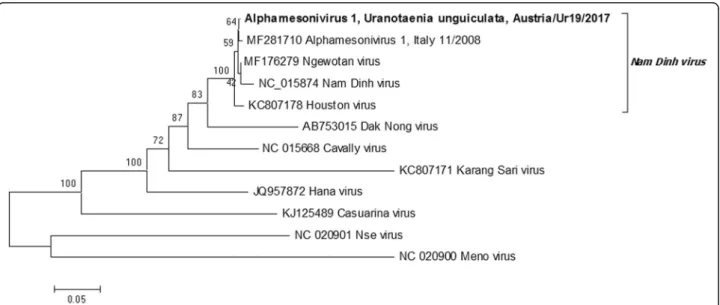
This passage (p2) was used for sequence characterization by PANA [21], as it did not produce amplicons by flavivirus-specific RT-qPCR. This passage did not produce CPE on Vero cells grown at 37 °C. Following PANA PCR cloning, the virus was determined to be an alphamesoni- virus, based on the sequence of 6 unique gene products ap- proximately 500–2000 bp long distributed throughout the genome. Primers were designed based on sequence similarity to previously characterized alphamesoni- viruses (Additional file 2: Table S1), and PCR ampli- cons were sequenced to create a nearly complete genome sequence (missing approximately 300 bases from the 5' and 50 bases from the 3' end) (GenBank:
MH215275). A phylogenetic tree of the alignment of the conserved putative spike protein (ORF 2a) showed that the isolated virus was closely related to Nam Dinh virus (GenBank: DQ458789), placing it within the species groupAlphamesonivirus 1, and more distantly related to the following representative species of the Mesoniviridae: Alphamesonivirus 2, Karang Sri virus (GenBank: KC807171); Alphamesonivirus 3, Dak Nong virus (AB753015); Alphamesonivirus 4, Casuarina virus (GenBank: KJ125489); Alphamesonivirus 5, Hana virus
(GenBank: JQ957872);Mesonvirus 1, Nse virus (GenBank:
JQ957874); and Mesonivirus 2, Meno virus (GenBank:
JQ957873) (Fig.2). The virus sequence had all features as- sociated with the genus Alphamesonivirus, including the reported ribosomal frame-shift site separating ORF1a and ORF1b, which encode the putative viral replicase [27,28].
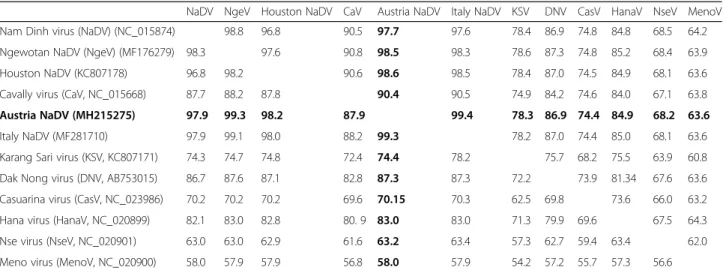
The virus isolate had the highest amino acid and nucleo- tide sequence identity (99.33 and 99.41%, respectively) to an alphamesonivirus identified inCx. pipiensin Italy, 2008 (GenBank: MF281710, Table3).
An RT-qPCR assay was designed to screen the other pools of male and female mosquitoes for the presence of alphamesonivirus. Ten pools from 2016 (five pools of fe- males) and nine pools from 2017 (seven pools of fe- males) were positive for alphamesonivirus (Table 2). A bias-corrected ML estimate for MFIR was 17.3 (95% CI:
7.89–35.28) for males and 54.19 (95% CI: 33.87–86.92) for femaleUr. unguiculataover both years.
Mosquito infection and virus transmission
A total of 362 female Ur. unguiculatamosquitoes were collected over four nights around Lake Neusiedl in June 2018. The legs and wings were dissected and the remaining bodies were pooled in 44 pools for the detec- tion of virus (Table 4). The presence of WNV-lin. 4c was detected in nine of these pools, corresponding to 225 mosquitoes from which legs and wings had been pooled into 39 pools. WNV-lin. 4c was detected in six of the 39 pools. The potential for virus transmission was determined by detecting virus nucleic acid on honey-soaked FTA cards which had been placed into chambers with mosquitoes for feeding purposes prior to their dissection. WNV-lin. 4c nucleic acid was detected
Fig. 2Phylogenetic tree of the amino acid sequence of the putative spike protein (complete ORF2a) from selected species ofMesoniviridae, including a newly described isolate fromUranotaenia unguiculatain Austria (MH215275). Node support is based on 1000 bootstrap replicates
on one of nine honey cards, suggesting thatUr. unguicu- lataare capable of transmitting the virus. Alphamesoni- virus was detected in three pools of mosquito bodies, corresponding to 11 mosquitoes, from which the legs and wings had been placed into tubes individually. From these 11 mosquitoes, alphamesonivirus was detected in the legs and wings of two individuals, indicating a dis- seminated infection. The presence of alphamesonivirus nucleic acid was detected on three of the nine honey cards, suggesting the presence of the virus in saliva.
Discussion
Herein we report the first molecular identification of the hosts of Ur. unguiculata from the guts of blood-engorged flies. The hosts were identified as an- urans, and a similar host preference is known for other members of the genus Uranotaenia [7, 8]. Commonly encountered species of anurans at BSI during the study
period includedPelophylax sp., as well as Hyla arborea (Linnaeus, 1758) (Anura: Hylidae), Bufo bufo(Linnaeus, 1758) (Anura: Bufonidae) and Bombina bombina (Lin- naeus, 1761) (Anura: Discoglossidae) (viavisual encoun- ter during trap setup). It is unknown ifUr. unguiculata takes blood from animals other than frogs. Previous at- tempts to identify the hosts ofUr. unguiculata have re- lied on serological testing, wherein amphibian antiserum was not used or was unavailable, and the host identities were determined to be from reptiles [10] or a horse [11].
It has been reported thatUr. sapphirina(Osten Sacken, 1868) (Diptera: Culicidae), a Nearctic species, may also feed on mammals in addition to amphibians [7,29]. Fur- thermore, landing captures and captures from host-baited traps suggest that Ur. unguiculata may be attracted to mammals including humans [9, 11, 30] but not birds [3]. The relatively low efficiency of common mosquito collection methods (e.g. CO2-baited light traps) for the collection ofUr. unguiculatahas left many gaps in the knowledge of the behaviors of this species.
Efficient collection methods for a mosquito species reflect its host preference and common methods (e.g. CO2-baited light traps) are designed to collect mosquitoes based on for- aging behavior during appetential flight [31]. For example, trap height is an important factor for collecting ornithophi- licversusmammal-biting mosquitoes [32–34]. Sebesta et al.
[3] collectedUr. unguiculatain 1 m high CO2-baited CDC light traps, and none in 5-m high traps nor in pigeon-baited traps. We reasoned thatUr. unguiculatafor- aging preference would reflect host behavior, and therefore placed our traps near the water surface (0.5 m height). Al- though we did not compare the importance of trap height to collection efficiency ofUr. unguiculatadirectly, this trap placement was an improvement compared to other pub- lished records of Ur. unguiculata in the same trapping Table 3Sequence identity matrix of species ofMesoniviridae, including a new isolate inUranotaenia unguiculatafrom Austria
NaDV NgeV Houston NaDV CaV Austria NaDV Italy NaDV KSV DNV CasV HanaV NseV MenoV
Nam Dinh virus (NaDV) (NC_015874) 98.8 96.8 90.5 97.7 97.6 78.4 86.9 74.8 84.8 68.5 64.2
Ngewotan NaDV (NgeV) (MF176279) 98.3 97.6 90.8 98.5 98.3 78.6 87.3 74.8 85.2 68.4 63.9
Houston NaDV (KC807178) 96.8 98.2 90.6 98.6 98.5 78.4 87.0 74.5 84.9 68.1 63.6
Cavally virus (CaV, NC_015668) 87.7 88.2 87.8 90.4 90.5 74.9 84.2 74.6 84.0 67.1 63.8
Austria NaDV (MH215275) 97.9 99.3 98.2 87.9 99.4 78.3 86.9 74.4 84.9 68.2 63.6
Italy NaDV (MF281710) 97.9 99.1 98.0 88.2 99.3 78.2 87.0 74.4 85.0 68.1 63.6
Karang Sari virus (KSV, KC807171) 74.3 74.7 74.8 72.4 74.4 78.2 75.7 68.2 75.5 63.9 60.8
Dak Nong virus (DNV, AB753015) 86.7 87.6 87.1 82.8 87.3 87.3 72.2 73.9 81.34 67.6 63.6
Casuarina virus (CasV, NC_023986) 70.2 70.2 70.2 69.6 70.15 70.3 62.5 69.8 73.6 66.0 63.2
Hana virus (HanaV, NC_020899) 82.1 83.0 82.8 80. 9 83.0 83.0 71.3 79.9 69.6 67.5 64.3
Nse virus (NseV, NC_020901) 63.0 63.0 62.9 61.6 63.2 63.4 57.3 62.7 59.4 63.4 62.0
Meno virus (MenoV, NC_020900) 58.0 57.9 57.9 56.8 58.0 57.9 54.2 57.2 55.7 57.3 56.6
Percent nucleotide sequence identity (above the diagonal) and percent amino acid identity (below the diagonal) are shown (Austrian isolate in bold). GenBank accession numbers are listed in the row headings, and column heading abbreviations are defined in the row heading
Table 4Summary of infection and potential transmission of West Nile virus lineage 4c andAlphamesonivirus 1by Uranotaenia unguiculatamosquitoes
Virus Samplea Total Poolsb Positive poolsc
West Nile virus Body 362 44 9
Legs and wings 225 39 6
Honey card - 9 1
Alphamesonivirus 1 Body 362 38 3
Legs and wings 11 11 2
Honey card - 9 3
aMosquitoes were provided a 25% honey solution on Whatman® FTA® cards, then dissected, removing legs and wings from the body
bLegs and wings were stored separately from body, and samples were pooled into tubes of 2–50 (bodies) or 1–10 (legs and wings). Each honey card sampled between 20-50 mosquitoes
cPools were tested for the presence of virus by RT-PCR
locales [5,26] as well as in the broader surrounding biogeo- graphical regions [35] using common mosquito collection methods. Importantly, we collected both male and female Ur. unguiculata; the efficient collection of males to our knowledge has not yet been reported.
Although we provide evidence that Ur. unguiculata fe- males are attracted to sound, it is unknown if sound is used exclusively to locate hosts. The host species identified here, Pelophylaxsp. andB. bombinawere the only two species of anurans heard calling during the collection period. Sound attraction is known fromUr. lowiiTheobald, 1901 (Diptera:
Culicidae), a Neotropical species of mosquito [17], and sev- eral species ofUranotaeniafrom Japan [8]. Acoustic loca- tion of hosts by hematophagous dipterans is best known from the Corethrellidae (Wood & Borkent 1989), a family closely related to mosquitoes which feed on frogs [36–38].
The sound of the calling maleD. gratiosus, a Nearctic spe- cies of frog, is attractive toUr. lowiias well as to Neotrop- ical and Australian corethrellids. Therefore it has been hypothesized that acoustic location in corethrellids is per- formed by a sensory organ that detects frequencies that are similar to wing beat frequency (approximately 420–450 Hz), which match the dominant frequency of the call ofD.
gratiosus (Additional file 1: Figure S1) [37, 39, 40]. The Johnston’s organ is known to be the auditory organ of mos- quitoes, and is used to identify the wing beat frequency of conspecifics during mating, though may be sensitive up to 2 kHz in some species [41,42]. Further studies on acoustic location and sound preference of Ur. unguiculataare un- derway, and the attractiveness of native anuran calls will be evaluated.
Knowledge of mosquito host feeding behavior is very im- portant for understanding the epizootic potential of arbovi- ruses. The presence of WNV-lin. 4c nucleic acid was recently reported inUr. unguiculata from the study loca- tion here, as well as other sites in central Europe [4–6].
Here we provide evidence thatUr. unguiculataare feeding onPelophylaxsp. at a site where WNV-lin. 4c was detected in pools of conspecifics, including evidence that suggests the mosquitoes are both infected with the virus and are capable of transmitting the virus. Taken together with the detection of virus nucleic acid present in bothUr. unguicu- lataand also frogs (P. ridibundus) collected from a site in southern Russia [14], it is likely that this virus is maintained in a frog-mosquito transmission cycle. The first identifica- tion of WNV-lin. 4 was from a Dermacentor marginatus (Sulzer, 1776) (Acari: Ixodidae) tick in the Volgograd region, southern Russia, in 2003 (WNV-lin. 4a; GenBank:
AY277251) [12], and a similar virus has also been isolated from Cx. pipiens in Spain 2011 (WNV-lin. 4b; GenBank:
GU047875) [13]. In sum, there is further support for WNV-lin. 4c being ecologically and genetically distinct from other WNV lineages [43]. Since WNV-lin. 1 and WNV-lin. 2 are known pathogens to humans and animals,
it is important to understand the epizootic potential of the WNV-lin. 4c. Initial efforts have been successful in isolating this virus on C6/36 cells, and future efforts should characterize the pathogenicity of the virus. Additionally, the vector status ofUr. unguiculatafor WNV-lin. 4c must be determined in controlled experiments.
The genusUranotaeniahas few reports of infection with arboviruses. Eastern equine encephalitis virus was discov- ered in Ur. sapphirina in the southern USA [44], and a cyprovirus (Reoviridae) has also been isolated from this spe- cies [45]. A novel flavivirus, Nounane virus, was reported fromUr. mashonaensisTheobald, 1901 (Diptera: Culicidae) in Côte d’Ivoire [46]. Nounane virus bears closest similarity to Barkedji virus, a virus found inCx. perexiguusTheobald, 1903 (Diptera: Culicidae) mosquitoes in Israel [47], and Nhumirim virus, a virus found inCx. chidesteri Dyar, 1921 mosquitoes from Brazil [48]; the vertebrate host is unknown for these viruses, although all are genetically similar to mosquito-borne zoonotic flaviviruses and not to insect-specific flaviviruses. Herein we report a potentially en- dogenous flavivirus-derived genomic sequence, identical to the sequence reported by others in Ur. unguiculata [15], possibly reflected an ancient association betweenUranotae- niaand flaviviruses [49,50]. In addition, we isolated and se- quenced the genome of an alphamesonivirus which is widespread in the population at BSI. Another member of the family Mesoniviridae, Meno virus, was isolated from Côte d'Ivoire, 2004, where it was found inUr. chorleyiEd- wards, 1936 (Diptera: Culicidae) along with Cavally virus, the first insect-associated nidovirus [27,51]. Much remains unknown about this newly discovered family of insect vi- ruses, which are within theNidovirales[27,28]. Future ef- forts will further characterize the virus isolate reported herein, and the presence of this or another alphamesonvirus within other mosquito species in the region will be tested.
In general, the comparatively low efficiency of collection and lack of human-biting behavior have likely contributed to the gaps in knowledge about this mosquito species in the Western Palaearctic. The optimized sampling conditions re- ported here increased trapping success compared to previ- ous methods. This will aid in future studies of the habits of this elusive mosquito, and the transmission of arboviruses.
Conclusions
Improved collection techniques have yielded this first de- finitive report of the hosts ofUr. unguiculatausing molecu- lar methods, and have revealed the presence of a novel insect-specific flavivirus. The mosquitoes take blood from frogs, Pelophylax spp., and may transmit WNV-lin. 4c to frogs, although vector competence has not yet been estab- lished. It was discovered thatUr. unguiculataare attracted to sound, potentially as a method of acoustic location of hosts. Further research will focus on the ecological associ- ation between the mosquitoes and their hosts, as well as
the transmission ecology of WNV-lin. 4 and the newly-describedAlphamesonivirus 1isolate.
Additional file
Additional file 1:Figure S1.Frequency spectrogram of a single vocalization ofDryophytes gratiosus(Anura: Hylidae) used as an attractant in mosquito sound traps. The grayscale spectrogram was generated in Raven Lite version 1.0 [52]. The spectrogram displays sound frequencies over time where darker pixels indicate relatively louder tones. The dominant frequencies of the call are approximately 450 and 2000 Hz, with harmonics above and below the 2 kHz tone.
Charif, RA, DW Ponirakis, and TP Krein. 2006. Raven Lite 1.0 User’s Guide. Cornell Laboratory of Ornithology, Ithaca, NY. (TIF 66 kb) Additional file 2:Table S1.Primers used to generate a nearly full- length sequence of a newly-describedAlphamesonivirus 1(Mesoniviridae) inUranotaenia unguiculatamosquitoes found in Austria. (DOCX 20 kb)
Acknowledgements
The authors thank René Haider and Bernhard Wildom for assistance with field work. Silke Schweiger of the Austrian Museum of Natural History provided voucher specimens.
Funding
The study was funded by the University of Veterinary Medicine Vienna Postdoc Programme awarded to JVC and NN. Mosquito collections in Hungary were funded by the grant NKFIH K120118 awarded to TB.
Availability of data and materials
The datasets used and/or analyzed during the current study are available from the corresponding author on reasonable request.
Authors’contributions
JVC performed data collection, data analysis, and wrote the manuscript, ZS and TB provided specimens for analysis, TZ and NN facilitated field work and contributed to writing the manuscript, all authors read and approved the final manuscript.
Ethics approval and consent to participate Not applicable.
Consent for publication Not applicable.
Competing interests
The authors declare that they have no competing interests.
Publisher’s Note
Springer Nature remains neutral with regard to jurisdictional claims in published maps and institutional affiliations.
Author details
1Viral Zoonoses, Emerging and Vector-Borne Infections Group, Institute of Virology, University of Veterinary Medicine, Vienna, Austria.2Department of Microbiology and Infectious Diseases, University of Veterinary Medicine, Budapest, Hungary.3Lendület Ecosystem Services Research Group, MTA Centre for Ecological Research, Vácrátót, Hungary.4Hungarian Natural History Museum, Budapest, Hungary.5Biological Station Lake Neusiedl, Illmitz, Austria.6Department of Basic Medical Sciences, College of Medicine, Mohammed Bin Rashid University of Medicine and Health Sciences, Dubai, United Arab Emirates.
Received: 18 April 2018 Accepted: 23 July 2018
References
1. Becker N, Petric D, Zgomba M, Boase C, Madon M, Dahl C, et al. Mosquitoes and their control. Springer: Berlin-Heidelberg; 2010.
2. Filatov S. Little pigeons can carry great messages: potential distribution and ecology ofUranotaenia(Pseudoficalbia)unguiculataEdwards, 1913 (Diptera:
Culicidae), a lesser-known mosquito species from the Western Palaearctic.
Parasit Vectors. 2017;10:464.
3. Šebesta O, Halouzka J, Hubálek Z, Juřicová Z, Rudolf I,Šikutová S, et al.
Mosquito (Diptera: Culicidae) fauna in an area endemic for West Nile virus. J Vector Ecol. 2010;35:156–62.
4. Kemenesi G, Dallos B, Oldal M, Kutas A, Földes F, Németh V, et al. Putative novel lineage of West Nile virus inUranotaenia unguiculatamosquito, Hungary. Virus Dis. 2014;25:500–3.
5. Pachler K, Lebl K, Berer D, Rudolf I, Hubálek Z, Nowotny N. Putative new West Nile virus lineage inUranotaenia unguiculatamosquitoes, Austria, 2013. Emerg Infect Dis. 2014;20:2119–22.
6. Dinu S, Cotar AI, Pănculescu-Gătej IR, FălcuţăE, Prioteasa FL, Sîrbu A, et al.
West Nile virus circulation in south-eastern Romania, 2011 to 2013. Euro Surveill. 2015;20
7. Cupp EW, Zhang D, Yue X, Cupp MS, Guyer C, Sprenger TR, et al.
Identification of reptilian and amphibian blood meals from mosquitoes in an eastern equine encephalomyelitis virus focus in central Alabama. Am J Trop Med Hyg. 2004;71:272–6.
8. Toma T, Miyagi I, Tamashiro M. Blood meal identification and feeding habits ofUranotaeniaspecies collected in the Ryukyu Archipelago. J Am Mosq Control Assoc. 2014;30:215–8.
9. Ryba J, Hájková Z, Kaftan M. Occurrence ofUranotaenia unguiculataEdwards, 1913 (Diptera, Culicidae) in Czechoslovakia. Folia Parasitol. 1974;21:142.
10. Beier JC, Zimmerman JH, Kenawy MA, el Said S, Abbassy MM. Host-feeding patterns of the mosquito community (Diptera: Culicidae) in two Faiyum Governorate villages, Egypt. J Med Entomol. 1987;24:28–34.
11. Braverman Y, Kitron U, Killick-Kendrick R. Attractiveness of vertebrate hosts toCulex pipiens(Diptera: Culicidae) and other mosquitoes in Israel. J Med Entomol. 1991;28:133–8.
12. Lvov DK, Butenko AM, Gromashevsky VL, Kovtunov AI, Prilipov AG, Kinney R, et al. West Nile virus and other zoonotic viruses in Russia: examples of emerging-reemerging situations. Arch Virol Suppl. 2004;18:85–96.
13. Vazquez A, Sanchez-Seco MP, Ruiz S, Molero F, Hernandez L, Moreno J, et al. Putative new lineage of West Nile virus, Spain. Emerg Infect Dis. 2010;16:549–52.
14. Shopenskaya TA, Fedorova MV, Karan L, Frolov AY, Malenko GV, Levina LS, et al. New variant of West Nile virus and its potential epizootic and epidemic importance. Epidemiol Infect Dis. 2008;5:38–44.
15. Ergünay K, Litzba N, Brinkmann A, Günay F, Sarıkaya Y, Kar S, et al. Co- circulation of West Nile virus and distinct insect-specific flaviviruses in Turkey. Parasit Vectors. 2017;10:149.
16. Edman JD, Evans FD, Williams JA. Development of a diurnal resting box to collectCuliseta melanura(COQ.). Am J Trop Med Hyg. 1968;17:451–6.
17. Borkent A, Belton P. Attraction of femaleUranotaenia lowii(Diptera:
Culicidae) to frog calls in Costa Rica. Can Entomol. 2006;138:91–4.
18. Kitano T, Umetsu K, Tian W, Osawa M. Two universal primer sets for species identification among vertebrates. Int J Legal Med. 2007;121:423–7.
19. Moureau G, Temmam S, Gonzalez JP, Charrel RN, Grard G, de Lamballerie X.
A real-time RT-PCR method for the universal detection and identification of flaviviruses. Vector Borne Zoon Dis. 2007;7:467–77.
20. Maher-Sturgess SL, Forrester NL, Wayper PJ, Gould EA, Hall RA, Barnard RT, et al. Universal primers that amplify RNA from all three flavivirus subgroups.
Virol J. 2008;5:16.
21. Stang A, Korn K, Wildner O, Uberla K. Characterization of virus isolates by particle-associated nucleic acid PCR. J Clin Microbiol. 2005;43:716–20.
22. Tamura K, Stecher G, Peterson D, Filipski A, Kumar S. MEGA6: Molecular Evolutionary Genetics Analysis version 6.0. Mol Biol Evol. 2013;30:2725–9.
23. Edgar RC. MUSCLE: multiple sequence alignment with high accuracy and high throughput. Nucleic Acids Res. 2004;32:1792–7.
24. Ident and Sim.http://www.bioinformatics.org/sms2/ident_sim.html.
Accessed 3 Apr 2018.
25. Mosquito Surveillance Software. West Nile Virus. CDC.https://www.cdc.gov/
westnile/resourcepages/mosqSurvSoft.html. Accessed 3 Apr 2018.
26. Lebl K, Zittra C, Silbermayr K, Obwaller A, Berer D, Brugger K, et al.
Mosquitoes (Diptera: Culicidae) and their relevance as disease vectors in the city of Vienna, Austria. Parasitol Res. 2015;114:707–13.
27. Zirkel F, Roth H, Kurth A, Drosten C, Ziebuhr J, Junglen S. Identification and characterization of genetically divergent members of the newly established family Mesoniviridae. J Virol. 2013;87:6346–58.
28. Vasilakis N, Guzman H, Firth C, Forrester NL, Widen SG, Wood TG, et al.
Mesoniviruses are mosquito-specific viruses with extensive geographic distribution and host range. Virol J. 2014;11:97.
29. Molaei G, Andreadis TG, Armstrong PM, Diuk-Wasser M. Host-feeding patterns of potential mosquito vectors in Connecticut, USA:
molecular analysis of bloodmeals from 23 species ofAedes, Anopheles,Culex,Coquillettidia,Psorophora, andUranotaenia. J Med Entomol. 2008;45:1143–51.
30. Bagirov GA, Gadzhibekova EA, Alirzraev GU. The attack activity of Uranotaenia unguiculataEdwards, 1913 mosquitoes on man. Med Parazitol (Moskva). 1994;3:39–40 (In Russian).
31. Bidlingmayer WL. The measurement of adult mosquito population changes - some considerations. J Am Mosq Control Assoc. 1985;1:328–48.
32. Swanson DA, Adler PH. Vertical distribution of haematophagous Diptera in temperate forests of the southeastern USA. Med Vet Entomol. 2010;24:182–8.
33. Johnston E, Weinstein P, Slaney D, Flies AS, Fricker S, Williams C.
Mosquito communities with trap height and urban-rural gradient in Adelaide, South Australia: implications for disease vector surveillance.
J Vector Ecol. 2014;39:48–55.
34. Jansen CC, Zborowski P, Ritchie SA, van den Hurk AF. Efficacy of bird-baited traps placed at different heights for collecting ornithophilic mosquitoes in eastern Queensland, Australia. Aust J Entomol. 2009;48:53–9.
35. Engler O, Savini G, Papa A, Figuerola J, Groschup MH, Kampen H, et al.
European surveillance for West Nile virus in mosquito populations. Int J Environ Res Public Health. 2013;10:4869–95.
36. McKeever S, Hartberg WK. An effective method for trapping adult female Corethrella(Diptera: Chaoboridae). Mosq News. 1980;40:111–2.
37. Camp JV. Host attraction and host selection in the family Corethrellidae (Wood And Borkent) (Diptera). Statesboro: Georgia Southern University; 2006.http://digitalcommons.georgiasouthern.
edu/etd/728
38. Camp JV, Irby WS. Molecular confirmation of frogs (Anura) as hosts of Corethrellidae (Diptera) in the southeastern United States. J Insect Sci. 2017;
17https://doi.org/10.1093/jisesa/iex068.
39. Bernal XE, de Silva P. Cues used in host-seeking behavior by frog-biting midges (Corethrellaspp. Coquillett). J Vector Ecol. 2015;40:122–8.
40. Bernal XE, Rand AS, Ryan MJ. Acoustic preferences and localization performance of blood-sucking flies (CorethrellaCoquillett) to túngara frog calls. Behav Ecol. 2006;17:709–15.
41. Cator LJ, Arthur BJ, Harrington LC, Hoy RR. Harmonic convergence in the love songs of the dengue vector mosquito. Science. 2009;323:1077–9.
42. Göpfert MC, Robert D. Nanometre-range acoustic sensitivity in male and female mosquitoes. Proc R Soc Lond B. 2000;267:453–7.
43. Rizzoli A, Jimenez-Clavero MA, Barzon L, Cordioli P, Figuerola J, Koraka P, et al. The challenge of West Nile virus in Europe: knowledge gaps and research priorities. Euro Surveill. 2015;20
44. Cupp EW, Klingler K, Hassan HK, Viguers LM, Unnasch TR. Transmission of eastern equine encephalomyelitis virus in central Alabama. Am J Trop Med Hyg. 2003;68:495–500.
45. Shapiro A, Green T, Rao S, White S, Carner G, Mertens PPC, et al.
Morphological and molecular characterization of a Cypovirus (Reoviridae) from the mosquitoUranotaenia sapphirina(Diptera:
Culicidae). J Virol. 2005;79:9430–8.
46. Junglen S, Kopp A, Kurth A, Pauli G, Ellerbrok H, Leendertz FH. A new flavivirus and a new vector: characterization of a novel flavivirus isolated fromUranotaeniamosquitoes from a tropical rain forest. J Virol. 2009;83:4462–8.
47. Kolodziejek J, Pachler K, Bin H, Mendelson E, Shulman L, Orshan L, et al. Barkedji virus, a novel mosquito-borne flavivirus identified inCulex perexiguusmosquitoes, Israel, 2011. J Gen Virol. 2013;94:2449–57.
48. Pauvolid-Corrêa A, Solberg O, Couto-Lima D, Kenney J, Serra-Freire N, Brault A, et al. Nhumirim virus, a novel flavivirus isolated from mosquitoes from the Pantanal, Brazil. Arch Virol. 2015;160:21–7.
49. Lequime S, Lambrechts L. Discovery of flavivirus-derived endogenous viral elements inAnophelesmosquito genomes supports the existence of Anopheles-associated insect-specific flaviviruses. Virus Evol. 2017;3:vew035.
50. Crochu S, Cook S, Attoui H, Charrel RN, De Chesse R, Belhouchet M, et al. Sequences of flavivirus-related RNA viruses persist in DNA form
integrated in the genome ofAedesspp. mosquitoes. J Gen Virol.
2004;85:1971–80.
51. Zirkel F, Kurth A, Quan P-L, Briese T, Ellerbrok H, Pauli G, et al. An insect nidovirus emerging from a primary tropical rainforest. mBio.
2011;2:e00077–11.
52. Charif RA, Ponirakis DW, Krein TP. Raven Lite 1.0 User’s Guide. Ithaca: Cornell Laboratory of Ornithology; 2006.