DOCTORAL DISSERTATION
KACHLEK MARIAM LILIA
UNIVERSITY OF KAPOSVÁR
FACULTY OF AGRICULTURAL AND ENVIRONMENTAL SCIENCES
2017
DOI: 10.17166/KE2018.003
KAPOSVÁR UNIVERSITY
FACULTY OF AGRICULTURAL AND ENVIRONMENTAL SCIENCES
Mycotoxins in the Food Chain Research Group Head of Doctoral School:
Prof. Dr. Kovács Melinda
Corresponding Member of the Hungarian Academy of Sciences
Supervisor:
Prof. Dr. Kovács Melinda
Corresponding Member of the Hungarian Academy of Sciences
Co-Supervisor:
Dr. Szabó-Fodor Judit Ph.D., Senior research fellow
THE DOSE AND TIME DEPENDENT, SINGLE AND COMBINED CYTO- AND GENOTOXIC EFFECTS OF MYCOTOXINS FUMONISIN B
1, DEOXYNIVALENOL AND ZEARALENONE
Written by:
KACHLEK MARIAM LILIA
KAPOSVÁR 2017
DOI: 10.17166/KE2018.003
ii
LIST OF ABBREVIATIONS iv
1. INTRODUCTION 1
2. LITERATURE REVIEW 3
2.1
Fusarium mycotoxins3
2.1.1 Fumonisin B1 3
2.1.2 Deoxynivalenol 4
2.1.3 Zearalenone 6
2.1.4 Cyto- and genotoxic effects of FB1, DON and ZEN 8
2.2 Mycotoxin co-occurrence 12
2.3 Combined effects of mycotoxins 15
2.3.1 In vivo studies 19
2.3.2 In vitro studies 21
2.4 Mathematical and statistical analysis of the interactions 23 2.5 Alleviation of mycotoxin adverse effects by medicinal plants 24
3. MATERIALS AND METHODS 27
3.1
In vivo experiments27
3.1.1 Effects of dietary exposure to three Fusarium mycotoxins 27 3.1.2 Effects of DON and possible protective effect of Carduus marianus in
growing rabbits 35
3.2
In vitro experiments42
3.2.1 Chemicals 42
3.2.2 Experimental design 42
3.2.3 Lymphocytes’ isolation and cell enumeration 43
3.2.4 Assays 43
3.2.5 Calculations and statistical analysis 44
4. RESULTS and DISCUSSION 46
4.1 In vivo experiments
46
4.1.1 Effect of multi-toxic dietary exposure on rabbit bucks 46 4.1.2 Effects of DON and possible alleviation from Carduus marianus on
growing rabbits 63
4.2
In vitro experiments79
4.2.1 Cytotoxicity assay 79
iii
4.2.2 Comet assay 84
5. CONCLUSIONS AND RECOMMENDATIONS 88
6. NEW SCIENTIFIC RESULTS 91
7. SUMMARY 92
8. ÖSSZEFOGLALÁS (SUMMARY IN HUNGARIAN) 96
9. ACKNOWLEDGMENTS 100
10. REFERENCES 102
11. SCIENTIFIC PAPERS AND LECTURES ON THE SUBJECT
OF THE DISSERTATION 121
11.1 Peer-reviewed papers published in foreign scientific journals 121 11.2 Peer-reviewed papers published in Hungarian scientific journal in
English 121
11.3 Proceedings in English 122
11.4 Abstracts in English 122
12. OTHER PUBLICATIONS 123
12.1 Peer-reviewed papers published in foreign scientific journals 123
12.2 Proceedings in Hungarian 123
12.3 Abstracts in English 123
12.4 Abstracts in Hungarian 124
13. CURRICULUM VITAE 125
iv
LIST OF ABBREVIATIONS
ALB Albumin
ALKP Alkaline phosphatase ALT Alanine aminotransferase
APCI Atmospheric-pressure chemical ionization AST Aspartate aminotransferase
BEA Beauvericin
BIL Bilirubin
BW Body weight
CCK8 Cell counting kit-8
CD Conjugated dienes
CFU-GM Colony forming unit-granulocytes and monocytes
CREA Creatinine
CT Conjugated trienes
DNA Deoxyribonucleic acid
DON Deoxynivalenol
ENB Enniatin B
ESI Electrospray ionization
FA Fumonisin A
FB Fumonisin B
FC Fumonisin C
FP Fumonisin P
FB1 Fumonisin B1
FB2 Fumonisin B2
FB3 Fumonisin B3
FB4 Fumonisin B4
FB5 Fumonisin B5
FB6 Fumonisin B6
v
FrA Fructosamine
FUMs Fumonisins
GGT Gamma-glutamyl transferase
GLOB Globulin
GLU Glucose
Gn-RH Gonadotropin-releasing hormone
GPx Glutathione peroxidase
GSH Reduced glutathione
IL- Interleukin
LDH Lactate dehydrogenase
LMP Low melting point
MCP-1 Monocyte chemoattractant protein-1
MDA Malondialdehyde
MN Micronuclei
mRNA Messenger ribonucleic acid MTT 3- (4,5-dimethylthiazol-2-yl)-2,5-
diphenyltetrazolium bromide
NIV Nivalenol
NMP Normal melting point
qPCR Quantitative (Real time) PCR
PA Phagocytic activity
PBS Phosphate buffered saline
PBMC Peripheral blood mononuclear cells
PCR Polymerase Chain Reaction
PTP Permeability transition pore RBCH Red blood cell hemolysate
ROS Reactive oxygen species
RT-PCR Reverse transcriptase PCR
vi tCHOL Total cholesterol
Sa/So Sphinganine/Sphingosine SCFA Short chain fatty acids
TBARS Thiobarbituric acid reactive substances TDI Tolerable daily intake
TG Triglyceride
TNFα Tumour necrosis factor α
TP Total protein
WST Water soluble tetrazolium salt
ZEN Zearalenone
1
1. INTRODUCTION
Secondary metabolites of fungi are natural products which can be harmful/undesirable or beneficial to humans (Demain and Fang, 2000). They are termed secondary because they do not play a role in the primary metabolism; they may be necessary for sporulation (Calvo et al., 2002). Mycotoxins belong to the harmful/undesirable secondary fungal metabolites.
Ergotism is the oldest mycotoxicosis known and it is referred as St Anthony’s fire as well. This mycotoxicosis has as a causative agent the fungus genus Claviceps. Claviceps fungi invade the plant by replacing the ovary with a mass of fungal tissue (in the female part of the host plant) which is called sclerotium (CAST, 2003). Although ergotism was described during the Middle Ages the foundation of modern toxicology did not take place until the 1960’s when the Turkey X disease occurred. Thousands of turkey poults died in England; later it was discovered that the cause was the exposure to aflatoxins, mycotoxins produced by fungi of the Aspergillus genus, although later it was disputed involving another mycotoxin cyclopiazonic acid in the etiology of the disease (Cole, 1986).
Since then approximately 400 fungal secondary metabolites have been characterized (Bennet and Klich, 2003). The most important toxin producing genera are Fusarium, Aspergillus, Penicillium, Claviceps and Alternaria (CAST, 2003).
The most important and most thoroughly studied mycotoxins and mycotoxin classes are fumonisins, trichothecenes, aflatoxins, ochratoxins and zearalenone. For the clarification of the erroneously and unequivocally used terminology in the year 2014, a new, systematic definition criterion has been worked out, in which mycotoxins are differentiated in free and unmodified, matrix-associated and chemically modified (Rychlik et al., 2014). The occurrence of mycotoxins worldwide is a very important issue in the aspect of both food and feed safety. The Commission of European Union has set guidance levels (76/2006/5EC) for several mycotoxins in products intended for animal feeding (European Commission, 2006).
In Europe, and in particular in the EU, regulatory and scientific interest in mycotoxins has undergone a development in the last decade from autonomous national activities towards more EU-driven activities with a structural and network
2
character. Harmonized EU limits now exist for 40 mycotoxin-food combinations (van Egmond et al., 2007).
Fusarium mycotoxins are the most frequently occurring mycotoxins worldwide and more specifically fumonisin B1 (FB1), deoxynivalenol (DON) and zearalenone (ZEN) are the most likely to co-occur (Griessler et al., 2010; Rodrigues and Naehrer, 2012; BIOMIN 2015; 2016; Smith et al., 2016). Thus these mycotoxins were chosen in order to investigate their single and combined effects. To the best of our knowledge, there are not many studies on their binary and especially ternary mixtures both in vivo and in vitro. Investigation of the interactive effects is very crucial for risk assessment since it has been confirmed - as aforementioned - that mycotoxin co-occurrence is a fact. Hence the single and combined effects of FB1, DON and ZEN were investigated both in vivo and in vitro in monogastric animals.
3
2. LITERATURE REVIEW 2.1 Fusarium mycotoxins 2.1.1 Fumonisin B1
The 28 fumonisin analogues that have been characterized since 1988 (Gelderblom et al., 1988) can be separated into four main groups identified as the fumonisin A, B, C, and P series (Rheeder, 2002). Among the four primary series of fumonisins (FA, FB, FC and FP), the toxicologically most important compounds are the FB analogues. Fumonisin B group comprises of six compounds; fumonisin B1, B2, B3, B4, B5 and B6 (FB1, FB2, FB3, FB4, FB5 and FB6 respectively), FB1 is the major metabolite (Kubena et al., 1997; Månsson et al., 2010, Bartók et al., 2013).
FB1 is generally found in the highest concentrations in maize and maize-based products. The main producers of FB1 are Fusarium verticillioides (previously F.
moniliforme) and F. proliferatum, which mainly occur in corn. FB1 is chemically a diester of propane-1, 2, 3- tricarboxylic acid (tricarballylic acid, TCA) and 2-amino- 12, 16- dimethyl- 3, 5, 10, 14, 15-pentahydroxyeicosane, in which the C14 and C15
hydroxyl groups are esterified with the terminal carboxyl group of TCA (Marasas, 2001; Heidtmann-Bemvenuti et al., 2011). The primary amino group of FB1 is essential for its toxicity. This is proven by the fact that acetylation of FA1 to FB1
inhibits its cytotoxic capability (Stockmann-Juvala and Savolainen, 2008).
The main mechanism of action of FB1 is a result of its similarity (long-chain hydrocarbon unit) to sphingosine and sphinganine, thus inhibiting the ceramide synthase enzyme. As a consequence FB1 disturbs the metabolism of sphingolipids resulting in accumulation of free sphingoid bases, altered sphinganine (Sa) to sphingosine (So) ratio (Sa/So), and depletion of complex sphingolipids. This was hypothesized by Wang et al. (1991) and was further confirmed by several research groups (Yoo et al., 1992; Wang et al., 1999; Enongene et al., 2000). These effects essentially lead to impairment of cell membrane function (Riley et al., 2001). Apart from inhibiting ceramide synthase, FB1 is suggested to stimulate apoptosis in cells but the mechanism is not clear. Some possible explanations could be the induction of lipid peroxidation and the decreased concentrations of antioxidants such as
4
glutathione (GSH) (Surai and Dvorsrka, 2005). Gelderblom et al. (1997) found altered lipid constituent proportions in exposed rats; in particular, phosphatidylethanolamines were significantly increased in the mitochondrial and even in the plasma membrane fractions. The main targets of FB1 are the liver and kidney (Stockmann-Juvala and Savolainen, 2008; Voss et al., 2007).
FB1 is confirmed causative factor of several diseases in livestock. In Equidae, neurotoxic and hepatotoxic effects have been reproduced experimentally.
Moreover, FB1 has been shown to induce cardiovascular problems to horses (Smith et al., 2002). The most important disease in horses, caused by FB1 is equine leukoencephalomalacia (ELEM) (Marasas et al., 1988; Kellerman et al., 1990; Ross et al., 1993). In swine, FB1 induces pulmonary oedema (porcine pulmonary oedema, PPE) syndrome (Colvin and Harisson, 1992). FB1 can also induce hepatotoxicity to swine (Gumprecht et al., 1998). Elevated serum cholesterol is another characteristic of FB1 toxicity (Rotter et al., 1996b; Gumprecht et al., 1998). Poultry and cattle are less sensitive to FB1 (Bolger et al., 2001). In lambs, acute hepatic and renal toxicity was reported by Edrington et al. (1995).
Carcinogenicity due to FB1 has been reported in laboratory animals as well.
Gelderblom et al. (1994) reported FB1-induced hepatocellular carcinoma in rats. FB1
is possibly carcinogenic to humans; it has been classified as a possible carcinogen (group 2B) by IARC (2002). FB1 has been associated with cancer of the oesophagus in rural regions of Southern Africa and China (Sydenham et al., 1990; Yoshizawa et al., 1994). FB1 can be teratogenic as well. It can cause neural tube defects (NTD) to embryos (Marasas et al., 2004).
Fumonisins (FUMs) are regulated in the European Union and the levels vary according to the species’ sensitivity. In the case of fumonisins (and the rest fusariotoxins), there are guidance (not maximum limits like for aflatoxins) values in both feed and food (European Commission, 2006).
2.1.2 Deoxynivalenol
DON is a member of the trichothecene family, which is consisted of approximately 200 compounds (Grove 2000; Pestka 2007, 2010). Trichothecenes
5
are structurally related sesquiterpenes which include four basic types, from which the most important representatives belong to types A, B and D (Pestka, 2007). The chemical name of DON is 12, 13-epoxy-3α, 7α, 15-trihydroxythichothec-9-en-8on (C5H20O6). The epoxide at the positions C12,13 makes trichothecenes toxic (Heidtmann-Bemvenuti et al., 2011). The ketone group which is in C8 is a characteristic of type B compounds (Ueno, 1984). The high melting point of DON gives its resistance to heat (cooking, cereal processing) (Bonnet et al., 2012). DON is produced by Fusarium graminearum and F. culmorum and despite being the least toxic trichothecene; its ubiquitous occurrence in grains increases its importance for food and feed safety.
Like the rest trichothecenes, DON inhibits the protein synthesis (the initiation and/or elongation of the polypeptide chain is inhibited) because of its ability to bind to eukaryotic ribosomes (Ueno et al., 1968; Ueno, 1984).
In pigs, DON is absorbed rapidly and almost completely through the stomach and proximal intestine (Dänicke et al., 2004). After DON is metabolized a chemical compound is formed; de-epoxy-DON.
The main symptoms of DON are diarrhoea, nausea, reduced feed intake and weight gain (Bonnet et al., 2012). The pig is the most sensitive while ruminants are the least sensitive animal species to DON. Monogastric animals are extremely prone to growth and body weight gain suppression upon DON exposure. In swine, DON is causing feed refusal already at 1 mg/kg, and emesis (vomiting; the minimum oral dose is 100μg/kg of body weight), thus given the trivial name vomitoxin (Vesonder et al., 1973; Ueno, 1984; Forsell et al., 1986). It has been demonstrated that DON can cause histopathological alterations to the gastrointestinal tract to pigs (Zielonka et al., 2009). The effect of DON on the intestine of pigs was observed by Lessard et al. (2015). They reported down-regulation of the genes important for integrity and barrier function. In chickens, DON can cause alterations to the small intestine for both weight and morphology (Awad et al., 2005). Changes in liver morphology (possible hepatic impairment) can be also induced by DON in rats (Bracarense et al., 2017).
DON, as well as other trichothecenes, has immunomodulatory abilities (Pestka and Smolinski, 2005). It was suggested by Rotter et al. (1996a) that the
6
immunosuppression induced by trichothecenes is due to the inhibition of translation (Bamburg, 1983), whereas the mechanism of immunostimulation is not so clear.
Even low DON doses can downregulate the gene expression of cytokines like interleukin 1β (IL-1β) or tumor necrosis factor α (TNFα) in the small intestine (Becker et al., 2011). A recent study in rats demonstrated the immunosuppressive properties of DON by means of histopathological examination of lymphoid organs (Bracarense et al., 2017). Poultry is quite resistant to the negative effects of DON.
Despite this, prolonged exposure to low doses of DON can cause problems related to productivity and immunity (Awad et al., 2013).
Reproductive system can be affected by DON as well. In rats, DON caused significant alterations to hormones like luteinizing (LH) and follicle stimulating (FSH) and testosterone in rats. Furthermore, DON affected the weights of the prostate and of the epididymal and seminal vesicle. Abnormal tail incidents were also increased along with a decrease in the numbers of spermatid and cauda epididymal sperm (Sprando et al., 2005).
As mentioned before, there are guidance values for all fusariotoxins including DON (European Commission, 2006). DON has not been classified as carcinogenic yet (Group 3 according to IARC, 1993).
2.1.3 Zearalenone
Chemically ZEN is a resocyclic acid lactone 6-(10-hydroxy-6-oxo-trans-1- undecenyl])-β-resocyclic acid lactone (Urry et al., 1966). ZEN was isolated by Christensen et al. (1965) who named it F-2 toxin. The next year, Urry et al. (1966) characterised its chemical structure and gave its present name zearalenone. ZEN is produced by various species of Fusarium genus, namely Fusarium graminearum (Giberella zea), F. culmorum, F. equiseti, F. sambucinum and F. crokwellense (Placinta et al., 1999; CAST, 2003). ZEN very often is co-occurring with DON since two of the producers of both toxins are the same i.e. F. graminearum and F.
culmorum.
ZEN is absorbed rapidly after oral administration (Zinedine et al., 2007).
Olsen et al. (1981) suggested two pathways for biotransformation of ZEN, hydroxylation and conjugation. After hydroxylation two metabolites are formed, α-
7
and β-ZOL with the latter sometimes having a higher potency (Frizzell et al., 2011).
These two metabolites are isomers and can be produced by fungi as well but in much lower concentrations (Zinedine et al., 2007). The main site of ZEN’s metabolization is the liver (Kiessling and Pettersson, 1978).
ZEN is not considered as a real toxin but rather a mycoestrogen due to its similar chemical structure with oestrogens. This similarity leads to a competitive binding to oestrogenic receptors which affect the reproductive system of both male and female animals (Abdelhamid et al., 1992; Nakaido et al., 2004). In the case of ZEN swine is the most sensitive species as well (Tiemann and Dänicke, 2007).
In female animals, ZEN can induce an early onset of puberty and can alter uterus weight and the morphology of the genital tract (Christensen et al., 1965;
Abdelhamid et al., 1992). As reviewed in Kanora and Maes (2009), ZEN can induce swelling of the vagina and the vulva and cause anoestrous or prolonged cycle in pigs. Furthermore, ZEN in high concentration (>25 ppm) can cause stillbirth or neonatal mortality. ZEN can affect male pigs as well; testicular weight decreases, epididymis and vesicular glands decrease in size, libido drops and spermatozoa do not bind as effectively to the zona pellucida (Christensen et al., 1972; Ruhr et al., 1983; Tsakmakidis et al., 2007). The effects of ZEN in male animals have been studied mainly in rats, mice and rabbits. It has been demonstrated that ZEN affects sperm cells, spermatogenesis, testosterone concentration and germ cells. As regards to spermatozoa, ZEN can affect the number, morphology and motility of the spermatozoa (Zatecka et al., 2014; Boeira et al., 2015). Furthermore, spermatogenesis can be adversely affected and one group has investigated the expression of genes essential for spermatogenesis (Cho et al., 2011; Zatecka et al., 2014). ZEN-induced germ cell degeneration has also been reported (Kim et al., 2003).
ZEN despite its pronounced ability to affect the reproductive system may cause effects in non-reproductive systems of the organisms (in vitro and in vivo).
ZEN has been shown to exert cytotoxic and genotoxic effects (Ouanes et al., 2003;
Abid-Essefi et al., 2004; Vlata et al., 2006; Gao et al., 2013). ZEN has been reported to be immunotoxic to rats (Hueza et al., 2014) and to exert immunomodulating effects to piglets (Marin et al., 2010). ZEN can be hepatotoxic as well, which has
8
been reported in gilts, rats and rabbits (Marin et al., 2013b). Oxidative stress is one of the mechanisms associated with genotoxicity. In the following chapter (2.1.4.2), studies regarding ZEN-induced oxidative stress will be discussed.
ZEN has not yet been classified as a carcinogen (Group 3) for humans by IARC (1993). The presence of ZEN, like FB1 and DON in animal feed is regulated in Europe (European Commission, 2006).
2.1.4 Cyto- and genotoxic effects of FB
1, DON and ZEN
The most popular endpoints used for the investigation of mycotoxin effects in vitro are cytotoxicity and genotoxicity.
As regards cytotoxicity, several parameters have been employed; the most frequently used are cell proliferation, metabolism and viability, cell membrane integrity as well as macromolecule synthesis (protein and DNA). Less frequently, cell cycle is investigated along with cell morphology. The most commonly used cell lines are intestinal cell lines of human (Caco-2, HT-29, HCT116) and animal origin (IPECJ-2), peripheral blood lymphocytes isolated usually from either humans or pigs and kidney (porcine, monkey, bovine, canine) cell lines. Genotoxicity has been studied as well but in a lesser extent, especially in vitro. Genotoxicity can be assessed studying different parameters like DNA damage, oxidative stress/status, micronuclei (MN) and DNA adducts formation, and sister chromatid exchange.
2.1.4.1 Studies on cytotoxic effects
The effects of mycotoxins on immunity are very important since low immunity can predispose humans and animals to infectious diseases. FB1 has been shown to be cytotoxic to both human and porcine peripheral blood mononuclear cells (PBMC) (Meky et al., 2001; Marin et al., 2007; Stoev et al., 2009; Mulunda and Dutton, 2014). Similarly to FB1, DON – known for its immunomodulation properties - has exerted cytotoxic effects to human and porcine PBMC (Lautraite et al., 1997; Thuvander et al., 1999; Meky et al., 2001; Goyarts et al., 2006a, Marin et al., 2006a; Baltriukiene et al., 2007; Taranu et al., 2010). Minervini et al. (2004) studied the effects of FB1 and DON in human erythroleukemia cell line (K562).
9
ZEN’s cytotoxic effects have been demonstrated only in one study using human PBMC by Vlata et al. (2006).
Cytotoxicity due to FB1 has been exerted to human (Caco-2 and HT-29 cells) and porcine (IPEC-J2 cells) intestinal cells as well (Clarke et al., 2014;
Kouadio et al., 2005; Wan et al., 2013a; Minervini et al., 2014). DON’s cytotoxic effects has been assessed in both human (Caco-2 and HCT116) and porcine (IPEC- J2) intestinal cells (Kouadio et al., 2005; Vandenbroucke et al., 2011; Wan et al., 2013a; Bensassi et al., 2014; Manda et al., 2015; Springler et al., 2016). On the other hand, the cytotoxic effects of ZEN have been studied only in human epithelial cells (Abid-Essefi et al., 2003; Abid-Essefi et al., 2004; Kouadio et al., 2005; Wan et al., 2013a; Bensassi et al., 2014).
Another important tissue in mycotoxin research is kidney, thus several studies have been performed in kidney cell lines of different origin. More specifically, FB1 exerted cytotoxic effects on monkey, porcine and bovine kidney lines (Abado-Becognee et al., 1998; Šegvić Klarić et al., 2008; Clarke et al., 2014).
In a study from the late 80’s the cytotoxic effects of DON on monkey, porcine and canine kidney cell lines were investigated (Reubel et al., 1989). On the other hand, ZEN’s cytotoxic effects on kidney in vitro have been assessed by the same group in three different studies (Abid-Essefi et al., 2003; Abid-Essefi et al., 2012; Bouaziz et al., 2013).
The liver is the main site for the metabolism of any substances entering the body of animals or humans. Despite this, only a few studies have been performed on the in vitro effects of FB1, DON and ZEN in the liver. The cytotoxic effects of FB1
have been studied in liver cells as well (primary hepatocytes; Riedel et al., 2016).
The only study on the cytotoxic in vitro effects of DON in the kidney is from Ueno et al. (1973). The effects of ZEN in human liver cells (HepG2) have been studied by Hassen et al. (2007).
Cytotoxicity has been scarcely studied in vivo. One of the few studies in cows was performed by Dänicke et al. (2011). Lymphocytes were isolated from the DON-treated cows for the assessment of the cytotoxicity (ex vivo) and non-treated cows for exposure to DON (in vitro) using 3-(4, 5-Dimethylthiazol-2-Yl)-2, 5- Diphenyltetrazolium Bromide (MTT) test. In the ex vivo experiment, viability was
10
decreased by 18%. On the other hand for the in vitro cell viability was seriously affected (50-80% decrease) when DON concentration was over 0.27 μM (0.08μg/ml). In the study of Marin et al. (2013a) piglets were exposed to ZEN for 18 days. At the end of the trial, lymphocytes were isolated from blood, spleen and lymph nodes and their proliferation was measured by the [methyl-3H]-thymidine proliferation assay. After 72h of incubation, viability decreased by 66.15% in lymphocytes isolated from blood as compared to control cells. On the other hand, no effect was observed in lymphocytes derived from spleen or lymph nodes.
2.1.4.2 Studies on genotoxic effects
One of the most popular methods for the assessment of genotoxic effects is comet assay, which is assessing the DNA damage. Other methods are micronuclei (MN) formation assay, DNA fragmentation assessment and sister chromatid exchange (SCE) assay. Oxidative stress has been investigated in many studies since it has been proposed as a possible mechanism of genotoxicity. In order to assess the oxidative stress, various endpoints are used such as the production of reactive oxygen species (ROS) and the level of thiobarbituric acid-reactive substances (TBARS).
FB1 has been shown to induce MN formation a dose-dependent manner to porcine kidney epithelial (PK15) cells (Šegvić Klarić et al., 2008). In the study of Galvano et al. (2002), FB1 induced DNA damage to human fibroblasts. Ehrilch et al.
(2002) studied the genotoxic effects of FB1 in HepG2 cells and found a dose- and time-dependent effect on the induction of DNA damage and MN formation. DON’s genotoxic effects have been studied in human epithelial cells. Bony et al. (2006) observed the genotoxic effects of DON on Caco-2 cells with comet assay. HepG2 cells were used by Zhang et al. (2009) for the assessment of genotoxicity caused by DON. Oxidative stress was correlated with the induced DNA damage by measuring the production of ROS and the level of TBARS. ZEN’s genotoxic effects have been studied by the same group in monkey (Vero) kidney and human (Caco-2 and Hep- G2) cell lines (Abid-Essefi et al., 2003; Ouanes et al., 2003; Hassen et al., 2007;
Abid-Essefi et al., 2012; Bouaziz et al., 2013). In most of the studies, DNA damage
11
was assessed by comet assay and was correlated with the production of ROS. The formation of MN in Vero cells has been also assessed in one of the studies (Ouanes et al., 2003). In their early trial, DNA fragmentation in Caco-2 was studied (Abid- Essefi et al., 2003). Apart from this group’s studies, there is another study on DNA damage and ROS production due to ZEN exposure in the human embryonic cell line (Gao et al., 2013).
In several in vivo trials, genotoxic effects are studied in parallel with oxidative stress. Aranda et al. (2000) assessed the genotoxic effect of FB1 in mice.
They studied the formation of MN in the bone marrow of the treated mice. Another study on the genotoxicity of FB1 was performed by Theumer et al. (2010). DNA damage was assessed by comet assay and MN formation test in spleen mononuclear cells of male Wistar rats. Additionally, malondialdehyde (MDA) was measured in order to assess the effect of oxidative stress with genotoxicity. Kouadio et al. (2013) investigated the effects of low doses of dietary DON and FB1 on DNA fragmentation of blood lymphocytes in mice. The DON-induced DNA damage was assessed on spleen leukocytes of chickens (Frankic et al., 2006). DON fed to broiler chickens induced DNA damage to blood lymphocytes of broiler chickens (Awad et al., 2014). Apart from the DNA damage, the level of oxidative stress (TBARS) was measured in plasma, heart, kidney and segments of the large intestine (duodenum and jejunum). In the study of Payros et al. (2017) DON exacerbated the genotoxicity of the rats colonized with Escherichia coli. Regarding ZEN’s genotoxic effects there is only one study in bone marrow and spleen lymphocytes of mice (Ben Salah- Abbès et al., 2009). Two studies on ZEN assessed oxidative stress in chickens and mice (Zourgui et al., 2008; Grešáková et al., 2012).
One of the most important factors determining the toxicity of a mycotoxin in the body of an animal is the metabolism (biotransformation pathways, processes of elimination, microbial transformation) which can explain the different sensitivity among farm animals (D’Mello et al., 1999; Minervini and Dell’ Aquila, 2008). The dose levels as well as the duration of the exposure are also affecting differently the final effects of mycotoxins (Forsell et al., 1986). A very good example regarding differences in doses and exposure time are two trichothecene mycotoxins. Although the acute toxicity of T-2 mycotoxin is much higher than that of DON, in practice T-2
12
occurs in much lower levels hence the chronic exposure of animals to the ubiquitous occurring DON can lead to more adverse effects in the long run. All the aforementioned differences are highlighted by the fact that the same mycotoxin - e.g. FB1 - results in different syndromes in different animal species as mentioned in previous sections of the literature review. At a cellular level different mechanisms of actions, to name a few, compromising of the cell membrane integrity, production of reactive oxygen species (ROS), binding to the DNA or to receptors, inhibiting macromolecular synthesis; cause the manifestation of different effects among cell lines. The issue becomes more complicated when the animals/cells are exposed to combined mycotoxins because all the aforementioned factors are interacting.
2.2 Mycotoxin co-occurrence
As aforementioned the various mycotoxins are regulated worldwide with maximum or recommended limits. However, single mycotoxin exposure is the exception than the rule, and this is a result of several factors. Firstly, many fungal species can produce more than one mycotoxin simultaneously. In addition to that, crops can be infected by different genera of fungi at the same time and last but not least, complete feed is prepared by various commodities. This mycotoxin co- occurrence is reported in several reviews and surveys (Speijers and Speijers, 2004;
Griessler et al., 2010; Monbaliu et al., 2010; Rodrigues and Naehrer, 2012, Streit et al., 2013; BIOMIN, 2015, 2016; Smith et al., 2016). In a survey conducted over a period of 4.5 years in countries of Southern Europe (Portugal, Spain, Italy, Greece and Cyprus), the Fusarium mycotoxins were found to be the major contaminants (fumonisins, type B trichothecenes and ZEN) of feedstuffs and compound feed samples (Griessler et al., 2010). In the survey of Monbaliu et al. (2010) 75% of the samples were contaminated with at least one mycotoxin. The most frequently occurring mycotoxins were type B trichothecenes and fumonisins. The highest incidence of co-occurrence was in maize (41 samples). In the survey of Streit et al.
(2013) that lasted from 2004 to 2012, all (83) the samples were found to be co- contaminated with 7 to 69 metabolites with 28 being the most frequent number of metabolites per sample. Similarly to the other surveys, Fusarium mycotoxins were
13
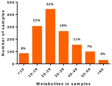
detected most often. In a most recent survey conducted by BIOMIN (Figure 1) 94%
of the samples tested worldwide were contaminated with more than ten mycotoxins, with DON, ZEN and FUMs being present in over 50% (BIOMIN, 2016). Samples were collected from 81 countries and 99066 analyses were performed in 16511 samples.
Figure 1: Number of metabolites in samples of cereals, corn and finished feed from around the globe (adapted from BIOMIN mycotoxin survey 2016).
Furthermore, in Europe the most frequently occurring mycotoxins were DON, ZEN and FB1 with an average of 70%, 48% and 48% contaminated samples (finished feed, corn and cereals), respectively (BIOMIN, 2016). The level of contamination, median and average concentations of the positive samples as well as maximum concentrations of FB1, DON and ZEN in Europe are shown in Table 1.
M e t a b o lit e s in s a m p le s
Number of samples
<10 10-19
20-29 30-39
40-49 50-59
>60 0
1 0 0 2 0 0 3 0 0 4 0 0 5 0 0
6%
2 2 % 3 2 %
1 9 %
1 1 % 7%
2%
14
Table 1: Fusarium mycotoxins contamination levels in main commodities in Europe (adapted from BIOMIN mycotoxin survey 2016).
Commodities Mycotoxins
Finished feed FB1 DON ZEN
Average of positives (μg/kg) 792 420 85
Median of positives (μg/kg) 164 138 20
Maximum (μg/kg) 33970 21300 7400
Corn FB1 DON ZEN
Average of positives (μg/kg) 2057 790 155
Median of positives (μg/kg) 845 422 44
Maximum (μg/kg) 42000 9816 2652
Cereals FB1 DON ZEN
Average of positives (μg/kg) 560 1023 78
Median of positives (μg/kg) 36 400 26
Maximum (μg/kg) 13360 37640 3757
Guidance values for FB1, DON and ZEN have been recommended by European Commision (Table 2; 2006/576/EC). In the following table only levels for pigs are given because they are the most sensitive species.
Table 2: Guidance values of FB1, DON and ZEN in European Union (adapted from the Commission Recommendation 2006/576/EC).
Commodity
Mycotoxins Complementary and complete feedings stuffs for pigs (12%
moisture content, mg/kg of feed)
FB1 5
DON 0.9
ZEN 0.25
In their review, Smith et al. (2016) compiled data from 107 articles and concluded that the most frequent mixtures of fusariotoxins worldwide were DON+ZEN, FUM+ZEN, DON+ nivalenol (NIV), DON+T-2.
According to the aforementioned surveys, the mycotoxin concentrations are usually low. Acute toxicity is rarely occurring thus chronic exposure to low concentrations can be of high importance for human and animal health (Streit et al., 2013).
As it can be concluded from the aforementioned, Fusarium toxins (i.e.
trichothecenes, fumonisins and zearalenone) are of particular interest, because their occurrence is ubiquitous in cereal grains, such wheat and corn, and additionally they co-occur very frequently.
15
The legislation is enacted based on risk assessment studies, which depend on the toxicity and exposure data of single mycotoxins. However, the effect of co- occurring mycotoxins cannot be predicted based on the effects of the single toxins.
When mycotoxins interact with each other, they may interact. Thus the determination of the interactive effects of mycotoxins and especially in low concentrations is essential for the establishment or revision of tolerable daily intake (TDI) (Speijers and Speijers, 2004) and/or maximum/guidance levels (Verstraete, 2006). Recently, the European Food Safety Authority (EFSA) funded a new project for the risk assessment of mycotoxin mixtures in food and feed (EFSA, 2017).
2.3 Combined effects of mycotoxins
There are several studies about interactions between mycotoxins (Grenier and Oswald, 2011; Alassane-Kpembi et al., 2017; Smith et al., 2016), but fewer report the combined effects of three specific Fusarium mycotoxins, which are more likely to co-occur; i.e. FB1, DON and ZEN (Tables 3, 4). Most of the studies about the interactions of FB1, DON and ZEN regard binary mixtures, although there are some ternary mixture studies (Forsell et al., 1986; Pestka et al., 1987; Harvey et al., 1996; Kubena et al., 1997; Grenier et al., 2011; Bracarense et al., 2012; Ficheux et al., 2012; Bensassi et al., 2014; Cortinovis et al., 2014; Kouadio et al., 2007; Wan et al., 2013a, b; Szabó-Fodor et al., 2015; Albonico et al., 2016; Tables 3, 4).
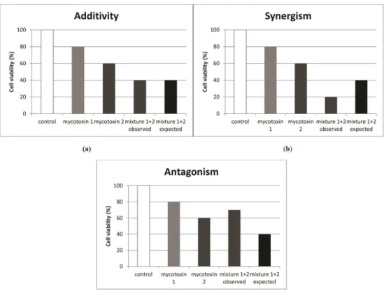
Interactive effects can fall in one of the three categories; addition, synergism and antagonism. In vitro, the definition is more clearly evident since we assess a specific endpoint and after calculating the expected value we compare it with the observed one and make our conclusions (Figure 2).
16
Figure 2: Graphical representation of mycotoxin interactions on cell viability in vitro (adapted from Smith et al., 2016).
17
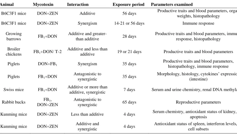
Table 3: In vivo studies on combined effects of fumonisin B1 (FB1), deoxynivalenol (DON) and zearalenone (ZEN)
Animal Mycotoxin Interaction Exposure period Parameters examined References
B6C3F1 mice DON+ZEN Additive 56 days Productive traits and blood parameters, organ
weights, histopathology Forsell et al., 1986
B6C3F1 mice DON+ZEN Synergism 14-21 or 56 days Immune response Pestka et al., 1987
Growing
barrows FB1+DON Additive and greater-
than additive 28 days Productive traits and blood parameters, immune
response, histopathology Harvey et al., 1996 Broiler
chickens FB1+DON/ T-2 Additive and less than
additive 19 or 21 days Productive traits and blood parameters Kubena et al., 1997 Piglets DON+FB1 Synergism 35 days Productive traits and blood parameters,
histopathology, immune response Grenier et al., 2011 Piglets FB1+DON Antagonistic to
synergistic 35 days Morphology, histology, cytokines’ expression
(intestine) Bracarense et al., 2012 Swiss mice FB1+DON Additive or more than
additive, synergistic 7 days Serum and urine chemistry, renal DNA methylation Kouadio et al., 2013 Rabbit bucks FB1,
DON+ZEN
Antagonistic to
synergistic 65 days Reproductive parameters Szabó-Fodor et al., 2015
Kunming mice DON+ZEN Less than additive 4 days Serum chemistry, antioxidant status of kidney, cell
apoptosis Liang et al., 2015
Kunming mice DON+ZEN Additive and
synergistic 4 days Antioxidant status of spleen, interferon levels, T-
cell subsets Ren et al., 2016
18
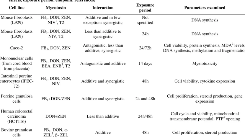
Table 4: In vitro studies on combined effects of fumonisin B1 (FB1), deoxynivalenol (DON) and zearalenone (ZEN); cell line, mycotoxins, effects, exposure period, endpoint, references)
Cell line Mycotoxin Interaction Exposure
period Parameters examined References
Mouse fibroblasts (L929)
FB1, DON, ZEN, NIV1, T2
Additive and in few exceptions synergistic
Not
specified DNA synthesis Groten et al., 1998
Mouse fibroblasts (L929)
FB1, DON, ZEN, NIV, T2
Less than additive to
synergistic 24h DNA synthesis Tajima et al., 2002
Caco-2 FB1, DON, ZEN Antagonistic, less than
additive, synergistic 24/72h Cell viability, protein synthesis, MDA2 levels,
DNA synthesis, methylation and fragmentation Kouadio et al., 2007 Mononuclear cells
(from cord blood from placenta)
FB1, DON, ZEN,
BEA, ENB3, T2 Antagonistic and additive 14 days Myelotoxicity Ficheux et al., 2012 Intestinal porcine
enterocytes (IPEC- J2)
FB1, DON, ZEN,
NIV Additive and synergistic 48h Cell viability, cytokine expression Wan et al., 2013a, b Porcine granulosa
cells FB1+DON/ZEN Additive and synergistic 24 and 48h Cell proliferation, steroid production, gene expression
Cortinovis et al., 2014 Human colorectal
carcinoma (HCT116)
DON+ZEN Less than additive 24h/48h Cell cycle and viability, mitochondrial
transmembrane potential, PTP4 opening Bensassi et al., 2014 Bovine granulosa
cells
FB1, DON, α-
ZEL5, β- ZEL Additive 48h Cell proliferation, steroid production Albonico et al., 2016
1nivalenol; 2 malondialdehyde; 3enniatin-B; 4 permeability transition pore; 5α-Zearalenol
19
2.3.1
In vivo studiesAnimal experiments with combined Fusarium toxins have been performed on swine, poultry, mice, and rabbits. In the following paragraphs, an overview of these studies will be provided.
Mice were used in the early studies (Forsell et al., 1986; Pestka et al., 1987;
Table 1). In the study of Forsell et al. (1986) weanling female mice (B6C3F1) were exposed to dietary DON and ZEN (5 and 10mg/kg of feed respectively). No interaction was observed for any of the physiological traits (production and blood parameters, organ weights, histopathology and immune parameters).
Pestka et al. (1987) studied the interactions of DON and ZEN on the immune function of B6C3F1 mice that were infected with the bacteria Listeria monocytogenes. The mycotoxin combination resulted in a decreased resistance to the bacteria suggesting synergism. Kouadio et al. (2013) performed a study on Swiss mice, which superseded their in vitro study about FB1, DON and ZEN on Caco-2 cell line (Kouadio et al., 2007). Differences among male and female mice were also investigated. The combination of DON [45 μg/kg of body weight (BW)] and FB1
(110 μg/kg of BW) for 7 days, exerted an additive or more than additive effect on the kidney of female mice regarding DNA methylation. Moreover, for both male and female mice the renal creatinine clearance was higher when the two toxins were combined showing a synergistic effect. The most recent study on mice regarding mycotoxins’ interactions was performed by Ren et al. (2016). Female Kunming mice were used for the investigation of the combined effects of DON and ZEN (intraperitoneal injection in both studies) by Liang et al. (2015) and Ren et al.
(2016). In the study of Liang et al. (2015) the target organ was the kidney. The endpoints used were serum chemistry, the antioxidant capacity of kidneys and cell apoptosis. The combination of DON (1.5 mg/kg of BW) and ZEN (20 mg/kg of BW) resulted in sub-additive nephrotoxic effect. Ren et al. (2016) used the spleen as a target organ. DON was administered at doses of 1.5 and 2.5 mg/kg of BW and ZEN was administered at doses of 20 and 30 mg/kg of BW. The endpoints used were the antioxidant status of the spleen, interferon levels and T-cell subsets. The combined effects were additive and synergistic.
20
Swine is an animal species quite suitable for mycotoxin research, because of its confirmed sensitivity to Fusarium mycotoxins (FB1, DON and ZEN) (Colvin and Harisson, 1992; Pestka, 2007; Marin et al., 2010; Cortinovis et al., 2013). The first study was conducted by Harvey et al. (1996) on growing barrows (Table 1). The barrows were fed FB1 (56 mg/kg of feed) and DON (3.6 mg/kg of feed) for 28 days.
The interactions were additive and more than additive for most of the investigated endpoints. Ingestion of subclinical doses of DON (3.1 mg/kg of feed or 130 μg/kg of BW) and FBs (6.5 mg/kg of feed- 4.5 mg/kg FB1 and 2.0 mg/kg of FB2 or 260 μg/kg of BW) respectively for 5 weeks by pigs induced greater histological damages and higher immunosuppression when these two toxins were consumed simultaneously, as compared to the single toxin effect (Grenier et al., 2011). From the same research group, a similar study (using the same toxin concentrations) revealed interactions which varied from synergistic to antagonistic, depending on the parameters examined (Bracarense et al., 2012). For example in jejunum, the villi height was significantly lower for the combined mycotoxins than FB1 alone (synergistic), whereas in the ileum there was no significant difference among the groups (less than additive). The lymphocytic infiltration was decreased in the ileum of the animals consuming DON+FB1 (antagonistic). The authors’ conclusion was that chronic ingestion of mycotoxins in low doses could predispose farm animals to infections caused by enteric pathogens due to alterations in the intestine.
Poultry is also commonly used in mycotoxins research because it is frequently exposed to mycotoxins due to their cereal based feeds. Although they are not as sensitive as swine, stress and potential high concentrations of mycotoxins due to special weather conditions could lead to hazardous health effects (Kubena et al., 1997). There are several studies concerning combined effects of mycotoxins on poultry (Grenier and Oswald, 2011), but regarding FB1, DON and ZEN there is only one (Kubena et al., 1997). In this trial, the effects of the combination of FB1 and DON (300 mg/kg and 15 mg/kg of feed respectively) were studied in a dietary exposure which lasted from hatching till 21 days of age. The exposure interactions observed were synergistic (serum chemistry), less than additive (body weight gain) and antagonistic (relative heart weight).
21
Rabbits are widely used as model animals in toxicological studies due to their high reproduction rate and the facilitation of measurement of various physiological parameters (Kachlek et al., 2016). Thus they have also been used in mycotoxin research, although mycotoxicoses occur less frequently than in other animal species.
In some of the studies other fusariotoxins like trichothecenes (T-2 and NIV), metabolites of ZEN (α-ZEN) and emerging mycotoxins [Beauvericin (BEA), enniatin B (ENB)] were used. The toxins are only referred in the tables (Tables 3, 4) for the convenience of the readers, but they will not be discussed since they are not in the scope of my studies.
2.3.2
In vitro studiesIn vivo studies are important to determine the effect on complex systems as living organisms. Still unraveling the mechanisms of actions in in vitro level is equally important. In addition, spearing animal experiments is in agreement with the Three R guides (replacement, reduction and refinement; Fenwick et al., 2009).
Bensassi et al. (2014) studied the interaction of DON and ZEN in the human colon carcinoma (HCT116) cell line. The endpoints used were cell viability, cycle analysis, mitochondrial apoptosis (mitochondrial membrane potential and permeability transition pore (PTP) opening). The combination of the toxins increased the cell proliferation as compared to the individual toxins thus showing an antagonistic effect on cytotoxicity, whereas a subadditive effect was observed for the mitochondrial apoptosis.
Kouadio et al. (2007) studied the combined effects of FB1, DON and ZEN in binary and ternary mixtures using the human intestinal cell line Caco-2. In this study, a lot of endpoints were investigated: cytotoxicity, protein synthesis, MDA levels and DNA synthesis, methylation and fragmentation. The least cytotoxic mixture was the FB1+ZEN (far less than additive) and the most cytotoxic was FB1+DON+ZEN (synergism). The binary mixtures of DON with ZEN or FB1
increased lipid peroxidation in a synergistic manner. The binary mixtures acted synergistically since they induced greater DNA damage than that of the individual
22
toxins. On the other hand, the percentage of inhibition of DNA synthesis of the ternary mixture was lower (25%) than any of the three mycotoxins individually (45, 70 and 43% for ZEN, DON and FB1 respectively), indicating antagonism.
Two studies by the same research group have been performed on swine jejunal epithelial cells concerning cytotoxicity and the expression of pro- inflammatory cytokines, respectively (Wan et al., 2013a,b). The study about cytotoxicity revealed that even at non-cytotoxic individual mycotoxin concentrations the different combinations of FB1, DON, and ZEN were already cytotoxic.
Particularly, the greatest loss of viability was induced by the quaternary mixture of DON, NIV, ZEN and FB1 which exerted a synergistic effect (Wan et al., 2013a).
The second study investigated the mRNA expression of pro-inflammatory cytokine [(interleukins α, β, 6 and 8- IL1α, IL1β, IL6, IL8), tumour necrosis factor α (TNFα) and monocyte chemoattractant protein-1 (MCP-1)] genes. Non-cytotoxic and cytotoxic concentrations of the toxins were used. Surprisingly, the mixtures in non- cytotoxic concentrations acted in a synergistic manner causing a significant upregulation of pro-inflammatory cytokine mRNA, which can lead to immunostimulation (Wan et al., 2013b).
A recent study on porcine granulosa cells exposed to FB1 alone and in combination with DON or α-ZEN was performed by Cortinovis et al. (2014). DON inhibited cell proliferation, but when it was combined with FB1, no significant difference was detected (additive effect). The same was observed regarding progesterone and oestradiol synthesis. For the first time, the interaction of two Fusarium mycotoxins was studied on bovine granulosa cells. The combination of FB1 (30 ng/ml and 100 ng/ml) and DON (100 ng/ml) was additive for either cell proliferation or steroid (progesterone and oestradiol) production (Albonico et al., 2016).
Despite the classic cell lines, there are studies using less common cell lines to assess the adverse effects of mycotoxins. In the study of Ficheux et al. (2012), human hematopoietic progenitors were used for the assessment of in vitro myelotoxicity [assessed by colony forming unit granulocyte and macrophage (CFU- GM) assay] after co-exposure to six Fusarium mycotoxins (BEA, ENB, T-2, FB1,
23
DON and ZEN) in binary mixtures. DON+ZEN and DON+ FB1 showed additive and antagonistic myelotoxic effects respectively.
The aforementioned studies focused either on binary or ternary mixtures. In the studies of the same research group i.e. Groten et al. (1998) and Tajima et al.
(2002), the interactions of five Fusarium toxins (FB1, DON, NIV, T-2 and ZEN) were investigated using DNA synthesis as the only endpoint on mouse fibroblast (L929) cells. The observed effects were mostly addition and in a lesser extent synergism (Groten et al., 1998). In the other study, five components produced less than additive effect and other four toxins revealed significant synergistic interactions (Tajima et al., 2002). These studies were mainly performed to prove the importance of the use of a robust mathematical design (further details in the following chapter) so details of the interactions are not mentioned here.
2.4 Mathematical and statistical analysis of the interactions
A quite crucial issue regarding toxicological interactions is the experimental design in order to be able to characterize the nature of the interaction(s) (Chou, 2006; Šegvić Klarić, 2012). The selection of the right experimental design is essential for accurate mathematical - statistical analysis.
The simplest and most commonly used mathematical model is the arithmetic definition of additivity. This model is mostly used for binary and ternary mixtures.
The expected combined effect is the sum of the effects of the individual toxins [cytotoxic effect (mycotoxin 1+ mycotoxin 2) = cytotoxic effect (mycotoxin 1) + cytotoxic effect (mycotoxin 2)]. The arithmetic definition of additivity is a widely used model (Kouadio et al., 2007; Ficheux et al., 2012, Bensassi et al., 2014;
Cortinovis et al., 2014).
Factorial (fractional, full, central composite) designs are used when many concentrations and/or combinations of mycotoxins have to be assessed. It is thus dramatically decreasing time and costs since the combinations tested are far less than the originally calculated ones (Groten et al., 1998; Tajima et al., 2002). The interactions occuring in factorial desings are determined by univariate analyses of variance; significant differences indicate synergistic or antagonistic effects (Wan et al., 2013a, b).
24
2.5 Alleviation of mycotoxin adverse effects by medicinal plants
In order to counteract the effects of mycotoxins, various strategies have been implemented. Several agricultural practices are used in order to prevent fungal infestation of the plants and subsequently mycotoxin production (Jouany, 2007).When prevention of mycotoxin production is not successful, several dietary strategies are applied, such as binding agents (e.g. clay like minerals), mycotoxin degrading enzymes or additives, like antioxidant compounds (vitamin E, selenium) or plant extracts (Galvano et al., 2001; Jouany, 2007).
In traditional medicine, plants have been widely used for thousands of years in many countries such as China, India and several African countries (Fabricant and Farnsworth, 2001). In ancient literature, approximately 500 medicinal plants are mentioned (Verma and Singh, 2008). Nowadays, medicinal plants are often used to improve the health status of animals, especially after the ban of antibiotics in the European Union (European Union Commission, 2005).
Oxidative stress is one of the main pathways for cell damage (cytotoxicity, genotoxicity and immunotoxicity) thus many plants that possess antioxidant properties have been used in studies investigating their possible protective effect against mycotoxins (Abdel-Fattah et al., 2010; Abid-Essefi et al., 2012). Carduus marianus (syn. Silybum marianum) is a member of Asteraceae (Compositae) family which contains several species used in herbal medicine e.g. Echinacea (Gao et al., 2010). One of the most popular colloquial names of C. marianus is milk thistle (Dunnick and Nyska, 2012). The main extract of C. marianus is silymarin, a mixture of polyphenolic compounds, from which the main subclass consists of seven flavonolignans, i.e. silychristins A and B, silydianin, silybins A and B and isosylibins A and B (Shibano et al., 2007). C. marianus has been widely used in traditional medicine in Africa and it is known for its hepatoprotective (Flora et al., 1998) as well as for its antioxidant effect (Henning et al., 2014). Some studies suggest a potential positive effect in inhibition of chemically induced diabetes (Shakeel and Yar, 2014) or anti-atherosclerotic effect (Białecka, 1997). The antioxidant effect of silybins was observed in the study of Trappoliere et al. (2009) on human hepatic stellate cells. Its antioxidant effects could be a key factor in
25
protecting against mycotoxins which are known to induce oxidative stress as a mechanism of action.
C. marianus has been used as a protective agent against dietary administered mycotoxins (AFB1 and FB1) in several trials with pigeons, mice, rats and broiler chickens. The plant was included in the diet in two different forms, either the pure extract (silymarin alone or in conjunction with phospholipids) or the seeds in powder form (He et al., 2004; Tedesco et al., 2004; Grizzle et al., 2009; Chand et al., 2011; El-Adawi et al., 2011; Muhammad et al., 2012; Amiridumari et al., 2013, 2014; Malekinejad et al., 2015).
Although one of the main protective effects of C. marianus derives from its antioxidant properties, in only one from the above-mentioned studies (El-Adawi et al., 2011) this parameter was investigated. Most of the studies investigated the possible protective effects of the plant on post mortem lesions and serum biochemistry (hepatoprotective effect) of the treated animals. In most of the trials, C.
marianus exerted protective effects against AFB1 and FB1 on productive performance parameters, lesional scores of tissues and liver enzymes’ concentration.
Detailed information on the experimental design and the effects of C. marianus in each study can be found in Chapter 4.1.2.
In conclusion it is evident that FB1, DON and ZEN mycotoxins are of worldwide importance and their co-occurrence is increasing every year. Despite this, their mixtures and especially the ternary one have not been studied thoroughly (in vitro). Furthermore, it is very interesting to investigate how their combinations in low concentrations (in vivo). Therefore the following aims were determined.
The aims of the present work were the following:
1.) Investigation of the single and combined effect of low doses of FB1, DON and ZEN in rabbit bucks focusing on the effects on reproduction.
2.) Determination of the effect of high dose of DON in growing rabbits on production parameters, blood indices, oxidative status, histopathology,
26
immunity, and genotoxicity. Furthermore, investigation of the possible protective effect of the medicinal plant Carduus marianus against DON.
3.) Determination of the IC50 value (half maximal inhibitory concentration) for FB1, DON and ZEN using porcine lymphocytes. Additionally, the in vitro investigation of their combinations in binary and ternary mixtures on cytotoxicity and genotoxicity.
27
3. MATERIALS AND METHODS
In order to reach the aims mentioned above two in vivo and a series of in vitro experiments were conducted.
Regarding in vivo experiments both research protocols were reviewed by Food Chain Safety and Animal Health Directorate of the Somogy County Agricultural Office, under permission number SOI/31/1679-11/2014.
All of the data have been published.
3.1 In vivo experiments
3.1.1 Effects of dietary exposure to three Fusarium mycotoxins 3.1.1.1 Experimental animals, housing and diets
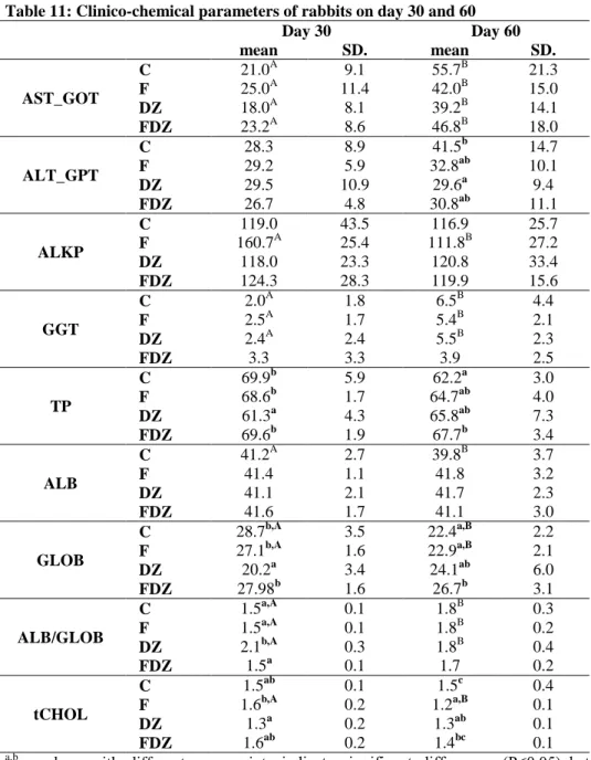
The experimental animals were Pannon White rabbit breeding bucks (24 weeks of age, 4.0 ± 0.5 kg mean body weight, n=60). The bucks were individually housed in wire mesh cages (42 × 50 cm) in a closed building and the light cycle was 16h light /day. Average temperature ranged from 16°C to 18°C and the farm had overpressure ventilation.
The animals received a commercial diet containing 10.3 MJ digestible energy/kg, 15.5% crude protein, 4.0% crude fat and 14.7% crude fibre for a total of 65 feeding days. The feedstuffs provided were available ad libitum, and the rabbits also had free access to drinking water.
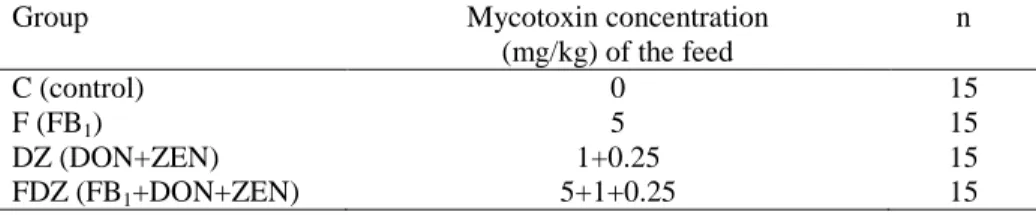
Bucks were divided into 4 experimental groups. One group of the experimental animals served as control. The feed of the other three groups contained fungal culture in a predefined concentration (Table 5).
Table 5: Experimental groups
Group Mycotoxin concentration
(mg/kg) of the feed
n
C (control) 0 15
F (FB1) 5 15
DZ (DON+ZEN) 1+0.25 15
FDZ (FB1+DON+ZEN) 5+1+0.25 15