Physiological and pathological adaptation of veins to hemodynamic stress
Theses
Dr. Gergely Gősi
Semmelweis University
Doctoral School of Clinical Medicine
Tutor: Dr. György Acsády, M.D., Ph.D., D.Sc.
Opponents: Dr. Tamás Sándor, M.D., Ph.D.
Dr. Attila Szabó, M.D., Ph.D.
Chairman of the panel: Dr. Ferenc Perner, M.D., Ph.D., D.Sc.
Members of the panel: Dr. Zoltán Járai, M.D., Ph.D.
Dr. István Sugár, M.D., Ph.D.
Budapest
2015
2 1. Introduction
Antigravity defence reactions probably represent one of the oldest adaptation mechanisms of terrestrial animals. The gravitational load is especially high for humans, the pressure load in the lower limbs can increase ten times in the upright body position. Significant data are available of adaptation mechanisms against short-term gravitational load. Acute regulation of the capacity is one of the most important functions of the veins. This was experimentally proved by the unchanged venous diameter observed after increasing the intraluminal pressure. Still remarkably less systemic publications deal with the long-term vascular adaptation processes supporting the orthostatic tolerance in humans.
On the adaptation mechanisms against gravitational stress, our group could draw significant conclusions in earlier studies. After long-term orthostatic load venous capacity increased, wall thickness remained unchanged and the distensibility decreased. Human and animal studies proved that the myogenic tone of human great saphenous vein is significantly higher than that of the vascular segments of dogs or rats. The pressure-induced myogenic tone has increased after orthostatic load.
Quantitative electron microscopic (EM) and immuno-histochemistry studies revealed that two weeks experimental orthostatic stress could significantly increase the adventitial sympathetic innervation density. We also examined the endothelial vesicles after two weeks load on EM images. Immuno-histochemistry proved endothelin and platelet derived growth factor (PDGF) content in these dark secretory granules. The proportion of vesicular area to that of the endothelial cells on EM images decreased after orthostatic load. The microvascular network has also changed after long-term gravitational load in animal models. In the superficial venous system of hind limbs we could observe decreased branching angles and increased passive diameter.
The clinical significance of venous hemodynamic load is unequivocal. Chronic venous insufficiency (CVI) and its superficial type, the varicosity are widespread diseases. The frequency of deep venous type CVI is about 7% for women and 9% for men, while the average incidence of varicosity is 26-29% in females and 13-15% in males. The prevalence of varicosity is even higher in Hungary. Advanced CVI does
3
not only deteriorates quality of life but also has remarkable economical consequences.
Expenses directly or indirectly spent on venous disorders can achieve 1.5-2% of health care budget in several European Union countries.
Venous bypasses after vascular surgical procedures are also exposed to significant hemodynamic stress. In arterial circumstances the dynamic, structural and biochemical adaptation of veins can lead to long-term changes of lumen calibre, wall thickness and structure. The hemodynamic load on veins consists of shear stress originated from the flow and tangential stress related to the intramural pressure. Shear stress mostly regulates the lumen. When the graft diameter increases the level of shear stress can get back to the normal range. As a consequence of high intramural pressure wall thickness increases. According to Laplace-Frank law, because of thicker wall tangential stress can decrease to normal. Pathological adaptation mechanisms can lead to the stenosis and damage of the venous graft that can result in bypass occlusion.
The objective of our investigations was to evaluate the biomechanical and structural responses of human veins to chronic gravitational load.
4 2. Aims
Numerous investigations have dealt with the biomechanical properties of veins affected by different chronic hemodynamic load. However, only a few comprehensive studies of this field can be found in the literature. Evaluating the properties of different vascular regions, in our experimental studies we were seeking the answers for the following questions:
1. How do the passive and active biomechanical properties of veins affected by different hemodynamic stress differ?
2. Can the experienced biomechanical differences be explained by adaptation mechanisms to gravitational load?
3. Do the differences in histological properties correspond with the biomechanical differences?
In a previous study, our group quantitatively determined the amount of endothelial vesicles using electron microscopy in rat limb veins. The density of these vesicles within the endothelial cells decreased significantly after long-term orthostasis.
There is scarce information in the literature about the density and geometry of such vesicles in human veins. We investigated the properties of these vesicles by electron microscopic studies that raised the following questions:
4. What is the density of endothelial secretory vesicles similar to that of the animal studies in human veins?
5. Does the vesicular density of veins affected by different hemodynamic stress differ?
6. Is there a difference between the geometry of venous vesicles originated from various regions?
5 3. Methods
For all the tests small superficial veins with the length of 8-10 mm and with the diameter of 1-2 mm were excised from the site of surgery.
3.1. In vitro biomechanical tests
Segments were cannulated at both ends and immersed in the tissue bath of the in vitro pressure angiograph. After incubation pressure-diameter plots were taken, between 2-30 mmHg in control Krebs-Ringer (nKR) solution without any agonist added. After that 10 µM norepinephrine (NE) was added to the bath to induce maximum contraction. Following 10 min incubation, the contracted pressure-diameter plots were recorded. With the NE still in the bath, acetylcholine (Ach) 10 µM, was added and the plot recorded again to test the extent of endothelial dilation. Nitrogen monoxide (NO) component of relaxation was tested by repeating the measurements with 100 µM L-nitro-arginin-methylester (L-NAME) in the bath. Using Ca-free solution we examined the passive biomechanical properties at the end. Diameter measurements were made by freezing the monitor picture and measuring the distance between outer and inner contours of the segments.
As a sum eleven neck vein samples (side-branches of external jugular veins) were excised during carotid surgery and eleven leg vein segments (side-branches of great saphenous veins) were removed during lower limb arterial reconstruction or coronary bypass.
3.2. Histochemistry and immuno-histochemistry tests
To visualize elastic sheets resorcin-fuchsin stainings were used. Immuno- histochemistry for smooth muscle actin (SMA) were made.
Eight small neck vein and seven saphenous vein samples of similar sizes were collected for histochemical studies.
6 3.3. Analysis of endothelial vesicle density
For quantitative determination of endothelial vesicles morphometric analysis of the electron micrographs was applied. One to twenty electron microscopic pictures of each sample with magnification ranging from 2,500× to 25,000× were taken, digitalized and then analyzed. The contour of endothelial cells and that of the dense vesicles were outlined and total cross-sectional areas were measured. These data were used to calculate the vesicular density for each patient and expressed as vesicular area relative to the total cross-sectional area of the endothelial cells. We analyzed nearly two hundred electron micrographs with this method.
The samples of these small veins of the upper limb were removed from the forearm during arterio-venous fistula formation for hemodialysis access. Vein segments of the lower extremity were excised from the groin or leg during arterial reconstruction surgery. Altogether 25 vessel samples were collected from 25 patients, 10 samples were taken from the upper and 15 samples from the lower extremities. We divided the lower limb samples into two subgroups, 7 veins were excised from the groin and 8 from the leg.
3.4. Vesicular geometry in the endothelium
Long and short axes of granule cross sections were measured on digitalized electron micrographs. Examining the geometry of the dense vesicles in leg and arm samples we measured the ratio of long and short axes of 2540 granules. We demonstrated the difference of the relative frequencies of granules with different short and long axises of leg and arm veins on colour-coded diagram.
Five upper limb and seven leg samples were examined in this study.
7 4. Results
4.1. Differences in the biomechanics of neck and leg veins
4.1.1. Geometry
Venous segments of similar range were collected from the neck and the leg.
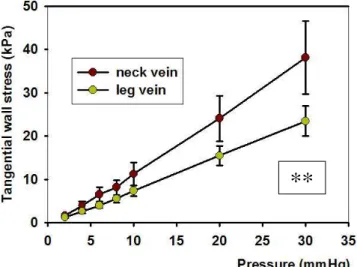
However, as an effect of higher spontaneous tone, diameters of leg veins were smaller in oxygenated nKR and they had a higher wall thickness. As a consequence of such differences in diameter and wall thickness, isobaric wall stress was significantly lower in leg veins than on the neck (Figure 1.).
Figure 1.: Tangential wall stress-intraluminal pressure plots taken in oxygenated nKR.** p<0.01, in two-way anova test.
4.1.2. Elasticity
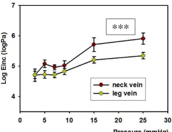
Incremental distensibilities differed in the two groups of veins in the relaxed state. Lower extremity veins were more distensible at moderate and higher pressures.
Similar statement can be made based on the relaxed elastic modulus-pressure plots.
With the exception of the lower pressures the upper body veins had higher elastic moduli (Figure 2.).
**
8
Figure 2.: Incremental elastic modulus as a function of intraluminal pressure in the relaxed state. , *** p<0.001 in two-way anova test.
4.1.3. Contractility
Spontaneous tone (% reduction of diameter in KR solution) was significantly higher in lower body veins. Norepinephrine induced (maximum) contractions were also more pronounced in the lower body vessels (Figure 3.).
Figure 3.: Spontaneous contraction in oxygenated nKR. Expressed as ratio of relaxed radius. *** p<0.001, in two-way anova tests
***
***
9 4.1.4. Endothelial dilation
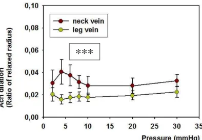
Acetylcholine dilation related to the relaxed state was more effective in upper body segments (Figure 4.). L-NAME induced contractions, however, did not differ in the two groups.
Figure 4.: Dilation in response to 10 micromoles/l acetylcholine. Ratio of relaxed radius.*** p<0.001, in two-way anova tests
4.2. Histochemistry of neck and leg veins
In leg veins elastic sheets were more continuous and higher in number (Figure 5.).
A B
***
10
Figure 5.: Resorcin-fuchsin staining. A: neck vein, B: leg vein.
Smooth muscle actin staining revealed more smooth muscle cell layers (Figure 6.).
A B
Figure 6.: Immuno-histochemical reaction for SMA. A: neck vein, B: leg vein.
4.3. Volume of endothelial dense vesicles of the upper and lower extremity
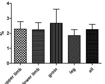
The proportion of the total area of the dense vesicles to that of the endothelial cross section in all samples was 2.26 ±0.34%. These values calculated for the upper extremity and for lower limb did not show statistically significant difference. We calculated the vesicular densities of areas with different chronic orthostatic stress (upper extremity vs. leg) that also proved to be statistically insignificant (Figure 7.).
11
Figure 7.: Territory occupied by the vesicles in the section of endothelial cells in different body areas.
4.4. Differences in geometry of dense vesicles
We calculated the difference of the relative frequencies of granules with different short and long axises of leg and arm veins. We found the vesicles from arm samples to be smaller and more circular (blue spots) and granules from the endothelium of leg veins bigger and more frequently elongated (red spots) (Figure 8.)
Figure 8.: Difference of relative frequencies of granules with different short and long axes of leg and arm veins.
Short axis (nm)
Long axis (nm)
12 5. Conclusions
We investigated the structural and functional properties of human veins from different regions that were affected by different orthostatic load. We could conclude the following results:
1. Our data demonstrate that there is significant quantitative difference in biomechanical properties of veins affected by different orthostatic loading. Wall thickness is higher in leg samples than in neck veins, so the isobaric wall stress of cervical veins is higher than that of the leg. Distensibility of leg veins is lower in the higher pressure range, so neck veins proved to be more rigid.
Elastic modulus is also higher in neck samples. Spontaneous myogenic tone and NE-induced contractility are significantly higher in the leg samples. Endothelial dilation is more pronounced in the jugular veins. We could not find difference in the contraction induced by L-NAME between the two groups, that means that Ach dilation was partly NO independent. Different vasodilator agents (eg.
prostanoids) are possibly in the background of the process.
2. All of our biomechanical results refer to the chronic adaptation mechanisms to gravitational load. Thicker wall and therefore lower isobaric wall stress of leg veins correspond with higher resistance against higher transmural pressures.
More pronounced distensibility, elasticity and higher levels of myogenic tone and maximal contractions of leg veins are all part of their adaptation to usually higher and changing orthostatic loading.
3. Histological and immuno-histochemistry studies proved that elastic sheets are more continuous and smooth muscle cell layers are higher in number in leg vein samples. Since elastic fibres are responsible for elasticity of vascular wall and smooth muscle causes contraction, the connection between the biomechanical properties and the histological characteristics of lower body veins affected by higher gravitational load is unquestionable.
4. The overall mean ratio of the vesicular area to that of the endothelium proved to be 2.26% in human superficial veins. Regarding all the samples a stable 1.8-
13
2.7% ratio can be observed that raises the possibility of a yet unknown regulating mechanism in the background.
5. Comparing the ratios of different regions with similar gravitational load (upper and lower extremities) we have not found significant difference between the results. When we calculated the vesicular densities of areas with different orthostatic stress (upper limb vs. leg) the difference was also statistically insignificant. The lack of difference might be attributed to earlier (evolutional) adaptation or exposure of dynamic changes of hydrostatic pressure during daily activity in human.
6. Remarkable differences have been identified in granule geometry between areas exposed to different gravitational stresses. We found the vesicles from leg samples to be bigger and more elongated than that of the neck veins. It is probable that leg granules develop into more elongated ones. There also can be differences in secretory content of the vesicles from different regions. Since these granules contain mediators of biomechanical regulation and agents of hemostasis, we assume that the differences in our results can contribute to the differences in thrombotic and hemodynamic behaviour of upper and lower extremity veins.
14 Publications of the author
Publications related to the theses
Gősi G, Monori-Kiss A, Nádasy Gy, Durkó A, Tőkés A, Monos E, Acsády Gy. (2014) Comaparison of passive and active biomechanical properties of human cervical and leg veins. Int Angiol, 33: 348-356.
Gősi G, Nádasy Gy, Raffai G, Fehér E, Szeberin Z, Monos E, Acsády Gy. (2014) Regional differences in statistical geometry of endothelial dense granules in human extremity veins. Plebology, pii: 0268355514547715.
Independent publications
Gősi G, Huoránszki F. (2001) Sigmoidoscopically induced pneumatosis cystoides coli in Crohn’s disease manifested by collar subcutaneous emphysema. Endoscopy, 33:
293.
Gősi G, Münch Z. (2002) A Fournier-gangrénáról; a korai diagnózis a legjobb ellenszer. Magy Seb, 55: 27-30.
Gősi G, Pencz Z, Laczkó Á, Acsády Gy. (2005) Takayasu arteritis; ritka kórkép, különleges megfontolások. Magy Seb, 58: 9-15.
Gősi G, Járányi Zs, Entz L, Acsády Gy. (2005) Congenitalis arterio-venosus fistula talaján kialakult artéria brachialis álaneurysma esete. Magy Seb, 58: 328-330.
Gősi G, Járányi Zs, Gálffy I, Bérczi V, Entz L. (2007) Alsó végtagi obliteratív elváltozások egyidejű, katéteres és sebészeti megoldása. Érbetegségek, 14: 17-20.
Gősi G. (2010) Kommentár: Risk of deep vein thrombosis following a single negative whole-leg compression ultrasound. JAMA, 2010, 303(5): 438-445. Motesz, XVIII: 52- 54.
Gősi G. (2014) A diabétesz és a perifériás érbetegség-az alapellátás jelentősége.
Magyar Családorvosok Lapja, 3: 3-4.
15
Mihók A , Imreh D, Gősi G, Benyó I. (1999) Iatrogen colorectalis perforációk – az elmúlt 20 év eseteinek áttekintése. Magy Seb, 52: 75-79.
Imreh D, Mihók A, Gősi G, Benyó I. (1999) A gastrointestinalis carcinoid tumorokról.
Magy Seb, 52: 39-45.
Polner K, Gősi G, Vas SI, Kalina I, Acsády Gy. (2009) Management of abdominal and iliac artery aneurysms by stent-graft implantation in a patient on CAPD. Clin Nephrol, 71: 359-362.
Kovács H, Fehérvári M, Forgó B, Gősi G, Oláh Z, Csobay-Novák Cs, Entz L, Szeberin Z. (2014) Elektiv infrarenalis aortaaneurysma sebészi kezelésének korai és késői mortalitása és morbiditása. Magy Seb, 67: 297-303.