0
HEMODYNAMIC AND ENDOCRINE FACTORS IN THE BIOMECHANICAL REMODELING OF THE
VASCULAR WALL
PhD thesis
dr. Judit Réka Hetthéssy
Basic and Translational Medicine Doctoral School Semmelweis University
Supervisor: György László Nádasy MD, Ph.D Official reviewers: Miklós Szokoly MD, PhD
Zoltán Németh MD, Ph.D
Head of the Final Examination Committee: László Entz, MD, Ph.D, D.Sc
Members of the Final Examination Committee:
Sándor Frenyó, MD, Ph.D,D.Sc Éva Toronyi, MD, Ph.D,D.Sc
Budapest
2019
1
Table of contents
I.Abbreviations……… 3
II. Introduction……….………. 7
1. Hemodynamic control of vessel wall biomechanics ………. 9
2. Humoral control of the biomechanical properties of the vessel wall ……. 16
3. Biomechanical remodeling of resistance arteries in hypertension, sex differences, effects of sex hormones………..…….. 22
4. The effects of venous hypertension on the geometrical and biomechanical characteristics of the venous wall ………. 38
5. Mechanisms leading to the development of varicose veins……… 40
6. Development of varicose veins, biomechanical alterations in varicosity.. 41
7. Morphology, geometry and the development of network collaterals……. 49
III. Aims……… 51
IV. Methods……….. 52
1. Chronic angiotensin II infusion; Ovariectomy, Estrogen therapy in rats………..…. 55
2. Partial chronic occlusion (‘‘clipping’’) of the saphenous vein in rats….. 56
3. In vitro pressure-angiometry………. 58
4. Intravital videomicroscopy and hemodynamic measurements ……….… 61
5. Histology and immune-histology of the main branch following chronic partial clipping of the saphenous branch in high-pressure low-flow venous model……….…. 62
6. Batson 17 plastic casts of main branch ………..…… 63
7. Statistical analysis………. 65
V. Results………. 66
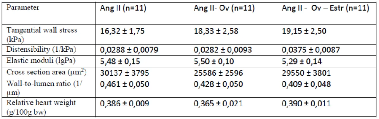
1. Sex differences in the remodeling of rat coronary resistance arteries in angiotensin II hypertension………..………. 66
2. Remodeling of rat coronary resistance arteries in angiotensin II hypertension ovariectomy vs. ovariectomy and estrogen replacement ….. 69 3. The effects of high-pressure low-flow conditions induced by chronic
stricture of the main branch of the saphenous vein on the
2
hemodynamics of the saphenous main branch. Biomechanical alterations of the main branch following partial clipping of the main
branch of the saphenous vein……….………… 73
4. The effects of high-pressure low-flow conditions induced by chronic stricture of the main branch of the saphenous vein on the collateral system of the saphenous main branch……… 79
5. The effects of high-pressure low-flow conditions induced by chronic stricture of the main branch of the saphenous vein on the histological characteristics of the saphenous main branch……….. 81
VI. Discussion……….……….. 89
1. Sex Differences in the Biomechanics Intramural Coronary Arteries in Angiotensin II–Induced Hypertension……….… 89
2. Estrogen therapy may counterbalance eutrophic remodeling of coronary arteries and increase bradykinin relaxation in a rat model of menopausal hypertension……….... 92
3. Remodeling of the venous wall in high pressure-low flow conditions... 93
4. Early stage varicose disease……… 95
VII. Conclusions………. 97
VIII. Summary……… 99
IX. Összefoglaló……… 100
X. References ……… 101
XI. Publications by the Author in this field………... 119
XII. Acknowledgements……… 120
3
I. Abbreviations
5-HT2 5-hydroxytryptamine
α Alpha
AA Arachidonic acid
Ach Acetylcholine
ADP Adenosine diphosphate
AngII Angiotensin II
ASK1 Apoptosis signal-regulating kinase 1
ATP Adenosine triphosphate
AT1R Angiotensin II receptor type 1
β Beta
BK Bradykinin
bw Body weight
C Capacitance
CV Vascular Compliance
Ca2 Calcium
CaCl Calcium chloride
cAMP Cyclic adenosine monophosphate CD68 Cluster of Differentiation 68 protein cGMP Cyclic guanosine monophosphate
CD18 Integrin beta-2
COX Cyclooxygenase
CSE Cystathionine γ-lyase
c-SRC Proto-oncogene tyrosine-protein kinase
D Distensibility
4
d Diameter
DAG Diacylglycerol
E Elastic modulus
E Estrogen
ECM Extracellular matrix
EDRF Endothelium derived relaxing factor
EDHF Endothelium derived hyperpolarizing factor EDTA Ethylenediaminetetraacetic acid
g Gram
h Wall thickness
H Histamine
I Current
i.p. Intra peritoneal
IP3 1,4,5-trisphosphate
IM Intramuscular
JNK Jun N-terminal kinases
k Proportionality constant
K+ Potassium
KCl Potassium chloride
kg Kilogram
kPa Kilopascal
l Length
m Meter
M Muscarinic
MgSO4 Magnesium sulfate
MBP Mean Blood Pressure
5
MAP kinase Mitogen-activated protein kinase
min Minute
mm Millimeter
mmHg Millimeters of mercury
μm Micrometers
MMP Matrix metalloproteinase
MPa Megapascal
ng nanogram
Na+ Sodium
NA Norepinephrine
NaCl Sodium chloride
NAD(P)H Nicotinamide adenine dinucleotide phosphate
NaHCO3 Sodim bicarbonate
NaH2PO4 Sodium phosphate
nKR Normal Krebs-Ringer solution
NO Nitric oxide
NOS Nitric oxide synthase
O2 Oxygen
OV Ovariectomy
OVX Ovariectomy
P Pressure
PG Prostaglandin
PGH2 Prostaglandin H2 PGI2 Prostaglandin I2
Q Flow
r Radius
6
R Resistance
RF Resorchin-fuchsin
SD Standard deviation
SHR Spontaneously hypertensive rat SEM Standard error of mean
SMA Smooth muscle actin
SVR Systemic vascular resistance
TRX Thioredoxin
TxA2 Thromboxane A2
TXNIP Thioredoxin-Binding Protein
U Voltage
U46619 9,11-Dideoxy-9alpha,11alpha-methanoepoxy prostaglandin
V Volume
VCAM1 Vascular cell adhesion protein 1
7
II. Introduction
The vascular system plays a diverse role in the human body. It acts as a conduit, transferring material, energy and information (functions of “conduction”). It is capable of mechanical adaptation, plays a role in the formation, conduction and dissipation of hemodynamic waves. It has receptor functions neurotransmitters are released in them and are important components of synthesis of different tissues. It plays a role in the biochemical conversion of substances in blood plasma in hemostasis and transmural transport of different substances.
Hypertension is a well-recognized risk factor for cardiovascular and renal diseases and may lead to increased risk of stroke (Bergan (Bergan, Pascarella, &
Schmid-Schönbein, 2008). A study of men and women aged 45 to 83 years showed the age-standardized prevalence of hypertension at baseline was 74.3% for men and 70.2%
for women (Lacruz ., 2015) . Small resistance arteries play a key role in the control of blood pressure. These segments are responsible for the most significant decrease of hydrostatic pressure along the circulatory system. The rise in blood pressure affects the structure and morphology of the vessel wall (Hayashi & Naiki, 2009). Angiotensin II is greatly implicated in vascular remodeling of small-resistance arteries (Intengan &
Schiffrin, 2000; Neves et al., 2004; Neves, Virdis, & Schiffrin, 2003). Sex differences in hypertension have been described, and are at least in part thought to be due to the effects of estradiol and testosterone (Barton, Prossnitz, & Meyer, 2012), but little is known about the mechanisms through which sex hormones affect the remodeling of coronary resistance arteries.
A cross-sectional study of 2,211 men and women revealed that varicose veins affect 28% of the adult female population compared with in 15% of the adult males (Raffetto et al., 2010).The primary cause of the development of lower limb varicosity has not been established, but pressure loading, vein valve dysfunction, diabetes, obesity, smoking and age, pregnancy, standing for long periods of time have been implicated as major contributing factors to this to the progression of this disease. (Raffetto & Khalil, 2008; Raffetto et al., 2010). The pathomechanisms underlying the development of varicose vein is still inconclusive, and little is known about the effects of flow disturbances on venous pathology.
8 Histology and physiology of the vessel wall
Histology of blood vessels (Figure 1.).
Figure 1.
Histology of veins and arteries.
(Figure from - (Torres-Vázquez, Kamei, & Weinstein, 2003)) Veins and arteries and veins are both composed of similar tissue layers, an inner endothelium (tunica intima) surrounded by internal elastic tissue, a layer of smooth muscle cells (tunica media), external elastic tissue, and fibrous connective tissue (tunica adventitia).
Arteries and veins have are composed of the same three layers, the tunica intima, the tunica media and the tunica adventitia. The tunica intima lines the lumen of the blood vessels. It consists of simple squamous epithelium and a thin basal membrane.
The simple squamous epithelium provides a smooth surface for unperturbed blood flow.
The tunica intima is collectively referred to as the endothelium. The tunica media is made of smooth muscle and elastic fibers. It is responsible for vasodialation and vasoconstriction of the blood vessels. The tunica adventitia is the most superficial layer.
It is made of collagen fibers running in all directions for strength in many different directions. In arterioles the tunica intima consists of a single layer of squamous epithelium, the tunica media a few or single layer of smooth muscle cells and the tunica adventitia is quite this, and may megre with surrounding tissue.
9
Arteries and veins are exposed to different pressures of blood flow. These differences are reflected in the structure of the vessels. Arteries experience a pressure wave as blood is pumped from the heart therefore the walls of arteries are much thicker than those of veins. In addition, the muscular tunica media is much thicker in arteries than in veins. As a result, arteries have a more uniform, circular shape. Veins are not exposed to pressure waves, therefore the vessel walls of veins are thinner than arteries and do not have as much tunica media. The tunica media is smaller in relation to the lumen than in arteries. Veins are more irregular in shape.(Hall, 2015; Torres-Vázquez et al., 2003)
Vascular endothelial cells in the resistant arteries are constantly exposed to the dynamic changes of blood flow (Fig. 2). The hemodynamic forces can be resolved into three components: (1) shear stress, the tangential frictional force acting at the endothelial cell surface, (2) hydrostatic pressure, the perpendicular force acting on the vascular wall, and (3) cyclic strain, the circumferential stretch of vessel wall (Hsiai &
Wu, 2008).
Figure 2.
Figure from (Hsiai & Wu, 2008)
Hemodynamic forces acting on blood vessels
Diagram of hemodynamic forces acting on endothelial cells and smooth muscle cells in the blood vessel wall. (a) Fluid shear stress which is the tangential frictional force by virtue of blood viscosity, acts on endothelial cells mainly. (b) Cyclic strain exerts a circumferential stretch on arterial wall in response to cardiac contraction. Hydrostatic pressure acts perpendicularly on endothelial cells. (c) Endothelial cells are constantly
10
exposed to both biomechanical and biochemical stimuli, which modulate endothelial functional phenotype. The biochemical stimuli include hormones, growth factors, cytokines that may be delivered via the blood or via autocrine or paracrine mechanisms (Hsiai & Wu, 2008).
II./1. Hemodynamic control of vessel wall biomechanics
It has long been established that the conduction and wave forming/conducting/dissipating functions are determined by the geometrical and elastic properties of vessels. These characteristics, however may adapt to a set of physiological and pathological factors (Monos, 1986).
Short term adaptation
While alterations in tissue composition are usually seen in long term adaptation, short term adaptation or regulation of the vessel wall is most commonly achieved through setting of the myogenic tone of the vessel. Changes in geometrical and elastic properties are key elements in this process (Monos, 1986). The diameter of the vessel is determined by transmural pressure as well as by the elastic and contractile elements of the wall. Vessel tone (which may be measured by comparing vessel diameters in an active and passive state) is set by several factors.
The intrinsic tone, which can be observed on isolated vessel segments is modulated by the endothelium and by metabolic, endorine, neural and hemodynamic factors. All these factors therefore play a role in the control of peripheral resistance (Hall, 2015; Mulvany & Aalkjaer, 1990).
Myogenic tone is considered to be a key factor in the local regulation of blood flow (Bayliss, 1902)(Figure 3.). Myogenic response is defined as active vessel contraction to an increase of intravascular pressure (Bayliss, 1902; Kuo, Chilian, & Davis, 1991; Kuo, Davis, & Chilian, 1991; Meininger & Davis, 1992; Rajagopalan, Dube, & Canty, 1995) Mulvany and Aaljaker (Mulvany & Aalkjaer, 1990) described that this myogenic response is regulated by stretch or pressure response, mostly in small arteries and arterioles (<500 µm and <100 µm, respectively). Elevation of cytoplasmic Ca2+ ion
11
levels, due mechanically sensitive Ca2+ membrane channels have been described to be involved in these mechanisms (Hill, Yang, Ella, Davis, & Braun, 2010).
Figure 3.
Myogenic tone - active vessel contraction to an increase of intravascular pressure
Myogenic response may be studied in cannulated and pressurized vessel segment preparations in vitro. This way other variables can be controlled (neurohumoral, metabolic factors etc.) (Kuo, Davis, & Chilian, 1988; Meininger &
Davis, 1992). Resistance artery control of local tissue flow, partially due to the myogenic tone is very effective according to the Hagen-Poiseuille law. In a thin, rigid tube, if flow is laminar the intensity of flow (Q) is directly proportional to the pressure difference between the points of the tube (P1-P2) and also proportional to the fourth power of the inner radius (r). In analogy with the Ohm’s law used in electrodynamics, we can compute hydrodynamic resistance, and applying the Hagen-Poiseuille law, R=
(P1-P2)/Q=8xLη/πr4. Thus vascular resistance is directly proportional to the length of the vessel and the viscosity of blood, and is inversely proportional to the fourth power of the radius of the vessel. In consequence, a key factor in the acute regulation of blood flow is vessel diameter (Fonyó, 2011; Hall, 2015). As the intensity of flow changes in correspondence with the fourth power of the radius of the vessel wall, minor alterations in diameter lead to significant changes in flow. Every factor that influences the smooth muscle tone and reactivity of vessels may have short-term regulatory effects on hemodynamic adaptation (Monos, 1986). Flow autoregulation is implemented at the level of precapillary resistance arteries.
12
In addition to pressure and wall stretch, flow, which, in the arteries is pulsatile flow, also induces both long and short term reactions within the vessel wall. Release of vasoactive agents and immediate alterations in vessel diameter will be resulted (Cronenwett & Johnston, 2014). In case the endothelium is damaged there is a direct action of blood flow shear stress on the smooth muscel cells and fibroblast on the vessel wall. This may come from secretion, laminar, pulsatile, and oscillating flow shear stresses. These stresses may lead to alterations in alignment, contraction, proliferation, apoptosis, differentiation, and migration in the remaining cells of the vascular wall.
Smooth muscle cells, and fibroblast both posses a high degree of plasticity that allows large scale yet reversible changes within the cell in response to alterations in local environmental factors. This is why smooth muscle cells, and fibroblasts may play a crucial role in vascular repair and remodeling (Shi & Tarbell, 2011). In a physiological state they are arranged in distinct patterns within the vessel wall. There is evidence that different types of mechanical stimuli regulate vascular cell morphology. Fluid shear may lead to the orientation of the endothelial cells parallel with flow (Chiu, Usami, &
Chien, 2009). Laminar shear stress may induce perpendicular alignment of the smooth muscle cells. The alignment of smooth muscle cells semmed to dependend on and cytoskeleton-based mechanisms (Lee, Graham, Dela Cruz, Ratcliffe, & Karlon, 2002).
Pulsatile strain and shear stress resulted in a circumferential pattern alignment of the smooth muscle cells. It has been found that non-uniform blood flow causes the smooth muscle cells to align perpendicular to luminal flow (parallel to transmural flow) and migrate toward the lumen, while uniform shear stress does not significantly affect smooth muscle cell orientation and migration. Circumferential alignment of the smooth muscle cells alloes the blood vessels to better resist tangential stresses induced by blood pressure (Shi & Tarbell, 2011).
Long-term adaptation
It has long been established that the alterations in tissue composition due to changes in hemodynamic forces may be part of physiological adaptation mechanisms, but also can be pathological processes (Monos, 1986). It is also known that the vascular wall is composed of elastic and of non-elastic components, and all these determine its biomechanical properties (Mulvany & Aalkjaer, 1990). Strain produced by stress
13
appears instantaneously in purely elastic body. The materials, where there is time gap before the strain appears are defined as viscoelastic (Milnor, 1972). The main building components of the vessel wall are elastin, collagen and smooth muscle cells. Wall composition will play a central role in determining the elastic response of the vessel in response to intraluminal pressure changes.
Elastin (Figure 4.) is a highly elastic biological material, a protein that allows many tissues to resume their shape after stretching or contracting. For elastin, an almost linear stress-strain relationship is characteristic. Collagens (Figure 4.) are astructural proteins found in a variety of tissue in abundance. The vital importance of collagen as a scaffold demands a manifold of essential characteristics, including thermal stability, mechanical strength, and the ability to engage in specific interactions with other biomolecules. The collagen molecule consists of helically wound chains of amino-acids.
These helices are woven (Shoulders & Raines, 2009) into microfibrils, which weave together into subfibrils and fibrils. Due to this structural characteristic, this “waviness”
the stress—strain relationship shows a very low stiffness at small stretch ratios. The stiffness increases fast once the fibers are deformed into straight lines.
a, b,
Figure 4.
Main contractile and force-bearing structural proteins of the vascular wall (a) collagen, (b) elastin.
(https://www.tes.com/lessons/jezmMDoMqDP_kw/connective-tissue-collagen, https://step1.medbullets.com/biochemistry/102079/elastin,
Any transmural pressure within vessel exerts a force on the vessel wall that would tend to rip the wall apart were it not for the opposing forces supplied by the muscle and connective tissue of the vessel wall. This force is equal to the product of the transmural pressure and the vessel’s inner radius, and it is defined as circumferential or
14
tangential wall tension. The relationship is defined by the Law of Laplace. The Law of Laplace applies directly only to cylinders with thin walls. Blood vessel walls are sufficiently thick, so in vascular mechanics the tangential wall stress is preferably used (Rhoades & Bell, 2012). Tangential wall stress of the vessel (σ) is determined by transmural pressure (P), vessel inner radius (ri) and wall thickness (h). σ=Pri/h (Laplace- Frank equation). Changes in wall thickness therefore lead to alterations in wall stress.
Blood vessels that have thick walls relative to their radius are able to withstand higher pressure than vessels with small ratios because in the former wall stress is lower. Not pressure, but but wall stress is what must be overcome to contract a blood vessel (Rhoades & Bell, 2012).
Blood vessels are constantly exposed to mechanical forces in the form of cyclic strain and shear stress. The main source of cyclic strain is blood pressure as radial and tangential forces in the vessel wall work to counteract the intraluminal pressure.
Hemodynamic forces have long been recognized as key modulators of protein synthesis, cell morphology migration, differentiation and proliferation. The Laplace-Frank equation describes the tension per unit of thickness of the wall, which represents the stress exerted on the wall in the circumferential direction. Circumferential stress affects all cell types in the vessel wall, whereas shear stress principally acts on the endothelium. Pulsatile flow also plays a role in the long term adaptation of the vessel wall. Chronic changes in mechanical forces lead to vascular remodeling and adaptive alterations in vessel shape and composition over time (Cronenwett & Johnston, 2014)
Vascular remodeling may occur as a result of changes in pressure, radius or blood flow. Smooth muscle cell hypertrophy and elevation in collagen and elastin production have been shown to accompany increased circumferential stress (Prado &
Rossi, 2006). The opposite has been demonstrated as well, that a decrease in circumferential wall stress leads to wall atrophy (Cronenwett & Johnston, 2014).
Studies have shown that when the diameter of a vessel increases, the number of lamellar units and the overall thickness of the vessel wall increase also in order to maintain circumferential wall stress. In elastic arteries the adaptive response physiologically works to normalize tensile stress.
In a tube with rigid, inflexible walls the volume of fluid is the same independent of the pressure difference between the inside and the outside of the tube, however blood
15
vessels are viscoelastic. Consequently, the volume contained within them is a function of both the pressure difference across their wall (transmural pressure - defined as the difference of pressure inside versus outside the vessel wall) and the degree of flexibility/elasticity of the vessel wall itself (Rhoades & Bell, 2012). Studies have revealed that average circumferential (tangential) wall stress increases with increased pressure and vessel size (Bérczi, Tóth, Kovách, & Monos, 1990; Monos, 1986; Monos
& Kovách, 1980; Szekeres et al., 1998).
Young's modulus, also known as the elastic modulus, is a measure of the stiffness of a solid material. Constant elastic modulus (independent of strain) characterizes the linear elastic solid materials. This modulus defines the relationship between stress (force per unit area) and strain (proportional deformation) in a material (Bergel, 1961). The vessel wall is not a linear elastic solid material: the stress/strain ratio increases with increasing strain. Elastic modulus thus can be defined for a short range of the stress/strain relationship for given values of stress (in case of vessels, intraluminal pressure) only. Such moduli are called incremental elastic moduli and are plotted against stress or pressure (Monos 1986). Vascular stiffness is often expressed by the circumferential incremental elastic modulus, computed from the equation Einc= ΔP⁄Δ ro x 2ri2
x ro ⁄(ro2
x ri2
), where Einc is the incremental elastic modulus, ri is the inner radius, ro is the outer radius, and Δ ro is the change in the outer radius in response to an intraluminal pressure change of P. When the parameters defined at specific pressure levels, different values may be expected at different pressure levels because pressure- diameter relationships of vessels are nonlinear (Hayashi & Naiki, 2009).
One problem with compliance is that different-sized vessels can not be compared. For example, a large stiff -walled vessel may have a higher compliance value than a tiny flexible vessel. For this reason, the percentage increase in volume for a given increase in pressure may be used as a means of comparing distensibility between vessels and segments of the vasculature of different sizes. This value is called vascular distensibility D= ΔV/ Vo ΔP (Rhoades & Bell, 2012).
The composition of the vessel wall determines its elasticity. From the elastic parameters used distensibility will depend both on vessel geometry and wall elasticity, while elastic modulus, especially it is expressed as a function of wall stress will be an inherent property of the wall material (Hayashi, 2003).
16
II./2. Humoral control of the biomechanical properties of the vessel wall
There are a myriad of agents that have an effect on the vascular system. These may be direct physical forces, chemical agents, hormones, paracrine substances, and receptor-mediated hormonal and neurotransmitter agonists. These agents may have an effect on the endothelium or on the vascular smooth muscle. The vasoconstriction/relaxation may be receptor mediated or non-receptor mediated.
Vasoconstrictors: Non–receptor-mediated
Calcium (Ca2+): During vascular smooth muscle contraction actin and myosin filaments slide over one another. Compared with skeletal muscle, smooth muscle contracts very slowly, develops and maintains high forces for long periods of time. Also it uses a relatively low amount of adenosine triphosphate (ATP) in the process. This contraction is controlled by intracellular free calcium ion concentration. The calcium may enter the smooth muscle cells via voltage-gated calcium channels, receptor- dependent and mechanosensitive calcium channels, and by release from the sarcoplasmic reticulum. It is released from the endoplasmic reticulum by certain agents, IP3 being the most important secon messenger with such an effect. Calcium binds to calmodulin, which in turn activates myosin light chain kinase to initiate crossbridge attachments (Fonyó, 2011; Hall, 2015).
Potassium (K+): Potassium causes membrane depolarization, this in turn results in the activation of calcium channels.
Sodium (Na+): Decreased transmembrane sodium gradient leads to decreased activation of plasma membrane sodium/calcium exchange and increased intracellular calcium levels. The Na+/ K+ pump is responsible for the base membrane potential in cells. The inward movement of sodium ions and the outward movement of potassium ions are passive and the reverse movements against the electrochemical gradients require the activity of an ATP-driven Na+/ K+ pump. Maintenance of a normal electrical environment in the cell requires that the Na+/ K+ ATP-ase. The Na+/ K+ pump is regulated by several factors including the intracellular sodium concentration and the neuromediators norepinephrine and acetylcholine. The Na+/ K+ ATP-ase alters the membrane potential and influences vessel function in an essential way.(Hall, 2015;
Vassalle, 1987)
17
Increased transmural pressure: Increased transmural pressure leads to the activation of stretch-dependent calcium channels. Alteration of vessel wall tension may lead to a myogenic response that does not require the presence of the endothelium. It is accompanied by a membrane depolarization and an increase of the intracellular Ca2+
concentration, which depends largely on an influx of extracellular calcium via voltage- operated calcium channels.(Schubert & Mulvany, 1999)
Vasoconstrictors: Receptor-mediated
Norepinephrine (NA): Norepinephrine binds to either alpha or beta adrenergic receptors. Alpha receptors are divided into subtypes α1 and α2; beta receptors into subtypes β1, β2, and β3. All of these function as G protein-coupled receptors, meaning that they exert their effects via a complex second messenger system.
Norepinephrine is a powerful vasoconstrictor. When the sympathetic nervous system is activated (through stress or exercise) the sympathetic nerve endings release norepinephrine into the tissues. This surge of norepinephrine has a stimulative effect on the heart, contracts the veins and arterioles. Through a dual control system norepinephrine may have an effect on tissues not only through direct simulation of the nerves, but through the indirect effects of circulating norepinephrine. (Hall, 2015) Norepinephrine binds to α-adrenoceptors on the cell membrane, which is linked to a membrane-bound regulatory protein, heterotrimeric G-protein (guanine nucleotide binding protein) with q/11 type alfa subunits. This activates phospholipase C, leading to hydrolysis of membrane-bound phosphatidyl inositol bisphosphate to yield inositol triphosphate and diacylglycerol. Inositol bisphosphate then causes the release of calcium from the sarcoplasmic reticulum, which ultimately leads to smooth muscle contraction reduction in vessel radius and thus increase in vascular resistance.
Norepinephrine plays an important role in the regulation of myocardial perfusion, as has an effect on the vascular tone of coronaries (Quillen, Sellke, Banitt, & Harrison, 1992).
There are both α and β receptors present on coronaries. α receptors cause vasoconstriction and β receptors vasodilation. The α1 receptors and α2 receptors found on coronary vascular smooth muscle cells cause vasoconstriction through smooth muscle contraction. There are α2 receptors on endothelial cells, these cause vasodilation through the release of nitric-oxide (NO) (Bassenge & Heusch, 1990; Furchgott &
18
Vanhoutte, 1989). There are β2 receptors on coronary resistance arteries, here norepinephrine causes vasodilation through increasing cyclic adenosine monophosphate (cAMP) levels in vascular smooth muscle cells (Bassenge & Heusch, 1990; Hall, 2015)
Acetylcholine (Ach): Acetylcholine is a parasympathetic neurotransmitter. It activates a range of muscarinic M receptors. In endothelial cells it may activate M1, M2, or M3 receptors, which opens plasma membrane calcium channels, activates phospholipase C and increases the production of IP3 and Diacylglycerol (DAG). Most of the effect of acetylcholine in vessels is endothelium dependent. If the endothelium is damaged, acetylcholine induced vasodilation (see next section) turns into vasoconstriction in isolated aorta preparations.(Nasa, Kume, & Takeo, 1997;
Tangsucharit et al., 2016)
Serotonin may cause vasoconstriction through the activation of 5- hydroxytryptamine (5-HT2) receptors which open receptor-operated plasma membrane calcium channels. The constrictor action of the serotonine may be due to a direct activation of vascular smooth muscle, augmentation of the action of other endogenous vasoconstrictors such as catecholamines, angiotensin II and the release of norepinephrine from adrenergic nerves. (Ayme-Dietrich, Aubertin-Kirch, Maroteaux, &
Monassier, 2017)
Vasopressin: Vasopressin is a powerful vasoconstrictor in the body when applied in higher concentrations. It is formed in nerve cells in the hypothalamus, is transported to the posterior pituitary gland, and from here it is secreted into the blood. It is a key factor in fluid volume control under physiological conditions. Direct vascular control is a less important function of vasopressin in physiological concentrations.
Angiotensin II (AngII): Angiotensin II is one of the most powerful vasoconstrictor substance known. It causes significant constriction of the small arteries and arterioles. It thereby plays an important role in determining total peripheral resistance, and increases arterial pressure. It activates angiotensin II receptors, and induces G protein-dependent signaling pathways (IP3 /DAG pathways). IP3 binds to its receptor on sarcoplasmic reticulum, opening a channel that allows calcium influx into the cytoplasm. Ca2+ then binds to calmodulin and activates the myosin light chain kinase, which in turn phosphorylates the myosin light chain and enhances the
19
interaction between actin and myosin, causing smooth muscle cell contraction (Mehta &
Griendling, 2007).
Endothelin: Endothelin is yet another potent vasoconstrictor. It may be found in the endothelium of the vasculature. The most typical stimulus for release is damage to the endothelium. Subtypes of endothelin receptors have been identifies, certain subtypes mediate vasoconstriction while other receptors mediate both vasoconstriction and vasodilatation. ETA is a subtype for vasoconstriction found in the smooth muscle cells.
It activates receptors, which stimulate of phospholipase C, formation of IP3, and release of calcium from the sarcoplasmic reticulum.(La & Reid, 1995)
Thromboxane A2 (TxA2) is an extremely potent vasoconstrictor (Yamada et al., 2003) derived from the metabolism of arachidonic acid. (Scornik & Toro, 1992) Thromboxane (TxA2) is a vasoactive agent. It is produced in response to a myriad of stimuli, from the cell membrane. There are different types of receptors that mediate the effects of thromboxane along with prostaglandin on coronary vascular smooth muscle.
The high-affinity (kD: > or = 1 nM) Prostaglandin H2 PGH2/TxA2 receptors mediate the contractile actions of TxA2. Through different G-proteins these receptors activate intracellular signal transduction pathways which control intracellular calcium level Adenosine triphosphate (ATP)-dependent K-channels are regulated by TxA2 and may be involved in the determination of coronary smooth muscle tone. (Schrör, 1993). TxA2 (Yamada et al., 2003) also has an effect on vascular smooth muscle, as it stimulates mitogenic growth.
Vasodilators: Direct
Nitric oxide (NO): Agents that control tissue blood flow locally typically only dilate only the small resistance arteries as they can only reach these, and not the larger caliber vessels upstream. Even so, when blood flow increases in the microvascular area, it leads to secondary mechanisms that have a vasodilatative effect on larger arteries. The endothelium in the small resistance arteries synthesizes substances that induce vasodilation. The key vasodilator substance is endothelium derived relaxing factor (EDRF). This mainly consists of nitric oxide. This has a very short half-life in the blood stream - only 6 seconds (Cocks, Angus, Campbell, & Campbell, 1985). Rapid flow of blood through the vessels causes shear stress on the endothelium, which through the
20
mechanical stretch leads to a significant increase in the release of nitric oxide, which leads to vasodilation of the vessels. This is fortunate because this mechanism causes vasodilation in the upstream arterial blood vessels whenever microvascular blood flow increases downstream. Without this mechanism the effectiveness of local blood flow control would be significantly decreased as a significant part of the resistance to blood flow is in the upstream small arteries. NO stimulates guanylate cyclase of the vascular smooth muscle, formation of cGMP, calcium removal and hyperpolarization. It also inhibits voltage-gated calcium channels.(Johns, 1991)
Hyperkalemia: Vascular smooth muscle membrane is depolarized in highly hyperkalemic solution and a contraction will be the result. However, in in vivo conditions, local hyperkalemia will mostly result vasodilation.
Magnesium: An increase in magnesium ion concentration causes powerful vasodilation because magnesium ions inhibit smooth muscle contraction. This is thought to be a direct action of magnesium, independent of NO.(Teragawa, Kato, Yamagata, Matsuura, & Kajiyama, 2001)
Vasodilators: Receptor-mediated
Epinephrine: Epinephrine leads to vasodilation through β2 receptor-mediated activation of adenyl cyclase resulting in the formation of cAMP (cyclic adenosine monophosphate).
Adenosine: Adenosine is produced within the myocardial cell. It plays a key role in the local metabolic regulation of coronary circulation. Adenosine acts on purinergic receptors. A1 receptor subtypes are found in the myocardium, and A2 receptor subtypes are found in the vasculature. Adenosine acts through receptor activation of ATP- dependent potassium channels. This leads to membrane hyperpolarization and closure of voltage-gated calcium channels. The vascular A2 receptors found on vascular smooth muscle cells stimulate the formation of cAMP. This leads to vasodilation in the coronaries. It also leads to hyperpolarization resulting from opening of potassium channels. Adenosine is also an endothelium-independent vasodilator (Kuo, Davis, &
Chilian, 1995)
Histamine (H): Histamine acts on the H2 receptor as avasodilator. H2 receptors are spread throuout the resistance arteries and may be found on vasuclar smooth muscle
21
cells. Vasodilator effect is mediated through cyclic Adenosine monophosphate (cAMP);
H1 receptors may be found on endothelial cells, and their stimulation leads to the formation of NO (Ebeigbe & Talabi, 2014).
Bradykinin (BK): The group of agents called kinins are potent vasodilators.
They are small polypeptides that are split away by proteolytic enzymes from alpha2- globulins in the plasma or tissue fluids. One of the key enzymes is kallikrein. It is present in the bloodstream and the tissue fluids in its inactive form, and may be activated by a myriad of factors, such as inflammation, chemical or physical effects.
When it is activated, kallikrein acts on alpha microglobulin to release a kinin called kallidin. Kallidin is then converted into bradykinin by tissue enzymes. Bradykinin has a short life, only a few minutes, as it is inactivated by carboxypeptidase or by the converting enzyme, which activates angiotensin. Kallikrein enzyme is deactivated by the the kallikrein inhibitor. Bradykinin causes vasodilation and increased capillary permeability (Hall, 2015). BK has been shown to play a role in the physiological regulation of coronary microcirculation. BK is produced in both the myocardium and in the vascular tissue. BK may act on different receptors. Receptors may be found either on the endothelium or on vascular smooth muscle cells. Bradykinin causes endothelium-dependent vasodilation on constitutive B2 receptors located on endothelial cells of the vascular wall (Okamura & Toda, 1989), and vasoconstriction on smooth muscle cells, which is mediated by B1 receptors. The effects of bradykinin are thereby usually a combination of an endothelium–dependent vasodilator and a direct smooth muscle- dependent vasoconstrictor effect. The endothelium-dependent action of bradykinin through the activation of nitric - oxide synthase (Kuo, Chilian, & Davis, 1991). There is also evidence, that bradykinin has an NO-idependent vasodilation effect which may be mediated by prostacyclin and endothelium-derived hyperpolarizing factor (EDHF) (Okamura & Toda, 1989). There is a body of evidence to support that bradykinin is a potent vasodilator on coronary arteries of different species and humans both on isolated vessels and heart preparations, such as in situ (Dézsi, Szekeres, &
Schiszler, 1995; Rajagopalan et al., 1995). Bradykinin induced vasodilation appears to be a key mechanism in coronary microcirculation (Kuo et al., 1991; Rajagopalan et al., 1995).
22
Comparison of metabolic control mechanisms in coronaries and veins
Metabolic control mechanisms in coronary blood flow.
Myocardial metabolic demand is matched by an increase in coronary blood flow under physiological conditions. This process appears to be largely regulated by local humoral agents (Muller, Davis, & Chilian, 1996)(Figure 5.).
Figure 5.
Control mechanisms of coronary blood flow. Humoral agnets play a paramount role in the regulation of coronary blood flow.
(Figure- http://www.sci.utah.edu/~macleod/bioen/be6000/notes/L12-control-circ.pdf)
Adenosine – considerations in the coronary arteries. Adenosine is a substance that plays a cardinal role in the regulation of coronary blood flow adaptations during rapid changes in cardiac performance (Bassenge & Heusch, 1990). Adenosine is produced within the myocardial cell, when myocardial metabolism increases. It diffuses to the vessels, leading to vasodilation (Bassenge & Heusch, 1990). There is evidence to suggest, that small coronary arteries are the primary target for the effects of adenosine (Bassenge & Heusch, 1990; Kuo et al., 1995; Muller et al., 1996). When the myocardium becomes “underperfused” due to a rise in metabolic demand, hypoxia, hypercapnia, and acidosis also contribute to vasodilation. Metabolism becomes anaerobic, lactic acid is released and acidosis develops (Hall, 2015). Other agents, such as prostaglandins and bradykinin may also be released, and may contribute to the vasodilation. The activation of ATP sensitive potassium channels may also be an
23
important regulatory mechanism in the humoral control of microvascular resistance (Muller et al., 1996). Neural stimuli may have both direct and indirect effects on coronary microcirculation. Norepinephrine from the sympathetic nerves act directly through the release of neurotransmitters on the coronaries. Indirect effects are caused by decreased or increased activity of the heart (Hall, 2015). Therefore sympathetic neurotransmitters can elicit either vasoconstrictor or vasodilator effect, depending on the presence of receptors. In the coronaries we find both α and β receptors with the predominance of β receptors (Juhász-Nagy & Szentiványi, 1973). There are also α2 receptors in the endothelium, which are responsible for the release of NO and therefore have vasodilatative effects.
Endothelium dependent control mechanisms
The endothelium is the active participant of more than one regulatory mechanism (Fiugre 6.). One of the most important vasodilator agents is produced by and released from the endothelium. It is the endothelium derived relaxing factor (EDRF) which is composed principally of nitric oxide. Blood flow in the vessels causes shear stress, leading to contortion of the endothelial cells leading to an increased release of nitric oxide. (Bassenge & Heusch, 1990; Furchgott & Vanhoutte, 1989; Kuo et al., 1991; Kuo, Davis, et al., 1990)
Figure 6.
Figure from (Wang, 2009)
Molecular basis for endothelium-dependent vasorelaxation
24
NO is produced from the endothelium by NO synthase (NOS), cystathionine γ-lyase (CSE), and cyclooxygenase-1 (COX-1), respectively. They are released from the endothelium and act on the adjacent smooth muscle cells to induce relaxation. NO and H2S are also produced in smooth muscle cells. AA - arachidonic acid; EDHF - endothelium-derived hyperpolarizing factor. Prostacyclin (PGI2) is produced from the endothelium through cyclooxygenase-1, and binds to specific receptors in smooth muscle cells and activates adenylate cyclase. Thus, increased cAMP levels in smooth muscel cells relax the cells ((Wang, 2009)).
Furchgott and Zawadki (Furchgott & Zawadzki, 1980) demonstrated that acetylcholine relaxation in isolated arteries is endothelium dependent. Therefore the agent was named endothelium derived relaxing factor (EDRF). Endothelium dependent relaxation of blood vessels is produced by several substances (i.e. acetylcholine, ADP, bradykinin, histamine etc.). NO stimulates guanylate cyclase of the vascular smooth muscle, with the resulting increase in cGMP activating relaxation. Other relaxing factors include prostaglandin I2 and endothelium derived hyperpolarizing factor (EDHF) (Bassenge & Heusch, 1990; Furchgott & Vanhoutte, 1989; Shepherd & Katusić, 1991).
Several agents (acetylcholine, bradykinin, histamine, ATP) acting on endothelial receptors may release endothelium derives relaxing factors causing vascular relaxation (Bassenge & Heusch, 1990; Furchgott & Vanhoutte, 1989) The direct action of these agents on smooth muscle cells may cause vasoconstriction. The resulting vascular effect depends on the equilibrium of vasodilator and vasoconstrictive effects. If the endothelium is damaged vasoconstriction may occur by the stimulation if otherwise vasodilator agonists (i.e. acetylcholine) (Bassenge & Heusch, 1990).
Humoral control of veins
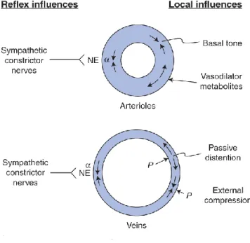
While both arteries and veins are composed of similar cellular components, there are substantial differences. In terms of biological activity of the endothelium arteries and veins differ from each other (de Sousa et al., 2005) (Figure 7.).
25 Figure 7.
Figure from David E. Morman Cardiovascular Physiology 8th Edition
As seen from the figure due to the structural and hemodynamical differences between arteries and veins, there are differences in regulations as well.
During experiments regarding arterial and venous biomechanics usually the endothelium-dependent vasodilator response is also tested (Haas et al., 2007). This response may be elicited by infusing substances that promote synthesis or release of NO from the endothelium. Bradykinin is one of these substances. Venous endothelial cells express constitutive B2 kinin receptors. These receptors may be activated by bradykinin amongst other substances. These receptors activate phospholipase C, leading to an increased level of intracellular calcium, which in turn stimulates NO synthesis. Here, the effect of bradykinin enhancing endothelium-dependent vasodilatation through the release of NO (Dachman, Ford, Blaschke, & Hoffman, 1993).
Previous studies where acetylcholine was administered to elicit endothelium- dependent vasodilator response demonstrated that high-dose infusions of acetylcholine may cause vasoconstriction. There has been more than one study, focusing on the dose- dependent and endothelium-dependent dilator effect of acetylcholine in veins. They suggested that vasodilation mediated by acetylcholine in veins occurs only via NO, similarly to arteries (Vallance, Collier, & Moncada, 1989).
26
Several studies have demonstrated that both acetylcholine and bradykinin can be used for evaluation of endothelium-dependent venodilation (Rabelo et al., 2008).
Alpha 1 adrenoreceptors that have been described to be present in several venous segments, such as vena cava, saphenous vein, pulmonary vein etc. promote vasoconstriction (Muramatsu, Ohmura, & Kigoshi, 1995).
II./3. Biomechanical remodeling of resistance arteries in hypertension, sex differences, effects of sex hormones
The effect of hypertension on the geometric and biomechanical parameters of resistance arteries.
Hypertension is a well established risk factor for several cardiovascular diseases.
Clinically hypertension is defined as systolic blood pressure higher than 140 mmHg and/or diastolic pressure higher than 90 mmHg for systemic arteries (Bergan et al., 2008).
The rise in blood pressure affects the structure and morphology of the vessel wall. The mechanical properties and therefore the contractility of the vessels may also be altered (Hayashi & Naiki, 2009)
Small resistance arteries play a key role in the control of blood pressure. These segments are responsible for the most significant decrease of hydrostatic pressure along the circulatory system. Peripheral resistance small arteries are defined as having a lumen diameter of <350 μm and arterioles as having lumen diameter of <100 μm. These two types of vessels are major contributors to total peripheral resistance. According to Poiseuille’s law resistance is inversely proportional to the forth power of the radius, consequently small changes in lumen - either functional or structural - lead to significant alteration in arterial resistance along these vessel segments (Laurent &
Boutouyrie, 2015; Mulvany & Aalkjaer, 1990; Michael John Mulvany et al., 1996;
Rizzoni & Agabiti-Rosei, 2012).
Living tissue may adapt to mechanical demands by altering structure, geometry and biomechanical properties. Mechanical stress is a key factor. In blood vessels the most relevant stresses are: tangential stress induced by blood pressure (circumferential
27
direction), wall shear stress caused by blood flow, and axial stress elicited by axial elongation (Hayashi & Naiki, 2009). These forces will induce the long-range adaptive processes that affect the geometry (lumen size and wall thickness), tissue composition and elasticity of the resistance vessels. In addition, we can not neglect the nonhemodynamical neural and humoral effects, which are also altered in case of hypertension (epinephrine, norepinephrine, angiotensin II, etc.)
Small Artery Remodeling in Essential Hypertension
According to several studies some of the most essential findings in resistance arteries in case of essential hypertension are vasoconstriction, eutrophic remodeling with increased medial thickness: lumen ratio, altered distensibility and a decrease in the vasodilation reserve (Laurent & Boutouyrie, 2015; Mulvany & Aalkjaer, 1990;
Mulvany et al., 1996; Rizzoni & Agabiti-Rosei, 2012)
Increased systemic vascular resistance (SVR), and small artery remodeling are typical findings in established essential hypertension (Eftekhari et al., 2012). It has been described that small artery structure is predictive of cardiovascular events (Mathiassen et al., 2007; Rizzoni et al., 2003). In hypertension, a typical biomechanical manifestation of arterial wall adaptation is hypertrophy. This adaptation has several advantages. First of all, it restores circumferential wall stress (tangential stress) at in vivo operating pressure to a normal value. It also optimizes arterial stiffness (Fridez et al., 2001; Hayashi & Naiki, 2009). Through these mechanisms in hypertension arterial elasticity is gradually optimized at in vivo working pressures. In hypertension, an increase in vascular tone is seen (Fridez et al., 2001; Mulvany & Aalkjaer, 1990).
Eutrophic inward remodeling may be typical in small arteries (Mulvany, 2008; Mulvany
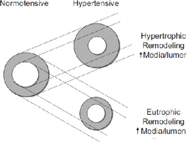
& Aalkjaer, 1990, Intengan et al. 1999; Rizzoni & Agabiti-Rosei, 2012). When inward eutrophic remodeling occurs, a thickening of the media, a reduction in lumen and outer diameter, and increased medial thickness: lumen ratio are seen. This occurs without significant alteration to the overall amount of wall tissue or media cross-sectional area (Mulvany et al., 1996; Rizzoni & Agabiti-Rosei, 2012)(Figure 8.).
28 Figure 8.
Figure from (Schiffrin, 2004)
The cross-sectional area of the media, will remain normal in eutrophic remodeling.
However in hypertrophic remodeling. the cross-sectional area of the media will be increased. The lumen diameter is decreased in remodeled small arteries, and the exterior diameter may be reduced in eutrophic remodeling.
In eutrophic remodeling, the outer diameter and lumen are both reduced, however the cross-section area of the media is maintained. Although the media cross- section does not undergo hpyertrophy, the smooth muscle cells themselves rearrange around a smaller lumen. This in turn leads to an increase in media width. Because the lumen diameter is reduced, the media–lumen ratio is increased, which should not be confused with hypertrophy (Schiffrin, 2004).
According to the Laplace-Frank equation (circumferential wall stress, σ=MBP×R/h, where MBP is mean BP, R is the radius, and h is the wall thickness), if the same amount of wall material is organized around a smaller lumen without net cell growth, it is an effective way to normalize circumferential wall stress.
In contrast to some types of secondary hypertension, where hypertrophic remodeling may be seen, we see inward eutrophic remodeling in the early stages of essential hypertension (Rizzoni et al., 2000). As essential hypertension reaches later stages, it has been theorized that eutrophic remodeling may turn into hypertrophic (Mulvany et al., 1996) In essential hypertension, inward eutrophic remodeling may
29
present a protective mechanism against the rise in blood pressure, thereby preventing the rise in circumferential wall stress at the level of arterioles and capillaries which are ill equipped to withstand such loads. Another typical finding in essential hypertension is a decrease in the vasodilation reserve (the ability to increase blood flow with maximal vasodilatation) (Laurent & Boutouyrie, 2015).
Vascular smooth muscle cells are activated by hypertension (Hayashi & Naiki, 2009). Smooth muscle cells play a role in vascular remodeling in hypertension. Along with collagen and elastin, smooth muscle cells are cardinal structural elements in the vascular wall, and may therefore be essential factors in hypertensive remodeling. The changes in vascular tone and contractility seen in hypertension are linked to smooth muscle cell activity. Vascular smooth muscle cells are regarded as the sensory and effector elements in the adaptation processes, and it is thought that they are activated during the early stages of arterial adaptation to hypertension (Schiffrin, 2004).
Structural changes along the small arteries are accompanied with changes in myogenic tone. At higher pressure loads, increased myogenic tone reduces lumen diameter (Izzard, Rizzoni, Agabiti-Rosei, & Heagerty, 2005). This plays an important role in the autoregulation of blood flow, and the regulation of capillary pressure.
Resistance arteries have the greatest capacity for myogenic response. This response to increased circumferential wall stress is inversely related to the diameter of the vessel (Allen et al., 1997). This way circumferential wall stress is optimized at normal pressure loads (Prewitt et al., 2002). Even though increased wall stress is a stimulus for growth, a myogenic response can prevent this process despite an increased blood pressure. By preventing growth, eutrophic remodeling is maintained. Therefore in case of impaired myogenic tone, as seen in certain types of secondary hypertension, the rise in wall stress would not be counteracted, and thus hypertrophy develops (Schofield, Malik, Izzard, Austin, & Heagerty, 2002). Therefore, the maintained myogenic properties of the vessels play an essential role in determining further structural changes in hypertension. Beside altered myogenic tone, vasoconstriction related to chronic neurohumoral stimuli (contributing to induction of hypertension) may also play a role in the adaptation mechanisms seen in hypertension.
As noninvasive ultrasonographic measurement techniques advanced, it became possible to measure wall thickness, diameter in human patients accurately even in
30
smaller vessels (Hayashi & Naiki, 2009), which is a prerequisite for calculating biomechanical properties. Significantly larger intimal medial thickness was found in hypertensive patients compared to the normotensive group, the internal diameters were less affected. Hypertensive patients had an increased ratio of wall thickness to internal diameter, increased elastic modulus and lower vascular compliance compared to normotensives. There is a decrease in the elastin to collagen ratio which seems to be connected with changes of elastic modulus (Berry & Greenwald, 1976). According to these studies the restoration of normal wall stress by hypertrophy in hypertension in animals may also be observed in human hypertensive patients.
There is also a strain hypothesis regarding hypertensive remodeling (Takamizawa & Hayashi, 1987), according to altered strain is more important than altered stress in inducing the remodeling process (Guo & Kassab, 2004). Remodeling of the ECM adjusting it to elevated myogenic tone of the smooth muscle should explain the morphological hypertensive remodeling according to that theory.
Coronary microvascular dysfunction characterized by abnormal structure and function of the coronaries has been linked to systemic hypertension (Hayashi & Naiki, 2009). Although several studies have investigated these fields, the pathogenic mechanisms underlying coronary microvascular remodeling in hypertension are incompletely understood (Mancini et al., 2013).
The role of inflammation in hypertension
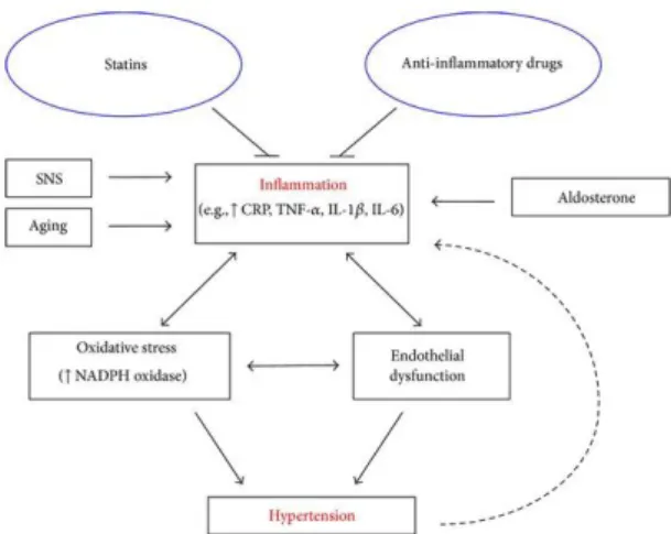
Hypertension is associated with inflammation; however, whether inflammation is a cause or effect of hypertension is not well understood. Human and animal studies have suggested that inflammation leads to the development of hypertension, and oxidative stress and endothelial dysfunction and other potential proinflammatory conditions such as activation of the sympathetic nervous system, aging, and elevated aldosterone may play a role (Figure 9.).
31 Figure 9.
Figure from (Dinh, Drummond, Sobey, & Chrissobolis, 2014).
This figure demonstrates the relationship between inflammation and hypertension and the contributing factors involved. According to this theorem anti-inflammatory drugs and statins may be effective antihypertensive due to their anti-inflammatory properties
CRP can stimulate monocytes to release proinflammatory cytokines such as interleukin-6 (IL-6), interleukin-1 beta (IL-1β), and tumour necrosis factor alpha (TNF- α) and also endothelial cells to express intracellular adhesion molecule (ICAM)-1 and vascular cell adhesion molecule (VCAM)-1. This in effect will further promote inflammation.CRP is considered the inflammatory marker with the strongest association with hypertension. It has been demonstrated in numerous clinical trials that hypertensive patients commonly have increased plasma CRP levels.
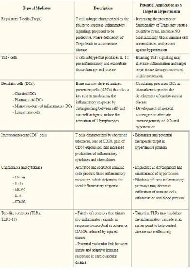
Experimental studies have focused upon the pathophysiological role of individual T-cell subsets, dendritic cells, TNF-α, IL-17, MCP-1, and IL-6, an chemokins in hypertension. They have found that innate and adaptive immunity both play a significant role in the pathogenesis of hypertension. Specific immune cell types, cytokines, toll like receptors, and components of inflammasomes may all pose novel targets for antihypertensive therapy (De Miguel, Rudemiller, Abais, & Mattson, 2015) (Table 1.).
32 Table 1.
Table from (De Miguel et al., 2015).
Table showing the components of the immune system that may play a role in the development of hypertension. Possible mechanisms, and potential targets for therapy.
A single nucleotide polymorphism in the gene SH2B3 encoding the lymphocyte adaptor protein, LNK has been shown to play a role in the development of hypertension.
Loss of LNK leads to profound inflammation in renal and vascular tissues as well as increased IFNγ leading to hypertension and end-organ damage (Dale & Madhur, 2016).
The role of Angiotensin II in vascular remodeling
Angiotensin II is greatly implicated in vascular remodeling of small-resistance arteries. A characteristic change seen in Angiotensin II induced hypertension is
33
increased medial thickness to lumen ratio. This may result from a reduced outer diameter. In this case the lumen is narrowed without net growth (eutrophic remodeling).
The media to lumen ratio may also increase, or a thicker media may be formed (hypertrophic remodeling). Increased arterial stiffness is a hallmark of structural alteration typical of angiotensin II induced hypertension. (Intengan & Schiffrin, 2000;
Neves et al., 2004; Neves, Virdis, & Schiffrin, 2003)
Changes in the extracellular matrix (including collagen type I and III elastin and fibronectin) may also be seen. The overall stiffness of the arterial wall depends on the ratio between distensible components (elastin) and the less distensible components (collagen, fibronectin) (Intengan & Schiffrin, 2000). Elevations in collagen and fibronectin content and decreased elastin have been shown in the media of small arteries from AngII-infused animals. (Brassard, Amiri, & Schiffrin, 2005; Neves et al., 2004;
Neves et al., 2003). It has been demonstrated in immunohistochemical and histochemical assays that angiotensin II has the ability to increase collagen type I and fibronectin content and decrease the amount of elastin in small resistance arteries (Brassard, Amiri, & Schiffrin, 2005; Neves et al., 2003)
This suggests, that in Angiotensin II induced hypertension smooth muscle cells are not the only elements of adaptation, but other cells, such as fibroblasts and/or myofibroblasts may play a role in adaptation. The adaptation of the extracellular matrix is fundamental in stiffening the arterial wall. Collagen plays a key role in this process, as it is found in abundance in the vascular wall. Fibrinectin may accumulate in the media of the small resistance arteries. The process of fibrosis may be influenced by increased activity of local growth factors such as transforming growth factor β, platelet- derived growth factor and other growth factors, such as insulin-like growth factor and basic fibroblast growth factor. (Intengan & Schiffrin, 2001)
Angiotensin II binds to receptors on the vascular smooth muscle cells.
Angiotensin type 1 receptor (AT1 receptor) is a G-protein coupled receptor, and has been described to mediate the cardiovascular effects of angiontensin II (Singh &
Karnik, 2016). This receptor is predominantly expressed in cardiovascular cells, such as vascular smooth muscle cells, and activates various signalling molecules, including G- protein-derived second messengers, protein kinases and small G-proteins (Higuchi et al., 2007). AT1 receptor signalling is mediated through G-proteins, G-protein
34
independent β-arrestin, reactive oxygen species, non-receptor type tyrosine kinases, small G-proteins, transactivation of receptor tyrosine kinases. Furthermore, interacting scaffold, mechanical stress, heterodimerization; and signalling through phosphorylation, desensitization, and internalization may also be involved. It has been described that non-physiologocal activation of AT1 receptor may lead to a number of pathologies including cardiovascular remodeling and hypertrophy, vascular inflammation and atherosclerosis, endothelial dysfunction, oxidative stress, extra cellular matrix deposition, angiogenesis (Singh & Karnik, 2016). The AT1 receptor activates tyrosine kinases, epidermal growth factor receptor, platelet-derived growth factor receptor, and insulin-like growth factor-1 receptor, and nonreceptor tyrosine kinases, such as c-SRC.
As a result, activation of NAD(P)H (nicotinamide adenine dinucleotide phosphate) oxidase may occur, resulting in intracellular generation of reactive oxygen species, which influences redox-sensitive signaling molecules, such as mitogen-activated protein kinases, transcription factors, and matrix metalloproteinases (Schiffrin & Touyz, 2004;
Tabet et al., 2008).The attachment of the extracellular matrix to the smooth muscle may also be affected via the adhesion molecules that mediate the anchoring of vascular smooth muscle cells to ECM components (Intengan et al., 1999).
Sex differences in hypertension
Sex differences in hypertension have been described, and are at least in part thought to be due to the effects of estradiol and testosterone (Barton et al., 2012). One of the effects of testosterone is an increase in angiotensin levels (Yanes et al., 2009). It has been described, that vasoconstrictors and vasodilators have different effects in the the presence of male and female hormones. Chromosome-linked genetic differences have been assumed to be a factor contributing to sex differences in hypertension (Ely et al., 2010). Differences have also been reported in therapy and blood pressure control (Thoenes et al., 2010).
Sex differences in the early stages of hypertensive vascular adaptation in the heart and in the intramural small coronary arteries that are fundamentally responsible for the blood supply of the heart muscle have not been studied thoroughly. The literature is also scarce regarding sex differences in the biomechanical adaptation, and changes in the pharmacologic reactivity of the vascular wall of intramural coronaries in