The Continuity of the Chloroplast in Euglena
JEROME A. SCHIFF AND H. T. EPSTEIN
Department of Biology, Brandeis University, Waltham, Massachusetts
I. Introduction—the Two Aspects of Organelle Continuity
Eukaryotic cells contain several organelles conspicuously delimited from the surrounding cytoplasm at the level of resolution of the light micro- scope. This characteristic separates the vast bulk of living organisms from the prekaryotic group represented by blue-green algae and bacteria, and provides us with the problem of the origins of various organelles such as chloroplasts and mitochondria.
For convenience, two aspects of the problem can be distinguished. T h e first concerns the developmental origins of the organelle in question in- cluding a description of its formation from simpler precursor bodies to- gether with a consideration of the control mechanisms governing the inception and orderly programming of the process. A second aspect con- cerns the continuity of the organelle from generation to generation and raises questions concerning the localization of information for the forma- tion of organelle constituents and the mode of inheritance of this informa- tion during the reproductive cycle. Carried far enough back in time, this aspect includes speculations regarding the evolutionary origins of the organelle.
In discussing these questions, we will be primarily concerned with organelle continuity in Euglena gracilis var. bacillaris and with the chloroplast of this organism. Chloroplast development and inheritance in other organisms, particularly higher plants, has been reviewed extensively (Gibor and Granick, 1964; Granick, 1961, 1963; Rhoades, 1946; Cleland, 1962).
Much of the pioneering work on Euglena has been performed by Pringsheim, Lwoff, Hutner, Provasoli, and their collaborators. Through their efforts, the two strains of Euglena most popular for research were isolated and obtained in axenic culture; defined media were devised which
131
132 JEROME A. SCHIFF AND H. T. EPSTEIN
support luxuriant growth of the organism in a wide p H range (pH 3-9) on a variety of substrates. Their work also emphasized the suitability of Euglena for studies of chloroplast development by demonstrating that the formation of this organelle was light-dependent and that various agents would interfere with the ability of the cells to form plastids. This work is ably summarized in reviews (Hutner and Provasoli, 1951, 1955). T h e preceding applies only to two strains of Euglena gracilis since many other species of Euglena do not posses these characteristics. These two strains are Euglena gracilis var. bacillaris and Euglena gracilis, Z strain. Since the strains are very similar, Euglena will be used to designate them in the sub- sequent discussion whenever there is no reason to think that the findings reported are unique to one strain or the other. A useful Euglena bibliog- raphy will be found in Wolken (1961).
II. The Developmental Aspect of Chloroplast Continuity
A. The Euglena Chloroplast
The fully developed chloroplast characteristic of light-grown cells of Euglena consists of about 12 lamellae each composed of 2 to 4 closely appressed pairs of membranes called discs (Gibbs, 1960; Epstein and Schiff, 1961; Ben-Shaul et al, 1964) as illustrated in Figures 1 and 4. As is characteristic of algal chloroplasts in general, there is a centrally located pyrenoid region which appears more dense in electron micrographs and in which the lamellae appear to be somewhat reduced in thickness. Plates of paramylum, the carbohydrate reserve of Euglena (a ß-l:3 glucan) (Kreger and Meeuse, 1952; Clarke and Stone, 1960) flank the exterior of the pyrenoid and the entire plastid is surrounded by a double membrane.
In the fluorescence microscope (where, under illumination with blue light, the chlorophyll emits a red fluorescence) most of the chlorophyll appears to be distributed in the lamellar regions with very little or no fluorescence within the pyrenoid region. Figure 1 also shows another algal
FIG. 1. Cell of wild-type Euglena grown in the light. (Ben-Shaul et al., 1964.) Abbreviations used here and in subsequent figures: B, basal body; C, chloroplast;
D, disc; E, endosome; ER, endoplasmic reticulum; G, Golgi body; GU, gullet; L, lamella;
M, mitochondrion; N, nucleus; NP, nuclear pore; P, pyrenoid; PE, pellicle; PM, para- mylum; PP, proplastid; PV, pinocytotic vesicle. Marker indicates 1 μ. Here and in sub- sequent figures, the organism is Euglena gracilis var. bacillaris.
FIG. 2. Wild-type Euglena grown in the dark. (Ben-Shaul et al., 1965.) (Key to ab- breviations in legend of Fig. 1.)
' ■ ■ * , * ** : c
"
4m
w^W
755
134 JEROME A. SCHIFF AND H. T. EPSTEIN
characteristic shared by Euglena, the absence of grana regions in the chloroplast characteristic of higher plant chloroplasts.
B. The Light Requirement for Chlorophyll and Chloroplast Formation
We are accustomed to thinking of the formation of chlorophyll and chloroplasts in plants as a light-dependent process but in fact, many or- ganisms form chlorophyll and chloroplasts in the dark. The angiosperms, in general, and Euglena and Ochromonas (Gibbs, 1962) require light for the formation of plastid structures and chlorophyll, but certain gymno- sperms and several algae such as Chlorella and Chlamydomonas do not.
Indeed it may be true that the majority of algae resemble Chlorella and Chlamydomonas in this respect although an experimental test of this requires that the organism in question be capable of organotrophic growth on a reduced carbon source in the dark. Organisms which cannot utilize organic substrates cannot be adequately tested.
Those organisms which require light for chloroplast development and chlorophyll formation all appear to utilize light energy for the con- version of protochlorophyll(ide) a to chlorophyll(ide) a and careful action spectra in higher plants match the absorption spectrum of protochloro- phyll(ide). (In those cases where careful studies have been carried out pro- tochlorophyllide has been shown to be the compound converted photo- chemically to chlorophyllide a. In Euglena, however, and several other organisms, it has not been determined whether protochlorophyll or proto- chlorophyllide is the photochemically reduced precursor. While it is likely that protochlorophyllide will also turn out to be the precursor in these cases, the designation "protochlorophyllide)" will be used to indicate the ambiguity.) This photochemical step has been studied in a holochrome particle extracted from beans, and much of the pioneering work was done in Smith's laboratory (J. H. C. Smith, 1958). While the action spectra measured for Euglena are consistent with those for higher plants, (Nishimura and Huzisige, 1959; Wolken et al., 1955) more data are necessary to be certain. Due to the small amounts of protochlorophyll(ide) in dark-grown organisms, most action spectra have measured the effective- ness of various wavelengths in bringing about an accumulation of chloro-
FIG. 3. Wild-type Euglena grown in the dark. (Liss et al., 1965.) (Key to abbrevia- tions in legend of Fig. 1.)
FIG. 4. Wild-type Euglena grown in the light. (Liss et al, 1965.) (Key to abbrevia- tions in legend of Fig. 1.)
136 J E R O M E A. SCHIFF AND H . T. EPSTEIN
phyll over a period of time. It would be very desirable, however, to have action spectra for the transformation of the initial protochlorophyllide to chlorophyllide since other photochemical transformations (possibly with different action spectra) may subsequently intervene.
From organisms which do not require light for chloroplast and chloro- phyll formation, mutants can be isolated which require light. Thus, mutants of Chlamydomonas and Chlorella are known which are yellow when grown in the dark and become green only on exposure to light (Sager, 1958). If it is assumed that these mutants are the result of a single mutation in each case, it seems likely that the wild-type organisms contain both the standard light-induced protochlorophyll(ide) to chlorophyll(ide) step and an enzyme system for carrying out this transformation in the dark. Mutation would then have eliminated the dark enzyme leaving only the light-induced alternative, and rendering the mutant light-dependent for chloroplast and chlorophyll formation.
C. The Proplastid in Dark-grown Euglena
After many generations of growth in the dark, Euglena cells lose all of their chlorophyll and contain only protochlorophyll(ide) (Nishimura and Huzisige, 1959; Schiff et al, 1961b) as the detectable green pigment. Early electron micrographs revealed that these cells contain proplastids about 1 μ in diameter which seem to lack extensive internal structure (Epstein and Schiff, 1961; Ben-Shaul et al, 1964), (Fig. 2). Improvement of fixation procedures, however, has permitted the detection of one or two internal membranes in many of the proplastids which are occasionally organized for part of their length into a more complex structure (Fig. 3), and particles the size of ribosomes can also be seen (Liss et al., 1965). Rough counts with the fluorescence microscope indicate that there are about 30 proplastids per cell (Epstein and Schiff, 1961; Epstein et al., 1960).
Extensive tubular structures, such as the prolamellar body found in cer- tain proplastids of higher plants (von Wettstein, 1961) seem to be absent (Ben-Shaul et al, 1964).
D. Chloroplast Development in Euglena
When dark-grown cells of Euglena are exposed to light, chloroplast development ensues whether the cells are dividing or not. By keeping the cells under nondividing conditions on a "resting" medium it has been possible to study chloroplast development uncomplicated by cell division (Stern et al, 1964a).
The morphological events of this process may be summarized as follows
THE CONTINUITY OF THE CHLOROPLAST IN EuglenCL 137
(Epstein and Schiff, 1961; Ben-Shaul et al, 1964): (1) Membranes (which later form discs) are invaginated from the inner proplastid membrane; (2) These discs eventually fuse with each other along their length to form lamellae consisting of 2-4 discs; (3) T h e pyrenoid region differentiates after about 18-24 hours of development; (4) The maximum number of lamellae is formed by about 72 hours of development and chloroplast development is complete. The kinetics of this process are summarized in Fig. 5. The rise and fall in the number of discs reflects the initial forma- tion of discs reaching a maximum number, cessation of disc formation, and fusion of discs to form lamellae. Lamella formation is linear from about 14 hours to maturity.
All parameters seem to increase by about a factor of 3 between 10 and 14 hours of development. This has been interpreted as a fusion of three proplastids to form one chloroplast at this point in development, which is consistent with some radiation data to be subsequently presented. The pattern of development in Euglena appears to be quite different from that in higher plants and other algae which have been studied (Gibbs, 1962;
von Wettstein, 1961; Sager, 1958).
E. Developmental Physiology of the Chloroplast in Euglena
1. Pigments. After its initial formation on light induction of dark- grown cells (Nishimura and Huzisige, 1959; Schiff et al., 1961b), chloro- phyll a increases slowly up to about 10 hours (Stern et al., 1964a). After 10 hours (Fig. 6) the rate of chlorophyll a formation increases dramatically and becomes essentially parallel to the rate at which lamellae are being formed. Chlorophyll b is difficult to measure because even in fully devel- oped cells it constitutes only a small proportion of the total chlorophyll.
The dark-grown cell contains appreciable carotenoid (Stern et al., 1964a; Krinsky and Goldsmith, 1960) although the proportion of the total cell content localized in the proplastid is not known. On light induction of chloroplast development the increase of carotenoids parallels that of chlorophyll a (Fig. 6) (Stern et al, 1964a). T h e carotenoids of the dark- grown cell are: ß-carotene, echinenone, euglenanone, cryptoxanthin, zeaxanthin, iram-antheraxanthin, m-antheraxanthin, trollein, and hy- droxyechinenone (Krinsky and Goldsmith, 1960). At about 4 hours of chloroplast development neoxanthin appears for the first time and continues to increase during development (Krinsky et al., 1964). The fact that neoxanthin appears approximately at the inception point of photo- synthetic competence is pregnant with speculative possibilities.
2. Photosynthesis. The first event which can be measured by gas
138 J E R O M E A . S C H I F F AND H . T . EPSTEIN
exchange is the large irreversible increase in the rate of oxygen uptake when dark-grown cells are exposed to light (Schiff, 1963). This un- doubtedly represents an increase in respiration for mobilization of energy for the synthesis of chloroplast constituents. Superimposed on this large irreversible change is a small reversible photo-induced uptake of oxygen.
As development of the chloroplast proceeds, this reversible uptake is
24 36 4 8 TIME IN HOURS
FIG. 5. Kinetics of the formation of discs and lamellae; and plastid lengths and widths during chloroplast development in Euglena. Zero time represents dark-grown cells immediately before induction of development with light. Time is measured from hours of development after dark-grown cells are exposed to light. In all cases, the points represent the means of several observations and the flags show the 95% confidence in- tervals of the means. T h e dotted line was fitted by least squares to the linear portion of lamella development. (Ben-Shaul et al., 1964.)
THE CONTINUITY OF THE CHLOROPLAST IN Euglena 139 gradually compensated until by about 4 hours of development it has
become balanced by photo-induced oxygen evolution. From this point onwards there is an increasing net photosynthetic oxygen evolution (Fig. 6). The inception point for photosynthesis, therefore, can be assumed to be somewhat prior to 4 hours of development. Photosynthetic carbon dioxide fixation also becomes apparent at about 6 hours of development (Stern et al., 1964a). By 15 hours (Fig. 6) of development the photo- synthetic quotient is 1.0 and remains at this value during the remainder
x
a. o
Û UJ
x
LL 3 0 -
25-
20-
15-
10-
5- 24-
| 20-
51 6.
_l _l UJ | 2 -
CO O 8-
4-
• C 02 o 02
* CHLOROPHYLL
* CAR0TEN0ID
L·./
• / A
4/
A · 9 m
ξ/^** Α
/ »o
< ■>
-15 X I-
°
33
■12 O X -<
-9 Γ" Γ
o m
■σ ta -3 ^
Ci >
3J o
H m z o o h2 r
0 20 40 60 80 100 TIME AT 100-150 FC. (HR)
FIG. 6. Kinetics of the appearance of chlorophyll, carotenoids, photosynthetic oxygen evolution, and photosynthetic carbon dioxide fixation during chloroplast development in Euglena. As in Fig. 5 (data on comparable cells), zero time represents measurements on dark-grown cells; time is measured from inception of light-induced chloroplast development. (Stern et al., 1964a.)
of development. In general, the rate of photosynthesis during develop- ment parallels the formation of pigments and lamellae (Stern et al., 1964a) (Figs. 5 and 6). The optimum light intensity for development in Euglena is about 100 ft-c; higher or lower intensities yield lower amounts of chlorophyll. Low intensities also lead to abnormal develpment (Stern et al., 1964b; Ben-Shaul et al., 1964).
3. Protein Synthesis. Since chloroplast development takes place nor- mally on a medium devoid of nitrogen and carbon sources, it is of interest to know whether the formation of the chloroplast involves de novo protein synthesis or, alternatively, whether the proteins of the chloroplasts already exist in the dark-grown cells and are merely packaged
140 J E R O M E A. SCHIFF AND H . T . EPSTEIN
into chloroplasts on light induction. Several enzyme activities have been shown to be associated with the chloroplast fraction of light-grown cells (Smillie, 1963).
One way of attacking the problem is to measure enzyme activities during development. TPN-Triose phosphate dehydrogenase (Fuller and Gibbs, 1959; Brawerman and Königsberg, 1960), TPN-transhydrogenase (Lazzarini and Woodruff, 1964; Lazzarini and San Pietro, 1963), and ribulose diphosphate carboxylase (Fuller and Gibbs, 1959) all appear during development. By spectroscopic determination, it has been possible to show that cytochrome 552, the chloroplast-localized cytochrome of Euglena is formed during development (Nishimura, 1959; Wolken and Gross, 1963; Perini et al., 1964a,b) at a rate which maintains the ratio of cytochrome to chlorophyll at 1:400, the value characteristic of the mature chloroplast (Perini et al., 1964b). This suggests that photosynthetic units are being constructed and completed as individual units during develop- ment. The number of chlorophyll molecules per cytochrome is consistent with the currently accepted size of the photosynthetic unit.
Another approach involves the preparation of antibodies to the proteins of fully mature chloroplasts by injecting acetone powders of purified chloroplasts into rabbits and isolating the antiserum (Lewis et al., 1965). It is then possible to detect a number of antigen-antibody reactions by means of the agar diffusion methods developed by Ouchterlony. Using these methods, investigations have shown that there is a progressive in- crease in the number of antigens in the cell which react with the anti- chloroplast antibodies as chloroplast development proceeds. These results suggest that new proteins are being formed during chloroplast develop- ment (Lewis et al., 1965). Considered together, this evidence suggests that the induction of chloroplast development by light resembles a mass induction of adaptive enzymes. Many new proteins and enzyme activities appear during development and since this occurs on a medium lacking carbon and nitrogen, these molecules must be synthesized either from internal pools of intermediates or from the breakdown of existing macro- molecules.
4. RNA and Ribosomes during Development. Extensive work on this problem has been performed using the Z strain (Smillie and Krotkov, 1960). Previous findings had indicated that exposure of dark-grown organisms to light brings about synthesis of chloroplast proteins and an increase and change in cellular RNA (Brawerman et al., 1962a; A. O.
Pogo et al., 1962). Further explorations showed that the chloroplasts have distinctive ribosomes (Brawerman, 1963) which can be distinguished from
THE CONTINUITY OF THE CHLOROPLAST IN Euglena 141 their cytoplasmic counterparts on the basis of base composition and sedi-
mentation characteristics. It was further shown that intact chloroplasts from Euglena can incorporate amino acids into protein (Eisenstadt and Brawerman, 1964a,b). Ribosomes isolated from both chloroplasts and cytoplasm could incorporate amino acids into protein when provided with necessary cofactors and intermediates (Eisenstadt and Brawerman, 1964a,b). The evidence suggests that both the cytoplasm and chloroplasts of Euglena have protein synthesizing systems of the usual type found in other systems.
5. Miscellaneous Compounds. Differences in lipid content and type of lipid have been found between light-grown and dark-grown cells (Rosen- berg, 1963). a-Linolenic acid seems to be characteristic of the Euglena chloroplast since it is found in that fraction of the light-grown cells, but is absent or present only in a very small amount in dark-grown cells and in cells which cannot form plastid structures (Erwin and Bloch, 1962).
Ergosterol, on the other hand, appears not to be chloroplast-associated since it is found in comparable amounts in dark-grown, light-grown, and mutant cells (Stern et al., 1960). As in other systems, iron is necessary for chloroplast development and iron deficiency can impede chlorophyll formation (Price and Carell, 1964). Aminotriazole inhibits chlorophyll formation in Euglena (Aaronson and Scher, 1960). Utilizable carbon sources appear to repress chloroplast development to some extent (App and Jagendorf, 1963).
6. Control Mechanisms. Possible control mechanisms for chloroplast development will be considered in the last section of this paper.
F. The Return of the Chloroplast to the Proplastid Condition
This is a process most easily studied in unicellular organisms since in multicellular organisms the plastids exist in a highly determinate structure, the leaf. In Euglena, however, the chloroplast is capable of returning to the proplastid condition on dark adaptation (Ben-Shaul et al., 1965). In dividing cells, lamellae dissociate into discs which are progressively lost from the plastids at a rate of about 0.3 per generation, somewhat less than the rate predicted from simple dilution of chloroplast constituents among daughter cells (0.5 per generation). Chlorophyll, how- ever, is initially lost at a rate of about 0.5 per generation. This observation suggests that when the light is turned off, chlorophyll synthesis stops immediately (as required by the known light requirement for the proto- chlorophyll(ide) to chlorophyll (ide) step) but that some discs and lamellae
142 J E R O M E A. S C H I F F AND H . T . EPSTEIN
continue to be made at a low rate. This evidence leads to the interpreta- tion that synthesis of the messenger RNA for the production of the con- stituents of these structures may be repressed in darkness, but that the messenger has a sufficiently long lifetime to persist for a generation or two to permit the synthesis of constituents at a diminishing rate as the avail- able messenger becomes diluted among daughter cells. After about 144 hours of darkness (8 generations) the chloroplast has regressed all the way to the proplastid condition (Ben-Shaul et al., 1965).
Under nondividing conditions a different situation prevails (Ben-Shaul et al., 1965). There is virtually no loss of structure over the course of 144 hours even though about 38% of the chlorophyll has been lost completely and another 50% has been converted to pheophytin (see Greenblatt and Schiff, 1959; Brown, 1963). This suggests that chlorophyll per se is not a determinant of the lamellar structures, a conclusion also reached by other workers with different material (von Wettstein, 1961).
Thus, chloroplast development from the proplastid on light induction takes place in both dividing and nondividing cells in an identical manner.
The reverse process, conversion of chloroplasts to proplastids in darkness, takes place in dividing cells but not in nondividing cells. An explanation of these differences can be sought in control mechanisms which adapt Euglena to its ecological situation. The organism is a facultative photo- troph or organotroph growing equally well by photosynthetic fixation of carbon dioxide or at the expense of reduced organic compounds in the medium. The dark-grown organisms containing only proplastids can live and multiply if a reduced carbon source is available to them. If no carbon source is available they will cease to divide but will live for quite a while.
It is of great adaptive advantage for the organism, under these conditions, to be able to form chloroplasts as soon as light is available and to adopt a phototrophic mode of existence. Even in the presence of organic com- pounds, when the organism can divide, photosynthesis might still be a more efficient method of energy utilization. Thus, the organism is capable of forming chloroplasts either under dividing or nondividing conditions.
Consider now the organism with fully developed chloroplasts. If light becomes limited two alternatives are available. If the medium is devoid of reduced carbon sources and growth is impossible in darkness the organism maintains its plastids which would be advantageous at the first reappear- ance of light. Should the medium contain reduced sources of carbon the organism divides and rapidly loses the excess baggage of the chloroplast, by returning them to the proplastid condition, while it exists as an efficient organotroph. During the course of evolution control mechanisms
THE CONTINUITY OF THE CHLOROPLAST IN Ellglend 143
have apparently been selected in Euglena which efficiently adapt it to prevailing environmental conditions (Ben-Shaul et al., 1965).
III. T h e Replicative Aspect of Chloroplast Continuity A. Definition of the Problem
On the basis of available information from many organisms, it is possible to make some general models of chloroplast inheritance. Figure 7 shows three extremes for the purposes of discussion. In the first alternative information for the construction of a plastid resides in the nucleus which
I 2 3 FIG. 7. Simplified hypothetical schemes for possible genetic interactions between organelles. In all cases, the circular structure represents the nucleus and the ellipse depicts the chloroplast. In alternative 1, an informational unit in the nucleus codes for the proteins of the entire cell including the chloroplast. Alternative 3 represents the other extreme in which the nucleus and chloroplast have independent informational units, the nuclear unit codes for generalized cell protein, and the chloroplast unit de- termines chloroplast proteins. Alternative 2 is necessitated by genetic studies with higher plants and shows two additional modes of interaction. T h e nucleus may code for a protein(s) which manufacture nutrients (X) required by the chloroplast. Alterna- tively, the nucleus may manufacture a mutagen (M) which irreversibly mutates the chloroplast informational unit.
codes for production of everything else in the cell as well. This model predicts that the chloroplast (or proplastid, or any other organelle) is constructed de novo in each generation from information supplied by the nucleus. The large numbers of mutations in maize, barley, and other organisms which affect chloroplast phenotypes and which behave in a perfectly Mendelian fashion can be used in support of this model. As we will see later, however, other interpretations of this data are possible.
The third alternative visualizes independent informational units in the nucleus and in the plastid. The unit in the nucleus codes for proteins produced in the nucleus and cytoplasm exclusive of the organelle in question. The organelle itself contains an informational unit which codes for its own proteins. When the cell replicates, the two informational units
144 J E R O M E A. S C H I F F AND H . T. EPSTEIN
are replicated independently leading to the possibility of autonomous chloroplast division.
In between these two extremes of interpretation are the possibilities for genetic interaction between the nucleus and the organelle shown in the second alternative of Fig. 7. These possibilities have been suggested by experiments with higher plants. Rhoades (1946) discovered a mutant affecting chloroplast phenotype in maize which he called iojap. The iojap gene is chromosomal and behaves in a Mendelian manner. Plants homo- zygous for the mutant gene produce abnormal chloroplasts. These ab- normalities persist and are perpetuated, however, when the chloroplasts are crossed back into plants having a normal genetic constitution.
Rhoades suggested, therefore, that the nucleus and plastid might have different genomes but that a mutant gene in the nucleus could produce a mutagen which irreversibly mutated the genome of the plastids which then continue to replicate the abnormality even after the nuclear constitu- tion was returned to normal. Similar and even more complex interactions have been found in other plants, particularly Oenothera (Cleland, 1962).
Nuclear mutations affecting chloroplast phenotypes but which are en- tirely normal in Mendelian behavior (cited above in connection with alternative 1) can also be reinterpreted here. It is possible that the nucleus and plastid have independent genomes but that during the course of evolution the plastid has become nutritionally dependent on the rest of the cell for one or more metabolites (represented by "X" in alternative 2).
A nuclear mutation which prevented the formation of these nutrients would lead to chloroplast abnormalities even though there was no direct informational dependence of the chloroplast on the nucleus.
In any case, the weight of the evidence seems to suggest separate genomes in nucleus and plastid. The rest of this paper will be concerned with evidence that the behavior of the Euglena system is consistent with this interpretation.
B. Cytological Evidence for Chloroplast Division
The fact that chloroplasts of algae divide has been known for many years and is well documented (Bold, 1951). In Euglena deses, for example, Gojdics was able to show that the cell divided first, apportioning the chloroplast complement approximately equally to the two daughter cells (Gojdics, 1934). After cell division was completed each chloroplast divided to restore the original plastid complement. An electron micrograph of a dividing chloroplast in Euglena gracilis var. bacillaris is shown in Fig. 8.
Since the chloroplast returns to the proplastid condition in this organism,
THE CONTINUITY OF THE CHLOROPLAST IN EugleYld 145
and the number of proplastids remains approximately constant from gener- ation to generation in the dark and since the dark-grown cells are always capable of chloroplast formation, it seems reasonable to assume that the proplastids in this species are also capable of division.
FIG. 8. Wild-type Euglena grown in the light in which a dividing chloroplast may be seen.
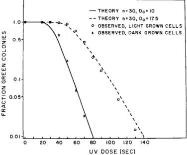
C. Blockage of Green Colony-Forming Ability by Ultraviolet Light If light-grown or dark-grown cells of Euglena gracilis var. bacillaris are exposed to ultraviolet light and are plated under nonphotoreactivating conditions, but with light induction to permit chloroplast formation, the number of green colonies formed decreases as the ultraviolet dose is increased (Pringsheim, 1958; Lyman et al, 1959, 1961). Increasing num- bers of nongreen colonies replace the green colonies which are lost (Fig. 9).
Thus, ultraviolet light brings about a loss of the ability to become green in the progeny of cells which had received the radiation. It is possible to bring about 100% conversion of the progeny to the nongreen condition at ultraviolet doses which are too low to have any effect whatsoever on
146 J E R O M E A. S C H I F F AND H . T. EPSTEIN
viability. Loss of viability begins at doses 10 times greater than the maxi- mum doses required for inactivation of green colony-forming ability (compare Figs. 10 and 12) (Lyman et al., 1961; Hill et al., 1965a).
The first question which arose concerned the numbers of ultraviolet- sensitive entities controlling this process. Target analysis of the inactiva- tion curves revealed that the curves for dark-grown and light-grown cells both fitted multiplicities of about 30 (Fig. 9) (Lyman et al, 1961). This
— THEORY n = 3 0 , D0= 10 - - T H E O R Y n = 3 0 , D0= I 7 . 5
o OBSERVED, LIGHT GROWN C E L L S x OBSERVED, DARK GROWN C E L L S Z o
_J o o z
UJ LU
a:
0 . 0 5 H
o.oi
8 0 100 120 140
UV DOSE (SEC)
FIG. 9. Inactivation of green colony-forming ability in Euglena by ultraviolet light.
T h e points represent experimental findings and the curves were calculated from target theory, multiplicity (w) of 30. DQ is the dose required for a single inactivation. Light- grown and dark-grown cells show the same multiplicity but differ in DQ. (Lyman et al, 1961.)
has since been verified by computer analysis of many experiments (Hill et al., 1965a). This number agreed very nicely with the number of proplastids in the dark-grown cells estimated by fluorescence microscopy.
The fact that light-grown cells having about 10 chloroplasts also showed ultraviolet multiplicities of 30 led us to the hypothesis that in the forma- tion of 10 chloroplasts from 30 proplastids during light-induced develop- ment, proplastids fused in threes to form single chloroplasts conserving the ultraviolet-sensitive entities present in each proplastid. Evidence consist- ent with this interpretation was presented above in the discussion of
THE CONTINUITY OF THE CHLOROPLAST IN EugleUd 147
chloroplast development. On the basis of these facts and the lack of lethality in this ultraviolet range, we suggested that the ultraviolet-sensi- tive sites were cytoplasmic (Lyman et al., 1961). Gibor and Granick (1962b) confirmed this by elegant experiments with an ultraviolet micro- beam and were able to show that high doses of ultraviolet delivered selectively to the nucleus killed the cells, but low doses delivered to the surrounding cytoplasm, with the nucleus shielded, reproduced the phenom- ena we had described. It appears likely then, that the ultraviolet-sensi-
10;
O P 1.0!
(J <
U-
z
I 0I ;
ID
0.01-
0 10 2 0 30 4 0 5 0 UV DOSE (MIN)
FIG. 10. Ultraviolet inactivation and photoreactivation of viability (colony-forming ability) in Euglena. The extrapolations indicate a multiplicity of about 8 for inactiva- tion in this particular experiment. (Hill et al., 1965a.)
tive entities affecting green colony-forming ability are localized in the plastids themselves.
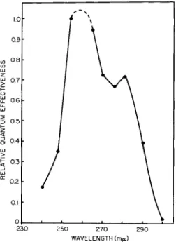
The second question concerned the chemical nature of the ultraviolet- sensitive chromophore. An action spectrum for the ultraviolet inactiva- tion revealed that the absorbing chromophore had peaks in the regions of 260 and 280 ιημ suggesting the participation of a nucleoprotein (Fig. 11) (Lyman et al., 1961).
As is true of ultraviolet inactivation in many other systems, the inactiva- tion of green colony-forming ability in Euglena can be reversed by treat- ment with long wavelength ultraviolet and blue light if given soon after
w \ \ \ \
\ \ DOSE RATE 1.3 ergs /mm 2/ s e c
\ v o PH0T0REACTIVÄTED
• N0NPH0T0REACTIVATED
148 J E R O M E A. SCHIFF AND H. T. EPSTEIN
inactivating ultraviolet (Schiff et al, 1961a). This photoreactivation of green colony-forming ability can result in 100% reversal in Euglena (Fig.
12). This is a very high efficiency when compared with other systems (Jagger> 1958), and with the photoreactivation of viability in Euglena itself (Fig. 10) (Hill et al, 1965a), and suggests that processes resembling multiplicity reactivation in bacteriophages may be occurring, resulting in cooperation among damaged entities in a single cell. This interpretation
i.o 0.9 en 0.8
Ixl
ω 0.7 F o iÜ 0.6 u_ liJ
I 0.5
I -
<
8 0.4
LÜ >
£ 0.3
_J UJ K 0.2
0.1 0,
230 250 270 290 WAVELENGTH (m/i)
FIG. 11. Action spectrum for ultraviolet inactivation of green colony-forming ability in Euglena. (Lyman et al, 1961.)
and others are discussed fully elsewhere (Hill et al, 1965a,b,c). (For some anomalies in the Z strain, see Cook, 1963.)
Figure 10 also shows that multiplicities for inactivation of viability in Euglena are of the order of four to eight, consistent with other unpub- lished data from our laboratory using X rays. This suggests that the nuclear chromosomal complement in our strain of Euglena gracilis var.
bacillaris is polyploid and helps to explain why the plastid system is so much more sensitive to ultraviolet than is viability.
The action spectrum for photoreactivation of green colony-forming ability in Euglena (Fig. 13) (Schiff et al, 1961a) is very similar to that
20 40 SECONDS OF UV
FIG. 12. Ultraviolet inactivation and photoreactivation of green colony-forming ability in light-grown cells of Euglena (lower curves). T h e upper part of the figure shows that the ultraviolet doses employed have no effect on cell viability as measured by viable cell numbers estimated from total colony count ("cell no." in upper part of figure).
(Lyman et al., 1961.)
X- COLI (J.andL.) o- EUGLENA
330 350 370 390 410 430 450 470 WAVELENGTH (m/*)
FIG. 13. Action spectrum for photoreactivation of green colony-forming ability in Euglena compared with photoreactivation in Escherichia colt measured by Jagger and Latarjet (1956). T h e rest of the visible spectrum (green to red) is completely inactive.
(Schiff et al, 1961a.)
149
150 JEROME A. SCHIFF AND H. T. EPSTEIN
described for photoreactivation of viability of E. coli and of photoreactiva- tion of ultraviolet-inactivated bacteriophage T2 in the same organism (Keiner, 1951; Jagger and Latarjet, 1956). Since this system is known to involve the inactivation and repair of DNA it became possible to consider the possibility of plastid-localized DNA nucleoproteins which control the ability of the cells to produce green colonies.
It became important to know whether ultraviolet acted by preventing the replication of plastid entities or whether it merely prevented the
FIG. 14a. Y3BUD, a yellow ultraviolet-induced mutant of Euglena grown in the dark.
Note investment of pellicle by the endoplasmic reticulum and its continuation centrally.
(Liss et al., 1965.) (Key to abbreviations in legend of Fig. 1.)
FIG. 14b. Y3BUD, a yellow ultraviolet-induced mutant of Euglena grown in the light. Note connection of endoplasmic reticulum and nuclear membrane, and endo- plasmic reticulum investment of pellicles of the exterior and of the gullet. (Liss et al., 1965.) (Key to abbreviations in legend of Fig. 1.)
FIG. 14C. W3BUL, a white ultraviolet-induced mutant of Euglena grown in the light.
Pinocytotic vesicles can be seen in association with the gullet. T h e questionable struc- tures are believed (on the basis of many observations) to be either abnormal mito- chondria or amyloplasts. (Liss et al., 1965.) (Key to abbreviations in legend of Fig. 1.)
152 J E R O M E A. SCHIFF AND H . T. EPSTEIN
development of proplastids into chloroplasts. An experiment was devised in which dark-grown cells were irradiated with a dose of ultraviolet which would produce 100% inactivation of green colony-forming ability when plated under nonphotoreactivating conditions. An aliquot of these cells was placed on a resting medium which prevented cell division and the cells were exposed to red light which induces chloroplast and chlorophyll formation without causing photoreactivation. Fluorescence microscopy revealed that all of the irradiated cells produced normal chlorophyll and chloroplasts indicating that chloroplast development was not blocked by ultraviolet. When the cells which had formed chloroplasts were plated however, they formed 100% nongreen colonies indicating that ultraviolet blocks the replication of chloroplast-forming entities at the time of cell division (Schiff et al., 1961b).
The experiments with ultraviolet and photoreactivation lead to the assumption that the dark-grown cells contain about 30 DNA-protein entities localized in the approximately 30 proplastids. On light induction, the 30 proplastids develop into 10 chloroplasts by fusion in threes with the conservation of the 30 DNA-protein entities now localized 3 to a chloroplast. Irradiation of either the dark-grown or light-grown cells with ultraviolet results in blockage of either proplastid or chloroplast replica- tion, respectively, at the time of cell division. This would predict then, that ultraviolet inactivation and loss of green colony-forming ability by a cell should be synonymous with the loss of that cell's plastids and their contained DNA entities.
D. Cytology of Ultraviolet Matant Cells
Cells incapable of chloroplast and chlorophyll production produced through ultraviolet treatment have never reverted to chloroplast-forming competence in the 8 years these cells have been carried in culture. In addition, fluorescence microscopy reveals no red-fluorescing structures of any kind indicating the absence of protochlorophyll(ide) as well.
T o confirm the absence of plastid structures, an extensive program of
FIG. 15. W3BUL, a white ultraviolet-induced mutant of Euglena grown in the dark.
Structures marked with question marks are unknown but do not appear (on the basis of many observations) to be proplastids. (Liss et al., 1965.) (Key to abbreviations in legend of Fig. 1.)
FIG. 16. W3BUL, a white ultraviolet-induced mutant of Euglena grown in the light.
Note diversity of mitochondrial morphology, nuclear pores, and the absence of plastid- related structures. (Liss et al., 1965.) (Key to abbreviations in legend of Fig. 1.)
153
JEROME A. SCHIFF AND H. T. EPSTEIN
electron microscopy of Euglena mutants was undertaken. Improvement of fixation and embedding procedures (Liss et al., 1965) now results in an image of proplastids and chloroplasts which stand out clearly and show enough individuality not to be confused with other organelles (Figs. 3 and 4). Other structures improve in resolution as well, including the endoplas- mic reticulum (Fig. 14) (Liss et al., 1965). Using these new methods, we have carefully examined the light-grown cells and dark-grown cells of all of our mutants and find that in the case of chlorophyll-free mutants induced by ultraviolet, streptomycin, and heat, there are no detectable chloroplasts or proplastids (Liss et al., 1965). Representative pictures are shown in Figs. 15-20. Gibor and Granick (1962a) have treated mutants of this type from the Z strain with δ-aminolevulinic acid by freeze thawing the cells in a solution of the compound. They report that red-fluorescing centers can be detected in the cells after this treatment and interpret this to mean that plastids, or remnants of their structure, persist in the mutants. (See also Siegesmund et al., 1962, and Moriber et al., 1963.) No emission spectra are reported, however, for these red-fluorescing centers and it remains possible that they are mitochondrial sites of protoporphyrin synthesis for heme and cytochrome production rather than centers which form magnesium-containing tetrapyrroles, since both classes of compounds fluoresce red.
On the basis of our own work cited above and to be detailed below, we consider it unlikely that plastid or proplastid structures persist in clones of certain mutants of bacillaris induced with ultraviolet, heat, or strepto- mycin.
In addition to these mutants induced by ultraviolet, streptomycin, or heat which lack the ability to form any plastid structures or chlorophyll, others have been isolated which are blocked at some stage of development.
All of these have proplastids in the dark (like wild type, Fig. 4) but display interesting abnormalities which should be of considerable use in studying the physiological basis of development (Liss et al., 1965). For example, pale-green mutants (Pj) have been isolated with lack patches of lamellae and make less chlorophyll than wild type (Figs. 21 and 22) (Stern et al.,
FIG. 17. W1()BSmL, a white streptomycin-induced mutant of Euglena grown in the dark. Note absence of plastid-related structures and the extensive smooth endoplasmic reticulum. (Liss et al., 1965.) (Key to abbreviations in legend of Fig. 1.)
FIG. 18. W10BSmL, a white streptomycin-induced mutant of Euglena grown in the light. Note absence of plastid-related structures. (Liss et al., 1965.) (Key to abbrevia- tions in legend of Fig. 1.)
154
ISS
156 JEROME A. SCHIFF AND H. T. EPSTEIN
1964a). Olive-green mutants have abnormally large plastids with discon- tinuous lamellae and many unfused discs as in Ox (Figs. 23 and 24).
Another olive-green mutant, 02 (Figs. 25 and 26) produces structures strangely reminiscent of the grana of higher plant chloroplasts. Yellow mutants have been obtained which are blocked rather early in develop- ment and form only small abnormal plastids. Yx and Y3 (Figs. 27-30) are examples (Stern et al., 1964a).
The mutant cells also display a phenomenon noted for dark-grown cells (Lefort, 1964); there is a great hypertrophy of mitochondrial structures of bewildering diversity. It is possible that the presence or absence of an active chloroplast controls the development of mitochondrial structures for compensatory respiratory activity.
It is also possible that the mutagenic agent used in each of the cases to produce mutants in chloroplast phenotype also produced mutations in some of the mitochondria yielding some abnormal forms. Evidence for mitochondrial DNA will be discussed below.
E. DNA of the Chloroplast and Other Organelles
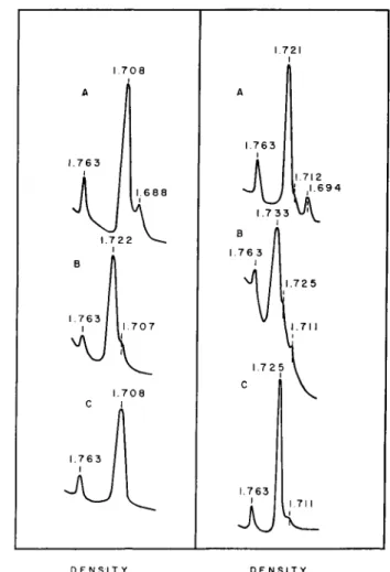
The experiments with ultraviolet light described above suggested very strongly that a plastid-localized species of DNA exists in Euglena. Evi- dence in support of this was forthcoming from a comparison of the band- ing profiles of Euglena DNA in cesium chloride density gradients (Leff et ai, 1963). As may be seen in Fig. 31, light-grown cells display two bands of different densities, one at 1.708 and a satellite band at 1.688. Both of these are double-stranded since heat denaturation brings about the expected shifts to higher densities. Corresponding DNA preparations from ultraviolet mutants (Fig. 31) which lack plastid structures entirely, retained the main band at density 1.708 but showed no detectable satellite DNA. This suggested that the main band (1.708) is probably nuclear DNA while the satellite (1.688) is probably chloroplast-associated DNA.
A similar situation was found in a comparison of Chlamydomonas DNA with its aplastidic counterpart, Polytoma (Fig. 31) (Leff et al., 1963).
FIG. 19. WgBHL, a white heat-induced mutant of Euglena grown in the dark. On the basis of many observations, the structures with question marks do not resemble proplastids; they may be amyloplasts. (Liss et al., 1965.) (Key to abbreviations in legend of Fig. 1.)
FIG. 20. WgBHL, a white heat-induced mutant of Euglena grown in the light. Note absence of plastid-related structures and presence of basal body, connection of endo- plasmic reticulum and nuclear envelope, and Golgi lamellae with dense intercisternal elements. (Liss et al., 1965.) (Key to abbreviations appears in legend of Fig. 1.)
l^»ÊÉÈÈÈÈi^\
~!ÏÊÊ&W£&^U.
M
157
158 J E R O M E A. S C H I F F AND H . T . EPSTEIN
FIG. 21. Proplastids of PjBXL, a pale-green X ray-induced mutant of Euglena grown in the dark. (Liss et al., 1965.) (Key to abbreviations in legend of Fig. 1.)
FIG. 22. Plastids of PjBXL, a pale-green X ray-induced mutant of Euglena grown in the light. (Liss et al., 1965.) (Key to abbreviations in legend of Fig. 1.)
'&'·^ ':
" ^ : Λ « ;
At';·
mM
,*■*;
« * * · & * »
t * :
259
160 JEROME A. SCHIFF AND H. T. EPSTEIN
FIG. 23. Proplastids of CLBS, an olive-green spontaneous mutant of Euglena grown in the dark. (Liss et al., 1965.) (Key to abbreviations in legend of Fig. 1.)
FIG. 24. Plastids of (XBS, an olive-green spontaneous mutant of Euglena grown in the light. Note junctions between plastids on right. (Liss et al., 1965.) (Key to abbrevia- tions in legend of Fig. 1.)
161
162 J E R O M E A. S C H I F F AND H . T. EPSTEIN
FIGS. 25 and 26. Two representative examples of plastids in 02B X , an olive-green X ray-induced mutant of Euglena grown in the light. (Liss et al., 1965.) (Key to abbrevia- tions in the legend of Fig. 1.)
163
164 JEROME A. SCHIFF AND H. T. EPSTEIN
FIG. 27. Proplastids of YjBXD, a yellow X ray-induced mutant of Euglena grown in the dark. (Liss et al., 1965.) (Key to abbreviations in legend of Fig. 1.)
FIGS. 28a and 28b. Representative plastids of Y ^ X D , a yellow X ray-induced mutant of Euglena grown in the light. Note that the limiting membrane of the three plastids in Fig. 28a is continuous. (Liss et al., 1965.) (Key to abbreviations in legend of Fig. 1.)
■.i?&?
165
166 JEROME A. SCHIFF AND H. T. EPSTEIN
FIG. 29. Proplastids of Y3BUD, a yellow ultraviolet-induced mutant of Euglena grown in the dark. (Liss et al., 1965.) (Key to abbreviations in legend of Fig. 1.)
FIG. 30. Plastids of Y3BUD, a yellow ultraviolet-induced mutant of Euglena grown in the light. (Liss et al., 1965.) (Key to abbreviations in legend of Fig. 1.)
ta®*»
PM
767
168 JEROME A. SCHIFF AND H. T. EPSTEIN
>
1.763
1.763
1.763
1.763
XI
D E N S I T Y D E N S I T Y
FIG. 31. Left: Microdensitometer tracings of ultraviolet photographs of DNA from Euglena gracilis var. bacillaris banded in cesium chloride density gradients. A, Light- grown wild-type cells, native DNA (13 μg); B, Light-grown wild-type cells, heat de- natured DNA (13 μg); C, W3BUL, native DNA (11 μg) (mutant lacking chloroplasts produced by treatment with ultraviolet). Right: Microdensitometer tracings of ultra- violet photographs of DNA banded in cesium chloride density gradients. A, Chlamydo- monas Reinhardt Yv native DNA (6.4 μg).: B, Chlamydomonas Reinhardt Υχ, heat- denatured DNA (6.4 μg); C, Polytoma obtusum, native DNA (7 μg).
In all cases, the band of density 1.763 is an added DNA of known density to calibrate the gradient. (Leff et al, 1963.)
THE CONTINUITY OF THE CHLOROPLAST IN Euglena 169
This interpretation was further strengthened by the finding that dark- grown cells in Euglena which contain proplastids also contain the 1.688 satellite band (Fig. 32) (Edelman et al, 1964).
Soon after this initial demonstration of satellite DNA, it became pos- sible to show that the 1.688 satellite was highly enriched in the chloroplast fraction of light-grown cells of both the bacillaris (Fig. 33) (Edelman et al.,
1964; Ray and Hanawalt, 1964) and Z strains (Brawerman and Eisen- stadt, 1964b).
DARK-GROWN CELLS
DENSITY
FIG. 32. Microdensitometer tracing of ultraviolet photographs of DNA from dark- grown wild-type cells of Euglena gracilis var. bacillaris. The band at 1.763 is added DNA of known density to calibrate the gradient. (Edelman et al., 1964.)
It then became apparent that there were actually two DNA satellites in light-grown and dark-grown cells of wild-type Euglena (Fig. 34) (Edelman et al., 1965; Ray and Hanawalt, 1965). The satellite at density 1.691 had been overlooked previously because the concentrations of DNA used in ultracentrifugation had been too low to permit its detection in the ultra- violet mutant cells. Wild-type Euglena cells contain, therefore, three types of DNA; main band DNA (1.707) which is associated with the nucleus, and two satellites: Sc (1.686) associated with the chloroplast fraction and with the ability of the cells to make chloroplasts, and another satellite Sx
(1.691) which is associated with the small particle fraction of the cells containing the mitochondrial cytochromes (Edelman et al, 1965). As may be seen from Fig. 35 (column A, unstarred), all strains of Euglena capable
Ld O
z <
CO or o (Λ CO <
>
170 JEROME A. SCHIFF AND H. T. EPSTEIN
of forming proplastids or a partial chloroplast, contain Sc in addition to Sx and main band. Treatment of any of these strains with ultraviolet and isolation of colorless clones results in a loss of Sc (Fig. 35, column A, starred). All strains incapable of forming plastid structures lack Sc but contain Sx and main band (Fig. 35, column B, unstarred). As expected,
7 0 6
LIGHT-GROWN CELLS
CHLOROPLASTS
DENSITY
FIG. 33. Enrichment of satellite DNA of density 1.685 in the chloroplast fraction of light-grown cells of Euglena gracilis var. bacillaris compared with whole cell DNA. T h e curves show microdensitometer tracings of the ultraviolet absorption photographs of DNA separated in cesium chloride density gradients. In both cases, 1.742 represents added DNA of known density to calibrate the gradient. (Edelman et al., 1964.)
THE CONTINUITY OF THE CHLOROPLAST IN Euglena 171 treatment of these cells with ultraviolet produces no further alteration.
This demonstrates that Sc is definitely correlated with the cells' ability to produce proplastids and chloroplasts and is consistent with the cytological evidence presented before for plastid loss in mutant cells produced by ultraviolet (Edelman et al., 1965).
DENSITY
FIG. 34. Microdensitometer tracing of ultraviolet absorption photograph of DNA from light-grown cells of Euglena gracilis var. hacillaris separated on a cesium chloride density gradient. T h e moderate overloading of DNA in the gradient permits the resolu- tion of two satellites. T h e band at 1.743 is added density standard DNA to calibrate the gradient. T h e overloaded band is main band DNA. (Edelman et al., 1965.)
Table I summarizes the properties of the three types of DNA from Euglena cells. The hacillaris and Z strains are quite similar in densities to those found for main band and Sc. In hacillaris, the base compositions and molecular weights as isolated vary considerably among the three types of DNA.
172 JEROME A. SCHIFF AND H. T. EPSTEIN
Consistent with these findings, bromouracil incorporation by Euglena brings about an increase in plastid mutations (Scher and Collinge, 1965).
Azathymine incorporation affords ultraviolet protection, presumably by reducing the probability of thymine dimer formation (Lyman and Smillie,
1963).
STRAIN
WILD TYPE WILD TYPE*
Y,BXD Y,BXD*
0,BS 0 , B S *
02BX 02B X *
P,BXL P,BXL*
ABSORPTION PHOTOGRAPHS
1.686 1.743 1.691 !
STRAIN
W3BUL W3BUL*
W8BHL W8BHL*
W,0BSmL W,0BSml_*
W3 0B S W3 0B S *
Y2BUL Y2BUL*
ABSORPTION PHOTOGRAPHS
1.686 1.743 1.691 !
FIG. 35. The DNA's of Euglena mutants. Band of density 1.743 is density standard DNA added to calibrate gradient. All strains in column A are capable of developing at least a partial chloroplast and possess both satellite bands, Sx (1.691) and Sc (1.686). All strains in column B are incapable of even partial chloroplast development and contain only satellite band Sx (1.691). All strains in both columns A and B when subjected to treatment with ultraviolet light (indicated by asterisk) yield strains which lack Sc
(1.686) but retain Sx (1.691). (Edelman et al, 1965.)
F. Organelle-Associated DNA in Other Species
Since the original demonstration by Ris and Plaut (1962) of DNA fibrils in chloroplasts, satellite DNA's from many chloroplast-containing species have been described. These are summarized in Table II. As may be seen, the base compositions of satellite DNA's attributed to the chloroplast vary widely from species to species as do the main band
TABLE I
DNA OF Euglena gracilis M
Ci O z
H
5
G H
o
*i H H r>
S r O *
O
*ü r1
>
Source and method Bacillaris (density)
Bacillaris (thermal denaturation) Bacillaris (analysis)
Z strain (density) &
Z strain (analysis)
Z strain (thermal denaturation) &
Main A + T
52 45 47 51 49 47-52
band G + O
48 55 53 49 51 48-53
Sc A + T
74 70 76 76 75 74-79
G + C 26 30 24 24 25 21-26
A + T 69
—
— 67
—
—
Sx G + C
31
—
— 33
—
—
Reference
Molecular weight, as isolated (sedimentation) Denaturation studies Density (gm/cm3)
Edelman et al, 1964, 1965 Edelman et al., 1964 Ray and Hanawalt, 1964 Brawerman and Eisenstadt, 1964b Brawerman and Eisenstadt, 1964b Brawerman and Eisenstadt, 1964b 20-40 X 106 20-40 X 106 2.6-3.6 X 10⁶ Ray and Hanawalt, 1964, 1965 Double-stranded Double-stranded Double-stranded Edelman et al., 1965
1.707 1.686 1.691 See above references a Includes approximately 2.3%
& Calculated from data given in
methyl cytosine (Ray and Hanawalt, 1964; Brawerman et al., 1962b).
reference.
T A B L E I I
D N A O F C H L O R O P L A S T - C O N T A I N I N G SPECIES AND R E L A T E D O R G A N I S M S
vr
Main band" Satellites
Group Species Density
1.707
1.708
1.726(?)
1.723
1.721
1.725
1.716 A + T
52
51
38
36
38
35
43 G + C
48
49
62
64
62
65
57
Density
1.686&
1.684&
1.702&^
1.695e
1.694e
—
1.695&
A + T
74
75
61
64
65
—
64 G + C
26
25
39
36
35
—
36
Density
1.691c
1.692
—
—
1.712
1.711
A + T
69
67
—
—
47
48 G + C
31
33
—
—
53
52
References
T a b l e I
T a b l e I
Sager and Ishida, 1963
Chun et al., 1963
Leff et al., 1963
Leff et al., 1963
Chun et al., 1963;
Iwamura and Kuwashi- ma, 1964;
Iwamura, 1960
w
0 M
>
c/5
n S
>
Ö X H
H
S
2 E u g l e n o p h y t a Euglena gracilis
v a r . bacillaris Euglena gracilis
v a r . Z
C h l o r o p h y t a Chlamydomonas Reinhardt
Chlamydomonas Reinhardt
Chlamydomonas Reinhardt
Polytoma obtusum
Chlorella ellipsoidea