Gender differences in sport-adaptation of intramural coronary resistance arteries
Ph.D. thesis
Marianna Török MD Doctoral School of Clinical Medicine
Semmelweis University
Supervisor: Szabolcs Várbíró, MD, Ph.D.
Official reviewers: Prof. Gábor Pavlik, MD, D.Sc Attila Szendrői, MD, Ph.D
Head of the Complex Examination Committee:
Prof. László Gerő, MD, Ph.D., Ds.C.
Members of the Complex Examination Committee:
Gabriella Lengyel, MD, Ph.D., med. habil.
István Sziller, MD, Ph.D., med. habil.
Budapest 2020
1
1. Introduction
Beneficial effects of regular exercise on the body, especially on the the cardiovascular system are now well established. The risks of sedentary lifestyle such as obesity, diabetes and consequent cardiovascular morbidity and mortality are also known. Sex differences are manifested not only in risk factors of many diseases, illnesses (especially cardiovascular disease), but in health conditions as well. The cardiovascular morbidity and mortality differ in males and females, it is lower in females compared to males until menopause. The study of gender differences in physiological (especially athlete’s heart) and pathological (e.g. heart failure, diabetes) conditions are at the forefront of international research. We have far less knowledge about the sport adaptation of the vascular system than about the sport adaptation of heart. The adaptation of vessels is not uniform in the entire vascular network. There is a significant difference in vascular adaptation depending on the location of and size of the vessels. Vessels, supplying peripheral exercising muscles are affected by different hemodynamic forces than other vessels (e.g. coronary vessels). Different vascular adaptation is expected from peripheral muscle vessels (where flow is increased periodically), from a visceral vessels (in which the flow decreases during sport activities) and from intramural coronary vessels (where the heart muscle is constantly, rhytmically active during exercise). Sport adaptation of the vascular system is created by hemodynamic changes during exercise, such as changes in blood pressure and endothelial shear stress. Although regular sport does not elevate the mean arterial pressure, but systolic pressure will
2
rise in response to physical activity, in case of moderate activity, especially in trained persons, there is hardly any rise in diastolic pressure, even a drop in that parameter can occur.
This results an elevation in pulse pressure. As an effect of increased pulse pressure, the vascular wall tension elevates periodically. The cyclic wall tension and repeated shear stress increase the endothelial nitric oxide synthase (eNOS) expression and eNOS phosphorylation, resulting in increased nitric-oxide-dependent vasodilatation. Regular exercise induces hypertrophy and remodeling of the left ventricular myocardium. The elevated myocardial mass requires increased perfusion, which can be provided only by remodeling of the coronary circulation. This implies that the coronary arteries must also adapt to the changed hemodynamic situation as a result of exercise. Most studies describe functional adaptation on gender differences in sport adaptation of vascular system.
Testosterone and estrogen are also involved in regulating vascular endothelial and smooth muscle cell function.
2. Objectives
The cardiovascular morbidity and mortality differ in males and females, these are lower in females compared to males until menopause. This is clearly due to the protective effect of estrogen. Regular sports also have a protective effect on cardiovascular risk. Long-term regular exercise induces hypertrophy and remodeling of the left ventricular myocardium. Cardiac hypertrophy following intensive sport is a physiological condition. The elevated myocardial mass requires increased perfusion, which will be provided by remodeling of the coronary circulation. The present study investigated whether the resistance coronary arteries are adapted structurally and functionally to long-term intensive
3
exercise during the process in which the ventricular myocardium is transformed into ‘athlete’s heart.’ We aimed to determine the sex differences in the adaptation process of intramural coronary resistance arterioles to long term physical exercise.
Based on these, we aimed to answer the following questions:
1) Are here gender dependent differences in the morphology and function of rat intramural coronary arterioles?
2) Do these characteristics change as a result of intense swim-training?
3) Are there gender differences in sport adaptation?
3. Methods 3.1. Animals
The animals were housed in a room with constant temperature (22 ± 2°C) with a 12-hour light-dark cycle. They were kept on a standard laboratory rat diet provided ad libitum and had free access to water. All procedures and handling of the animals during the study were approved by the Animal Care Committee of Semmelweis University as well as by state authorities (permission number: PEI/001/2374-4/2015). After one week of acclimatization, young adult (12 weeks old) male and female Wistar rats were randomly divided into four groups: male exercised (MEx, n=8), female exercised (FEx, n=8), male sedentary control (MSe, n=8) and female sedentary control (FSe, n=8).
4
3.2. The intensive swim training protocol
The groups in training (MEx and FEx) underwent a graded, intense swimming exercise protocol. Rats were placed in a container of water (separately). The program started with 15 minutes of swimming per day, and the time of exercise was raised every second day by an additional 15 minutes until the duration of swimming reached a total of 200 minutes, which was then maintained throughout the experiment. Trained rats swam for a total of 12 weeks with 5 days of swimming + 2 days of rest per week. The control sedentary groups (MSe and FSe) were put in the water for only 5 minutes daily, 5 days/week, in parallel with the 12-week-long training program of the swimmers.
3.3. Echocardiography
Echocardiographic assessments were performed after completion of the training program under isoflurane anesthesia. Standard two-dimensional short-axis records were acquired (at the mid-papillary level). The stored pictures were analyzed by blinded investigators. The computed parameters were relative heart weight (heart weight/body weight ratio), fractional shortening and ejection fraction.
3.4. In vitro pressure arteriography of intramural coronary arteries
At the end of week 12, under pentobarbital anesthesia (45 mg/kg body weight, given intraperitoneally), the heart was removed, its weight measured. After that the heart was placed in cold Krebs solution (nKR) The composition of the nKR solution was as follows (mM): 119 NaCl, 4.7 KCl, 1.2 NaH2PO4, 1.17 MgSO4, 24 NaHCO3, 2.5 CaCl2, 5.5 glucose and 0.0345 EDTA. The biomechanical and functional properties of the intramural coronary resistance arteries were
5
investigated by pressure microarteriography. The arteriolar segment with a length of approximately 2 mm was placed in a tissue bath (Experimetria LTD, Budapest, Hungary) with a glass bottom, filled with a normal Krebs-Ringer solution (37°C, pH 7.4, 5% CO2, 20% O2 és 75% N2). The arteriolar segment was cannulated at both ends with plastic microcannulas. The segments were pressurized using servo- controlled roller pumps, in order to maintain 50 mmHg intraluminal pressure during incubation, and to change it between 0-150 mmHg. All experiments were performed under no flow condition. Pictures were taken regularly and stored.
The analysis of the pictures was performed with specific image-analyzing software (ScopePhoto). The inner and outer diameters and wall thicknesses were measured. A length calibration was performed with a micrometer etalon (Wild, Heerbrugg, Switzerland).
3.5. Vascular protocols
Resistance sized small arteries, with outer diameter at preparation around 200 micrometer from sedentary and swim- trained male and female rats were taken and incubated in Krebs buffer solution at 50 mmHg of intraluminal pressure for 30 minutes. Resistance-sized arteries develop spontaneous contraction if they are incubated in an oxygenated medium. To induce the initial spontaneous contraction, no contracting agent was added. A pressure diameter curve was then determined by increasing the pressure from 0 to 150 mmHg in 10 mmHg. The steady-state diameter was measured at each step. Thereafter, bradykinin (BK) was added in cumulative concentrations (10-8, 10-7 and 10-6 M, each concentration lasted for 10 minutes), and the diameters were measured. Then, the NO synthase blocker nitro-L-arginine methyl ester hydrochloride (L-NAME) was added (10-5 M) for 20 minutes, and the diameters were measured again. Drugs were washed out, and after a 10-minute rest, the
6
original diameter was restored, and we added U46619, a TxA2
receptor agonist, (at a concentration of 10-7 M) to the bath; the vessel was then incubated for 10 minutes, and the pressure diameter curves were recorded repeatedly. Finally, the segments were incubated for 30 minutes in a buffer without Ca2+ and passive, relaxed pressure diameter curves were recorded repeatedly. The calcium-free Krebs solution contained 92 NaCl, 4.7 KCl, 1.18 NaH2PO4, 20 MgCl2, 1.17 MgSO4, 24 NaHCO3, 5.5 glucose, 2.0 EGTA and 0.025 EDTA.
3.6. Calculations
The wall stress was calculated according to the Laplace-Frank equation and the incremental tangential elastic modulus was calculated based on the formula of RH Cox. The functional parameters were calculated from outer radius. Spontaneous tone and active tone were calculated as follows:
Spontaneous tone (%) = (roCa-free-ronKR)/roCa-free*100
Bradykinin relaxation (%) = (roBK-ronKR)/ ronKR*100
L-NAME effect (%) = (roBK-roL-NAME)/ronKR*100
U46619 contraction (%) = (ronKR-roU46619)/ronKR*100
3.7. Histology studies
The segments used for the biomechanical measurements were removed and placed in 4% formaldehyde for fixation (n=4-4).
After dehydration, they were embedded in paraffin, and 5- micrometer-thick sections were cut. The elastin fibersfibers were stained with resorcin-fuchsin (RF) on the coronary segments. The resorcin-fuchsin-stained sections were analyzed by colorimetric techniques to evaluate the inner elastic membrane. The noncalibrated optical density of specific staining was measured by ImageJ software (NIH, Bethesda, MA, USA). Immunohistochemistry was performed on paraffin
7
embedded tissue sections of coronary arteries of heart against TXA2 receptor using polyclonal rabbit anti-TXA2 receptor.
3.8. Statistical evaluation
Normal distribution was tested with the Shapiro-Wilks method, , a logarithmic transformation of data was used if feasible to achive normal distribution.Data are presented as the mean ± SEM. A two-way analysis of variance (ANOVA) with the factors ‘training’ and ‘sex’ was performed. As a post hoc test, Tukey’s test was used. P <0.05 was accepted as statistically significant difference.
4. Results
4.1. Morphology and function of the heart
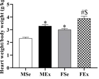
The heart weight/body weight ratio was significantly higher in exercised and sedentary female rats than in exercised and sedentary male animals. Postmortem measured heart weight values in proportion to body weight were higher in both exercised groups (Figure 1). The systolic function of the enlarged ventricle (determined by the ejection fraction and fractional shortening) was significantly elevated in both males and females by long-term intensive swim training (p<0.05, two-way ANOVA, Tukey post-hoc test. Ejection fraction (%):
MSe: 74 ± 1.1 vs. MEx: 80 ± 1.6 vs. FSe: 76 ± 1.0 vs. FEx: 81
± 0.8. Fractional shortening (%): MSe: 44 ± 1.0 vs. MEx: 51 ± 1.7 vs. FSe: 46 ± 0.9 vs. FEx: 51 ± 0.9).
8
Figure 1. Heart weight/body weight ratio
Two-way ANOVA with post hoc Tukey’s test. Values are the means ± SEM. *P < 0.05 vs. male sedentary; #P < 0.05 vs. female sedentary; $P <
0.05 vs. male exercised
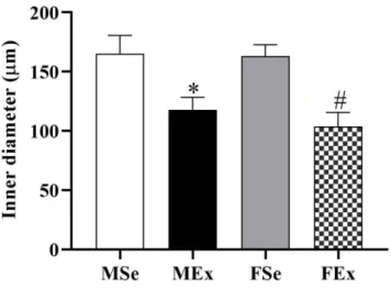
4.2. Morphology and biomechanical parameters of intramural coronary resistance arteries In intensively trained male and female rats, the inner diameters of the vessels were reduced in the relaxed state and the ratio of wall thickness to lumen diameter were elevated in both MEx and FEx animals (Figure 2 and 3). The tangential wall stress was significantly lower in FSe rats than in MSe rats at physiological pressures (at 50 mmHg). This value significantly decreased in the MEx group approaching the level of that in the female animals (p<0.05, two-way ANOVA, Tukey post- hoc test. Tangential wall stress (kPa): MSe: 27± 3.4 vs. MEx:
15 ± 2.7 vs. FSe: 14 ± 1.4 vs. FEx: 12 ± 2.1).
9
Figure 2. Inner diameter
Two-way ANOVA with post hoc Tukey’s test. Values are the means ± SEM. *P < 0.05 vs. male sedentary; #P < 0.05 vs. female sedentary
Figure 3. The ratio of wall thickness to lumen diameter
Two-way ANOVA with post hoc Tukey’s test. Values are the means ± SEM. *P < 0.05 vs. male sedentary; #P < 0.05 vs. female sedentary
10
4.3. Resorcin-fuchsin staining and the incremental elastic moduli
The incremental elastic moduli increased in trained animals of both sexes (p<0.05, two-way ANOVA, Tukey’s post-hoc test.
The incremental elastic moduli (logkPa): MSe: 1.84± 0.17 vs.
MEx: 2.57 ± 0.12 vs. MSe: 2.21 ± 0.15 vs. MEx: 2.81 ± 0.1).
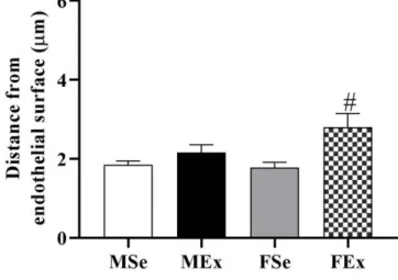
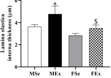
The distance of the lamina elastic interna layer with maximal intensity was located farther away from the endothelial surface in the trained female animals compared to that in the control female animals (Figure 4.). The inner elastic membrane became thicker in the MEx animals than that in the MSe and FEx animals. There seemed to be a tendency towards thickening of the inner elastic lamina in the trained female animals, but the difference did not reach the level of statistical significance (Figure 5). The optical density of noncontractile fibers on resorcin-fuchsin-stained sections was significantly lower in female animals (FSe and FEx) than in MSe rats, and this value was significantly reduced in the MEx group approaching the level of that in the female animals (p<0.05, two-way ANOVA, Tukey post-hoc test. optical density: MSe:
0.29 ± 0.02 vs. MEx: 0.19 ± 0.01 vs. FSe: 0.19 ± 0.01 vs. FEx:
0,23 ± 0.01).
11
Figure 4. Distance from endothelial surface
Two-way ANOVA with post hoc Tukey’s test. Values are the means ± SEM. #P < 0.05 vs. female sedentary
12
Figure 5. Lamina elastica interna thickness
Two-way ANOVA with post hoc Tukey’s test. Values are the means ± SEM. *P < 0.05 vs. male sedentary; $P < 0.05 vs. male exercised
4.4. Vascular reactivity
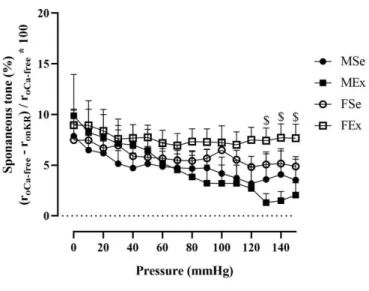
The spontaneous tone was significantly higher in trained female rats than in trained male animals at high pressure (Figure 6). A pronounced difference was found in U46619- induced vasoconstriction, as the contraction of trained female coronary vessels was much higher than that of trained male specimens almost in the entire pressure range (Figure 7).
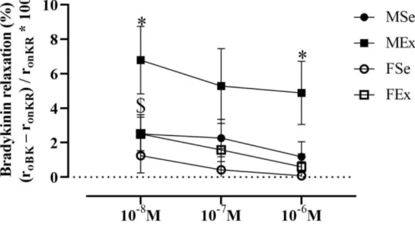
Training resulted in significantly better bradykinin-induced relaxation at 10-8 M and 10-6 concentrations in exercised male rats compared with control males. Bradykinin-induced relaxation was greater in trained males than in trained female rats at 10-8 M (Figure 8). The contraction caused by L-NAME was also higher in trained male rats than in control male and trained female rats (Figure 9).
13
Figure 6. Spontaneous tone
Two-way ANOVA with post hoc Tukey’s test. Values are the means ± SEM. $P < 0.05 vs. male exercised
Figure 7. U46619-induced constriction
Two-way ANOVA with post hoc Tukey’s test. Values are the means ± SEM. $P < 0.05 vs. male exercised
14
Figure 8. Bradykinin relaxation
Two-way ANOVA with post hoc Tukey’s test. Values are the means ± SEM. *P < 0.05 vs. male sedentary; $P < 0.05 vs. male exercised
Figure 9. Effect of L-NAME
Two-way ANOVA with post hoc Tukey’s test. Values are the means ± SEM. *P < 0.05 vs. male sedentary; $P < 0.05 vs. male exercised
15
4.5. TxA2 receptor immunohistochemistry
The percentage of positively stained area of TxA2R was significantly lower in the vessel wall of exercised males compared to exercised females, implying a marked exercised induced sex difference in the protein expression level of TxA2R in coronary arteries (Figure 10).
Figure 10. TxA2R immunohistochemistry
Two-way ANOVA with post hoc Tukey’s test. Values are the means ± SEM. $P < 0.05 vs. male exercised
16
5. Conclusions
In the present study, we aimed to answer the following questions:
1. Is there gender dependent differences of morphology and function in rat intramural coronary arterioles?
Based on our results, there is no significant difference in the morphology and function of intramural resistance coronary arteries in young adult male and female rats. Exercise- independent gender difference was found only in tangential wall stress. It was lower in female animals than in male sedentary animals.
2. Do these characteristics change as a result of intense swim-training?
We found that the intramural coronary resistance arteries of the rats were structurally and functionally transformed as a result of a long-term intensive exercise training program. While the inner diameter of these arterioles decreased, the ratio of the wall thickness to lumen diameter increased, and the incremental elastic moduli increased in trained animals of both sexes. Range of coronary vascular reactivity increased in both exercised male and female rats, but its’ mechanism was different depending on sex.
3. Are there gender differences in sport adaptation?
There was no significant difference in the morphological adaptation of the coronary arteries between the two exercised groups. The tangential wall stress was lower in female animals than in male animals in the sedentary groups, and after exercise, tangential wall stress was reduced in male exercised
17
animals to a level approaching that in female animals. The elevated incremental elastic modulus is due to the increase in the total cross-section of the lamina elastica interna. The lamina elastica remodeling was different in female and male animals in the exercise group. The distance of the layer with maximal intensity was located farther away from the endothelial surface in the trained female animals. The inner elastic membrane became thicker in the MEx animals. There is training-induced remodeling of coronary resistance artery function in both sexes, but in several aspects, this remodeling is different in both sexes. While spontaneous tone and U46619-induced contractility increased in trained female rats, endothelium-dependent dilatation increased in exercise-trained male rats.
18
6. Bibliography of the candidate’s publications Publications related to the thesis:
Torok M, Monori-Kiss A, Pal E, Horvath E, Josvai A, Merkely P, Barta BA, Matyas C, Olah A, Radovits T, Merkely B, Acs N, Nadasy GL, Varbiro S. (2020) Long-term exercise results in morphological and biomechanical changes in coronary resistance arterioles in male and female rats. Biol Sex Differ, 11: 7. IF: 3.267* (D1)
Torok M, Horváth EM, Monori-Kiss A, Pál É, Gerszi D, Merkely P, Sayour AA, Mátyás Cs, Oláh A, Radovits T, Merkely B, Ács N, Nádasy GyL and Várbíró S. (2020) Chronic swimming training resulted in more relaxed coronary arterioles in male and enchanced vasoconstrictor ability in female rats. J Sports Med Phys Fitness, IF: 1,432*
Publications not related to the thesis:
1. Olah A, Matyas C, Kellermayer D, Ruppert M, Barta BA, Sayour AA, Torok M, Koncsos G, Giricz Z, Ferdinandy P, Merkely B, Radovits T. (2019) Sex Differences in Morphological and Functional Aspects of Exercise-Induced Cardiac Hypertrophy in a Rat Model. Front Physiol, 10: 889.
IF: 3.367*
2. Mátyás C, Németh BT, Oláh A, Török M, Ruppert M, Kellermayer D, Barta BA, Szabó G, Kökény G, Horváth EM, Bódi B, Papp Z, Merkely B, Radovits T. (2017) Prevention of the development of heart failure with preserved ejection fraction by the phosphodiesterase-5A inhibitor vardenafil in rats with type 2 diabetes. Eur J Heart Fail, 19: 326-336. IF:
10.683
19
3. Olah A, Kellermayer D, Matyas C, Nemeth BT, Lux A, Szabo L, Torok M, Ruppert M, Meltzer A, Sayour AA, Benke K, Hartyanszky I, Merkely B, Radovits T. (2017) Complete Reversion of Cardiac Functional Adaptation Induced by Exercise Training. Med Sci Sports Exerc, 49: 420-429. IF:
4.291
4. Olah A, Nemeth BT, Matyas C, Hidi L, Lux A, Ruppert M, Kellermayer D, Sayour AA, Szabo L, Torok M, Meltzer A, Geller L, Merkely B, Radovits T. (2016) Physiological and pathological left ventricular hypertrophy of comparable degree is associated with characteristic differences of in vivo hemodynamics. Am J Physiol Heart Circ Physiol, 310: H587- 597. IF: 3.348
5. Kovacs A, Olah A, Lux A, Matyas C, Nemeth BT, Kellermayer D, Ruppert M, Torok M, Szabo L, Meltzer A, Assabiny A, Birtalan E, Merkely B, Radovits T. (2015) Strain and strain rate by speckle-tracking echocardiography correlate with pressure-volume loop-derived contractility indices in a rat model of athlete's heart. Am J Physiol Heart Circ Physiol, 308:
H743-748. IF: 3.324
* expected impact factor value
∑IF: 29.712