ARTICLE
Early Atherosclerosis and Chlamydia Pneumoniae Infection in the Coronary Arteries
Eszter HORTOVÁNYI, György ILLYÉS, Anna KÁDÁR
Semmelweis University, Faculty of Medicine, 2ndDepartment of Pathology, Budapest, Hungary
Introduction
Atherosclerosis has been classified as the disease of the intimal layer of the arteries.13However, there are some new aspects for approaching this problem if it is accepted that the intimal and medial layer of the vessels is a com- plex unit.14,15In the aorta and the left descending coronary artery (LAD) the occurrence of atheroma and the intima/
media ratio increased progressively from 15 to 34 years of age (WHO-PBDAY study).7In young people some arteri- al structural modifications –such as medial hypertrophy – could be a forerunner of atheroma, as medial hypertrophy
Received:Dec 21, 2002;accepted: Febr 10, 2003
Correspondence: Anna KÁDÁR, MD, PhD, DSc, Semmelweis University, Faculty of Medicine, Üllôi út 93., Budapest, 1091 Hun- gary, Tel: (36-1)-215-7300/3400 Fax: (36-1)-215-6921, e-mail: kadann@korb2.sote.hu
In non-atheromatous segments of coronary arteries a sequence of preatherosclerotic changes was iden- tified which consisted of medial thickening fol- lowed by intimal thickening. More recently, Chlamydia pneumoniae seropositivity was associat- ed with enhanced intima-media thickness of arter- ies. In the present study the intimal and medial thickness of coronary artery of young adults were measured, and were correlated with the presence of Chlamydia pneumoniae antigens. Proximal and dis- tal segments of the left anterior descending coro- naries (LAD) obtained at autopsy from young adults (15-34 years) were studied. The thickness and cellular density of the intima and of the media with- out clear-cut atherosclerotic changes were measured by image analysis. The hypertrophy index was cal- culated as the ratio of cell density and the thickness of the respective layer. Atherosclerotic lesions occurring elsewhere in the same coronary were
noted and graded by severity. The presence of Chlamydia pneumoniae verified by immunohisto- chemistry was correlated with the severity of lesions and with the hypertrophy index. In the prox- imal segments, atherosclerosis of LAD was associat- ed with the widening of both the intima and the media of lesion free-sites. In the distal coronary seg- ments the proportion of the intimal thickening had a significant association with atherosclerosis. Com- pared to non-infected arteries, Chlamydia pneumo- niae infection was associated with higher hypertro- phy index in the intima as well as in the media. The rate of Chlamydia pneumoniae positivity increased with the severity of lesions. As a conclusion: in the LAD coronary, the intimal thickening is the main preatherosclerotic change. Chlamydia pneumoniae may favour arterial wall hypertrophy and plays a role in lesion progression. (Pathology Oncology Research Vol 9, No 1, 42–46, 2003)
Keywords: chlamydia pneumoniae, early, atherosclerosis, hypertrophy
was found to be associated significantly with the appear- ance of atheromatous plaques.14The hypertrophic media could be the source of intimal smooth muscle cells.
A key event in the development of atherosclerosis is the accumulation of macrophages in the arterial intima. Its possible connection with a chronic inflammatory process that could thicken the arteries is not well clarified.11For a missing link the infection of the vasculature with Chlamy- dia pneumoniae is challenging, as it is more frequently found in atherosclerotic, than in unchanged arteries sup- porting its role in the pathogenesis of atherosclerosis.2,8 More recently, clinical investigations reported that seropositivity for Chlamydia pneumoniae correlates with intima-media thickness.5,9
In the present study we surveyed the presence of Chlamydia pneumoniae in segments of LAD in young population. We compared the infected and non-infected coronaries by measuring their hypertrophy index. Arterial
wall hypertrophy and cell density expressed by a compa- rable parameter were considered as preatherosclerotic markers.15To our knowledge, we are the first to investigate the correlation between immunohistochemically localized Chlamydia pneumoniae infection and the thickness of inti- ma and media.
Material and methods
Left anterior descending coronary artery (LAD) samples were examined. The specimens were obtained from a WHO study of sudden death cases. (See details in article 7). These were collected from 1, 5 and 7 cm away from the origin of the artery at the time of autopsies. Cases with
known atherosclerotic risk factors (hypertension, diabetes, smoking and alcohol consumption) were excluded from this study. Sampling, and most of the histology procedures were published elsewhere.6,7Intimal lesions were classi- fied in keeping with the suggestion of the American Heart Association.12
The following coronary changes were analysed: Type I structural pattern showing different grades of intimal thickening. Here we included also unchanged arteries.
Type II lesions, the intimal thickening with lipid laden foam cell accumulation (i.e. fatty streaks). Type III and Type IV are atheromatous lesions. Type I and Type II were included in a morphometric study. (See below). The arter- ial segments of Type III and Type IV were not considered for further morphometric analysis.
1. Morphometric analysis was performed on a total of 90 samples from 30 cases. These samples were divided further into two subgroups: A arteries: samples derived from cases that elsewhere in the coronary had atheroscle- rotic plaques, and non-A arteries: samples from those cases where changes of the coronary system (even far away from the measured site) did not progress to plaque formation. Image analysis was performed on a 4 µm thick H&E stained transversal coronary sections of selected A and non-A arterial samples. The thickness of the different layers (intima and media) was computed by measuring their areas and the length of the inner intimal surface by image analysis (Scion Image for Windows version Beta 4.0.2., Scion Corporation) at sites of the most flattened lamina elastica interna on the respective part of the section.
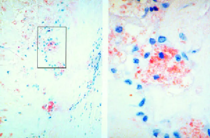
2. For the identification of Chlamydia pneumoniae anti- gens immunohistochemical reactions (TWAR monoclonal antibody RR-402) were performed in consecutive sections on coronary artery samples of 74 cases of the WHO study showing different intimal changes (Figure 1). These immunochemical reactions were evaluated semiquantita- tively (negative or positive).
3. Morphometric analysis was performed on 8 samples (4 positive and 4 negative for Chlamydia pneumoniae anti- gens) from 1 cm distally to the origin of the LAD arteries, showing only type I and type II lesions with no athero- sclerosis elsewhere (non-Aarteries) of the above 74 sam- ples. The thickness of the upper intima (intima without the myoelastic layer) and the media was computed as described above. For the measurement of hypertrophy index we worked out a new method in accordance with Tracy.15The area of the different layers was measured by image analysis (Scion Image for Windows), and nuclear density (cellularity) was determined for each of the layers.
Hypertrophy index was expressed by the ratio of the aver- age thickness and the cell density of that layer. The myoe- lastic layer, which is an unstable, and probably continu- ously transforming tissue defined by high cellularity at the Figure 1. Immunohistochemical detection of Chlamydia pneu-
moniae antibody RR-402 in coronary artery. Upper: positive monocytes are shown in the thickened intima (100X), in the squdred area foam cells are seen. Bottom: foam cells from the signed area above (400X).
Figure 2. Transversal section of distal LAD coronary. I: upper intima. ME: myoelastic layer. M: media. The thickness of the different layers was computed by dividing their areas and the length of the inner intimal border on the same part of the sec- tion. The nuclear density (cellularity) was measured in the upper intima and media as shown in the processed image on the left. 200 X
I
ME M
medial border of the intima was excluded from the above measurement of hypertrophy index (Figure 2). The thick- ness and the hypertrophy index of the both layer were cor- related with the results of Chlamydial immunohistochem- ical reactions.
ANOVA was used for statistical analysis followed by the pair wise comparison of the group means. Correlations of data were counted by Contingency analysis. The signifi- cance level of all tests was set to 0,05.
Results
At the proximal 1 cm segment of LAD both the intima and the media was thickened, when atherosclerosis was present elsewhere along the coronary (“A”). The relative intimal thickness is not predictive for atherosclerosis at the proximal segments of LAD. At the segments of 5 and 7 cm distal from the origin of LAD the relative intimal thickness increases in correlation with atherosclerosis elsewhere (Table 1).
Twenty five out of the 74 coronary arteries were positive for C. pneumoniae by immunohistochemistry (39%): 8 of 42 arteries with mild disease (19%), and 17 of 32 arteries with advanced lesions (53%) [Type I lesion: 1 out of 19 (5%), type II lesion: 7 out of 23 (30%) type III lesion: 6
out of 15 (40%) and type IV-VI: 11 out of 17 (65%)]
(Table 2). Advanced lesions were positive for C. pneumo- niae significantly more often than early lesions by contin- gency analysis (p<0,05).
The presence of Chlamydia pneumoniae antigens was correlated with an upper intimal and medial thickness as well as the hypertrophy index of both the intimal and the medial layer of the proximal segments of LAD (Table 3).
Upper intimal and medial thickening seems to be unin- volved in the process. Chlamydial infection is associated with higher hypertrophy index (increased intercellular matrix) in the upper intima as well as in the media. There were no similar changes in the myoelastic layer.
Discussion
Intimal thickenings (adaptive thickening) are thought to predispose to atheroma.12More recently, the thickening of the medial layer became in the centre of interest. Tracy at al.examined longitudinally opened right coronary arteries and found that coronary arteries that contain atheroma tend to possess a media of greater thickness than those that lack atheroma.14,15They declare that a fibroplastic thicken- ing of the intima throughout of the right coronary artery was preceded by medial hypertrophy. In our study thick- ened intima and/or media of the proximal coronaries were associated frequently with atheromatous changes. Howev- er, in the distal LAD we found only the intimal thickening as significant candidate for preatherosclerotic lesion that precedes the formation of a plaque.
These changes can be explained by increased synthesis of intercellular matrix and/or smooth muscle cell hyper- trophy and/or hyperplasia. In our study, the cell density in the upper intima and in the media did not increase related to atherosclerosis and this would suggest that the increase in thickness of the different vascular layers could be attrib- uted to a balanced cell proliferation and increase in the amount of extracellular matrix.
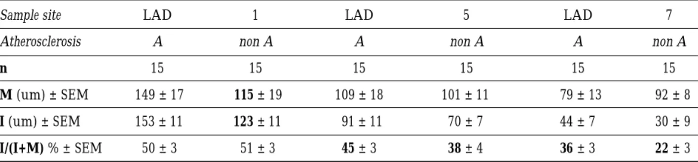
Table 1. Mean thickness of media and intima and the relative intimal thickness of coronary artery segments with and without atherosclerotic lesions
Sample site LAD 1 LAD 5 LAD 7
Atherosclerosis A non A A non A A non A
n 15 15 15 15 15 15
M (um) ± SEM 149 ± 17 115 ± 19 109 ± 18 101 ± 11 79 ± 13 92 ± 8
I(um) ± SEM 153 ± 11 123 ± 11 91 ± 11 70 ± 7 44 ± 7 30 ± 9
I/(I+M) % ± SEM 50 ± 3 51 ± 3 45± 3 38± 4 36± 3 22± 3
A: there is atherosclerosis elsewhere, non A: there is no atherosclerosis elsewhere in the coronary. Significant widening of each layer in the proximal segment and relative intimal thickness increase distally as a correlation with atherosclerosis (A versus nonA). Signif- icant value pairs (P< 0,05) are bold printed. M means media, I means intima, SEM (standard error of means).
Table 2. Chlamydial antigen positivity of different preatherosclerotic and atherosclerotic lesions in LAD Lesions Type I Type II Type III Type IV
Total N 19 23 15 17
positive 1 7 6 11
% positive 5 30 40 65
The rate of positivity increases significantly (P< 0,05) with the severity of lesions (from type I to IV). P< 0,05 of bold printed per- cent values.
The difference in the intensity of intimal and medial thickening in the proximal and distal LAD may be related to the different structure and function of these segments.
Proximal segments are more elastic and changes are resembled to those in the aorta, where medial thickening precedes intimal thickening. The phenomenon of medial growth was observed by us in the aorta and preceded sig- nificant intimal thickening and the occurrence of athero- sclerotic lesions (nonpublished data).
However, the answer to the question how medial hyper- trophy could be linked to atherosclerotic intimal changes is missing. It is not known whether changes of the medial smooth muscle cells influence the atherosclerotic process in the intima, or smooth muscle cells migrate and prolifer- ate in the intima – at sites defined as the myoelastic layer -or the fire-trap of atherosclerosis lays only in the intima and the medial changes being only the consequences of the intimal changes.
Over the last years the role of C. pneumoniae in the development of atherosclerosis has been widely dis- cussed.1,3-5,8,9In accordance with the literature, we found a significant correlation between advanced atherosclerotic changes and C. pneumoniae antigen positivity. However, there is no conclusive evidence yet that C. pneumoniae causes atherosclerosis or acute coronary syndromes. Clin- ical studies reported coronary vessel wall thickening in cases of C. seropositivity.5,9 Our data presented in this report show that there is a significant correlation between the C. pneumoniae infection and intimal and medial hyper- trophy. Chlamydial infection is associated with higher hypertrophy index – mainly increased intercellular matrix – in the upper intima as well as in the media. Thus, our results support that C. pneumoniae may have a role in the lesion progression. C. pneumoniae might disturb the nor-
mal regulatory mechanism in the vessels and may initiate a chronic inflammation. Strengthening this premise, C. pneumoniae has been found to upregulate the secretion of gelatinase by human macrophages,16 and induce the production of monocyte chemotactic protein and inter- leukin-8.10 Gelatinase is a metalloproteinase, which degrade collagen fibres. By degrading collagen, C. pneu- moniae facilitate the migration of monocytes from the blood to the intima, and probably facilitate the migration of smooth muscle cells from the media to the intima. The production of monocyte chemotactic protein and inter- leukin-8 also attract monocytes and smooth muscle cells.
Regarding our results, we hypothesize that C. pneumoniae may have a stimulatory effect on the smooth muscle cells both of the intima and media to induce extracellular matrix production. By such influences C. pneumoniae may trig- ger or accelerate atherosclerotic changes.
In conclusion in the proximal coronary both the intimal and medial thickening, and in the distal coronary only the intimal thickening are to be considered as early athero- sclerotic changes. C. pneumoniae is more frequently local- ized in plaques than in unchanged or preatherosclerotic intimas. Its presence is coincident with hypertrophy and increased extracellular matrix of the vascular segments around atherosclerotic lesions, which points to its roles in lesion development and progression.
Acknowledgements
The authors thank Mr. Csaba Lódi for the morphometric mea- surements and Mrs. Agnes Szik for the excellent technical help.
This work was supported by Hungarian National Grants OTKA 1082, and a Ph.D. grant (E.H.).
References
1. Berger M, Schroder B, Daeschlein G, Schneider Wet al.:
Chlamydia pneumoniae DNA in non-coronary atherosclerotic plaques and circulating leukocytes. J Lab Clin Med 136: 194- 200, 2000.
2. Davidson M, Kuo CC, Middaugh J et al.:Confirmed previous infection with Chlamydia pneumoniae (TWAR) and its pres- ence in early coronary atherosclerosis. Circulation 98: 628-33, 1998.
3. Gurfinkel E, Bozovich G, Darcoca A et al.: Randomized trial of roxithromycin in non-Q-wave coronary syndromes: ROXIS pilot study. Lancet 350: 404-7, 1997.
4. Ericson K, Saldeen TG, Lindquist O et al.:Relationship of Chlamydia pneumoniae infection to severity of human coro- nary atherosclerosis. Circulation 101: 2568-71, 2000.
5. Espinola-Klein C, Rupprecht HJ, Blankenberg S et al.:Are morphological or functional changes in the carotid artery wall associated with Chlamydia pneumoniae, Helicobacter pylori, cytomegalovirus, or herpes simplex virus infection? Stroke 31:2127-33, 2000.
6. Hortoványi E, Illyés Gy, Glasz T, Kádár A:Chlamydia pneu- moniae in different coronary artery segments in the young.
Pathol Res Pract 198: 19-23, 2002.
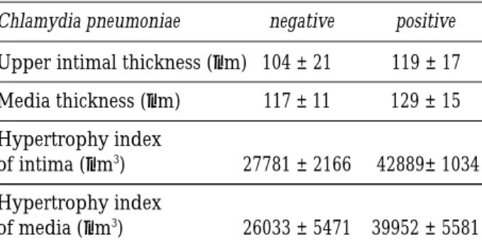
Table 3. Thickness and hypertrophy index of the seg- ments of positive and negative Chlamydia pneumoni- ae immunhistochemistry
Chlamydia pneumoniae negative positive Upper intimal thickness (µm) 104 ± 21 119 ± 17 Media thickness (µm) 117 ± 11 129 ± 15 Hypertrophy index
of intima (µm3) 27781 ± 2166 42889± 1034 Hypertrophy index
of media (µm3) 26033 ± 5471 39952 ± 5581 Increased average thickness of the upper intima and media is visible in Chlamydia positive cases. Hypertrophy index of both the intima and the media were significantly increased in Chlamydia positive versus Chlamydia negative coronaries. Sig- nificant value pairs (P< 0,05) are bold printed. ± values mean SEM (standard error of mean).
7. Kádár A., Mózes G, Illyés G et al.: Histomorphometry and his- tochemistry of atherosclerotic lesions in coronary arteries and the aorta in young population. World Health Organization (WHO) and the World Heart Federation (WHF) Pathobiologi- cal Determinants of Atherosclerosis in Youth Study (WHO/WHF PBDAY Study) 1986-1996. Nutr Metab Cardi- vasc Dis 9:220-227, 1999.
8. Kuo CC, Grayston JT, Campbell LA et al.: Chlamydia pneu- moniae (TWAR) in coronary arteries of young adults (15-34 years old) Proc Natl Acad Sci USA 92: 6911-14, 1995.
9. Mayr M, Kiechl S, Willeit Jet al.:Associations of antibodies to Chlamydia pneumoniae, Helicobacter pylori, and Cytomegalo- virus with immune reactions to Heat-shock protein 60 and carotid or femoral atherosclerosis. Circulation 102:833-39, 2000 10. Molestina RE, Dean D, Miller RD et al.:Characterization of a
strain of Chlamydia pneumoniae isolated from a coronary atheroma by analysis of the omp1 gene and biological activity in human endothelial cells. Infect Immun 66:1370-6, 1998,
11. Ross R:Atherosclerosis – an inflammatory disease. N Engl J Med 340: 115-25, 1999.
12. Stary HC: Composition and classification of human athero- sclerotic lesions. Virchows-Archiv-A-Pathol-Anat 421:277, 1992.
13. Stary HC: The Histological classification of atherosclerotic lesions in human coronary arteries. Atherosclerosis and Coro- nary Artery Disease Chapter 26 pp 463-474. Lippincott-Raven Publishers, Philadelphia, 1996.
14. Tracy RE:Medial thickness of coronary arteries as a correlate of atherosclerosis. Atherosclerosis 139:11-19. 1998.
15. Tracy RE:Medial thickenings of coronary artery and the aging risk factor for atherosclerosis. Atherosclerosis 155: 337–346, 2001.
16. Vehmaan-Kreula P, Puolakkainen M, Sarvas M et al.:Chlamy- dia pneumoniae proteins induce secretion of the 92-kDa gelati- nase by human monocyte-derived macrophages. Arterioscler Thromb Vasc Biol 21:1-8, 2001.