Control of myogenic tone and agonist induced contraction of intramural coronary resistance arterioles by cannabinoid type 1 receptors and endocannabinoids
Mária Szekeres1,2, György L. Nádasy1*, Eszter Soltész-Katona1 and László Hunyady1,3
1Department of Physiology, Faculty of Medicine, Semmelweis University 2Department of Morphology and Physiology, Faculty of Health Sciences, and 3Laboratory of Molecular Physiology at Semmelweis University, Hungarian Academy of Sciences, Budapest, Hungary
Number of words: 5803
*He is the corresponding author.
Address: P.O.Box 259, H-1444 Budapest, Hungary Phone: +36-1-4865814, +36-1-4191500 / 60456 Mobil:+36-20-586-1406
Fax: +36-1-266-6504
Email address: nadasy.gyorgy@med.semmelweis-univ.hu
Short title: Endocannabinoids moderate coronary arteriole tone via CB1R
Keywords: Angiotensin II, coronary artery, cannabinoid, vascular tone, diacylglycerol
LIST OF ABBREVIATIONS
Ach, acetylcholine; 2-AG, 2-arachidonoylglycerol; Ang II, angiotensin II; AT1R, type 1 angiotensin receptor; BK, bradykinin; CB1R, type 1 cannabinoid receptor; DAG, diacylglycerol; DAGL, diacylglycerol lipase; EC, endocannabinoid; ERK, extracellular signal-regulated kinase; HPLC, high performance liquid chromatography; KO, knockout; LAD, left anterior descending; LNA, Nω-nitro-L- arginine; MAG, monoacylglycerol; NO, nitric oxide; qRT-PCR, quantitative real-time PCR; PLC, phospholipase C; THC, Δ9-tetrahydro-cannabinol; THL, tetrahydrolipstatin; SNP, sodium-nitroprusside;
VSMC, vascular smooth muscle cell
INTRODUCTION
Hemodynamic resistance of intramural coronary arterioles with diameters below 200 µm determines local ventricular flow. Such vessels have a substantial spontaneous/myogenic tone which is kept reduced by metabolic factors from surrounding ventricular tissue, beta adrenergic effects, endothelial NO and also by vasodilatory prostanoids produced in the wall. Compared to resting coronary vascular flow, during heavy physical exercise and also in hypoxia a 4-5 times increase in cardiac blood flow (“coronary vasomotion”) can be achieved 1-7.
It has been revealed, that exogenous tetrahydro-cannabinol (THC)8, anandamide, 2- arachidonoilglycerol (2-AG)9-10 , different natural and synthetic agonists of the CB1R (e.g. WIN55212)11-
14 cause substantial vasodilation in several vascular beds, such as in coronary arteries 9-11, cerebral arteries11, the mesenteric vascular bed8 and in the aorta14. Several hemodynamically and clinically important resistance arteries also responded to cannabinoids 8,15. Most vasodilatory and hypotensive actions of cannabinoids seem to involve the CB1R receptors 11,15,16.
Endocannabinoids are produced in the wall of different vessels and by their vasodilatory actions they contribute to local vascular control15-22. However, the extent of this contribution can be much different in different vascular areas18-24. Exogenous cannabinoids increased coronary flow in isolated rodent hearts25, dilated larger coronary arteries9,10,17,26 and even cardioprotective and antiischemic effects27-29 have been attributed to them. In an earlier work from our laboratory16, we have demonstrated that continuous production of endocannabinoids (mostly 2-arachnoidylglycerol, 2-AG) in the wall of skeletal muscle resistance arterioles maintained a reduced spontaneous and agonist induced tone via vascular CB1 receptors. Pharmacological control of coronary resistance arteries might be much different from that of resistance arteries in other vascular areas3 and may even differ from larger coronary vessels5. Myogenic contraction and tone of resistance-sized arteries involves different agonist-induced and cellular mechanisms 30.
Taking into consideration the unique clinical significance of the coronary vascular system and the specific features of coronary resistance artery control it is of utmost importance to study whether
endocannabinoid-mediated vasodilatory mechanisms do exist in coronary resistance arteries and to what extent they are able to modulate the myogenic and agonist induced coronary vascular tone.
MATERIALS AND METHODS
Animals
Male Wistar rats were used (250-350g, Charles River Laboratories-Semmelweis University, Budapest). All animals were anaesthetized with pentobarbital sodium (Euthasol, ASTfarma 50 mg/kg intraperitoneally) and an extra dose (appr. 10 mg/kg) was additionally given, if necessary. Anesthetized animals were sacrificed by fast bleeding. The investigation conforms to the Guide for the Care and Use of Laboratory Animals (NIH, 8th edition, 2011) as well as National legal and institutional guidelines for animal care. They were approved by the Animal Care Committee of the Semmelweis University, Budapest and by Hungarian authorities (No. 263/003/2008 and No. 001/2139-4/2012).
Chemicals
Angiotensin II, bradykinin (BK), WIN55212 (a CB1R agonist), Nω-nitro-L-arginine (an inhibitor of nitric oxide synthase) and tetrahydrolipstatin (THL, a diacylglycerol lipase inhibitor) were purchased from Sigma-Aldrich (St. Louis, MO, US). The neutral CB1R antagonist O2050, the inverse CB1R agonist AM251 were purchased from Tocris Bioscience (Elliswille, MI, US). All other salts and chemicals were purchased from Sigma.
Immunohistochemistry
Hearts from rats and mice were fixed in 4% paraformaldehyde and placed in 10 and 20% sucrose solutions (15–18 hrs each) for cryoprotection. The tissues were quickly frozen on dry ice, embedding, sectioning and staining were executed as described 22,31. Immunostaining was performed with CB1R primary antibody (Cayman Chemicals, Ann Arbor, MI, overnight). The development of slides was made
using the biotin extravidin ABC method (Vector Labs, Burlington, CA). Diaminobenzidine (DAB) was used for visualization22,31. Antibody specificity was confirmed preparing aortic slides of wild and CB1R - /- knockout mice.
Western blots
Microdissected samples were washed twice in ice cold PBS solution (composition in mM, NaCl 137, KCl 2.7, Na2HPO4 10.1, KH2PO4 1.8, pH 7.4). They were homogenized in glass tubes and lysed in SDS lysis buffer containing 10 % merkapto-ethanol and protease inhibitor cocktails (Sigma-Aldrich). In the lysis buffer samples were sonicated, boiled and centrifuged. Proteins from tissue samples were separated with SDS-polyacrylamide gel electrophoresis and were blotted onto the PVDF membrane.
Membranes were treated with antibodies against CB1R (Cayman Chemicals) and beta-actin (Sigma- Aldrich) followed by the treatment with HRP-conjugated secondary antibodies. Visualization was made with SuperSignal West Pico reagent (Promega, Madison, WI) and results were quantitatively evaluated with densitometry.
RNA extraction and real-time PCR
Vascular tissues were removed by fast and careful microscopic dissection for RNA extraction16. In anesthesia, the chest was opened, the heart removed. Intramural coronary arterioles branching from the left anterior descendent coronary artery were isolated. Vessels were placed in cold sterile phosphate buffer solution (PBS as above). Total RNA was extracted (RNeasy mini kit, Qiagen) and reverse transcription was carried out according to the manufacturer’s instructions (Fermentas, Ontario, Canada).
Real-time PCR assays were performed on LightCycler 480 (Roche Applied Science, Indianapolis, IN) with the SYBR Green method. Primers were designed and synthesized by Sigma-Aldrich. Cycling conditions were: 10 min preincubation at 95 ºC, 45-50 cycles of 95 ºC 10 sec, 62 ºC 5 sec and 72 ºC 15 sec. Fluorescence data including melting curves were obtained. For normalization, the glyceraldehyde-3- phosphate dehydrogenase (gapdh) housekeeping gene was used (ENSRNOG00000004253). Efficiency
for each primer pair was determined byusing serial dilutions of the PCR product. Fold ratios of gene expression were calculated as follows:
Ratio=E ΔCt target gene / E ΔCt GAPDH
Ct was calculated by the second derivative method using LightCycler 480 Software. ΔCt is the difference in Ct values obtained between the reference and tested samples. Relative messenger RNA levels of cannabinoid receptor type 1 (Cnr1) were calculated (ENSRNOG00000008223). Primers were for Cnr1:
forward primer GGACTCAGACTGCCTGCACA, reverse primer ACAAAAGCAGCAGGCTCACA and for gapdh: forward primer CCTGCACCACCAACTGCTTAG, reverse primer CAGTCTTCTGAGTGGCAGTGATG. Tissue gene expression levels were plotted against gapdh expression level.
Isolation of rat coronary resistance arteries for video microarteriography
During anaesthesia of rats, the chest was quickly opened, the heart removed and placed into cold normal Krebs-Ringer (nKR) solution which contained in mM, NaCl 110, KCl 5, CaCl2 2,5, MgSO4 1,0, KH2PO4 1,0, NaHCO3 24 and glucose, 10 (pH 7.4). Intramural coronary arterioles branching from the left anterior descending (LAD) coronary artery were isolated by careful microdissection as described previously 5,32,33. Distal segments close to the apex with approximately 100-150 μm of inner diameter were prepared. One segment was prepared and measured from each animal. The segment was placed in a tissue bath filled with Krebs solution. It was cannulated at both ends with microcannulas, extended to its in situ length in the glass bottomed tissue bath of a pressure microarteriography chamber (Experimetria, Budapest). Clotted blood was washed out at low perfusion pressure, and then one of the cannulas was closed. Utilizing a pressure-servo syringe reservoir system (Living Systems, Burlington, VT, US) arterioles were pressurized in a no-flow condition. Vessels were checked for leaks by the stability of the inlet pressure when the servo function was turned off. Vessels with observable leaks were discarded. The
temperature of the chamber was maintained at 37 oC, bubbled with 21 % O2 and 5 % CO2 , balanced with N2, the pH was kept at 7.4. A continuous superfusion of the bath was applied at a rate of 2.5 mL/min.
The cannulated vessel was visualized by video-microscopy and the inner diameter was measured on frozen images (Leica inverted microscope, Leica DFC 320 digital camera, LeicaQWin software)5,6,16,32. Calibration was made with a micrometer etalon (Wilde, Heerbrugg, Switzerland). Intraluminal pressure was calibrated with a mercury manometer.
Experimental protocols of isolated vessel studies
Following a 60-min equilibration period, at 50 mmHg intraluminal pressure in nKR solution, pharmacological responses of the arterial segments were tested according to the specific protocols.
Agonists were administered in a dose-dependent manner into the chamber and steady-state diameter was recorded for each dose or in a single (submaximal) dose. 10-min washout periods were applied between drugs. Inhibitors were applied for at least 10 minutes prior to and during agonist administration. Each inhibitor was applied in separate experimental series, either the pressurized segments were in spontaneous myogenic contraction or their contraction was enhanced with Ang II. In coronary vessels, Ang II is suitable to produce precontraction, since it produces a stable contraction without observable desensitization response in contrast to other type of vessels like gracilis arterioles and aorta16,22. After Ang II precontraction (1 µM), SNP (1 µM) was applied to test endothelium-independent vasodilation.
Endothelial relaxation was tested by bradykinin (100 nM), which is an appropriate endothelium- dependent vasodilator in coronary resistance arteries of rats5. The experiments were terminated by obtaining passive (relaxed) vascular diameter in calcium-free Krebs solution.
In the first group of experiments (n=14), rat coronary arterioles were subjected to elevated doses of agonists, Ang II (0.1 nM-10 µM), WIN55212 (0.1 nM-1 µM), SNP (0.1 nM-10 µM) and BK (0.1 µM) with the CB1R neutral antagonist O2050 (1 μM) or only its vehicle being present in the bath. Since WIN55212 is lipid-soluble, it was applied prior to and after CB1R antagonist in separate vessel segments.
In the second group (n=5), concentration-response to Ang II was obtained before and during the
administration of the DAG lipase inhibitor tetrahidrolypstatin (1 μM) or its vehicle. In a third experimental set (n=6), the effect of CB1 receptor antagonist (inverse agonist) AM251 (1 μM) (or its vehicle) on the tone of coronary arterioles and on WIN55212- and SNP-induced vasodilations were tested. In a fifth set of experiments (n=5), the effect of Nω-nitro-L-arginine (an inhibitor of nitric oxide synthase, 50 μM) was applied 20 minutes prior and during the administration of CB1R agonist WIN55212 (1 μM) or the endothelium-dependent agonist BK (0.1 µM) in order to test the role of NO in the CB1R- mediated coronary functions.
Statistical analysis
Data are presented as means±SEM. Vascular tone was calculated as percent change from passive diameter. Changes in vascular diameter were also calculated as percent change from control value. For paired data we used the Student’s t-test, for multiple comparisons one-way and two-way ANOVAs were applied (SigmaStat). Concentration-response curves without and with inhibitors present in the solution were compared using the four parameter logistic function (SigmaPlot). Relative gene expression levels were plotted against reference control (gapdh). P<0.05 was considered significant for each comparisons.
RESULTS
Molecular and functional evidence of the expression and presence of CB1Rs in intramural coronary resistance arteries
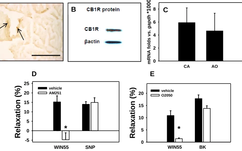
CB1R protein is present in the wall of these rodent microvessels as it has been demonstrated by immunohistochemistry of ventricular tissue. Immunostaining appeared in the wall of intramural coronary arterioles (Fig. 1A.). Western blots also proved the expression of the CB1R protein in the wall of rat small coronary arteries isolated by microdissection (Fig. 1B.). In addition, messenger RNA for CB1 receptors was also detected by qRT-PCR in amounts similar to those found in rat aortic tissue (Fig. 1C.). Segments
in myogenic contraction relaxed in response to the CB1R agonist WIN 55212 (1 µM). Further, this relaxation could be inhibited by the specific blockers O2050 (1 µM), and AM251 (1 µM). These CB1R antagonists, however, did not affect either endothelium independent (SNP, 1 µM) or endothelium dependent (BK, 0.1 µM) relaxations (tested on Ang II precontracted segments, Fig. 1D-E.).
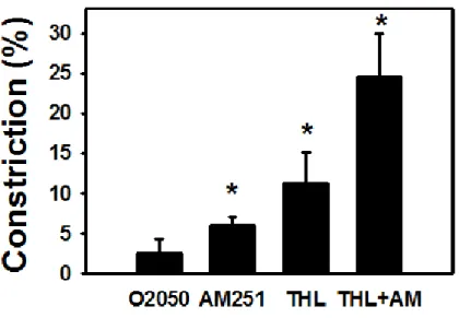
Evidence of existence of an intrinsic CB1R activation during spontaneous myogenic contraction
Coronary arteries when pressurized, exhibit a substantial spontaneous, myogenic tone in oxygenized nKR solution. This, with all probability mimics the in vivo situation 5,32,33. We tested whether a basal endocannabinoid release affects the spontaneous tone in these vessels. Vascular diameter of our segments during spontaneous contraction (following an equilibration period) was 138.7±6.6 µm (inner diameter) corresponding to 20.1±2.9 % (n=15) myogenic tone (compared with relaxed controls).
The neutral CB1 receptor antagonist (O2050, 1 µM) moderately (2.6±1.7 %, n.s.), the inverse CB1R agonist AM251 (1 µM) significantly increased coronary myogenic tone (6.0±1.0 %, p<0.05). The DAG lipase enzyme inhibitor THL (1 µM) effectively increased basal coronary tone (11.2±3.9 % contraction, p<0.05). This effect was additive with the similar effect of AM251: a substantial diameter reduction of 24.6±5.3% (p<0.05) was reached when both antagonists were applied together (Fig. 2.).
Products of the enzyme DAG lipase must be the main endocannabinoids in this tissue. These experiments indirectly prove that spontaneous myogenic tone of these vessels is kept under continuous control by endogenously produced 2-AG. This control can be considered fairly effective as the myogenic contraction of these vessels practically doubles if 2-AG synthesis and endocannabinoid action on CB1Rs are both inhibited at the same time.
Effect of exogenous cannabinoid on coronary arterial tone
Adding angiotensin II to the bath further contracts coronary arteries, this contraction is added to the myogenic one. The specific CB1R agonist WIN55212 very effectively relaxed coronary arteriole
segments precontracted with close-to-maximal concentrations of Ang II (1 µM). The concentration- dependent vasodilation by the CB1R agonist WIN 55212 is shown on Fig. 3A, which is effectively inhibited by the CB1R antagonist O2050. This record proves that agonist induced contraction of coronary resistance arterioles can be inhibited by exogenous stimulation of the cannabinoid receptors. However, the concentration-dependent vasodilation by the NO donor sodium nitroprusside (SNP) was not affected by these CB1R antagonists (Fig. 3B and D). Fig. 3. and Fig 1D demonstrate an interesting fact:
WIN55212, a CB1R agonist is an almost as effective a coronary vasodilator as the well-known NO donor SNP is.
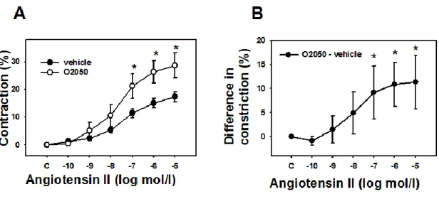
Agonist induced contraction goes on with parallel CB1R activation
Ang II elevated coronary arteriole tone in a dose-dependent manner, at maximal concentrations inducing about a 20% further reduction of diameter of segments in spontaneous contraction. This effect reached a maximum level around 10 µM of Ang II concentration. Inhibition of CB1 receptors by O2050, significantly enhanced Ang II-induced contractions of coronary arterioles (significant over 100 nM Ang II, p<0.05, Figure 4). The four parameter logistic analysis has shown that there was not significant change in logEC50% values for Ang II contraction (-7.36±0.19 vs. -7.54±0.21 without and with the antagonist, respectively, n.s.), while maximum contraction values significantly elevated (17.8±1.2 vs.
29.1±2.9%, p<0.05). These observations prove that during agonist induced contraction, a continuous and stimulated endocannabinoid production is present in the wall which modulates contraction due to its effect on CB1Rs.
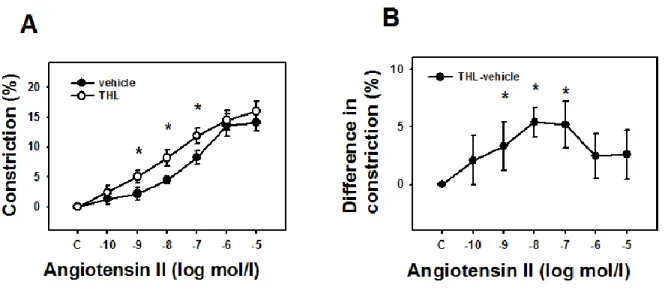
Effect of inhibition of DAG lipase enzyme on the agonist-induced response
The DAG lipase blocker THL also augmented Ang II-induced contractile response in the coronaries (Figure 5, p<0.05 at 1 nM-100 nM of Ang II). Based on this observation, it is highly probable that 2-AG is the endocannabinoid whose production is elevated by Ang II in this vascular preparation and the co-stimulation of CB1Rs produced limits then Ang II contraction22.
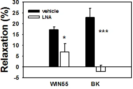
Role of endothelial nitric oxide in CB1R induced coronary vasodilation
The coronary dilatory effect of the CB1R agonist Win 55212 on Ang II precontracted segments was partially suspended by the NO synthase blocker Nω-nitro-L-arginine (50 µM), suggesting a partially NO mediated effect (Fig 6., p<0.05). By comparison, the dilatory effect of bradykinin, an endothelial coronary vasodilator, was fully diminished by the same concentration of LNA (p<0.001).
DISCUSSION
Substantial modulator effect found both in myogenic and in agonist induced contraction
Our observations demonstrated the existence and a substantial functional role of CB1Rs in rodent intramural coronary arterioles, vessels that basically determine local ventricular flow. The presence of CB1R protein (with immune-histochemistry and Western blot, Fig 1A. and 1B.) and of its mRNA (Fig.
1C.) have been first proven in this vessel-type. We have also proved that endocannabinoids acting on such receptors play an important role in forming myogenic and agonist induced tone of these vessels. The specific CB1R agonist WIN55212 induced a substantial concentration-dependent vasodilation (Figs. 3A.
and 3C.) which was prevented by the CB1R neutral antagonist O2050 (Fig. 3A.) and by the inverse agonist AM251 (Fig. 3C.). AM251 and the DAG lipase enzyme inhibitor THL substantially increased coronary arteriole myogenic tone (Fig. 2.). Ang II induced tone was augmented both by O2050 and THL (Figs.4. and 5.). We have a sound foundation to declare that a DAG lipase product endocannabinoid, with all probability 2-AG is continuously synthesized in the wall of these vessels, and the amount produced is enhanced by Ang II. Both the spontaneous myogenic and Ang II-induced contractions are attenuated by its effect. As neither SNP nor BK relaxations were effected by CB1R antagonists (Fig. 3B. and D.), we can conclude that simply altering the contractile state of a coronary arteriole segment will not automatically alter its endocannabinoid production.
Coronary cannabinoid-mediated relaxation
We have found a significant CB1R-mediated vasorelaxation in rat coronary arterioles (Fig. 3A, 3C.).
Our findings are in accord with an earlier publication by White9 who found that isolated rings prepared from the left anterior descendent coronary artery of the rat (not a resistance sized artery) and precontracted with 5-HT, dilated in response to exogenous anandamide. This dilation could be inhibited by the CB1R blocker SR14176A. It is interesting to note that he has found the more specific CB1R blocker AM251 less effective. There seems to be some contradiction also with the observations by Mair26. On main coronary artery branches of the rat, precontracted with U46619 (a thromboxane agonist), he has found that relaxation occurred through CB2Rs due to activation of the ERK1/2 pathway and vasodilatory prostanoids. Our opinion is that tissue composition and function of larger coronary arteries viable for wire myographic studies can be substantially different from the real resistance vessels studied by us using the pressure arteriography technique.
Coronary endocannabinoid production and relaxation
Endocannabinoid production has been detected in different vascular beds and also in heart15-22. Several types of cells of different vascular tissues can be sources of endocannabinoids: endothelial cells, smooth muscle cells, perivascular neurons, platelets, leukocytes, monocytes, macrophages etc.18,34-36. Endogenous release of 2-AG has been observed from human vascular endothelial cells. The 2-AG released was supposed to be an intrinsic vasomodulator37. In bovine coronary arteries, metacholine stimulates endothelial 2-AG release through PLC and DAGL activation and serves as an intermediate for vasodilatory eicosanoid release34. Not only endothelial cells but also vascular smooth muscle cells can release 2-AG22. We have found previously that Ang II-stimulated 2-AG-release from vascular smooth muscle cells has been attenuated by the inhibition of DAG lipase and augmented by monoacylglycerol (MAG) lipase inhibition22. In conformity with the vascular production of 2-AG, we have also found that inhibition of DAG lipase augmented, while inhibition of MAG lipase attenuated vasoconstriction of the
aorta. This indicates the role of locally released 2-AG in the control of vascular tone in this vessel22. Similarly, a previous observation found that 2-AG production in rat aorta was enhanced by carbachol38.
The contribution of the endocannabinoid mechanism to lumen control in these important vascular segments is substantial, we have a good reason to suppose a physiological impact. We have found here that inhibition of DAGL, the enzyme responsible for the production of a substantial part of tissue endocannabinoids, augmented coronary resistance artery tone by around 10% even without adding any stimulants (Fig. 5.). The double inhibition of the endocannabinoid effect (CB1R +DAGL inhibition) induces 24.6±5.3% contraction, that practically doubles the approximately 20% spontaneous myogenic tone (Fig. 2.). We can conclude that similarly to endogenous prostanoids and NO22, a continuous production of vasodilatory endocannabinoids in the resistance artery wall counterbalances the myogenic tone in coronary resistance arteries. The concentration-dependent Ang II-induced contraction of these segments has also been accompanied by a stepwise elevation of the moderating effects of endogenous cannabinoids. Both the CB1R antagonist O2050 and the DAG lipase blocker THL augmented Ang II- induced contraction when compared with contraction without the antagonist (Figs. 4. and 5.). This makes it highly probable, that “basal” endocannabinoid release (and its vasorelaxation effect) is increasing with increasing concentrations of the contracting agonist. According to a previous observation from our laboratory, in nonvascular cells, co-expressing the AT1-and CB1 receptors, Ang II-induced stimulation of AT1R via the activation of PLC and DAG-production led to the transactivation of CB1 receptors by endocannabinoid release38,39. The Ang II-induced CB1 receptor activity was inhibited by DAGL inhibitor suggesting, that 2-AG production from DAG plays an important role in the mediation of this action40. Similarly, our previous observation on skeletal muscle arterioles indicated that inhibition of CB1 receptors augmented Ang II-induced vasoconstriction by increasing both efficacy and the potency of the Ang II- induced responses16.
Considering the mechanism of this relaxing effect, our present studies on coronary arterioles suggest a mechanism analogous that found in skeletal muscle arterioles. The AT1R, Gq, PLC, DAG pathway produces a substrate for the DAG lipase, the 2-AG produced co-activates CB1Rs in the
neighborhood, inducing, by a not fully identified yet action, relaxation, limiting the contraction exerted.
How does the co-activation of CB1Rs relax these vessels? K+ channel-mediated cell hyperpolarization9,10,17,41, depression of Ca2+ influx41,42, modulation of endogenous prostanoid production9,10,26,35, activation of Gi/o or other G proteins3,9,40 are the mechanisms suggested in the literature by different authors.
The role of endothelium and endothelium-mediated nitric oxide in the cannabinoid-induced vasodilatory response seems to be different in different vascular territories. Previously, in gracilis arterioles we found that WIN55212-induced vasodilation was not attenuated by the blocker of NO synthase nitro-L-arginine, while it effectively inhibited acetylcholine-induced vasodilation16. Also, we have found that the removal of endothelium did not change the augmented vasoconstrictor effect induced by the CB1R blocker O2050 in gracilis vessels. Sources of vascular endocannabinoids can be heterogenous: 2-AG production has been detected both from endothelial cells and from vascular smooth muscle cells22,35,37,38
. These observations are in good accordance with our present observations on coronary resistance vessels, inhibition of NO synthesis caused a partial (by approx. 50%) attenuation of the CB1R agonist WIN55212-induced vasodilation (Fig. 6.).
Potential physiological role
We can remark here that such intrinsic “braking” feed-backs frequently occur in complex physiological control networks. In case of the coronary resistance artery control mechanisms, the endothelial NO and endogenous prostanoid production, stimulated by agonists, can be mentioned. We and other laboratories have also found earlier that myogenic and agonist-induced tone of coronary resistance arteries are fine-tuned by altered NO and vasoactive prostanoid production occurring in the wall of these vessels3,6,7 Now, we can add to this a similar new mechanism: endogenous cannabinoid production. One can theorize on the potential physiological benefits: Full contraction closing the lumen can be prevented by them or, certain contractile effects can be neutralized. Anyway, they yield a more
complex and more versatile lumen control mechanism, which is able to respond to diverse physiological needs in a more diverse manner.
Conclusion
Our studies have proven the existence of the CB1R protein, its mRNA and also its functional relevance to induce substantial vasodilation in the wall of intramural coronary resistance arteries, vessels that basically determine ventricular local flow. Further, we provide clear evidence that a vasorelaxant endocannabinoid system is present in these vessels, which substantially contributes to the physiologically important lumen controlling mechanisms here through vascular CB1 receptors. This mechanism is active both during spontaneous myogenic contraction and also in Ang II-induced contraction. We suggest, that to the known physiological mechanisms that reduce the substantial spontaneous/myogenic tone of coronary resistance vessels (metabolic factors, beta adrenergic stimuli, vasodilatory prostanoids and endothelial NO) the endogenous cannabinoids should be added.
ACKNOWLEDGEMENTS
The authors are grateful to Dr. Gábor Turu, Dr. Zsuzsanna E. Tóth (Semmelweis University, Budapest), Dr. Andreas Zimmer (University of Bonn) and Dr. Istvan Katona (Institute of Exp. Med. of the Hungarian Academy of Sciences, Budapest) for helpful discussion of the manuscript. The authors are grateful to Ildikó Oravecz, Judit Rácz, Ilona Oláh and Anikó Schulcz for their expert assistances. The authors declare that there is no conflict of interest that would prejudice the impartiality of the present work.
GRANTS
This work was supported by grants from the Hungarian National Science Foundation (OTKA NK- 100883, NK-72661) and the National Development Agency, Hungary (TÁMOP 4.2.1.B-09/1/KMR- 2010-0001).
DISCLOSURES, CONFLICTS OF INTERESTS None is declared.
AUTHOR CONTRIBUTIONS
M.Sz. and L.H.: conception and design of research. M.Sz. and G.L.N. performed experiments. M.Sz., G.L.N. and E.S-K. performed analysis. M.Sz. and G.L.N. interpreted results of experiments. M.Sz. and E.S-K. prepared figures. M.Sz. drafted manuscript. L. H. edited and revised manuscript. L. H., M.Sz., G.L.N. and E.S-K. approved final version of manuscript.
REFERENCES
1. Kuo L, Davis MJ, Chilian WM. Myogenic activity in isolated subepicardial and subendocardial coronary arterioles. Am J Physiol 1988;255:H1558-H1562.
2. Duncker DJ, Bache RJ. Regulation of coronary blood flow during exercise. Physiol Res 2008;88:1009- 1086.
3. Duncker DJ, Koller A, Merkus D, Canty JMJr. Regulation of coronary blood flow in health and ischemic heart disease. Progr Cardiovasc Dis 2015;57:409-422.
4. Laughlin MH, Bowles DK, Duncker DJ. The coronary circulation in exercise training. Am J Physiol Heart Circ Physiol 2012;302:H10-H23.
5. Szekeres M, Dezsi L, Nadasy GL, Kaley G, Koller A. Pharmacologic inhomogeneity between the reactivity of intramural coronary arteries and arterioles. J Cardiovasc Pharmacol 2001;38: 584-592.
6. Szekeres M, Nadasy GL, Kaley G, and Koller A. Nitric oxide and prostaglandins modulate pressure- induced myogenic responses of intramural coronary arterioles. J Cardiovasc Pharmacol 2004;43: 242- 249.
7. Toda N, Toda H. Coronary hemodynamic regulation by nitric oxide in experimental animals: Recent advances. Eur J Pharmacol 2011;667:41-49.
8. O'Sullivan SE, Kendall DA, and Randall MD. The effects of Delta9-tetrahydrocannabinol in rat mesenteric vasculature, and its interactions with the endocannabinoid anandamide. Br J Pharmacol 2005;145: 514-526.
9. White R, Ho WSV, Bottrill FE, Ford WR, and Hiley CR. Mechanisms of anandamide-induced vasorelaxation in rat isolated coronary arteries. Br J Pharmacol 2001;134: 921-929.
10. Grainger J and Boachie-Ansah G. Anandamide-induced relaxation of sheep coronary arteries: the role of the vascular endothelium, arachidonic acid metabolites and potassium channels. Br J Pharmacol 2011;134: 1003-1012.
11. Wagner JA, Jarai Z, Batkai S, and Kunos G. Hemodynamic effects of cannabinoids: coronary and cerebral vasodilation mediated by cannabinoid CB(1) receptors. Eur J Pharmacol 2001; 423: 203-210.
12. Randall MD, Harris D, Kendall DA, and Ralevic V. Cardiovascular effects of cannabinoids.
Pharmacol Ther 2002;95: 191-202.
13. Randall MD, Kendall DA, and O'Sullivan S. The complexities of the cardiovascular actions of cannabinoids. Br J Pharmacol 2004;142: 20-26.
14. Dannert MT, Alsasua A, Herradon E, Martin MI, and Lopez-Miranda V. Vasorelaxant effect of Win 55,212-2 in rat aorta: new mechanisms involved. Vascul Pharmacol 2007;46: 16-23.
15. Batkai S, Pacher P, Osei-Hyiaman D, Radaeva S, Liu J, Harvey-White J, Offertaler L, Mackie K, Rudd MA, Bukoski RD, and Kunos G. Endocannabinoids acting at cannabinoid-1 receptors regulate cardiovascular function in hypertension. Circulation 2004;110: 1996-2002.
16. Szekeres M, Nadasy GL, Turu G, Soltesz-Katona E, Toth ZE, Balla A, Catt KJ, and Hunyady L.
Angiotensin II Induces Vascular Endocannabinoid Release, Which Attenuates Its Vasoconstrictor Effect via CB1 Cannabinoid Receptors. J Biol Chem 287: 31540-31550, 2012.
17. Randall MD and Kendall DA. Involvement of a cannabinoid in endothelium-derived hyperpolarizing factor-mediated coronary vasorelaxation. Eur J Pharmacol 1997; 335: 205-209.
18. Hillard CJ. Endocannabinoids and vascular function. J Pharmacol Exp Ther 2000;294: 27-32.
19. Hiley CR. Endocannabinoids and the heart. J Cardiovasc Pharmacol 2009;53: 267-276.
20. Pacher P, Mukhopadhyay P, Mohanraj R, Godlewski G, Batkai S, and Kunos G. Modulation of the endocannabinoid system in cardiovascular disease: therapeutic potential and limitations. Hypertension 2008;52: 601-607.
21. Wagner JA, Hu K, Bauersachs J, Karcher J, Wiesler M, Goparaju SK, Kunos G, and Ertl G.
Endogenous cannabinoids mediate hypotension after experimental myocardial infarction. J Am Coll Cardiol 2001;38: 2048-2054.
22. Szekeres M, Nadasy GL, Turu G, Soltesz-Katona E, Benyo Z, Offermanns S, Ruisanchez E, Szabo E, Takats Z, Batkai S, Toth ZE, and Hunyady L. Endocannabinoid-mediated modulation of Gq/11 protein- coupled receptor signaling-induced vasoconstriction and hypertension. Mol Cell Endocrinol 2015;403:
46-56.
23. Stanley C, O’Sullivan SE. Vascular targets for cannabinoids: animal and human studies. Review. Br J Pharmacol 2014;171:1361-1378.
24. O’Sullivan SE. Endocannabinoids and the cardiovascular system in health and disease (Review) Handbook Exptl Pharmacol 2015;231:393-422.
25. Wagner JA, Abesser M, Karcher J, Laser M, Kunos G. Coronary vasodilator effects of endogenous cannabinoids in vasopressin-preconstricted unpaced rat isolated hearts. J Cardiovasc Pharmacol 2005;46:348-355.
26. Mair KM, Robinson E, Kane KA, Pyne S, Brett RR, Pyne NJ, and Kennedy S. Interaction between anandamide and sphingosine-1-phosphate in mediating vasorelaxation in rat coronary artery. Br J Pharmacol 2010;161: 176-192.
27. Zimmer A, Zimmer AM, Hohmann AG, Herkenham M, and Bonner TI. Increased mortality, hypoactivity, and hypoalgesia in cannabinoid CB1 receptor knockout mice. Proc Natl Acad Sci U S A 1999;96: 5780-5785.
28. Bouchard JF, Lepicier P, and Lamontagne D. Contribution of endocannabinoids in the endothelial protection afforded by ischemic preconditioning in the isolated rat heart. Life Sci 2003;72: 1859-1870.
29. Wagner JA, Abesser M, Harvey-White J, and Ertl G. 2-Arachidonylglycerol acting on CB1 cannabinoid receptors mediates delayed cardioprotection induced by nitric oxide in rat isolated hearts. J Cardiovasc Pharmacol 2006;47: 650-655.
30. Kim EC, Choi SK, Lim M, Yeon SI, Lee YH. Role of endogenous ENaC and TRP channels in the myogenic response of rat posterior cerebral arteries. PLOS One 2013;8(12)e84194):1-9.
31. Toth ZE, Mezey E. Simultaneous visualization of multiple antigens with tyramide signal amplification using antibodies from the same species. J Histochem Cytochem 2007;55: 545-554.
32. Nadasy GL, Szekeres M, Dezsi L, Varbiro S, Szekacs B, and Monos E. Preparation of intramural small coronary artery and arteriole segments and resistance artery networks from the rat heart for microarteriography and for in situ perfusion video mapping. Microvasc Res 2001;61: 282-286.
33.Szekeres M, Nadasy GL, Dezsi L, Orosz M, Tôkés A, Monos E: Segmental differences in geometric, elastic and contractile characteristics of small intramural coronary arteries J Vasc Res 1998; 35:332-344.
34. Gauthier KM, Baewer DV, Hittner S, Hillard CJ, Nithipatikom K, Reddy DS, Falck JR, and Campbell WB. Endothelium-derived 2-arachidonylglycerol: an intermediate in vasodilatory eicosanoid release in bovine coronary arteries. Am J Physiol Heart Circ Physiol 2005;288: H1344-1351.
35. Lipez-Miranda V, Herradon E, and Martin MI. Vasorelaxation caused by cannabinoids: mechanisms in different vascular beds. Curr Vasc Pharmacol 2008;6: 335-346.
36.
Pacher P, Batkai S, and Kunos G. Cardiovascular pharmacology of cannabinoids. Handb Exp
Pharmacol 2005; 599-625.37. Sugiura T, Kodaka T, Nakane S, Kishimoto S, Kondo S, and Waku K. Detection of an endogenous cannabimimetic molecule, 2-arachidonoylglycerol, and cannabinoid CB1 receptor mRNA in human vascular cells: is 2-arachidonoylglycerol a possible vasomodulator?
Biochem Biophys Res Commun 1998;243: 838-843.38. Mechoulam R, Fride E, Ben-Shabat S, Meiri U, and Horowitz M. Carbachol, an acetylcholine receptor agonist, enhances production in rat aorta of 2-arachidonoyl glycerol, a hypotensive endocannabinoid. Eur J Pharmacol 1998;362: R1-R3.
39. Turu G, Simon A, Gyombolai P, Szidonya L, Bagdy G, Lenkei Z, and Hunyady L. The role
of diacylglycerol lipase in constitutive and angiotensin AT1 receptor-stimulated cannabinoid
CB1 receptor activity. J Biol Chem 2007;282: 7753-7757.
40. Turu G, Varnai P, Gyombolai P, Szidonya L, Offertaler L, Bagdy G, Kunos G, and Hunyady L. Paracrine transactivation of the CB1 cannabinoid receptor by AT1 angiotensin and other Gq/11 protein-coupled receptors. J Biol Chem 2009;284: 16914-16921.
41
. Turu G and Hunyady L. Signal transduction of the CB1 cannabinoid receptor. J Mol Endocrinol 2010;44: 75-85.42.
Gebremedhin D, Lange AR, Campbell WB, Hillard CJ, and Harder DR. Cannabinoid CB1
receptor of cat cerebral arterial muscle functions to inhibit L-type Ca2+ channel current.
Am J Physiol 1999;276: H2085-2093.Fig. 1. Molecular and functional evidence of the presence of the CB1R in the intramural coronary resistance arterioles of the rat. A. Immunohistochemical localization in histological sections of ventricular tissue pieces containing the arterioles. Stainings of CB1Rs are indicated by arrows. Bar shows 100 µm. B. Western blot of CB1R protein from homogenates of microsurgically isolated resistance artery specimens. C. Expression of mRNA for CB1R in coronary resistance artery specimens (CA) and in the aorta (AO) of rats as determined by qRT-PCR (n=5). Expression fold vs. gapdh houskeeping gene is plotted. D-E. Relaxation in response to CB1R agonist (WIN55212, 1μM), inhibition of relaxation by the specific CB1R antagonists O2050 and AM251 (1μM) of Ang II-precontracted coronary artery segments.
SNP-induced (1μM, an endothelium independent vasodilator, n=11) and bradykinin-induced (BK, 0.1μM, an endothelium dependent coronary vasodilator) vasodilations were not affected by CB1R antagonists (segments precontracted with 1μM Ang II, n=8). Mean ± SEM values are shown. *, P<0.05 between control and inhibitor-treated values.
Fig. 2. Intrinsic CB1R activity keeps myogenic tone reduced. The type 1 cannabinoid receptor (CB1R)- antagonists O2050 (a neutral antagonist, 1μM) and AM251 (an inverse agonist, 1μM) and also the diacylglycerol (DAG) lipase-inhibitor tetrahydrolypstatin (THL, 1μM, inhibits the production of 2-AG) substantially increase the spontaneous tone of pressurized rat coronary arteries (n= 5 or 6 for each group).
Mean ± SEM values are shown. *, P<0.05 between control and inhibitor-treated values.
Fig. 3. CB1R-mediated modulation of agonist-induced contraction. A. Concentration dependent dilation of Ang II (1μM) precontracted coronary arteriole segments in response to different concentrations of the CB1R agonist WIN55212. The relaxation is almost completely inhibited in the presence of the specific CB1R blocker O2050 (1μM). B. For comparison, concentration-dependent vasodilation effect of NO donor sodium nitroprusside on coronary artery segments, which was not affected by the CB1R antagonist treatment. C and D. Similar responses were detected with the CB1R inverse agonist AM251 (1μM).
Fig. 4. CB1R-mediated modulation of agonist-induced contraction. Enhancement of concentration dependent Ang II contraction in the presence of the cannabinoid receptor (CB1R) antagonist O2050 (1μM) in the tissue bath (n= 10 and 8). Mean ± SEM values are shown. *, P <0.05 between control and inhibitor-treated values.
Fig. 5. Identification of the vasodilatory effect of a diacylglycerol lipase product. Enhancement of concentration-dependent Ang II-induced contraction in the presence of the DAG lipase blocker tetrahydrolipstatin (THL, 1μM) in the tissue bath (n= 6 and 5). Mean ± SEM values are shown. *, P
<0.05 between control and inhibitor-treated values.
Fig. 6. Endothelial component of CB1-R vasodilation. The CB1R agonist WIN55212 (1μM)-induced vasodilation was partially blocked by the presence of Nω-nitro-L-arginine in the bath (50 µM, n= 5). For comparison, the full inhibition of BK induced relaxation by LNA is demonstrated to the right. Mean ± SEM values are shown. *, P <0.05 and *** P <0.01 between control and inhibitor-treated values.
Fig. 1.
A B
WIN55 SNP
Relaxation (%)
-5 0 5 10 15 20 25
vehicle AM251
* *
E
WIN55 BK
Relaxation (%)
0 5 10 15
20 vehicleO2050
* *
CA AO
mRNA folds vs. gapdh
*1000
0 2 4 6
8
C
D
O2050 AM251 THL THL+AM 0
5 10 15 20 25 30
Constriction (%)
*
*
*
Fig. 2.
Fig. 3.
Na-nitroprusside (log mol/l)
Relaxa tion (%)
C -9 -8 -7 -6 -5
0 5 10 15
20 without O2050
O2050
Relaxa tion (%)
C -9 -8 -7 -6 -5
0 5 10
15 without AM251
AM251
WIN55212 (log mol/l)
Relaxation (%)
control -10 -9 -8 -7 -6 -5
0 5 10 15 20
without O2050 O2050
Relaxation (%)
control -10 -9 -8 -7 -6 -5
0 5 10 15 20
without AM251 AM251
} *
} *
A B
C D
C -10 -9 -8 -7 -6 -5 0
5 10 15 20
O2050 - vehicle
Angiotensin II (log mol/l) Differe nce in constriction (%)
B
* * *
C -10 -9 -8 -7 -6 -5
0 10 20
30 vehicle
O2050
Angiotensin II (log mol/l)
Contrac tion (%)
*
* *
A
Fig. 4.
C -10 -9 -8 -7 -6 -5 0
5 10 15
20 vehicle
THL
Angiotensin II (log mol/l)
Constrict ion (%)
A
*
*
*
Fig. 5.
C -10 -9 -8 -7 -6 -5
0 5
10 THL-vehicle
Angiotensin II (log mol/l) Differe nce in constriction (%)
B
* * *
WIN55 BK
Relaxation (%)
-5 0 5 10 15 20 25 30
vehicle LNA