TRESK background potassium channel modifies the
TRPV1-mediated nociceptor excitability in sensory neurons
Miklos Lengyel 1,Dominika Hajdu2,Alice Dobolyi1,Judit Rosta2, Ga´bor Czirja´k1, Ma´ria Dux*2 and Peter Enyedi*1
Abstract
Background:TWIK-related spinal cord potassium channel (TRESK) background potassium channels have a key role in controlling resting membrane potential and excitability of sensory neurons. A frameshift mutation leading to complete loss of TRESK function has been identified in members of a family suffering from migraine with aura. In the present study, we examined the role of TRESK channels on nociceptor function in mice.
Methods: Calcium imaging was used to investigate the role of TRESK channels in the modulation of the response evoked by transient receptor potential vanilloid 1 (TRPV1) receptor stimulation in dorsal root ganglion neurons. Release of calcitonin gene-related peptide from trigeminal afferents and changes in meningeal blood flow were also measured.
Experiments were performed on wild-type and TRESK knockout animals.
Results:Inhibition of TRESK increased the TRPV1-mediated calcium signal in dorsal root ganglion neurons and poten- tiated capsaicin-induced increases in calcitonin gene-related peptide release and meningeal blood flow. Activation of TRESK decreased the capsaicin sensitivity of sensory neurons, leading to an attenuation of capsaicin-induced increase in meningeal blood flow. In TRESK knockout animals, TRPV1-mediated nociceptive reactions were unaffected by pretreat- ment with TRESK modulators.
Conclusions:Pharmacological manipulation of TRESK channels influences the TRPV1-mediated functions of nocicep- tors. Altered TRESK function might contribute to trigeminal nociceptor sensitization in migraine patients.
Keywords
TRESK, TRPV1, migraine, CGRP, nociception
Date received: 21 September 2020; revised: 3 November 2020; accepted: 14 December 2020
Introduction
The primary headache migraine is a complex neuro- vascular disorder, and its pathophysiology is not completely understood. The neuropeptide calcitonin gene-related peptide (CGRP) is a focus of migraine research, since inhibiting CGRP release or blocking CGRP receptors with antagonists or antibodies has therapeutic effects in different forms of migraine (1,2). CGRP is expressed in 40–50% of neurons in human and rodent trigeminal ganglia (3,4).
Furthermore, the proportion of CGRP-containing tri- geminal neurons innervating intracranial blood vessels
1Department of Physiology, Faculty of Medicine, Semmelweis University, Budapest, Hungary
2Department of Physiology, Faculty of Medicine, University of Szeged, Szeged, Hungary
*These authors contributed equally to this work
Corresponding author:
Peter Enyedi, Semmelweis University,Ull}€ oiut 26, 1085 Budapest, Hungary.
Email: enyedi.peter@med.semmelweis-univ.hu Cephalalgia
2021, Vol. 41(7) 827–838
!International Headache Society 2021 Article reuse guidelines:
sagepub.com/journals-permissions DOI: 10.1177/0333102421989261 journals.sagepub.com/home/cep
seems to be higher than that supplying extracranial structures (5).
A significant population of meningeal peptidergic sensory neurons corresponds to chemosensitive noci- ceptors, which express different members of the tran- sient receptor potential (TRP) receptor family; for example, transient receptor potential vanilloid 1 (TRPV1) or transient receptor potential ankyrin 1 (TRPA1) cation channels (6,7). Agonists of TRP recep- tors (e.g. inhaled irritants) activating trigeminal noci- ceptors may trigger headache attacks (8). Upon stimulation, CGRP can be released from both the cen- tral and peripheral terminals of primary sensory neu- rons. CGRP released from the central terminals contributes to the activation of second order brainstem neurons of the nociceptive pathway, while CGRP released by the peripheral terminals of meningeal affer- ents dilates blood vessels and increases meningeal blood flow (9,10).
Trigeminal nociceptors innervating the meningeal tissues are considered to play a significant role in both peripheral and central sensitization of the trigem- inal nociceptive pathway, leading to increased head- ache susceptibility in migraineurs (11).
Pathophysiological conditions enhancing produc- tion of inflammatory mediators, oxidative or nitrosa- tive stress may lead to sensitization of TRP channels by increasing the number of functional membrane recep- tors or modifying the structure of the channel protein, leading to an increased probability of channel opening (12–14). Sensitization of TRP channels may increase the amount of CGRP released by the peripheral and central terminals of the activated trigeminal neurons leading to enhanced nociceptive reactions.
TWIK-related spinal cord potassium channel (TRESK) is a member of the two pore domain (K2P) potassium channel family. TRESK is activated by increases in the cytoplasmic calcium signal, via dephos- phorylation by the calcium/calmodulin-dependent phosphatase calcineurin (15). The channel is highly expressed in dorsal root and trigeminal ganglion neu- rons and is a major determinant of the resting mem- brane potential and excitability of these neurons (16,17). In 2010, TRESK was reported to contribute to the pathogenesis of typical migraine with aura. A frameshift mutation leading to the complete loss of TRESK function was identified in members of a family suffering from migraine with aura, while the mutation was absent in the individuals not affected by migraine (18).
The present experiments were designed to reveal the effect of TRESK potassium channels on the TRPV1- mediated nociceptor functions, by applying the recently described selective TRESK activator, cloxyquin (19,20), and TRESK inhibitor, A2764 (21), in
wild-type and TRESK-deficient mice. We measured calcium signals induced by the TRPV1 receptor agonist capsaicin in dorsal root ganglion neurons isolated from these animals, in order to estimate the significance of TRESK potassium channels in trigeminal nociceptor sensitization. The capsaicin-induced release of CGRP from meningeal afferents and consequent changes in meningeal blood flow were also compared in wild- type and TRESK KO mice.
Methods
Animals
Wild-type FVB/Ant mice were obtained from the Institute of Experimental Medicine of the Hungarian Academy of Sciences (Budapest, Hungary). The gener- ation and characterization of the TRESK KO mouse line has been described previously (21). Adult mice (both male and female) 3–6 months in age were used for the experiments. The animals were maintained on a 12 hour light/dark cycle with free access to standard laboratory chow and water in a specific pathogen-free animal facility. All experimental procedures were con- ducted in accordance with the Guide for the Care and Use of Laboratory Animals as adopted by the NIH, local state laws and institutional regulations.
Experiments were approved by the Animal Care and Ethics Committee of Semmelweis University and the University of Szeged (approval ID: XIV-I-001/2154-4/
2012 and XIV/2435/2020, respectively).
Calcium imaging
Wild-type or TRESK KO mice were killed by CO2
exposure. Dorsal root ganglia (DRG) were dissected from the thoracic and lumbar levels of the spinal cord and collected in sterile phosphate buffered saline (PBS) containing (in mM): 137 NaCl, 2.7 KCl and 10 NaH2PO4, pH 7.4 at 4C. Ganglia were incubated in PBS containing 2 mg/ml collagenase enzyme (type I;
Worthingthon, USA) for 30 minutes with gentle shak- ing at 37C (for further details regarding the isolation and culturing of the cells, see (22)). Cells were plated on Ibidi 8-wellm-slides (Ibidi GmbH, Germany) pretreated with poly-L-lysine (Sigma-Aldrich, Germany). Calcium imaging experiments were performed the day after iso- lation of the neurons. Cells were loaded with Fura2- AM (2mM dissolved in recording solution, Molecular Probes, USA) for 45 min.
For experiments in which the effect of cloxyquin was examined, the loading solution also contained the cal- cineurin inhibitor cyclosporin A (1mM, Sigma-Aldrich, Germany). The composition of the recording solution was the following (in mM): 140 NaCl, 3.6 KCl, 0.5
MgCl2, 2 CaCl2, 11 glucose and 10 HEPES, pH 7.4.
After loading, cells were incubated in recording solu- tion containing a TRESK channel modulator (Cloxyquin, 30mM, Sigma-Aldrich, or A2764 30mM) or vehicle control (6.5 mM ethanol). The synthesis and chemical characterization of A2764 has been described previously (21). Images were acquired using an inverted microscope (Axio Observer D1, Zeiss, Germany) equipped with a 401.4 oil immersion objective (Fluar, Zeiss, Germany) and a Cascade II camera (Photometrics, USA). Excitation wavelengths were set to 340 and 380 nm by a random-access mono- chromator connected to a xenon arc lamp (DeltaRAM, Photon Technology International, UK). Images were acquired every 5 sec at an emission wavelength of 510 nm with MetaFluor software (Molecular Devices, USA). The acquired images were analysed using MetaFluor Offline software. Cells were challenged with increasing concentrations of capsaicin (from 2 to 200 nM, Sigma-Aldrich, Germany). At the end of the experiment, cells were depolarized with 60 mM KCl and cells not responding to this stimulus with a calcium signal were excluded from the analysis. Data presented were normalized to the F340/F380 ratio measured before the application of capsaicin. Cells that responded to capsaicin with an at least 10% increase in F340/F380 signal compared to the control value were considered to be sensitive to capsaicin. Data shown in the paper are derived from two animals for each examined condition (cell isolations were per- formed independently on different days).
Ex vivo measurement of CGRP release
We used an establishedex vivodura mater preparation to measure basal and stimulated CGRP release from meningeal afferents (23). Wild-type and TRESK KO mice were decapitated following cervical dislocation.
Skin and muscles were removed and the skull was divided into halves along the midline. The cerebral hemispheres were removed and the skull halves were washed at room temperature for 30 min in synthetic interstitial fluid (SIF) containing (in mM): 135 NaCl, 5 KCl, 1 MgCl2, 5 CaCl2, 10 glucose and 10 HEPES, pH 7.4. Skull halves were placed in a humid chamber and the cranial fossae were filled with 60ml SIF. For CGRP measurement, three samples of the superfusate were collected with a micropipette at periods of 5 min.
The two skull halves of an animal were processed in parallel according to the following protocol. From one of the skull halves the first sample was taken after incu- bating with SIF in order to determine basal CGRP release. Then a TRESK antagonist A2764 (30mM) was applied for 5 min followed by a third sample obtained in the combined presence of A2764 and
capsaicin (6 nM). From the other skull half, the first sample was taken under the same conditions as from the first skull half, while, the second and third samples were taken in the presence of the vehicle for A2764 (6.5 mM ethanol in SIF). The flowchart of the experi- ments is demonstrated inFigure 3(a). Sample amounts of 50ml diluted with 75ml enzyme-linked immunoassay (EIA) buffer were placed into Eppendorf cups and immediately frozen at 70C for subsequent analysis.
For measurement of the CGRP concentration of sam- ples, the EIA method was used (Bertin Pharma, France). The CGRP concentrations of the superfusates were measured in pg/ml. Changes induced in CGRP release by capsaicin were expressed as percentage changes relative to the basal release. The starting time of the experiments was the same on each experimental day. During the experiment, animals belonging to dif- ferent genotypes were used in an alternating manner.
The researchers performing the CGRP concentration determination were blinded to the genotype of the animals.
In vivo recordings of meningeal blood flow
Changes in meningeal blood flow were measured in a modified open cranial window preparation developed originally for rats (24). Wild-type and TRESK KO mice were anesthetized with intraperitoneal thiopental sodium (80 mg/kg, Braun, Spain). The body tempera- ture of the animals was kept at 37–37.5C with a heat- ing pad. Mice were breathing spontaneously throughout the experiment. The head of the animal was fixed in a stereotaxic frame and the skin overlying the skull was opened. A cranial window was drilled into the parietal bone to expose the dura mater. To prevent thermal damage of the underlying dura mater, the parietal bone was cooled with saline.
Blood flow was recorded over a branch of the exposed middle meningeal artery with a needle-type probe of a laser Doppler flowmeter (Perimed, Sweden). To avoid drying out of the dura mater, it was covered with SIF.
Stimulation of the dura mater was performed by topi- cal application of capsaicin (1 or 6 nM) to the exposed dura mater at a volume of 40ml for 5 min. Changes in meningeal blood flow induced by capsaicin were mea- sured before and after the preapplication of the TRESK inhibitor A2764 (30mM) or the TRESK acti- vator cloxyquin (30mM) for 5 min. Meningeal blood flow was measured in perfusion units (PU); data on perfusion were processed with the Perisoft program (Perimed, Sweden). Basal blood flow was determined as the mean flow during a 3-min period prior to the stimulation of the dura mater. Percentage changes in meningeal blood flow in response to capsaicin was determined as mean flow values within the 5 min
application relative to the basal flow. During thein vivo blood flow measurements, the researcher performing the experimental protocol was aware of the genotype while during the data analysis blinding was achieved by assigning codes to recording that did not reveal the genotype of the animal.
Statistics
All values were expressed as mean valuesstandard error of the mean (SEM). Statistical analysis of the data was performed using Statistica 13 (StatSoft, USA). The effect of TRESK modulators on the calci- um responses evoked by capsaicin (data shown in Figures 1 and 2) was examined using the v2 test.
Analysis of the CGRP release and meningeal blood flow (data shown in Figures 3 and 4) was performed using the Wilcoxon test. A probability level ofp<0.05 was regarded as statistically significant.
Results
Pharmacological manipulation of TRESK channels modulates the sensitivity of sensory neurons to capsaicin
The role of TRESK channel in regulating the neuronal activity induced by TRPV1 activation was examined by performing ratiometric calcium imaging on DRG neu- rons isolated from wild-type and TRESK KO mice. In the first set of experiments, the effect of TRESK inhi- bition on TRPV1 receptor function was examined.
Neurons isolated from wild-type or TRESK KO ani- mals were incubated with the TRESK inhibitor A2764 (30mM) or vehicle control (6.5 mM ethanol) and sub- sequently challenged with increasing concentrations of capsaicin (2, 6 and 20 nM). Representative calcium imaging traces from cells incubated with A2764 or vehicle control are shown in Figure 1(a). The responses to the different capsaicin concentrations are summa- rized as a column graph in Figure 1(b) (control:
n¼114; A2764: n¼98 neurons) and also as scatter plots (Supplementary Figure 1(a)–(c): 2, 6 and 20 nM, respectively). Incubation with A2764 significantly increased the ratio of neurons responding to 6 and 20 nM capsaicin compared to vehicle control (p¼0.002 and p<0.001, respectively). In the case of 2 nM capsaicin, there was a similar tendency; however, it did not reach the level of statistical significance (p¼0.11).
The effect of A2764 on the capsaicin responses of TRESK KO DRG neurons was determined using the same strategy as in the case of the wild-type neurons (for representative recordings, see Figure 1(c)). The responses to the different capsaicin concentrations are
summarized as a column graph inFigure 1(d)(control:
n¼155; A2764: n¼176 neurons) and also as scatter plots (Supplementary Figure 1(a)–(c): 2, 6 and 20 nM, respectively). Incubation with A2764 did not signifi- cantly influence the ratio of capsaicin-responding cells (p¼0.26,p¼0.48 andp¼0.27 for 2, 6 and 20 nM cap- saicin concentrations, respectively).
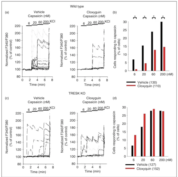
In the next set of experiments, the consequence of TRESK activation on the capsaicin-induced calcium responses of DRG neurons was determined using the TRESK activator cloxyquin. To maintain TRESK channels in their basal (phosphorylated) state, neurons were pretreated with the calcineurin inhibitor cyclo- sporin A (1mM) during the loading period.
Afterwards, neurons were incubated with cloxyquin (30mM) or vehicle control (6.5 mM ethanol) and sub- sequently challenged with increasing concentrations of capsaicin (6, 20, 60 and 200 nM). Representative calci- um imaging traces from wild-type neurons can be seen in Figure 2(a). The responses to the different capsaicin concentrations are summarized as a column graph in Figure 2(b) (control: n¼130, cloxyquin: n¼110 neu- rons) and also as scatter plots (Supplementary Figure 2 (a)–(d): 6, 20, 60 and 200 nM, respectively). Incubation with cloxyquin significantly decreased the ratio of neu- rons responding to capsaicin compared to vehicle con- trol at all tested concentrations (p¼0.020, p¼0.006, p¼0.028 andp¼0.003 for 6, 20, 60 and 200 nM cap- saicin, respectively).
The effects of cloxyquin on the capsaicin sensitivity of TRESK KO DRG neurons was also determined (for representative recordings, see Figure 2(c)). Calcium responses to the different capsaicin concentrations are summarized as a column graph inFigure 2(d)(control:
n¼127; cloxyquin: n¼152 neurons) and also as scatter plots (Supplementary Figure 2(a)–(d): 6, 20, 60 and 200 nM, respectively). In the TRESK-deficient DRG neurons, incubation with cloxyquin did not influence the ratio of capsaicin-responding cells (p¼0.100, p¼0.065, p¼0.43 and p¼0.30 for 6, 20, 60 and 200 nM capsaicin, respectively).
Inhibition of TRESK channels potentiates capsaicin- induced CGRP release
Basal release of CGRP in theex vivodura mater prep- aration of wild-type mice was 27.84.7 pg/ml (n¼15) and 294.6 pg/ml (n¼18) in TRESK KO mice.
Application of A2764 (30mM) or vehicle control (6.5 mM ethanol) had no effect on CGRP release (com- pare the columns labeled vehicle or A2764 to their respective controls in Figures 3(b) and 3(c)). After the solvent or A2764 pretreatment, capsaicin (6 nM) was applied to the dura mater preparation to stimulate CGRP release (see the columns labeled capsaicin in
Figures 3(b) and 3(c)). To determine the effect of A2764 on the capsaicin-evoked CGRP release, the CGRP concentrations measured after capsaicin stimu- lation were normalized to their respective basal values.
In wild-type animals, when the dura mater was pre- treated with vehicle, application of capsaicin increased the CGRP release by 7.38.2% (n¼15). However, when the dura mater was pretreated with A2764,
Wild type
(a) (b)
(c) (d)
TRESK KO Vehicle
Capsaicin (nM)
A2764 Capsaicin (nM) 2 6 20 KCI
0 2 4 6 8
Time (min) Normalized F340/F380 (% of control)
220 200 180 160 140 120 100 80
Vehicle Capsaicin (nM)
A2764 Capsaicin (nM) 2 6 20 KCI
0 2 4 6 8
Time (min) Normalized F340/F380 (% of control)
220 200 180 160 140 120 100 80
2 6 20 KCI
0 2 4 6 8
Time (min) Normalized F340/F380 (% of control)
220 200 180 160 140 120 100 80
2 6 20 KCI
* *
0 2 4 6 8
Time (min)
Normalized F340/F380 (% of control) Cells responding to capsaicin (% of cells)
220
30 25 20 15 10 5 0
2 6 20 (nM) Vehicle (114) A2764 (98)
Cells responding to capsaicin (% of cells) 30 25 20 15 10 5 0
2 6 20 (nM) Vehicle (155) A2764 (176) 200
180 160 140 120 100 80
Figure 1. Effect of the TRESK inhibitor A2764 on the calcium signal of dorsal root ganglion neurons evoked by capsaicin. Dorsal root ganglion neurons loaded with 2mM Fura2-AM were preincubated in control solution (containing ethanol, 6.5 mM) or A2764 (30mM).
Cells were stimulated with increasing concentrations (2, 6 and 20 nM) of capsaicin. As a positive control, at the end of the experiment, cells were depolarized by application of 60 mM KCl, unresponsive cells were excluded from the analysis. Data were normalized to the control value measured before application of capsaicin. (a) Representative calcium imaging traces of wild-type (WT) dorsal root ganglion neurons from a single microscopic field are shown; left panel: Control cells (n¼16), right panel: Cells pretreated with A2764 (n¼17). Application of consecutively increasing capsaicin concentrations and the 60 mM KCl test stimulus are marked with horizontal bars above the graphs. (b) The percentage of wild-type neurons responding to different concentrations of capsaicin are shown as a column graph (cells that responded to capsaicin with an at least 10% increase in F340/F380 signal compared to the control value were considered to be sensitive to capsaicin). Significant differences between the A2764 (red columns) and vehicle-treated groups (black columns) are marked with asterisks (*). (c) Representative calcium imaging traces of TRESK KO dorsal root ganglion neurons from a single microscopic field are shown; left panel: Control cells (n¼19), right panel: Cells pretreated with A2764 (n¼16). Application of consecutively increasing capsaicin concentrations and the 60 mM KCl test stimulus are marked with horizontal bars above the graphs.
(d) The percentage of TRESK KO neurons responding to different concentrations of capsaicin are shown as a column graph (cells that responded to capsaicin with an at least 10% increase in F340/F380 signal compared to the control value were considered to be sensitive to capsaicin).
capsaicin increased CGRP release by 41.013.4%
(n¼15). This increase in capsaicin-induced CGRP release was significantly higher (p¼0.004) than the capsaicin-induced CGRP release of the
vehicle-pretreated control (for a summary, see the right panel in Figure 3(b)). In TRESK KO animals, the increase in capsaicin-induced CGRP release after vehicle treatment was 2.97.4% (n¼18) and 12.2
Vehicle Capsaicin (nM) 6 20 60 200 KCI
0 2 4 6 8
Time (min) Normalized F340/F380 (% of control)
220
(a) (b)
(c) (d)
200 180 160 140 120 100 80
Vehicle Capsaicin (nM) 6 20 60 200 KCI
0 2 4 6 8
Time (min) Normalized F340/F380 (% of control)
220 200 180 160 140 120 100 80
Cloxyguin Capsaicin (nM) 6 20 60 200 KCI
0 2 4 6 8
Time (min) Normalized F340/F380 (% of control)
220 200 180 160 140 120 100 80
Cloxyguin Capsaicin (nM) 6 20 60 200 KCI
0 2 4 6
Time (min) Normalized F340/F380 (% of control)
220 200 180 160 140 120 100 80
* * * *
Cells responding to capsaicin (% of cells) 30 25 20 15 10 5 0
6 20 60 200 (nM) Vehicle (130)
Cloxyquin (110)
Cells responding to capsaicin (% of cells) 30 25 20 15 10 5 0
6 20 60 200 (nM) Vehicle (127)
Cloxyquin (152) Wild type
TRESK KO
Figure 2. Effect of the TRESK activator cloxyquin on the calcium signal of dorsal root ganglion neurons evoked by capsaicin. Dorsal root ganglion neurons loaded with 2mM Fura2-AM were preincubated in control solution (containing ethanol, 6.5 mM) or cloxyquin (30mM). Cells were stimulated with increasing concentrations (6, 20, 60 and 200 nM) of capsaicin. As a positive control, at the end of the experiment, cells were depolarized by application of 60 mM KCl, unresponsive cells were excluded from the analysis. Data were normalized to the control value measured before application of capsaicin. (a) Representative calcium imaging traces of wild-type (WT) dorsal root ganglion neurons from a single microscopic field are shown; left panel: Control cells (n¼20), right panel: Cells pretreated with cloxyquin (n¼13). Application of consecutively increasing capsaicin concentrations and the 60 mM KCl test stimulus are marked with horizontal bars above the graphs. (b) The percentage of wild-type neurons responding to different concentrations of capsaicin are shown as a column graph (cells that responded to capsaicin with an at least 10% increase in F340/F380 signal compared to the control value were considered to be sensitive to capsaicin). Significant differences between the cloxyquin (red columns) and vehicle treated groups (black columns) are marked with asterisks (*). (c) Representative calcium imaging traces of TRESK KO dorsal root ganglion neurons from a single microscopic field are shown; left panel: Control cells (n¼19), right panel: Cells pretreated with cloxyquin (n¼12). Application of consecutively increasing capsaicin concentrations and the 60 mM KCl test stimulus are marked with horizontal bars above. the graphs. (d) The percentage of TRESK KO neurons responding to different concentrations of capsaicin are shown as a column graph (cells that responded to capsaicin with an at least 10% increase in F340/F380 signal compared to the control value were considered to be sensitive to capsaicin).
9.9% after pretreatment with A2764 (for a summary, see the right panel in Figure 3(c)). The difference between the capsaicin-induced CGRP release of the vehicle and A2764 pretreated groups was not signifi- cant (p¼0.23).
Functional condition of TRESK channels modifies TRPV1-mediated changes in meningeal blood flow Basal meningeal blood flow in wild-type and TRESK KO animals measured with laser Doppler flowmetry
30’ Incubation (a)
(b)
(c)
5’ Basal 5’ Vehicle/A2764 5’ Vehicle/A2764 +Capsaicin
100 80 60
CGRP (pg/ml)
40 20
Basal Basal A2764 Vehicle A2764
A2764+Capsaicin Vehicle
Capsaicin 0
100
WT
TRESK KO
300 *
250 200 150 100 50 0 80
60
CGRP (pg/ml) Stimulation by 6 nM capsaicin (% of control)
40 20 0
100 80 60
CGRP (pg/ml)
40 20
Basal Basal A2764 Vehicle
A2764
A2764+Capsaicin Vehicle
Capsaicin 0
100 300
250 200 150 100 50 0 80
60
CGRP (pg/ml) Stimulation by 6 nM capsaicin (% of control)40
20 0
Figure 3. Inhibition of TRESK channels enhances capsaicin-induced CGRP release from trigeminal afferents. Hemisected mouse skull preparation with the adhering dura mater was incubatedex vivoand the CGRP release into the incubation medium was measured during three consecutive 5 min periods. (a) The experimental protocol is summarized as a flowchart. The 30 min preincubation was followed by a 5 min control period (basal release), thereafter solvent or A2764 (30mM) was applied for 5 min; finally, the preparation was stimulated by capsaicin (6 nM). (b) The CGRP release of wild-type (WT) mouse skulls (n¼15 animals) are summarized as box plots; left: Vehicle pretreatment, middle: A2764 pretreatment, right: Normalized values of the third, capsaicin challenge period. The difference in the capsaicin-evoked CGRP release between the vehicle and A2764 treated groups was statistically significant. (c) The CGRP release of TRESK KO mouse skulls (n¼18 animals) are summarized as box plots; left: Vehicle pretreatment, middle: A2764 pretreatment, right: Normalized values of the third, capsaicin challenge period. The difference in the capsaicin-evoked CGRP release between the vehicle and A2764 treated groups was not significant.
was in the same range, varying between 90–250 perfu- sion units (PU). Application of capsaicin (1 nM) failed to induce significant changes in meningeal blood flow (n¼11 and n¼9 measuring probes for wild-type and TRESK KO animals, respectively, see the columns labeled 1 nM capsaicin in Figure 4(a)). We measured a slight but not significant decrease in perfusion in both groups of animals. Meningeal blood flow was reduced to 97.31.6% (p¼0.13) and 94.72.6% (p¼0.08) of baseline in wild-type and TRESK KO animals, respec- tively. Pretreatment with A2764 (30mM) did not signif- icantly influence the meningeal blood flow. In TRESK KO animals, pretreatment with A2764 failed to change the slight vasoconstrictor effect of capsaicin; it reduced meningeal blood flow to 95.31.3% (p¼0.47) of the baseline, as seen in Figure 4(a). In wild-type animals application of capsaicin (1 nM) following pretreatment of the dura mater with A2764 increased meningeal blood flow to 1040.8% of the baseline (Figure 4 (a)). The capsaicin-induced increase in meningeal blood flow after A2764 pretreatment was significantly different from the effect of capsaicin without A2764 pretreatment (p<0.001).
To test the effect of TRESK channel activation on capsaicin-induced blood flow changes we used a higher capsaicin concentration (6 nM). Application of 6 nM capsaicin increased meningeal blood flow in both wild-type (104.50.8%, n¼7,p¼0.001) and TRESK KO animals (103.91.1%, n¼12,p¼0.005), as seen in Figure 4(b). Application of cloxyquin (30mM) had no significant effect on meningeal blood flow. In TRESK KO mice, capsaicin increased blood flow to 101.81.1% of the control value after the pretreat- ment with cloxyquin. This change was not significantly different from the effect of capsaicin without cloxyquin preapplication (p¼0.072).
However, in wild-type mice, when the dura mater was stimulated with capsaicin after pretreatment with cloxyquin, the meningeal blood flow was reduced to 98.52.4% of baseline (Figure 4(b)). This reduction in the effect of capsaicin was statistically significant (p¼0.036).
Discussion
The present study was initiated in an attempt to reveal possible functional cooperation between TRESK back- ground potassium channels and TRPV1 receptors expressed by nociceptors. Therefore wild-type and TRESK KO mice were studied by applying well estab- lished methods of experimental pain and headache research.
The non-selective cation channel TRPV1 is expressed by small-diameter dorsal root and trigeminal ganglion neurons with nociceptor function (25,26).
TRPV1 is an integrator of nociceptive stimuli; it can be activated by various exogenous and endogenous physical and chemical stimuli and it is also a target for pathophysiological processes leading to structural changes and consequent sensitization of the receptor (26). These features make the TRPV1 receptor an ideal contributor to peripheral and central sensitization of the nociceptive pathway. Although only 30–50% of primary sensory neurons express the vasodilator pep- tide CGRP (3,27), sensory neurogenic vasodilatation induced by CGRP release is regarded as a reliable indi- cator of the nociceptor activation. The beneficial effect of various anti-migraine drugs related to their CGRP release inhibiting effect or to the inhibition of the CGRP action supports the important role of this pep- tide in the trigeminal system and the pathophysiology of headaches (11).
The K2P channel TRESK is a major component of the background potassium current in the primary sen- sory neurons of the dorsal root and trigeminal ganglia.
Accordingly, an increased sensitivity to various painful stimuli have been described in TRESK KO animals in different regions of the body (28–32) and reduced abundance of TRESK was associated with pathologi- cal pain conditions (33,34). A recent report suggested that trigeminal neurons are particularly sensitive to altered TRESK activity (29). On the other hand, phar- macological activation (30) or overexpression (33,34) of the channel can reduce cellular excitability and pre- vent the development of increased sensitivity to differ- ent stimuli.
These results, in combination with the sensory neuron-specific expression of the channel (35–39) make TRESK an attractive drug target for the allevia- tion of pain in the disorders of nociception. TRESK was implicated in the pathophysiology of migraine when a dominant negative mutation of TRESK (F139WfsX24) was identified in a family, members of which suffer from migraine with aura (18). Since this first report, further dominant negative mutations of TRESK (W101R, C110R) have been identified in human genetic studies (40–42).
Single-cell RNA sequencing studies performed on mouse somatosensory neurons have shown that TRESK is present in cells also expressing the TRPV1 receptor and CGRP (37,38). Nociceptors of dorsal root and trigeminal ganglia share a lot of similarities regard- ing their receptors and peptide content (43,44). Hence, data obtained in dorsal root ganglion neurons may well also be applicable for trigeminal nociceptors.
Although the TRPV1 channel was originally consid- ered as a cation channel with no voltage sensitivity, patch-clamp studies revealed that temperature sensing is linked to voltage-dependent changes in TRPV1.
Activation of TRPV1 channels is facilitated upon
110
Increase in blood flow (% of baseline before capsaicin) Increase in blood flow (% of baseline before capsaicin)
100
90
80
110
100
90
80
1 nM Capsaicin
A2764+ 1 nM Capsaicin
1 nM Capsaicin
A2764+ 1 nM Capsaicin
110
Increase in blood flow (% of baseline before capsaicin) Increase in blood flow (% of baseline before capsaicin)
100
90
80
110
100
90
80
6 nM Capsaicin
Cloxyquin+ 6 nM Capsaicin
6 nM Capsaicin
Cloxyquin+ 6 nM Capsaicin
WT TRESK KO
WT TRESK KO
A2764 (a)
(b) Cloxyquin
*
*
Figure 4. TRESK channel activity modulates TRPV1-induced changes in meningeal blood flow. Changes in meningeal blood flow induced by capsaicin application (1 or 6 nM) were measured in wild-type (WT) and TRESK KO mice before and after pretreating the dura mater with A2764 (30mM) (a) or cloxyquin (30mM) (b). Significant differences in the effect of capsaicin before and after pretreatment are marked with asterisks (*). (a) In wild-type mice (left), application of A2764 to the dura mater was able to significantly potentiate the effect of 1 nM capsaicin on meningeal blood flow (n¼11 probe positions from n¼6 animals). In TRESK KO mice (right), application of 30mM A2764 did not influence the effect of capsaicin on the meningeal blood flow (n¼9 probe positions from n¼5 animals). (b) In wild-type mice (left), application of 30mM cloxyquin to the dura mater significantly decreased the effect of 6 nM capsaicin on meningeal blood flow (n¼11 probe positions from n¼5 animals). In TRESK KO mice (right), application of cloxyquin did not influence the effect of capsaicin on the meningeal blood flow (n¼12 probe positions from n¼8 animals).
membrane depolarization. As alterations in the mem- brane potential influence, the signaling of TRPV1 (45–
47), we wanted to test whether pharmacological manip- ulation of TRESK may influence the sensitivity of sen- sory neurons to capsaicin and regulate TRPV1- mediated nociceptor functions.
The sensitivity of DRG neurons from wild-type and TRESK KO animals to a potent TRPV1 agonist, cap- saicin, was determined by using cytoplasmic calcium imaging. There was no difference in the capsaicin sen- sitivity of wild-type and TRESK KO neurons, which is in good agreement with a recent study comparing the responses of wild-type and TRESK KO DRG neurons to various TRP channel agonists (32).
However, when wild-type DRG neurons were pre- treated with drugs modulating TRESK activity, the ratio of cells responding tosubmaximalcapsaicin chal- lenges significantly increased (in case of the TRESK inhibitor, A2764) or decreased (in case of the TRESK activator, cloxyquin). In DRGs of TRESK KO ani- mals, incubation with A2764 or cloxyquin did not influence the ratio of capsaicin-responding cells, indi- cating that the changes seen in wild-type neurons were consequences of the modified TRESK activity.
Accordingly, selective pharmacological manipulation of TRESK can regulate the sensitivity to submaximal concentrations of capsaicin in sensory neurons.
Stimulation of TRPV1 receptors releases the potent vasodilator peptide CGRP from nociceptors (6,48). In agreement with our calcium imaging data, in the ex vivo dura mater preparation, inhibition of TRESK by A2764 significantly increased the amount of CGRP released by capsaicin. Application of A2764 had no effect on the CGRP release in TRESK KO animals. Cloxyquin pretreatment failed to affect the capsaicin evoked CGRP release (data not shown). A possible explanation for the lack of effect is that cloxyquin does not stimulate TRESK further if it has been activated via the natural/phys- iological mechanism (19). During the processing of the dura preparation, a central axotomy of the tri- geminal neurons is unavoidable and the consequent calcium signal (and dephosphorylation of TRESK by calcineurin) may well lead to long-lasting activation of the channel (15). Since we could not guarantee the basal, phosphorylated state of TRESK channels in this experimental model, which would have been nec- essary for the efficient activation by cloxyquin in the ex vivo preparation, we did not pursue these experi- ments further.
Release of CGRP from meningeal afferents and the consequent vasodilatation are important components of migraine pathophysiology (10,49). In agreement with the results of our calcium imaging and CGRP
release experiments, inhibition of TRESK potentiated the blood flow increasing effect of capsaicin.
In these experiments, we used a low capsaicin con- centration (1 nM) that failed to increase meningeal blood flow in wild-type animals. Capsaicin application has a dual function in the trigeminovascular system.
Vasodilatation induced by sensory CGRP release and vasoconstriction reducing blood flow are parallel reac- tions observed in the trigeminovascular system upon capsaicin application (6). Vasoconstriction is the result of direct stimulation of vascular TRPV1 recep- tors leading to Ca2þinflow and contraction of the vas- cular smooth muscle cells (50). In wild-type mice the slightly stronger direct vasoconstrictor effect of capsa- icin was turned into a blood flow increasing effect after blocking the TRESK channels.
In our experiments testing the effect of TRESK channel activation by cloxyquin on TRPV1 function, we applied a higher capsaicin concentration (6 nM).
Increases in meningeal blood flow measured in wild- type animals indicate a higher CGRP release that over- rides the direct vasoconstrictor effect of capsaicin.
Pretreatment of the dura mater with cloxyquin reduced the amount of CGRP released by stimulation with cap- saicin, attenuating increases in meningeal blood flow in wild-type animals. In TRESK KO animals, modulators of the TRESK channel failed to affect TRPV1- mediat- ed changes in meningeal blood flow, indicating that the hemodynamic changes seen in wild-type animals were the consequences of the modified TRESK activity.
Although the role of TRESK in the pathogenesis of migraine remains controversial, recent studies in KO animals show that the genetic ablation of TRESK leads to an increased sensitivity to painful stimuli in animal models of migraine (29,30). In the present experiments, specific modulators of TRESK channel activity (both activator and inhibitor, cloxyquin and A2764, respectively) were applied in multiple systems (in vitro, ex vivo and in vivo) in wild-type animals. In addition, a TRESK KO strain was used in order to confirm the specificity of the applied drugs in all exper- imental systems.
Our results indicate that altering the TRESK activ- ity can modify the calcium signal generation of isolated nociceptive neurons, inhibition of TRESK activity increases the liberation of the vasoactive peptide CGRP in a tissue preparation, and the activation or inhibition of the channel also influences the meningeal vascular response to a nociceptive stimulus, capsaicin.
In conclusion, our data show that pharmacological modulation of TRESK activity can be used to manip- ulate nociceptor sensitivity; thus, in the future, selective and potent activators of TRESK may be considered as potential new therapeutics for the treatment of migraine and pain.
Key findings
• In primary sensory neurons, the functional condition of TRESK background potassium channels modifies the calcium signal generation of sensory neurons in response to capsaicin.
• Inhibition of TRESK channels potentiates CGRP release from trigeminal afferents induced by the acti- vation of TRPV1.
• Pharmacological modulation of TRESK channels modifies the TRPV1-mediated increases in meningeal blood flow.
Declaration of conflicting interests
The authors declared no potential conflicts of interest with respect to the research, authorship, and/or publication of this article.
Funding
The authors disclosed receipt of the following financial sup- port for the research, authorship, and/or publication of this article: This work was supported by research grants GINOP- 2.3.2-15-2016-00034, K119597 and K127988 projects of the Hungarian National Research, Development and Innovation Office (NKFIH) and the Higher Education Institutional Excellence Program of the Ministry of Human Capacities in Hungary, within the framework of the Molecular Biology thematic program.
References
1. Deen M, Correnti E, Kamm K, et al. Blocking CGRP in migraine patients – a review of pros and cons.J Headache Pain2017; 18: 96.
2. Edvinsson L, Haanes KA, Warfvinge K, et al. CGRP as the target of new migraine therapies – successful transla- tion from bench to clinic. Nat Rev Neurol 2018; 14:
338–350.
3. Lennerz JK, Ruhle V, Ceppa EP, et al. Calcitonin receptor-like receptor (CLR), receptor activity- modifying protein 1 (RAMP1), and calcitonin gene- related peptide (CGRP) immunoreactivity in the rat tri- geminovascular system: Differences between peripheral and central CGRP receptor distribution. J Comp Neurol2008; 507: 1277–1299.
4. Eftekhari S, Salvatore CA, Calamari A, et al. Differential distribution of calcitonin gene-related peptide and its receptor components in the human trigeminal ganglion.
Neuroscience2010; 169: 683–696.
5. O’Connor TP and van der Kooy D. Enrichment of a vasoactive neuropeptide (calcitonin gene related peptide) in the trigeminal sensory projection to the intracranial arteries.J Neurosci1988; 8: 2468–2476.
6. Dux M, Sa´ntha P and Jancso G. Capsaicin-sensitive neu- rogenic sensory vasodilatation in the dura mater of the rat.J Physiol2003; 552: 859–867.
7. Denner AC, Vogler B, Messlinger K, et al. Role of tran- sient receptor potential ankyrin 1 receptors in rodent models of meningeal nociception – experimentsin vitro.
Eur J Pain2017; 21: 843–854.
8. Edelmayer RM, Le LN, Yan J, et al. Activation of TRPA1 on dural afferents: A potential mechanism of headache pain.Pain2012; 153: 1949–1958.
9. Edvinsson L. The trigeminovascular pathway: Role of CGRP and CGRP receptors in migraine. Headache 2017; 57: 47–55.
10. Iyengar S, Johnson KW, Ossipov MH, et al. CGRP and the trigeminal system in migraine. Headache 2019; 59:
659–681.
11. Russo AF. Calcitonin gene-related peptide (CGRP): A new target for migraine. Annu Rev Pharmacol Toxicol 2015; 55: 533–552.
12. Mickle AD, Shepherd AJ and Mohapatra DP.
Nociceptive TRP channels: Sensory detectors and trans- ducers in multiple pain pathologies. Pharmaceuticals (Basel)2016; 9: 72.
13. Marics B, Peitl B, Varga A, et al. Diet-induced obesity alters dural CGRP release and potentiates TRPA1- mediated trigeminovascular responses. Cephalalgia 2017; 37: 581–591.
14. Dux M, Rosta J and Messlinger K. TRP channels in the focus of trigeminal nociceptor sensitization contributing to primary headaches.Int J Mol Sci2020; 21: 342.
15. Czirja´k G, Toth ZE and Enyedi P. The two-pore domain Kþ channel, TRESK, is activated by the cytoplasmic calcium signal through calcineurin. J Biol Chem 2004;
279: 18550–18558.
16. Kang D and Kim D. TREK-2 (K2P10.1) and TRESK (K2P18.1) are major background Kþchannels in dorsal root ganglion neurons.Am J Physiol Cell Physiol2006;
291: C138–C146.
17. Yoo S, Liu J, Sabbadini M, et al. Regional expression of the anesthetic-activated potassium channel TRESK in the rat nervous system.Neurosci Lett2009; 465: 79–84.
18. Lafreniere RG, Cader MZ, Poulin JF, et al. A dominant- negative mutation in the TRESK potassium channel is linked to familial migraine with aura.Nat Med2010; 16:
1157–1160.
19. Lengyel M, Dobolyi A, Czirja´k G, et al. Selective and state-dependent activation of TRESK (K2P 18.1) back- ground potassium channel by cloxyquin.Br J Pharmacol 2017; 174: 2102–2113.
20. Wright PD, Weir G, Cartland J, et al. Cloxyquin (5-chloroquinolin-8-ol) is an activator of the two-pore domain potassium channel TRESK. Biochem Biophys Res Comm2013; 441: 463–468.
21. Lengyel M, Erdelyi F, Pergel E, et al. Chemically modi- fied derivatives of the activator compound cloxyquin
exert inhibitory effect on TRESK (K2P18.1) background potassium channel.Mol Pharmacol2019; 95: 652–660.
22. Braun G, Lengyel M, Enyedi P, et al. Differential sensi- tivity of TREK-1, TREK-2 and TRAAK background potassium channels to the polycationic dye ruthenium red.Brit J Pharmacol2015; 172: 1728–1738.
23. Ebersberger A, Averbeck B, Messlinger K, et al. Release of substance P, calcitonin gene-related peptide and pros- taglandin E2 from rat dura mater encephali following electrical and chemical stimulation in vitro.
Neuroscience1999; 89: 901–907.
24. Kurosawa M, Messlinger K, Pawlak M, et al. Increase of meningeal blood flow after electrical stimulation of rat dura mater encephali: Mediation by calcitonin gene- related peptide.Br J Pharmacol1995; 114: 1397–1402.
25. Jancso G, Kira´ly E, Jo o F, et al. Selective degeneration by capsaicin of a subpopulation of primary sensory neu- rons in the adult rat.Neurosci Lett1985; 59: 209–214.
26. Tominaga M, Caterina MJ, Malmberg AB, et al. The cloned capsaicin receptor integrates multiple pain- producing stimuli.Neuron1998; 21: 531–543.
27. Hall AK, Ai X, Hickman GE, et al. The generation of neuronal heterogeneity in a rat sensory ganglion.
J Neurosci1997; 17: 2775–2784.
28. Castellanos A, Andres A, Bernal L, et al. Pyrethroids inhibit K2P channels and activate sensory neurons:
Basis of insecticide-induced paraesthesias. Pain 2018;
159: 92–105.
29. Guo Z, Qiu CS, Jiang X, et al. TRESK K(þ) channel activity regulates trigeminal nociception and headache.
eNeuro2019; 6 ENEURO.0236-19.2019 1–23.
30. Pettingill P, Weir GA, Wei T, et al. A causal role for TRESK loss of function in migraine mechanisms.Brain 2019; 142: 3852–3867.
31. Weir GA, Pettingill P, Wu Y, et al. The role of TRESK in discrete sensory neuron populations and somatosensory processing.Front Mol Neurosci2019; 12: 170.
32. Castellanos A, Pujol-Coma A, Andres-Bilbe A, et al.
TRESK background K(þ) channel deletion selectively uncovers enhanced mechanical and cold sensitivity.
J Physiol2020; 598: 1017–1038.
33. Tulleuda A, Cokic B, Callejo G, et al. TRESK channel contribution to nociceptive sensory neurons excitability:
modulation by nerve injury.Mol Pain2011; 7: 30.
34. Yang Y, Li S, Jin ZR, et al. Decreased abundance of TRESK two-pore domain potassium channels in sensory neurons underlies the pain associated with bone metasta- sis.Sci Signal2018; 11: eaao5150 1–20.
35. Manteniotis S, Lehmann R, Flegel C, et al.
Comprehensive RNA-Seq expression analysis of sensory
ganglia with a focus on ion channels and GPCRs in tri- geminal ganglia.PLoS One2013; 8: e79523.
36. Flegel C, Schobel N, Altmuller J, et al. RNA-Seq analysis of human trigeminal and dorsal root ganglia with a focus on chemoreceptors.PLoS One2015; 10: e0128951.
37. Usoskin D, Furlan A, Islam S, et al. Unbiased classifica- tion of sensory neuron types by large-scale single-cell RNA sequencing.Nat Neurosci2015; 18: 145–153.
38. Li CL, Li KC, Wu D, et al. Somatosensory neuron types identified by high-coverage single-cell RNA-sequencing and functional heterogeneity.Cell Res2016; 26: 83–102.
39. LaPaglia DM, Sapio MR, Burbelo PD, et al. RNA-Seq investigations of human post-mortem trigeminal ganglia.
Cephalalgia2017: 38: 912–932.
40. Andres-Enguix I, Shang L, Stansfeld PJ, et al. Functional analysis of missense variants in the TRESK (KCNK18) K channel.Sci Rep2012; 2: 237.
41. Imbrici P, Nematian-Ardestani E, Hasan S, et al. Altered functional properties of a missense variant in the TRESK K(þ) channel (KCNK18) associated with migraine and intellectual disability.Pflu¨gers Archiv – European Journal of Physiology2020; 472: 923–930.
42. Han JY, Jang JH, Park J, et al. Targeted next-generation sequencing of Korean patients with developmental delay and/or intellectual disability.Front Pediatr2018; 6: 391.
43. Kai-Kai MA. Cytochemistry of the trigeminal and dorsal root ganglia and spinal cord of the rat. Comp Biochem Physiol A Comp Physiol1989; 93: 183–193.
44. Waxman SG, Cummins TR, Dib-Hajj S, et al. Sodium channels, excitability of primary sensory neurons, and the molecular basis of pain. Muscle Nerve 1999; 22:
1177–1187.
45. Voets T, Droogmans G, Wissenbach U, et al. The prin- ciple of temperature-dependent gating in cold- and heat- sensitive TRP channels.Nature2004; 430: 748–754.
46. Gunthorpe MJ, Harries MH, Prinjha RK, et al. Voltage- and time-dependent properties of the recombinant rat vanilloid receptor (rVR1).J Physiol2000; 525: 747–759.
47. Nilius B, Talavera K, Owsianik G, et al. Gating of TRP channels: A voltage connection? J Physiol 2005; 567:
35–44.
48. Russell FA, King R, Smillie SJ, et al. Calcitonin gene- related peptide: physiology and pathophysiology.Physiol Rev2014; 94: 1099–1142.
49. Ho TW, Edvinsson L and Goadsby PJ. CGRP and its receptors provide new insights into migraine pathophys- iology.Nat Rev Neurol2010; 6: 573–582.
50. Kark T, Bagi Z, Lizanecz E, et al. Tissue-specific regula- tion of microvascular diameter: opposite functional roles of neuronal and smooth muscle located vanilloid recep- tor-1.Mol Pharmacol2008; 73: 1405–1412.