O R I G I N A L B A S I C S C I E N C E A R T I C L E
Capsaicin-induced rapid neutrophil leukocyte activation in the rat urinary bladder microcirculatory bed
Péter Járomi
1| Dénes Garab
2| Petra Hartmann
2| Dóra Bodnár
2|
Sándor Nyíri
2| Péter Sántha
3| Mihály Boros
2| Gábor Jancsó
3| Andrea Szabó
21Department of Urology, Bács-Kiskun County Teaching Hospital, Kecskemét, Hungary
2Institute of Surgical Research, University of Szeged, Szeged, Hungary
3Department of Physiology, University of Szeged, Szeged, Hungary
Correspondence
Andrea Szabó, Institute of Surgical Research, University of Szeged, Szőkefalvi- Nagy B 6, Szeged H-6720, Hungary.
Email: szabo.andrea.exp@med.u-szeged.hu
Funding information
European Structural and Investment Funds, Grant numbers: GINOP-2.3.2-15-2016- 00034, EFOP-3.6.2-16-2017-00006;
National Research, Development and Innovation Office, Grant number: NKFIH K116689
Aims:This study was initiated to investigate the involvement of neutrophil leukocyte activation in neurogenic inflammation, a process also involved in human urinary pathologies, elicited in the rat urinary bladder by the local administration of capsaicin, the archetypal TRPV1 agonist. The contribution of afferent nerves and sensory neuropeptides to leukocyte activation in the urinary bladder microcirculatory bed was examined.
Methods:Following a 15-min topical application of capsaicin (50μM), leukocyte- endothelial interactions were examined for an observation period of 45 min with intravital microscopy. Expression of adhesion molecules E-selectin and ICAM-1 implicated in these interactions was assessed by immunohistochemistry. Selective sensory denervation was performed by neonatal treatment with capsaicin. The role of the TRPV1 receptor and two sensory neuropeptides (CGRP and substance P [SP]) were studied using the selective antagonists capsazepine, CGRP8-37and RP67580, respectively.
Results:Capsaicin induced rapid increases in leukocyte rolling and adhesion and increased the expression of E-selectin and ICAM-1 in the postcapillary venules.
Sensory chemodenervation via capsaicin and also TRPV1 receptor antagonism effectively prevented these changes. A similar reduction was observed in leukocyte adhesion after topical application of CGRP8-34or RP67580, but only CGRP8-34
reduced the capsaicin-evoked leukocyte rolling.
Conclusions: Topical application of capsaicin induces early neurogenically mediated cellular microcirculatory inflammatory reactions via the activation of the TRPV1 receptor and the release of CGRP and SP from sensory nerves in the bladder.
Co-administration of SP and CGRP receptor antagonists may ameliorate microcirculatory inflammatory changes elicited by capsaicin in the urinary bladder.
K E Y W O R D S
capsaicin, CGRP, leukocyte rolling and adhesion, substance P
Karl-Erik Andersson led the peer-review process as the Associate Editor responsible for the paper.
Neurourology and Urodynamics.2017;1–9. wileyonlinelibrary.com/journal/nau © 2017 Wiley Periodicals, Inc.
|
11 | INTRODUCTION
C-fiber chemosensitive primary sensory neurons innervate a variety of organs and tissues and express the transient receptor potential vanilloid type 1 (TRPV1) receptor.1The activation of the TRPV1 receptor by noxious stimuli results not only in the generation of nociceptive impulses transmitted to the central nervous system, but also in the release of neuro- peptides from the stimulated sensory nerve endings. Neuro- peptides released from sensory nerves initiate an array of local tissue reactions, including vasodilatation and plasma extrav- asation, that is, a neurogenic inflammatory response. There is ample evidence of the role of chemosensitive primary afferents in the transmission of nociceptive impulses and mediation of neurogenic inflammation in a variety of organs and species, including man.1–3 Studies on the innervation of the rat urinary bladder revealed that the organ is richly served by capsaicin-sensitive afferent nerves, the majority of which contain peptides.1 This particular class of sensory nerves significantly contributes to the regulation of urinary bladder functions including the micturition reflexes in both animals in general and man.4,5 Moreover, several pathological con- ditions of the urinary bladder, including neurogenic detrusor overactivity, idiopathic detrusor overactivity, and bladder outlet obstruction involve changes in C-fiber afferent mechanisms.4–6 TRPV1 is involved in the transmission of urinary bladder pain and the application of TRPV1 agonists, and by eliminating capsaicin-sensitive sensory nerves, it has a therapeutic use in the treatment of pathological pain conditions of the urinary bladder. TRPV1 is also critically involved in inflammatory processes of the urinary bladder. In patients suffering from interstitial cystitis/bladder pain syndrome, the severity of the clinical symptoms and inflammation was correlated with the enhanced expression of TRPV1-immunoreactive nerve fibers in the urinary bladder.7 Hence, inflammatory reactions elicited through activation of the TRPV1 receptor by the local/intravesical application of vanilloid agonists, such as capsaicin or resiniferatoxin are of particular interest, as these agonists are used therapeutically to treat disorders such as neurogenic detrusor overactivity.5,8
TRPV1 activation is known to bring about characteristic microcirculatory reactions in several organs. The activation of the TRPV1 receptor, through the release of vasoactive peptides such as calcitonin gene related peptide (CGRP) and substance P (SP) from the sensory nerves, results in an extremely rapid elevation in blood flow and an increase in cutaneous and dural permeability.9,10 TRPV1-mediated vasodilatory effects are partially linked to the release of CGRP in the dura mater and the skin.9,11Another, indirect mechanism involves the enhanced activation of endothelial Ca2+-dependent nitric oxide synthase and consequent nitric oxide release elicited through a TRPV1-dependent rise in
intracellular Ca2+. This is supported by findings showing that the release of nitric oxide could be inhibited by pretreatment with the TRPV1 receptor antagonist capsazepine in vitro12 and by sensory denervation in vivo.13 Edema formation elicited by the activation of TRPV1 receptors, however, is linked to the release of SP and its binding to endothelial neurokinin-1 (NK1) receptors that lead to an increase in vascular permeability and polymorphonuclear leukocyte (PMN) infiltration,10 where the dose-dependent increase in plasma extravasation evoked by SP and capsaicin could be inhibited by capsazepine10,14 or by NK1 receptor antagonism.3
Even though TRPV1 agonists are used in the therapeutic management of urinary bladder disorders, the potential acute microcirculatory side effects of TRPV1 activation have not yet been examined. Therefore, the present study was designed to examine, by using intravital videomicroscopy and immunohistochemistry, the effects of capsaicin, the arche- typal TRPV1 agonist on PMN-endothelial cell interactions in the microcirculatory system of the rat urinary bladder. The contribution of sensory nerves and the sensory neuropeptides SP and CGRP to these inflammatory phenomena was studied with the aid of selective sensory chemodenervation and pharmacological antagonists.
2 | MATERIALS AND METHODS
The project was approved by the National Scientific Ethical Committee on Animal Experimentation (National Competent Authority), with the license number: V./144/2013. The study was performed in adherence with the EU Directive 2010/63/
EU on the protection of animals used for experimental and other scientific purposes and the National Institute of Health guidelines for the use of experimental animals.
2.1 | Neonatal capsaicin treatment
Newborn male Sprague-Dawley rats were injected subcuta- neously with a single dose of 50 mg kg−1 capsaicin or its vehicle (6% ethanol and 8% Tween 80 in saline) on day 2 of life under ether anesthesia. Neonatal capsaicin treatment has been shown to produce a selective degeneration of chemo- sensitive afferent nerves which are sensitive to capsaicin and express the TRPV1 receptor.1,15The eye-wiping test, using a 1% solution of zingerone dripped into the eyes of rats on the day of the experiment, was used to demonstrate the elimination of TRPV1-expressing afferent nerves.15
2.2 | Surgical procedures
Healthy adult Sprague-Dawley rats (and the average weight was 320 ± 10 g) were anesthetized intraperitoneally with an
initial dose of sodium pentobarbital (45 mg kg−1) and placed in a supine position on a heating pad to maintain the body temperature at 36-37 °C. The trachea, the carotid artery, and jugular vein were cannulated to facilitate spontaneous respiration, and for continuous measurement of blood pressure and the injection of fluorescent dyes, respectively.
There were no significant differences in the blood pressure among the different experimental groups (see later) at any time-point of the microcirculation measurements (data not shown).
The urinary bladder was prepared for intravital micro- scopic (IVM) examination using a modified method of Bajory et al.16 Following a midline laparotomy, the bladder was exposed and the median umbilical ligament resected. A polyethylene catheter (ID 0.28 mm, OD 0.61 mm; Smiths Medical, UK) was later inserted into the bladder dome and fixed by a ligature (via a 3-0 braided polyglycolic acid thread.
As the catheter insertion caused nearly complete emptying of the bladder, 0.5 mL of a body-warm 0.9% NaCl solution was injected to provide a similar, standard baseline filling state of the bladders in all of the experiments. In pilot studies, the resulting intravesical pressure (<7 mm Hg) did to cause any microcirculatory disturbances in the bladder. The remaining abdomen was covered with a Saran wrap to avoid fluid and heat loss and drying out of the surface of the abdominal organs.
2.3 | Experimental protocol
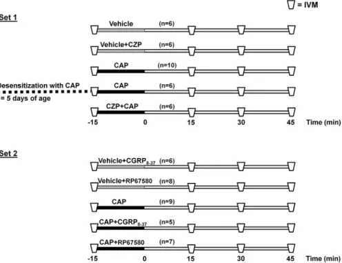
The aim of our experiments was to examine the microcircu- latory inflammatory consequences of a 15-min topical application of capsaicin on the bladder surface. We used two major sets (see Figure 1). In Set 1, the effects of TRPV1 receptor antagonism as well as the effects of ablation of TRPV1-positive nerves (achieved by the neonatal adminis- tration of capsaicin) were examined. In Set 2, the effects of selective CGRP and NK1 receptor antagonists were studied by monitoring the capsaicin-induced cellular inflammatory changes. All topical treatments were performed by placing pieces of Gelaspon®sponge (Chauvin Ankerpharm GmbH, Berlin, Germany) soaked in the respective compound(s) onto the anterior surface of the bladder for 15 min.
In the first set of experiments, the animals were allotted to one or the other of the following experimental groups: the anterior surface of the bladder was topically treated with capsaicin or its vehicle by dripping 100μL of the solution on a Gelaspon sponge which was placed on the bladder dome for 15 min in the first two groups (vehicle and capsaicin groups, n= 6, and n= 10, respectively). The effect of the specific TRPV1 inhibitor capsazepine on the microcirculatory changes in two groups where capsazepine was topically applied just before the administration of capsaicin or its vehicle (vehicle + capsazepine and capsaicin + capsazepine group, n= 6–6). These findings
were compared with those obtained in rats (n= 6) treated with capsaicin as neonates.15 In the second set of experi- ments, the effects of the capsaicin receptor antagonist capsazepine were studied on the capsaicin-induced inflam- matory changes. During these experiments (the second set), the microcirculatory effect of the CGRP and NK1 antagonism was examined and CGRP8-37 (10μM) and RP67580 (10μM) were applied on the bladder surface in the presence (capsaicin + CGRP8-37; n= 5) and capsai- cin + RP67580; n= 7) and in the absence of topical capsaicin (vehicle + CGRP8-37; n= 6), and vehicle + RP 67580;n= 8) (50μM). After a 15-min exposure, the sponge was removed from the bladder surface and thoroughly rinsed off with warm physiological saline and covered with a Saran wrap for the remainder of the duration. IVM recordings were made 15, 30, and 45 min after topical treatments. This overall 60-min observation period was chosen because accumulation of the fluorescence tracers in the urine with resulting deterioration in the contrast of the images did not permit a longer observation period.
2.4 | Intravital microscopy
The anterior surface of the bladder microcirculation was visualized by IVM (Zeiss Axiotech Vario 100HD microscope;
Carl Zeiss GmbH, Jena, Germany; 100-W HBO mercury lamp;
Acroplan 20× water immersion objective). The animals received intravenous injections of fluorescein isothiocyanate (FITC)-labeled erythrocytes17(0.2 mL; Sigma, St. Louis, MO) for the verification of the intact perfusion of the examined microvascular bed (using the 490/525 nm filter set of the microscope). Leukocytes were stained by injecting rhodamine 6G intravenously (0.2%, 0.1 mL; Sigma) and examined by a 526/555 nm filter set (Figure 2). Images were recorded with a charge-coupled device video camera (Teli CS8320Bi, Toshiba Teli Corporation, Osaka, Japan) attached to an S-VHS video- recorder (Panasonic AG-MD 830; Matsushita Electric Industrial Co., Tokyo, Japan) and a personal computer.
2.5 | Analysis of microcirculation data
A quantitative evaluation of the microcirculatory parameters was performed off-line by a frame-to-frame analysis of the videotaped images taken for IVM (IVM Software; Pictron Ltd, Budapest, Hungary). Leukocyte-endothelial cell interactions were analyzed in four or more postcapillary venules within a minimum of three observation fields per rat (choosing the most representative vessels). All of the evaluations were performed by two investigators. Rolling leukocytes were defined as cells moving with a velocity less than 40% of that of the erythrocytes in the centerline of the microvessel and passing through the observed vessel segment being observed within 30 s, and are expressed as the number of cells per mm per sec.18Adherent
leukocytes were defined as cells that did not move or detach from the endothelial lining within an observation period of 30 s and are expressed as the number of cells per mm2of endothelial surface, calculated using the diameter and length of the vessel segment. In this study, postcapillary venules with a diameter of 15–20 micrometers were used and the average vessel length was about 80 µm.
2.6 | Immunohistochemistry (E-selectin and ICAM-1)
E-selectin and intercellular adhesion molecule-1 (ICAM-1) are endothelium-derived adhesion molecules that play potential roles during the course of leukocyte rolling and adhesion onto the endothelium within the postcapillary FIGURE 1 The time sequence of treatments and measurements in experimental groups in Sets 1 and 2. The topical treatments with capsaicin (CAP, 50μM) or other compounds (see below) were performed at the anterior surfaces of the bladders for 15 min. In Set 1, the TRPV1 receptor antagonist capsazepine (CZP, 200μM topically) and ablation of the TRPV1-positive nerves (achieved by the neonatal administration of CAP) were applied. In Set 2, the consequences of selective CGRP and NK1 receptor antagonism were investigated by topically applying the selective CGRP receptor antagonist CGRP8-37 (10μM) and the NK1 receptor antagonists RP67580 (10μM) together with CAP. Here, IVM = intravital microscopic examinations of leukocyte-endothelial interactions in postcapillary venules of the bladder
FIGURE 2 Representative micrographs showing the sequence of PMN−endothelial interactions on three consecutive images (Panels A-C) recorded by using intravital microscopy (recording rate: 20 frames/s). A segment of the examined postcapillary vein in the urinary bladder is surrounded by lines in Panel A. Movement of rhodamine 6G-labeled PMNs (marked by a-e) is demonstrated frame-by-frame relative to a dashed line. Stationary (adhesive) leukocytes are marked by an ellipse. The bar in Panel C denotes a 100μM scale and this applies to all the
photomicrographs
venules; hence, we decided to measure their expression in the bladder in response to capsaicin. Forty-five minutes follow- ing topical application of capsaicin onto the urinary bladder, the organ was fixed by immersion in a fixative containing 4%
paraformaldehyde in a phosphate buffer (0.1 M, pH = 7.4) for 2 h at 4°C. Tissue samples were then transferred into phosphate buffer until further processing. Frozen sections 15 µm in thickness were cut with a cryostat and prepared for the demonstration of E-selectin and ICAM-1 immunoreac- tivity using the indirect double labeling immunohistochemi- cal technique. The sections were incubated overnight with mouse anti rat anti-ICAM-1 (clone 1A29; BD Pharmingen, San Jose, CA, 1:1000) and goat anti rat E-selectin (R&D Systems, Minneapolis, MN, 1:1200) primary antibodies dissolved in phosphate buffered saline containing 0.3%
Triton X-100. These sections were then incubated with dylight 488 conjugated donkey anti mouse and Cy3- conjugated donkey anti goat secondary antibodies (both from Jackson Immunoresearch Europe, Newmarket, UK).
They were covered with a ProlongGold mounting medium containing DAPI. Immunostained sections were then exam- ined under a Zeiss LSM 700 confocal microscope. One micrometer thin optical sections were collected via Z-stack capturing and the maximal intensity projection function was used to reconstruct an image of the 12 µm thick block of the tissue. Photomicrographs of control and capsaicin-exposed specimens were taken under identical conditions following a systemic random sampling method. Three bladders per group, three representative sections per bladder, and 5–9 regions per cryosection were selected for a quantitative evaluation. The observer obtained and evaluated photomicrographs of bladder specimens of the control and treated groups in a blinded manner. Relative pixel intensity data of postcapillary venule profiles (n= 24-30 for each experimental group) were collected by using the line profile measurement tool of the
ImagePro Plus 6 image analysis software package (Media Cybernetics, Rockville, MD).
2.7 | Statistical analysis
The minimum number of animals per group was determined based on the “PS: Power and Sample Size Calculator” software package. Data analysis was performed by means of the SigmaStat statistical software package (Jandel Corpora- tion, San Rafael, CA). Changes in microcirculation variables within and between groups were analyzed by two-way repeated measures ANOVA test, followed by the Holm-Sidak test. Immunohistochemistry data were analyzed using the two-way ANOVA test. All data values are expressed as means ± standard error of the mean (SEM), and P values
< 0.05 were considered statistically significant.
3 | RESULTS
The effect of selective sensory
chemodenervation and TRPV1 antagonism on the capsaicin-induced microcirculatory changes of the urinary bladder
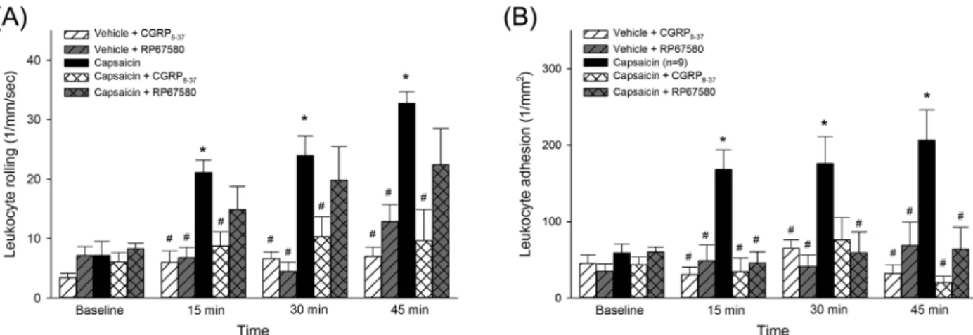
Topical application of capsaicin resulted in a marked increase in the degree of PMN-endothelial interactions in the postcapillary venules of the urinary bladder. Both primary (rolling, Figure 3A) and secondary interactions (adhesion, Figure 3B) were significantly enhanced within 15 min after topical treatment of capsaicin and remained elevated during the entire observation period of 45 min. Life-long sensory chemodenervation by neonatal capsaicin treatment completely prevented the capsaicin-induced vascular changes and indicated that the vascular effects of capsaicin are not direct, but are mediated through the release of vasoactive
FIGURE 3 Time course of changes in leukocyte rolling (A) and adhesion (B) in the postcapillary venules of the bladder after a 15-min topical exposure to capsaicin or vehicle. In other groups, TRPV1 receptor antagonism (via capsazepine, 200μM) and the ablation of TRPV1-positive nerves by neonatal capsaicin treatment were also applied. Values are presented as means ± SEM. Here, *P< 0.05 versus baseline; #P< 0.05 versus vehicle
agents from the sensory nerves. Likewise, the co- administration of capsazepine, a competitive TRPV1 anta- gonsist, markedly inhibited the intravascular inflammatory changes elicited by capsaicin. Last, neither capsazepine nor the vehicle for capsaicin produced any changes in PMN-mediated reactions.
The effects CGRP and NK1 receptor antagonists on TRPV1-activation-induced microcirculatory reactions of the urinary bladder
In the urinary bladder, the capsaicin-induced increase in leukocyte rolling was prevented by the topical co- administration of the specific CGRP receptor antagonist CGRP8-34 (see Figure 4A), but not by the NK1 receptor antagonist RP67580. However, PMN adhesion was signifi- cantly reduced by both the CGRP and the NK1 receptor antagonists (Figure 4B).
The effect of capsaicin on the expression of E-selectin and ICAM-1 in the urinary bladder microvasculature
In control urinary bladders, we noticed a faint-to-moderate staining of small venules with antibodies raised against E-selectin (Figures 5A and 5C) and ICAM-1 (Figures 5B and 5C). Topical application of capsaicin onto the urinary bladder resulted in a massive increase in both E-selectin (Figures 5D and 5F) and ICAM-1 (Figures 5E and 5F) immunoreactivities in small venules. A quantitative evalua- tion of the staining intensity revealed a significant increase in the relative pixel intensity for both E-selectin (control:
8.11 ± 2.97; capsaicin: 35.14 ± 3.45;P< 0.05) and ICAM-1
(control: 5.45 ± 2.71; capsaicin: 33.59 ± 3.24; P< 0.05) immunostaining.
4 | DISCUSSION
TRPV1 agonism represents one of the possible treatment modalities of overactive bladder syndrome,5,6 and the involvement of these afferent nerves in inflammatory processes of the urinary bladder was demonstrated earlier in both animal models6 and human diseases such as the painful bladder syndrome and interstitial cystitis.7 The cellular inflammatory processes and their dynamics associ- ated with the topical administration of TRPV1 agonists, such as capsaicin and resiniferatoxin, have not yet been investi- gated. The present study is the first to demonstrate the dynamics of leukocyte-endothelial interactions following TRPV1 receptor activation evoked by capsaicin in the bladder. An early increase in the frequency of endothelial- PMN interactions (both rolling and adhesion) was observed in the bladder postcapillary venules as early as 15 min after the administration of capsaicin and these inflammatory changes persisted throughout the entire experimental period (45 min).
The early onset of inflammatory changes following capsaicin administration is in line with previous observations that found a rapid development of NK1 receptor-dependent edema formation and PMN deposition after capsaicin in the skin10 and in the pancreas,14 and an even more rapid response occurring within minutes following an intratracheal adminis- tration of capsaicin.19
We have also demonstrated that the microvascular changes were brought about via a specific action of capsaicin on sensory nerves expressing TRPV1 receptors, as capsaicin- induced cellular inflammatory reactions were strongly inhibited by both TRPV1 receptor antagonism and the
FIGURE 4 Time course of changes in leukocyte rolling (A) and adhesion (B) in postcapillary venules of the bladder following a 15-min topical exposure to capsaicin or vehicle. The effects of the selective CGRP antagonist CGRP8-37 (10μM) and the NK1 receptor antagonists RP67580 (10μM) are also shown. Values are presented as means ± SEM. Here, *P< 0.05 versus baseline; #P< 0.05 versus vehicle
elimination of TRPV1-expressing nociceptive primary sensory neurons by neonatal capsaicin treatment. These observations suggest a neurogenic origin of the inflammatory process developing in the urinary bladder after the topical administration of capsaicin. The present findings also suggest that the effects of capsaicin are indirect and brought about by the release of pro-inflammatory neuropeptides known to be released from activated TRPV1-expressing sensory nerves.1,6 As SP and CGRP are the most abundant vasoactive peptides expressed in intact primary sensory neurons, we focused on the contribution of these neuropeptides to the capsaicin- induced microcirculatory reactions. The findings indicate that both CGRP and SP contribute to the capsaicin-induced endothelial adhesion of PMNs, but rolling is initiated only by CGRP. It is reasonable to assume therefore that TRPV1- induced CGRP release initiates the endothelial-PMN interaction by promoting leukocyte rolling, but adhesion is influenced by both peptides. A similar conclusion was drawn in another study, which showed that SP potentiated neutrophil accumulation in the skin only when it was released at a site where an inflammatory process had already been initiated.20
In the urinary bladder, the capsaicin-induced expression of endothelial adhesion molecules, which play an essential role in
microvascular inflammatory reactions, had not been examined up till now. In our study, the early increase in the expression of E-selectin and ICAM-1 immunoreactivities in the postcapillary venules of the urinary bladder occurred in parallel with the cellular microvascular changes. Previous studies on the human cutaneous vascular bed also demonstrated a very rapid PMN accumulation (in approximately 15 min) after intradermal injection of SP or CGRP21and this is linked to an increased expression of ICAM-1 mRNA (within 60 min after SP exposure).22 Enhanced ICAM-1 and E-selectin protein ex- pressions develop over a longer time-frame (these were typically examined 4 h or more after the topical application of SP in the skin).21–23 As for the urinary bladder, the NK1 receptor- mediated expression of ICAM-1 occurred as early as an hour after a local arterial injection of SP.24 Still, little is known regarding the subcellular regulatory mechanism by which the TRPV-related neuropeptide release modulates these changes and the pathway by which capsaicin achieved these effects remained basically unexplored. The remarkably rapid (less than 60 min) increase in the expression of endothelium-derived adhesion molecules demonstrated here probably cannot be explained by the most time-consuming Ca2+-dependent activa- tion of NF-κB pathways (as shown elsewhere).23 The involvement of mechanisms related to a capsaicin-induced FIGURE 5 Confocal photomicrographs illustrating the immunohistochemical localization of ICAM-1 (A, D) and E-selectin (B, E) in urinary bladders 45 min after exposure to the vehicle (A, C) and capsaicin (D, F). Note the marked capsaicin-induced increase in immunostaining of both ICAM-1 and E-selectin localized to venules on the lower panels. The merged photomicrographs (C, F) illustrate the close colocalization of ICAM-1 and E-selectin immunoreactivities. The bar in F denotes 50μM scale and this applies to all photomicrographs
intracellular free radical production, however, cannot be ruled out.24 Clearly, the mechanistic background of the particularly rapid adhesion molecule expression in the rat urinary bladder warrants a further in-depth investigation.
The present findings may also be of relevance to neurogenic inflammatory processes that develop in other organs under particular pathological conditions. The dynam- ics and extent of the inflammatory process may depend not only on the sensory innervation density and the distribution of TRPV1 receptors, but also on other factors such as the density of CGRP and SP receptors and the adhesion molecule expression of the particular tissue/organ.
Last, the potential clinical relevance of the present study should be mentioned, as TRPV1-expressing bladder sensory nerves are important therapeutic targets in the treatment of the overactive bladder syndrome and related disorders of the urinary bladder such as painful bladder syndrome and interstitial cystitis. Quite significantly, in patients with idiopathic detrusor overactivity, the TRPV1 agonist vanilloid compounds like resiniferatoxin and capsaicin are effective only in patients that display an overexpression of the TRPV1 receptor in the urinary bladder.7,25 The present findings suggest that, in the urinary bladder, the activation of TRPV1 receptor-expressing nociceptive afferent nerves results in a co-ordinated inflammatory process involving not only arteriolar vasodilatation and plasma extravasation, but also an increase in endothelial-leukocyte interactions that lead to the transmigration of leukocytes. Even though these neurogenic inflammatory changes may be transient due to ensuing rapid defunctionalization/chemodenervation of sen- sory nerves,9they may interfere with urinary bladder function following intravesical administration of vanilloid compounds for therapeutic purposes. Hence, the inhibition of this inflammatory process by using neuropeptide antagonists (such as CGRP antagonists) which are already approved for other human therapeutic purposes (eg, for migraine approved by the FDA) may be beneficial in the treatment of urinary bladder disorders that utilize vanilloid compounds.
ACKNOWLEDGMENTS
This study was supported by European Structural and Investment Fund grants GINOP-2.3.2-15-2016-00034, EFOP-3.6.2-16-2017-00006, and Hungarian National Research, Development and Innovation Office grant NKFIH K116689. The authors are grateful to Csilla Mester and Nikolett Beretka for their assistance.
CONFLICT OF INTEREST
The authors hereby declare that they had no conflict of interest, financial or otherwise, when they participated in this study.
REFERENCES
1. Szallasi A, Blumberg PM. Vanilloid (Capsaicin) receptors and mechanisms.Pharmacol Rev. 1999;51:159–212.
2. Moskowitz MA. Neurogenic inflammation in the pathophysiology and treatment of migraine.Neurology. 1993;43:16–20.
3. Nathan JD, Peng RY, Wang Y, McVey DC, Vigna SR, Liddle RA.
Primary sensory neurons: a common final pathway for inflamma- tion in experimental pancreatitis in rats.Am J Physiol. 2002;283:
G938–G946.
4. de Groat WC, Yoshimura N. Afferent nerve regulation of bladder function in health and disease. Handb Exp Pharmacol. 2009;
194:91–138.
5. Fowler CJ, Jewkes D, McDonald WI, Lynn B, de Groat WC.
Intravesical capsaicin for neurogenic bladder dysfunction.Lancet.
1992;339:1239.
6. Cruz F, Guimaräes M, Silva C, Reis M. Suppression of bladder hyperreflexia by intravesical resiniferatoxin. Lancet. 1997;350:
640–641.
7. Liu L, Mansfield KJ, Kristiana I, Vaux KJ, Millard RJ, Burcher E.
The molecular basis of urgency: regional difference of vanilloid receptor expression in the human urinary bladder. Neurourol Urodyn. 2007;26:433–438.
8. Kuo HC, Liu HT, Yang WC. Therapeutic effect of multiple resiniferatoxin intravesical instillations in patients with refractory detrusor overactivity: a randomized, double-blind, placebo controlled study.J Urol. 2006;176:641–645.
9. Dux M, Sántha P. Capsaicin-sensitive neurogenic sensory vasodilatation in the dura mater of the rat.J Physiol. 2003;552:
859–867.
10. Boros K, Dux G, Fekécs M, et al. Multiple impairments of cutaneous nociceptor function induced by cardiotoxic doses of Adriamycin in the rat.Naunyn Schmiedebergs Arch Pharmacol.
2016;389:1009–1020.
11. Williamson DJ, Hargreaves RJ, Hill RG, Shepheard SL. Intravital microscope studies on the effects of neurokinin agonists and calcitonin gene-related peptide on dural vessel diameter in the anaesthetized rat.Cephalalgia. 1997;17:518–524.
12. Ching LC, Kou YR, Shyue SK, et al. Molecular mechanisms of activation of endothelial nitric oxide synthase mediated by transient receptor potential vanilloid type 1. Cardiovasc Res. 2011;91:
492–501.
13. Poblete IM, Orliac ML, Briones R, Adler-Graschinsky E, Huidobro-Toro JP. Anandamide elicits an acute release of nitric oxide through endothelial TRPV1 receptor activation in the rat arterial mesenteric bed.J Physiol. 2005;568:539–551.
14. Grady EF, Yoshimi SK, Maa J, et al. Substance P mediates inflammatory oedema in acute pancreatitis via activation of the neurokinin-1 receptor in rats and mice. Br J Pharmacol. 2000;
130:505–512.
15. Bajory Z, Hutter J, Krombach F, Messmer K. New method: the intravital videomicroscopic characteristics of the microcircu- lation of the urinary bladder in rats. Urol Res. 2002;30:
148–152.
16. Jancsó G, Kiraly E, Jancsó-Gábor A. Pharmacologically induced selective degeneration of chemosensitive primary sensory neurones.Nature. 1977;270:741–743.
17. Ruh J, Ryschich E, Secchi A, et al. Measurement of blood flow in the main arteriole of the villi in rat small intestine with FITC-labeled erythrocytes.Microvasc Res. 1998;56:62–69.
18. Massberg S, Enders G, Leiderer R, et al. Platelet-endothelial cell interactions during ischemia/reperfusion: the role of P-selectin.
Blood. 1998;92:507–515.
19. Baluk P, Bertrand C, Geppetti P, McDonald DM, Nadel JA. NK1 receptors mediate leukocyte adhesion in neurogenic inflammation in the rat trachea.Am J Physiol. 1995;268:L263–L269.
20. Cao T, Pintér E, Al-Rashed S, Gerard N, Hoult JR, Brain SD.
Neurokinin-1 receptor agonists are involved in mediating neutrophil accumulation in the inflamed, but not normal, cutaneous microvas- culature: an in vivo study using neurokinin-1 receptor knockout mice.
J Immunol. 2000;164:5424–5429.
21. Smith CH, Barker JN, Morris RW, MacDonald DM, Lee TH.
Neuropeptides induce rapid expression of endothelial cell adhesion molecules and elicit granulocytic infiltration in human skin.
J Immunol. 1993;151:3274–3282.
22. Quinlan KL, Song IS, Bunnett NW, et al. Neuropeptide regulation of human dermal microvascular endothelial cell ICAM-1 expression and function. Am J Physiol. 1998;275:
C1580–C1590.
23. Lindsey KQ, Caughman SW, Olerud JE, Bunnett NW, Armstrong CA, Ansel JC. Neural regulation of endothelial cell-mediated inflammation.J Investig Dermatol Symp Proc. 2000;5:74–78.
24. Chien CT, Lin TB, Lai MK, Hsu SM. Substance P via NK1 receptor facilitates hyperactive bladder afferent signaling via action of ROS.Am J Physiol. 2003;284:F840–F851.
25. Kissin I, Szallasi A. Therapeutic targeting of TRPV1 by resiniferatoxin, from preclinical studies to clinical trials. Curr Top Med Chem. 2011;11:2159–2170.
SUPPORTING INFORMATION
Additional Supporting Information may be found online in the supporting information tab for this article.
How to cite this article:Járomi P, Garab D, Hartmann P, et al. Capsaicin-induced rapid neutrophil leukocyte activation in the rat urinary bladder microcirculatory bed.Neurourology and Urodynamics. 2017;1–9.
https://doi.org/10.1002/nau.23376