The Role of Diacylglycerol Lipase in Constitutive and Angiotensin AT 1 Receptor- stimulated Cannabinoid CB1 Receptor Activity *
Received for publication, December 26, 2006, and in revised form, January 11, 2007 Published, JBC Papers in Press, January 16, 2007, DOI 10.1074/jbc.C600318200
Ga´bor Turu‡§, Anne Simon¶, Pa´l Gyombolai‡, La´szlo´ Szidonya‡, Gyo¨rgy Bagdy储**, Zsolt Lenkei¶, and La´szlo´ Hunyady‡§1 From the‡Department of Physiology, Semmelweis University, Faculty of Medicine, H-1444 Budapest, Hungary, the§Laboratory of
Neurobiochemistry and Molecular Physiology, Hungarian Academy of Sciences and Semmelweis University, H-1444 Budapest, Hungary, the
¶Laboratoire Neurobiologie et Diversite´ Cellulaire, Ecole Supe´rieure de Physique et de Chimie Industrielles-Centre National de la Recherche Scientifique, Unite´ Mixte de Recherche 7637, 75013 Paris, France, the
**Laboratory of Neurochemistry and Experimental Medicine, National Institute of Psychiatry and Neurology, H-1021 Budapest, Hungary, and the
储Group of Neuropsychopharmacology, Hungarian Academy of Sciences and Semmelweis University, H-1089 Budapest, Hungary
The cannabinoid CB1 receptor (CB1R) is a G protein-coupled receptor, which couples to the Gi/ofamily of heterotrimeric G pro- teins. The receptor displays both basal and agonist-induced signal- ing and internalization. Although basal activity of CB1Rs is attrib- uted to constitutive (agonist-independent) receptor activity, studies in neurons suggested a role of postsynaptic endocannabi- noid (eCB) release in the persistent activity of presynaptic CB1Rs.
To elucidate the role of eCBs in basal CB1R activity, we have inves- tigated the role of diacylglycerol lipase (DAGL) in this process in Chinese hamster ovary (CHO) cells, which are not targeted specif- ically with eCBs. Agonist-induced G protein activation was deter- mined by detecting dissociation G protein subunits expressed in CHO cells with bioluminescence resonance energy transfer (BRET), after labeling the␣andsubunits with Renilla luciferase and enhanced yellow fluorescent protein (EYFP), respectively. Pre- incubation of the cells with tetrahydrolipstatin (THL), a known inhibitor of DAGLs, caused inhibition of the basal activity of CB1R.
Moreover, preincubation of CHO and cultured hippocampal neu- rons with THL increased the number of CB1Rs on the cell mem- brane, which reflects its inhibitory action on CB1R internalization in non-simulated cells. In CHO cells co-expressing CB1R and angiotensin AT1receptors, angiotensin II-induced Goprotein acti- vation that was blocked by both a CB1R antagonist and THL. These data indicate that cell-derived eCB mediators have a general role in the basal activity of CB1Rs in non-neural cells and neurons, and that this mechanism can be stimulated by AT1receptor activation.
The cannabinoid CB1 receptor (CB1R)2, a member of the G protein-coupled receptor (GPCR) superfamily (1), is activated by several lipid-derived endocannabinoid (eCB) ligands includ- ing anandamide and 2-arachydonyl-glycerol (2-AG) (2, 3).
Anandamide production in cells is a phospholipase D-depend- ent process, whereas 2-AG is generated by diacylglycerol lipases (DAGLs) from diacylglycerol (DAG) (4). The receptor is widely distributed in several tissues and is one of the most abun- dant GPCRs in the brain, being predominantly localized in axons of GABAergic neurons (5, 6). CB1R agonists are already used in chemotherapy-induced nausea and vomiting of cancer patients who have failed to respond adequately to conventional antiemetic therapy, whereas CB1R antagonists have recently been introduced in the therapy of obesity and nicotine addic- tion (4). Furthermore, modulating CB1R activity has therapeu- tical potential in a wide range of pathological conditions includ- ing mood and anxiety disorders, movement disorders, neuropathic pain, and multiple sclerosis, as well as cancer, car- diovascular diseases, obesity/metabolic syndrome, and muscu- loskeletal disorders (4, 7). CB1R couples mainly to the Gi/ofam- ily of heterotrimeric G proteins. However, recent data suggest that it can also couple to Gqproteins (4). CB1Rs mediate retro- grade signaling of activated neurons (4). Furthermore, in many cellular systems non-stimulated CB1Rs produce constitutive signal generation and internalization (8). Constitutive internal- ization of CB1Rs has been reported both in HEK-293 cells and in neurons (9, 10). This process can be blocked by inverse ago- nists and is required for the correct axonal localization of CB1Rs in neurons, which underlines the physiological impor- tance of this process (10). Although these effects are generally attributed to the constitutive activity of CB1Rs (8), continuous stimulation by eCBs can serve as an alternative mechanism to explain these data. Theoretically, a validated neutral CB1R antagonist,i.e.an antagonist without inverse agonist activity could arbitrate between these two alternative models, but no such molecule has been generated for CB1Rs. Recent papers proposed a role of continuous postsynaptic eCB release in the persistent inhibition of inhibitory postsynaptic currents in hip- pocampal and proopiomelanocortin neurons. This was shown to be a homosynaptic effect, based on findings that chelation of postsynaptic intracellular Ca2⫹with BAPTA interferes with the stimulatory effect of CB1R antagonists on presynaptic GABA release (11, 12). Similar findings were also reported in basolat- eral amygdala neurons (13). However, other studies showed that constitutive activity of expressed CB1Rs can also be detected in the absence of eCBs both in neurons and CHO cells, suggesting that the receptor is constitutively active (8, 14 –17).
Furthermore, constitutive internalization of CB1R is also
*This work was supported in part by grants from the Hungarian Science Foun- dation (OTKA T-046445, M-045341, and Ts-049851), the A´nyos Jedlik program (NKFP1– 010/2005), the Hungarian Ministry of Public Health (ETT 447/2006 and 460/2006), and the European Community (LSHM-CT-2004 –503474). The costs of publication of this article were defrayed in part by the payment of page charges. This article must therefore be hereby marked “advertisement” in accordance with 18 U.S.C. Section 1734 solely to indicate this fact.
1To whom correspondence should be addressed: Dept. of Physiology, Sem- melweis University, H-1444 Budapest, P. O. Box 259, Hungary. Tel.: 36-1- 266-9180; Fax: 36-1-266-6504; E-mail: Hunyady@puskin.sote.hu.
2The abbreviations used are: CB1R, cannabinoid CB1 receptor; 2-AG, 2-arachydonyl-glycerol; AngII, angiotensin II; AT1R, angiotensin AT1recep- tor; BRET, bioluminescence resonance energy transfer; GFP, green fluores- cent protein; EGFP, enhanced GFP; EYFP, enhanced yellow fluorescent pro- tein; CB1R-EYFP, EYFP-tagged CB1R; DAG, diacylglycerol; DAGL, diacylglycerol lipase; eCB, endocannabinoid; THL, tetrahydrolipstatin;
BAPTA, 1,2-bis(2-aminophenoxy)ethane-N,N,N⬘,N⬘-tetraacetic acid; GABA,
␥-aminobutyric acid; FBS, fetal bovine serum; CHO, Chinese hamster ovary.
THE JOURNAL OF BIOLOGICAL CHEMISTRY VOL. 282, NO. 11, pp. 7753–7757, March 16, 2007
© 2007 by The American Society for Biochemistry and Molecular Biology, Inc. Printed in the U.S.A.
at SEMMELWEIS UNIV OF MEDICINE, on March 24, 2012www.jbc.orgDownloaded from
detected in the somatodendritic region of isolated GABAergic neurons (10), which cannot be explained by postsynaptic regu- lation by cannabinoids and homosynaptic regulation.
To elucidate the intrinsic properties and the role of eCB release in the basal signaling activity and internalization of CB1Rs, we have expressed these receptors in CHO cells and cultured primary hippocampal neurons and studied the role of DAGLs in these processes. DAGLs are responsible for the pro- duction of 2-AG and are widely expressed in most tissues (18).
Therefore, we tested whether the inhibition of this enzyme can lead to interference with the basal activity of CB1R expressed in CHO cells. During retrograde signaling CB1R activity in pre- synaptic terminals can be regulated by eCB release caused by Ca2⫹ mobilizing agonists (4, 6). Since CB1R is expressed in various cells of the cardiovascular system (19), we also tested whether stimulation of co-expressed angiotensin AT1recep- tors (AT1Rs), which activate Gq, can increase the activity of CB1Rs in CHO cells.
Using energy transfer measurements between heterotrim- eric G protein subunits to detect G protein activation, here we show that the basal activity of CB1R in CHO cells is inhibited by THL, a known DAGL inhibitor. Basal CB1R internalization in CHO cells and cultured primary hippocampal neurons is also diminished following THL treatment. Moreover, we show that CB1R is activated following AT1R stimulation in CHO cells, and this activation is blocked by THL or a CB1R blocker. These data suggest the role of DAGLs in the basal activity of CB1R and support the model where this activity is driven by eCBs.
EXPERIMENTAL PROCEDURES
Materials—Rat␣o-CFP G protein subunit was kindly pro- vided by Dr. N. Gautam (20). Human1and ␥11G protein subunits were obtained from the UMR cDNA Resource Center.
Coelenterazine h, fetal bovine serum (FBS), Opti-MEM, Lipo- fectamine 2000, Neurobasal medium, and Versene were from Invitrogen. Candesartan was a gift from AstraZeneca (Mo¨lndal, Sweden). Unless otherwise stated, all other chemicals and reagents were from Sigma.
Plasmid Constructs and Transfection of CHO Cells—The cDNA of the rat CB1R and EYFP-tagged CB1R (CB1R-EYFP) were constructed as described previously (9). EYFP-1was gen- erated by subcloning human1subunit into the mammalian expression vector pEYFP-C1 (Clontech). ␣o-Rluc was con- structed by replacing the CFP coding region in␣o-CFP with Renillaluciferase. Rat HA-AT1R was constructed as described earlier (21). CHO cells were maintained in Ham’s F-12 supple- mented with 10% FBS, 100g/ml streptomycin, and 100 IU/ml penicillin. For BRET and confocal measurements, cells were grown in 6-well plates (on glass coverslips for confocal micros- copy) and transfected with the indicated constructs (2g of each DNA) using 2l/ml Lipofectamine 2000 in Opti-MEM.
Confocal Laser-scanning Microscopy and CB1R Endo- cytosis—CHO cells were grown on glass coverslips and trans- fected with CB1R-EYFP 48 h prior to measurement. Cells were serum-starved for 3 h with or without THL (1M) prior to stimulation with WIN55,212-2 (1M). Following stimulation, CHO cells were fixed with 4% paraformaldehide and were ana- lyzed by confocal microscopy in 20 cells in each experiment.
Images were taken approximately at the largest diameter of the nucleus, with the same settings in all internalization experi- ments, using a Zeiss LSM 510 confocal laser-scanning micro- scope. YFP was excited with an argon (488 nm) laser, and emit- ted fluorescences were detected using a 500 –530 bandpass filter. Membrane and intracellular fluorescence was measured using ImageJ (W. S. Rasband, ImageJ, United States National Institutes of Health, Bethesda, MD (rsb.info.nih.gov/ij/) 1997–
2006), and membrane fluorescence was divided by the intracel- lular fluorescence. Blind selection and analysis of the cells were performed to avoid any bias during the evaluation of the inter- nalization data.
Hippocampal Neuronal Cultures, Transfection, and Immunocytochemistry—Transfection, immunocytochemistry, and quantitative imaging of hippocampal neuronal cultures were performed as described previously (10). Treatments were identical to those in CHO cells (see above). For quantification of surface/total ratio of the somatodendritic region, high resolu- tion images of 14 –32 transfected neurons from two independ- ent experiments were used. In each series, acquisitions were performed under strictly identical conditions. The somatoden- dritic compartment was selected on the GFP image (total CB1R) and the mean fluorescence of the GFP and the Alexa-568 (surface CB1R) channels in this region was measured. The ratio of these two mean fluorescences gave to the surface/total ratio.
BRET Assay—Energy transfer between G protein subunits was measured using␣ofused withRenillaluciferase (␣o-Rluc) and1labeled with EYFP (EYFP-1). Medium was changed to FBS-supplemented Ham’s F-12 6 h following transfection and incubated overnight. Before the experiments the cells were serum-starved for 2 h in the absence or presence of THL, then detached with Versene and centrifuged. Cells were suspended in HEPES-buffered F-12 supplemented with 1 g/liter albumin and transferred to white 96-well plates. Coelenterazine h was added in HEPES-buffered Ham’s F-12 to a final concentration of 5M, and readings were collected using a Mithras LB 940 Multilabel Reader (Berthold Technologies, Bad Wildbad, Ger- many). The BRET ratio was defined as (emission at 530 nm)/
(emission at 485), and the normalized BRET ratio was calcu- lated as the BRET ratio for the co-expressed EYFP-1 and
␣o-Rluc constructs minus the BRET ratio for the co-expressed non-tagged1subunit and␣o-Rluc constructs. Data are shown as percent changes in normalized BRET ratios compared with the mean of the four control BRET ratio points before the first stimulation (BRET %).
Statistical Analysis—All data are presented as means⫾S.E.
Differences between groups were analyzed by one- or two-way repeated measures analysis of variance combined with Holm- Sidak test using the software SigmaStat for Windows 3.5 (Systat Software Inc., Richmond, CA). The value ofpless than 0.05 was considered significant.
RESULTS
Measurement of CB1R Activity with BRET—CB1R activity was measured by energy transfer between heterotrimeric G protein subunits. CB1R was co-expressed with␥11, ␣o-Rluc, and EYFP-1 subunits in CHO cells. Energy transfer was detected between␣o-Rluc and EYFP-1prior to stimulation, as
at SEMMELWEIS UNIV OF MEDICINE, on March 24, 2012www.jbc.orgDownloaded from
shown in Fig. 1A. Stimulation of cells with WIN55,212-2, a CB1R agonist, caused a decrease of the BRET signal, indicating that subunits were dissociated during activation of the hetero- trimeric Go protein. However, following the addition of AM251, an inverse agonist, the BRET signal increased, showing that its change was reversible. In contrast, when the inverse agonist was added first (Fig. 1A), it increased the BRET signal, indicating that non-stimulated CB1Rs exert basal activity in CHO cells. Application of WIN55,212-2 after inverse agonist treatment caused no decrease in BRET signal, as expected.
Constitutive Activity of CB1R Is Inhibited by DAGL Inhibitor—
The endogenous CB1R agonist 2-AG is generated from DAG by DAGLs␣and(18). As DAGLs are expressed in most tissues (18), they may produce 2-AG from DAG in non-stimulated cells, which could be responsible for basal CB1R activity. This hypothesis was tested using THL, a DAGL inhibitor (18, 22). Addition of THL to cells expressing CB1R and Goprotein subunits caused slow eleva- tion of BRET signal compared with control cells, which indicates the presence of DAGL-mediated eCB production in CHO cells (Fig. 1B). Moreover, additional treatment with AM251 caused a lower BRET ratio elevation compared with control cells but reached a similar level (Fig. 1B), indicating that the basal activity was reduced by THL. The time course of the BRET signal elevation could reflect the kinetics of ligand degradation. Addition of WIN55,212-2 to THL-pretreated cells led to activation of Gopro- tein (Fig. 1C). However, the inverse agonist effect of AM251 was much smaller than in cells without pretreatment (compare Fig. 1C with Fig. 1A). The percent changes of the BRET signal following stimulation are presented in Fig. 1D. AM251 treatment caused a smaller elevation of BRET ratio in THL-pretreated cells, and, as expected, the change of BRET ratio following WIN55,212-2 stim- ulation was augmented (Fig. 1D), demonstrating enhanced G pro- tein activation. These data show that basal Goprotein activity was strongly reduced in THL-pretreated cells, suggesting the role of DAGL in the constitutive activity of the CB1R. Similar results were also obtained in HEK-293 cells (data not shown), indicating that DAGL-dependent constitutive activity is not a CHO cell-specific mechanism.
THL Pretreatment Leads to Reduced Constitutive Internal- ization in CHO Cells and Cultured Hippocampal Neurons—
CB1Rs internalize constitutively both in HEK-293 and in neuronal cells, as described earlier (9, 10). The internaliza- tion can be further accelerated by agonists, whereas consti- tutive internalization is inhibited by inverse agonists (9, 23).
The effect of DAGL inhibition by THL treatment was deter- mined on constitutive internalization. First, CHO cells transfected with CB1R-EYFP were examined by confocal microscopy. In agreement with previous data (9), CB1R- EYFP is localized both to the plasma membrane and to intra- cellular vesicles in control cells (Fig. 2A), demonstrating that CB1R internalizes constitutively. 30-min stimulation with 1
MWIN55,212-2 enhanced the endocytosis of the receptor, as the fluorescence at the plasma membrane decreased, while more intracellular vesicles appeared (Fig. 2C). In con- trast, 3-h treatment with THL resulted in CB1R-EYFP trans- location from vesicles to the plasma membrane (Fig. 2B).
To demonstrate the relevance of this mechanism in pri- mary cells, we tested the effects of THL treatment in FLAG- FIGURE 1.CB1R activity measurement with BRET.BRET was measured in
CHO cells expressing the labeled G protein subunits. Control (A) and THL pretreated (C) cells were treated subsequently with WIN55,212-2 (1M) and AM251 (10M) (filled circles), AM251 (10M), and WIN55,212-2 (1M) (open triangles) or with vehicle (open square). InB, cells were treated with THL (1M) at 0 time, and AM251 (10M) was added before the end of experiment. The statistical analysis was performed from mean BRET% data after the first stim- ulation, and data are presented onD(**,p⬍0.01,n⫽4).
at SEMMELWEIS UNIV OF MEDICINE, on March 24, 2012www.jbc.orgDownloaded from
CB1R-EGFP-transfected primary hippocampal neurons.
Under control conditions, CB1Rs in the somatodendritic compartment were constitutively internalized, as indicated by their preferentially intracellular occurrence and the rela- tively low surface label density (Fig. 2,FandG), in accord- ance with previous data (10). The THL treatment resulted in up-regulation of CB1Rs on the somatodendritic cell mem- brane surface (Fig. 2,H–J).
Although constitutive internalization was inhibited, the WIN55,212-2 treatment induced similar internalization levels in control and THL-pretreated CHO cells and in neurons, showing that the internalization process is not attenuated due to nonspecific inhibition of endocytosis (Fig. 2,C–EandJ).
Angiotensin II (AngII)-induced Activation of CB1Rs—ECB production in CHO cells has not been observed previously (14). Therefore, we asked whether extracellular stimuli, which are expected to lead to DAG production and/or Ca2⫹
signal, can cause activation of expressed CB1Rs. Since Ca2⫹
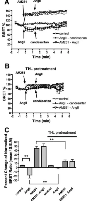
signal in neuronal tissues leads to eCB production (6), and CB1R is expressed in many tissues in the cardiovascular sys- tem (19), we tested whether CB1R activity can be enhanced by co-expression and stimulation of AT1R, a Gq-activating GPCR, which has a significant role in cardiovascular regula- tion (24, 25). Co-expression of AT1R with CB1R and stimu-
lation with AngII caused readily detectable Go activation (Fig. 3A). The AngII-dependent activation was blocked both by CB1R inverse agonist (Fig. 3,AandC) and by THL pre- treatment (Fig. 3, B and C), suggesting that it occurred through the DAGL-dependent activation of CB1R. The per- cent changes in BRET signals are shown on Fig. 3C. In con- trol experiments, stimulation of AT1R, expressed alone with the Goprotein subunits, led to some Goactivation (data not shown); however, this activation was not influenced by THL or AM251 treatment. Addition of candesartan, an inverse agonist of AT1R, after AngII stimulation reversed the stim- ulatory action of AngII. The slow kinetics of candesartan FIGURE 2.THL induces plasma membrane sequestration of CB1R in CHO
cells and cultured hippocampal neurons.CB1R-EYFP was expressed in CHO cells, and confocal microscopy images were taken of control (A), THL-pre- treated (B), WIN55,212-2 stimulated (C), and THL pretreated-WIN55,212-2 stimulated (D) cells (n⫽4 –5). THL pretreatment sequesters CB1R on the plasma membrane but does not inhibit internalization induced by WIN55,212-2. OnE, surface/intracellular fluorescence ratio is shown. FLAG- CB1R-EGFP was expressed in control (FandG) or THL-pretreated (HandI) cultured hippocampal neurons. Surface-localized CB1Rs were detected in live neurons with M1 anti-FLAG antibody. In control neurons, surface labeling of the somatodendritic region is weak (G,arrow) compared with axons (G,arrow- head), whereas THL pretreatment up-regulates somatodendritic surface label (I,arrow). OnJ, surface/total fluorescence ratio of 14 –31 neurons per condi- tion was counted from two independent experiments.Scale bar: 25m; *,p⬍ 0.05; **,p⬍0.01.
FIGURE 3.CB1R activity measurement with BRET in CHO cells co-express- ing CB1R and AT1R.BRET was measured in CHO cells expressing the labeled G protein subunits. Control (A) and THL-pretreated (B) cells were treated sub- sequently with AngII (100 nM) and candesartan (1M) (filled circles), AM251 (10M), and AngII (100 nM) (open triangles) or with vehicle (open square). The statistical analysis was performed from mean BRET signals after the first stim- ulation, and data are presented onC. (**,p⬍0.01,n⫽3– 4).
at SEMMELWEIS UNIV OF MEDICINE, on March 24, 2012www.jbc.orgDownloaded from
action, in contrast to the immediate elevation following WIN55,212-2 plus AM251 treatment (compare Fig. 3Awith Fig. 1A), is consistent with the higher agonist affinity of the AT1R. These data suggest that AT1R can transactivate co- expressed CB1Rs in CHO cells through DAGL-mediated eCB production.
DISCUSSION
DAGL activity, responsible for 2-AG production, is present in most tissues (8, 18), and DAG production by phospholipases C and D is a common process in various cell types. Therefore, we asked whether the basal DAG production and DAGL activ- ity can result in sufficient production of cannabinoid agonist(s) to maintain the constitutive activity of the CB1R. Here we show that DAGL inhibition in CHO cells leads to inhibition of the constitutive activity of CB1R, and similar results were obtained in HEK-293 cells. Moreover, activation of AT1R, a Gq-activat- ing GPCR, leads to activation of CB1R. These data suggest that CHO cells are able to produce a cannabinoid agonist and that this production occurs in non-stimulated cells and can be enhanced by AT1R, and presumably other GPCRs, which can activate Gq. The effect of DAGL inhibition on these processes suggests that these effects are mediated mainly by 2-AG (26, 27).
Basal CB1R activity has been thought to be agonist-indepen- dent, based on indirect evidence such as the lack of agonist production in unstimulated CB1R-expressing cells (8). In con- trast, our data show that CB1R constitutive activity is, at least in part, DAGL-dependent. These data are in good agreement with previous observations, which show that DAGLs are localized in somatodendritic region of the neurons in adult mouse brain (18) and CB1Rs internalize constitutively in this region but not in the axons (10), where the DAGLs are absent. Here we show that not only tonic G protein activation but also the constitutive internalization is inhibited by DAGL inhibitor, both in CHO cells and in the somatodendritic region of cultured hippocam- pal neurons. The basal activity of CB1Rs can be further acti- vated by stimulation of co-expressed Gq-coupled AT1R. Since the activity depends on endogenous cell-derived ligand(s), it can indicate continuous regulation of receptor operation, depending on other membrane receptor activity, phospholipid turnover, or Ca2⫹-related signals. In cells co-expressing the CB1R and AT1R, AngII stimulation leads to CB1R activation through DAGLs, and this could cause initiation of CB1R-spe- cific signal transduction pathways, which can be different from those regulated by Gqactivation. The CB1R activation after AngII stimulation is a novel mechanism of AT1R-mediated receptor transactivation, similar to the transactivation of EGF and other growth factor receptors reported in various target tissues of AngII (24).
These data raise the possibility of autocrine regulation of CB1R constitutive activity and internalization in non-neuronal tissues and possibly in somatodendritic region of neurons. Such a mechanism has wide implications for the pharmacological actions of drugs acting on CB1 receptors in the central nervous
system and the periphery. This type of regulation is in contrast to the model where CB1R is activated by circulating agonist in non-neuronal tissues, although the two type of activation could co-exist.
REFERENCES
1. Matsuda, L. A., Lolait, S. J., Brownstein, M. J., Young, A. C., and Bonner, T. I. (1990)Nature346,561–564
2. Devane, W. A., Hanus, L., Breuer, A., Pertwee, R. G., Stevenson, L. A., Griffin, G., Gibson, D., Mandelbaum, A., Etinger, A., and Mechoulam, R.
(1992)Science258,1946 –1949
3. Mechoulam, R., Ben-Shabat, S., Hanus, L., Ligumsky, M., Kaminski, N. E., Schatz, A. R., Gopher, A., Almog, S., Martin, B. R., Compton, D. R., Pert- wee, R. G., Griffin, G., Bayewitch, M., Barg, J., and Vogel, Z. (1995)Bio- chem. Pharmacol.50,83–90
4. Pacher, P., Batkai, S., and Kunos, G. (2006)Pharmacol. Rev.58,389 – 462 5. Howlett, A. C., Barth, F., Bonner, T. I., Cabral, G., Casellas, P., Devane, W. A., Felder, C. C., Herkenham, M., Mackie, K., Martin, B. R., Mechou- lam, R., and Pertwee, R. G. (2002)Pharmacol. Rev.54,161–202 6. Freund, T. F., Katona, I., and Piomelli, D. (2003) Physiol. Rev. 83,
1017–1066
7. Vinod, K. Y., and Hungund, B. L. (2006) Trends Pharmacol. Sci. 27, 539 –545
8. Pertwee, R. G. (2005)Life Sci.76,1307–1324
9. Leterrier, C., Bonnard, D., Carrel, D., Rossier, J., and Lenkei, Z. (2004) J. Biol. Chem.279,36013–36021
10. Leterrier, C., Laine, J., Darmon, M., Boudin, H., Rossier, J., and Lenkei, Z.
(2006)J. Neurosci.26,3141–3153
11. Neu, A., Foldy, C., and Soltesz, I. (2007)J. Physiol.(Lond.)578,233–247 12. Hentges, S. T., Low, M. J., and Williams, J. T. (2005)J. Neurosci.25,
9746 –9751
13. Zhu, P. J., and Lovinger, D. M. (2005)J. Neurosci.25,6199 – 6207 14. Bouaboula, M., Perrachon, S., Milligan, L., Canat, X., Rinaldi-Carmona,
M., Portier, M., Barth, F., Calandra, B., Pecceu, F., Lupker, J., Maffrand, J. P., Lefur, G., and Casellas, P. (1997)J. Biol. Chem.272,22330 –22339 15. MacLennan, S. J., Reynen, P. H., Kwan, J., and Bonhaus, D. W. (1998)Br. J.
Pharmacol.124,619 – 622
16. Pan, X., Ikeda, S. R., and Lewis, D. L. (1998) Mol. Pharmacol. 54, 1064 –1072
17. Vasquez, C., and Lewis, D. L. (1999)J. Neurosci.19,9271–9280 18. Bisogno, T., Howell, F., Williams, G., Minassi, A., Cascio, M. G., Ligresti,
A., Matias, I., Schiano-Moriello, A., Paul, P., Williams, E. J., Gangadharan, U., Hobbs, C., Di Marzo, V., and Doherty, P. (2003)J. Cell Biol.163, 463– 468
19. Randall, M. D., Kendall, D. A., and O’Sullivan, S. (2004)Br. J. Pharmacol.
142,20 –26
20. Azpiazu, I., and Gautam, N. (2004)J. Biol. Chem.279,27709 –27718 21. Turu, G., Szidonya, L., Ga´borik, Z., Buday, L., Spa¨t, A., Clark, A. J. L., and
Hunyady, L. (2006)FEBS Lett.580,41– 45
22. Bisogno, T., Cascio, M. G., Saha, B., Mahadevan, A., Urbani, P., Minassi, A., Appendino, G., Saturnino, C., Martin, B., Razdan, R., and Di Marzo, V.
(2006)Biochim. Biophys. Acta Mol. Cell. Biol. Lipids1761,205–212 23. Rinaldi-Carmona, M., Le Duigou, A., Oustric, D., Barth, F., Bouaboula, M.,
Carayon, P., Casellas, P., and Le Fur, G. (1998)J. Pharmacol. Exp. Ther.
287,1038 –1047
24. Hunyady, L., and Catt, K. J. (2006)Mol. Endocrinol.20,953–970 25. Spa¨t, A., and Hunyady, L. (2004)Physiol. Rev.84,489 –539
26. Bisogno, T., Ligresti, A., and Di Marzo, V. (2005)Pharmacol. Biochem.
Behav.81,224 –238
27. Makara, J. K., Mor, M., Fegley, D., Szabo, S. I., Kathuria, S., Astarita, G., Duranti, A., Tontini, A., Tarzia, G., Rivara, S., Freund, T. F., and Piomelli, D. (2005)Nat. Neurosci.8,1139 –1141
at SEMMELWEIS UNIV OF MEDICINE, on March 24, 2012www.jbc.orgDownloaded from