Differential β-arrestin2 requirements for constitutive and agonist-induced internalization of the CB1 cannabinoid receptor
Pál Gyombolaia,b, Eszter Borosa, László Hunyadya,b,#, Gábor Turua
aDepartment of Physiology, Faculty of Medicine, Semmelweis University, Budapest, Hungary;
pal.gyombolai@eok.sote.hu; boroseszter987@gmail.com; gabor.turu@eok.sote.hu;
Hunyady@eok.sote.hu
bLaboratory of Molecular Physiology, Hungarian Academy of Sciences and Semmelweis University, Budapest, Hungary
#Address correspondence to: Prof. Dr. László Hunyady, Department of Physiology, Faculty of Medicine, Semmelweis University, H-1444 Budapest, P. O. Box 259, Hungary, Fax: 36-1-266- 6504, Phone: 36-1-266-9180, E-mail: Hunyady@eok.sote.hu
*Manuscript
Click here to view linked References
Abstract
CB1 cannabinoid receptor (CB1R) undergoes both constitutive and agonist-induced internalization, but the underlying mechanisms of these processes and the role of β- arrestins in the regulation of CB1R function are not completely understood. In this study, we followed CB1R internalization using confocal microscopy and bioluminescence resonance energy transfer measurements in HeLa and Neuro-2a cells. We found that upon activation CB1R binds β-arrestin2 (β-arr2), but not β-arrestin1. Furthermore, both the expression of dominant-negative β-arr2 (β-arr2-V54D) and siRNA-mediated knock-down of β-arr2 impaired the agonist-induced internalization of CB1R. In contrast, neither β- arr2-V54D nor β-arr2-specific siRNA had a significant effect on the constitutive internalization of CB1R. However, both constitutive and agonist-induced internalization of CB1R were impaired by siRNA-mediated depletion of clathrin heavy chain. We conclude that although clathrin is required for both constitutive and agonist-stimulated internalization of CB1R, β-arr2 binding is only required for agonist-induced internalization of the receptor suggesting that the molecular mechanisms underlying constitutive and agonist-induced internalization of CB1R are different.
Keywords: β-arrestin, bioluminescence resonance energy transfer (BRET), CB1 cannabinoid receptor, constitutive internalisation, G protein-coupled receptors (GPCR), receptor endocytosis
1. Introduction
Internalization of G protein-coupled receptors (GPCRs) is an important process in the regulation of receptor function. Although its main function is the modulation of receptor number on the cell surface, thereby adjusting the sensitivity of the cell to external stimuli, it also plays role in the resensitization and signaling of GPCRs (Ferguson, 2001; Hunyady and Catt, 2006; Shenoy and Lefkowitz, 2011). At the molecular level, β-arrestins are key regulatory proteins of receptor internalization, as they can bind to activated GPCRs, as well as to clathrin and the adaptor protein AP-2, thus directing the receptor towards clathrin-mediated endocytosis (Shenoy and Lefkowitz, 2011). β-arrestins also mediate receptor desensitization, as their binding to the activated GPCRs causes the uncoupling of the receptor from its cognate G protein (Shenoy and Lefkowitz, 2011). Furthermore, they play important roles in the activation of G protein- independent signal transduction pathways, e.g. the activation of MAP kinases, phosphatidylinositol 3 kinase, Akt or the small GTP-ase RhoA (DeWire et al., 2007; Wei et al., 2003).
β-arrestin1 (β-arr1) and β-arrestin2 (β-arr2) are two ubiquitously expressed isoforms of β-arrestins (Ferguson, 2001). Although β-arrestin binding is a general property of most activated GPCRs, the selectivity and stability of these binding shows receptor specific differences. Namely, class A receptors (e.g. the β2 adrenergic receptor) bind β-arr2 with a higher affinity than β-arr1, and this binding is transient, i.e. it can only be detected at or near the plasma membrane. In contrast, class B GPCRs, such as the AT1
angiotensin receptor, bind both β-arr1 and β-arr2 with relatively high affinity and form
stable complexes with β-arrestins, so that β-arrestins remain bound to the receptor after internalization and can be detected on intracellular vesicles (Oakley et al., 2000).
The CB1 cannabinoid receptor (CB1R) belongs to the superfamily of G protein- coupled receptors (GPCRs). The receptor plays role in many important physiological processes, such as learning, thinking, nociception or the regulation of food-intake (Pacher et al., 2006). Activation of presynaptic CB1Rs by postsynaptic endocannabinoid release, which mediates retrograde transmission, is a key regulatory mechanism in the central nervous system, but paracrine activation of CB1Rs with a similar mechanism can also occur in extraneural tissues (Freund et al., 2003; Gyombolai et al., 2012; Sanchez et al., 2001; Turu et al., 2009; Szekeres et al., 2012). The cellular signaling events following CB1R activation are mainly associated with the activation of heterotrimeric Gi/o-proteins and include inhibition of adenylyl cyclases, activation of Kir channels, inhibition of Cav channels and phosphorylation and activation of different subtypes of mitogen-activated protein kinases (MAP kinases) (Turu and Hunyady, 2010). G protein-independent signaling events following CB1R stimulation have also been reported (Sanchez et al., 2001).
Similar to most GPCRs, CB1R internalizes upon agonist stimulation. This has been demonstrated in many cell lines, including CHO (Rinaldi-Carmona et al., 1998), AtT20 (Hsieh et al., 1999; Jin et al., 1999; Roche et al., 1999), F11 (Coutts et al., 2001), neuroblastoma N18TG2 (Keren and Sarne, 2003) and HEK293 (Keren and Sarne, 2003;
Leterrier et al., 2004) cells, as well as in hippocampal neurons, which naturally express CB1R (Coutts et al., 2001; Leterrier et al., 2006). According to different studies, this agonist-induced CB1R endocytosis occurs via clathrin- and/or caveolin-mediated
pathways in different cell types (Bari et al., 2008; Hsieh et al., 1999; Keren and Sarne, 2003; Wu et al., 2008).
Numerous studies suggest that β-arr2 is involved in the regulation of CB1R. Co- expression of both GRK3 and β-arr2 was needed for the proper desensitization of the receptor in Xenopus oocyte (Jin et al., 1999), and dominant negative GRK2 and β- arrestin constructs reduced CB1R desensitization in hippocampal neurons (Kouznetsova et al., 2002). In β-arr2 knockout mice desensitization and downregulation of CB1R were impaired in certain regions of the central nervous system (Nguyen et al., 2012).
Recruitment of β-arr2 to the activated CB1R has also been demonstrated (Daigle et al., 2008a). Mutation of amino acids S426 and S430 was shown to inhibit receptor desensitization as well as late phase receptor endocytosis, but not β-arrestin binding (Daigle et al., 2008b; Jin et al., 1999). It has been demonstrated that serine and threonine residues at the C-terminus of CB1R are involved in its β-arr2 binding and agonist-induced endocytosis (Daigle et al., 2008b; Hsieh et al., 1999). In contrast to the various studies that clearly point to an interaction between CB1R and β-arr2 upon agonist stimulation, no direct data have been hitherto presented concerning the β-arr1 binding of CB1R. In some structural studies, the association of β-arr1 with a synthesized CB1R C-terminus has been shown (Bakshi et al., 2007; Singh et al., 2011), however, such binding has not been demonstrated with the intact CB1R in living cells.
Constitutive internalization of CB1R (i.e. spontaneous internalization in the absence of CB1R agonists) has also been detected in hippocampal neurons, as well as in CHO and HEK cells (Leterrier et al., 2004, 2006; McDonald et al., 2007a; Turu et al., 2007). It has been suggested that constitutive CB1R internalization is the consequence of
its basal activity, since inverse agonist treatment or inhibition of basal activity with a DAG lipase inhibitor (e.g. tetrahydrolipstatin) interfered with this process (Leterrier et al., 2004, 2006; Rinaldi-Carmona et al., 1998; Turu et al., 2007). However, other studies have concluded that constitutive internalization occurs independently of receptor activity (McDonald et al., 2007a, 2007b; Kleyer et al., 2012). The latter statement raises the possibility that constitutive and agonist-induced internalization of CB1R may occur via distinct endocytic mechanisms (McDonald et al., 2007a). However, no evidence has been hitherto presented showing that these two processes are truly different in that they require distinct endocytic machinery to take place.
Our main goal was, considering the important consequences of β-arrestin recruitment in the regulation of cell function, to characterize the β-arrestin binding properties of CB1R and to reveal possible differences in constitutive and agonist-driven CB1R endocytosis by investigating the role of β-arrestins in these processes.
2. Materials and Methods
2.1. Materials
The cDNAs of the rat vascular CB1R and CB1R-eYFP were provided by Zsolt Lenkei (Centre National de la Recherche Scientificue, Paris). β-arrestin1, β-arrestin2 and β- arrestin2-eGFP cDNAs were kindly provided by Dr. Marc G. Caron (Duke University, Durham, NC). Molecular biology enzymes were obtained from Fermentas (Vilnius, Lithuania) and Stratagene (La Jolla, CA). pcDNA3.1 vector, coelenterazine h, fetal bovine serum (FBS), OptiMEM, Lipofectamine 2000, and PBS-EDTA were from
Invitrogen (Carlsbad, CA). WIN55,212-2 and AM251 were from Tocris (Bristol, UK).
Cell culture dishes and plates for BRET measurements were from Greiner (Kremsmunster, Austria). HaloTag® Alexa Fluor® 488 Ligand was from Promega (Madison, WI). Control siRNA, human β-arrestin2-specific siRNA, mouse β-arrestin2- specific siRNA and clathrin heavy chain-specific siRNA (with sequences 5’-
UUCUCCGAACGUGUCACGU-3’, 5’-GGACCGCAAAGUGUUUGUG-3’, 5’-
ACGUCCAUGUCACCAACAA-3’ and 5’-GAAAGAAUCUGUAGAGAAA-3’,
respectively) were from Eurofins MWG Operon (Ebersberg, Germany). HeLa and Neuro- 2a cells were from ATCC (American Type Culture Collection, Manassas, VA). Anti-β- arrestin2 and HRP-conjugated anti-rabbit and anti-mouse antibodies were from Cell Signaling Technology Inc. (Beverly, MA). Anti-clathrin heavy chain antibody was from Transduction Laboratories (Lexington, KY). Unless otherwise stated, all other chemicals and reagents were from Sigma (St. Louis, MO).
2.2. Plasmid constructs and site-directed mutagenesis
The mVenus-tagged rat AT1a receptor (AT1R-mVenus), human β2 adrenergic receptor (β2AR-mVenus) and rat CB1R (CB1R-mVenus) were created by exchanging the sequence of fluorescent proteins in AT1R-YFP (Turu et al., 2006), β2AR-Sluc (Toth et al., 2012) or CB1R-YFP, respectively, to the sequence of mVenus using AgeI and NotI restriction enzymes. The mCherry-tagged AT1R (AT1R-mCherry) and Cerulean-tagged β2AR (β2AR-Cerulean) were created similarly. β-arrestin2-Rluc was constructed as described previously (Turu et al., 2006). β-arrestin1-Rluc and β-arrestin1-GFP were generated from β-arrestin2-Rluc and β-arrestin2-GFP, respectively, by replacing the cDNA of β-arrestin2
with that of β-arrestin1. CB1R-mCherry and β-arrestin2-RFP were constructed by subcloning the cDNAs of CB1R or β-arrestin2 into mCherry or RFP containing vectors (provided by Dr.R. Tsien, University of California, San Diego, CA), respectively. For the construction of Halo-CB1R, the cDNA of HaloTag was first amplified from the HaloTag® pHT2 vector (Promega, Madison, WI) by PCR with the sense primer containing a cleavable signal sequence of influenza hemagglutinin (MKTIIALSYIFCLVFA) to achieve proper plasma membrane localization of the final construct (Guan et al., 1992). This product was inserted into a pEGFP-C1 vector (Clontech, Palo Alto, CA) in the place of eGFP sequence (pHalo-C1 vector). CB1R cDNA was then inserted into pHalo-C1 after the HaloTag cDNA to yield Halo-CB1R.
CB1R-Sluc was generated from CB1R-YFP by replacing the eYFP coding sequence with the cDNA of super Renilla luciferase (Woo and von Arnim, 2008). EYFP-tagged ICAM- 1 (ICAM-YFP) was constructed as described previously (Varnai and Balla, 2007). V54D mutation was inserted into the appropriate β-arrestin2 constructs by the QuikChange®
site-directed mutagenesis kit (Stratagene, La Jolla, CA) according to manufacturer’s suggestions. Sequences of all constructs were verified using automated DNA sequencing.
2.3. Cell cultures and transfection
HeLa and Neuro-2a cells were maintained in DMEM supplemented with 10% FBS, (Invitrogen, Carlsbad, CA), 100 μg/ml streptomycin, and 100 IU/ml penicillin in 5% CO2
at 37 °C. For confocal microscopy experiments, HeLa and Neuro-2a cells were grown on glass coverslips (coated with poly-L-lysine in the case of Neuro-2a cells) in 6-well plates and transfected with the indicated constructs using 1 μg/well of receptor constructs, 0.5
μg/well of β-arrestin constructs (as indicated) and/or 25 pmol/well of siRNA (as indicated), with 2 μl/well Lipofectamine 2000 in OptiMEM following the manufacturer’s instructions.
For BRET time course experiments, HeLa cells were grown on 6-well plates and transfected with the indicated constructs using 1 μg/well of CB1R constructs, 0.5 μg/well of β-arrestin constructs, 3 μg/well of ICAM-YFP and/or 25 pmol/well of siRNA (as indicated), with 2 μl/well Lipofectamine 2000 in OptiMEM following the manufacturer’s instructions. In the case of BRET titration experiments, HeLa cells were transfected with constant amounts of donor (β-arr1-Rluc or β-arr2-Rluc) and varying amounts of acceptor (CB1R-mVenus, β2AR-mVenus or AT1R-mVenus) constructs with 2 μl/well Lipofectamine 2000 in OptiMEM following the manufacturer’s instructions. The amount of total transfected DNA was held constant (2.5 μg/well) with addition of varying amounts of empty pcDNA3.1 plasmid.
2.4. Bioluminescence resonance energy transfer (BRET) measurements
The BRET assay measuring the effects of different CB1R variants on G protein activation was carried out as described previously (Turu et al., 2007). To measure BRET between β- arrestins and receptors, HeLa cells were transfected with Renilla luciferase-tagged β- arrestin isoforms (β-arr1-Rluc or β-arr2-Rluc) and mVenus-tagged AT1R, β2AR or CB1R, and measurements were performed on the day after transfection. To measure BRET between ICAM and CB1R, HeLa cells were transfected with eYFP-tagged ICAM-1, super Renilla luciferase-tagged CB1R and the indicated β-arr2 construct or siRNA, and measurements were performed two days after transfection. Before the experiments cells
were detached with PBS-EDTA and centrifuged. Cells were resuspended in a modified Krebs-Ringer buffer containing 120 mM NaCl, 4.7 mM KCl, 1.2 mM CaCl2, 0.7 mM MgSO4, 10 mM glucose, 0.1% BSA and Na-HEPES 10 mM, pH 7.4; and transferred to white 96-well plates. Measurements were performed at 37 °C, after the addition of the cell permeable substrate, coelenterazine h at a final concentration of 5 μM. Counts were recorded using a Mithras LB 940 multilabel reader (Berthold Technologies, Bad Wildbad, Germany) with filters at 485 (for Rluc) and 530 nm (for eYFP) wavelengths.
BRET ratios were calculated as a 530 nm/485 nm emission ratio. In the case of BRET titration experiments, mVenus/Rluc emission ratios were calculated by dividing average mVenus fluorescence counts (measured before the addition of coelenterazine h, with 485 nm excitation and 530 nm emission) by average Rluc bioluminescence counts (measured in non-stimulated cells after the addition of coelenterazine h). This was plotted against the average BRET ratio change measured 3 to 5 min after agonist stimulus. The measurements were done in triplicates, and the BRET records shown here are calculated from at least three independent experiments. BRET change was defined as the BRET ratio of a given time point minus the average BRET ratio of the initial (non-stimulated) time points. BRET change was baseline-corrected to the vehicle curve.
2.5. Confocal laser-scanning microscopy
Cells were grown on glass coverslips and transfected with the indicated constructs, as described above, 48 hours prior to measurement. EGFP (or Halo-Alexa488) and mCherry (or RFP) were excited with 488 nm argon, and 543 nm helium/neon lasers, respectively, and emitted fluorescence was detected with 500-550 bandpass and 560 longpass filters,
respectively, using a Zeiss LSM 510 confocal laser scanning microscope. For quantification of confocal measurements, 20 images of individual cells were taken from each sample, and intracellular and total cell fluorescence intensities of the images were determined using the ImageJ software (W. S. Rasband, ImageJ, United States National Institutes of Health, Bethesda, MD (rsb.info.nih.gov/ij/)). Intracellular fluorescence was divided by total cell fluorescence to yield IC/total cell fluorescence ratio. Blind selection and analysis of the cells were performed to avoid any bias during the evaluation of the internalization data.
2.6. Halo-labeling protocols
To measure agonist-induced internalization of the receptor, Halo-CB1R-transfected cells were stained with Halo-Alexa488 ligand diluted 1:20 000 in DMEM, and incubated at 37°C, 5% CO2 for 15 minutes. Cells were then washed twice with DMEM to remove unbound Halo-Alexa488, and treated with DMSO, WIN55,212-2 (10 μM) or WIN55,212-2+AM251 (10 μM+30 μM, respectively) at 37°C, 5% CO2 for 30 minutes.
At the end of stimulation period, cells were washed twice with PBS and fixed with 4%
formaldehyde at 4°C for 15 minutes. Fixed cells were analyzed under confocal microscope as described above.
To measure constitutive internalization of the receptor, Halo-CB1R-transfected cells were stained with Halo-Alexa488 ligand diluted 1:20 000 in DMEM, and incubated at 37°C, 5% CO2 for 15 minutes. Cells were then washed twice with DMEM to remove unbound Halo-Alexa488 and incubated in DMEM at 37°C, 5% CO2 for 5 hours and 45 minutes without any further treatment or in the presence of DMSO, WIN55,212-2 (10
μM) or AM251 (30 μM). To control the effectiveness of siRNA transfection during the 6 hours of incubation, parallel samples were subjected to the same washing procedures, however, without the staining period at the beginning. For these cells, an ‘agonist- induced’ protocol (15 min staining + 30 min WIN55 stimulus, see above) was applied 45 minutes before the end of the experiment. At the end of the 6 hours, all cells were washed twice with PBS and fixed with 4% formaldehyde for 15 minutes at 4°C. Fixed cells were analyzed under confocal microscope as described above.
2.7. Western blot analysis
Cells for Western blot analysis were transfected on 6-well plates parallel to the cells transfected for confocal analysis. On the day of experiment, cells were placed on ice and washed with ice-cold PBS. Cells were scraped into SDS sample buffer, briefly sonicated, boiled at 95°C for 5 min, centrifuged at 4°C for 10 min and separated on SDS polyacrilamide gels. The proteins were transferred to PVDF membranes, blocked (30 min, 5% fat-free milk powder in PBS with 0.05% Tween 20 (PBST)) and incubated with rabbit anti-β-arrestin2 primary antibody or mouse anti-clathrin heavy chain primary antibody (1 h, diluted 1:1000 or 1:4000, respectively, in PBST containing 5% fat-free milk powder) and HRP-linked goat anti-rabbit or anti-mouse (respectively) secondary antibody (30 min, diluted 1:2000 in PBST containing 5% fat-free milk powder). To control the total protein amount of cell samples, antibodies were removed with standard stripping buffer and a second immunostaining procedure was carried out with mouse anti- β-actin primary antibody (1 h, 1:10 000) and HRP-linked goat anti-mouse secondary antibody (30 min, 1:10 000). The antibodies were visualized using SuperSignal West
Pico reagent (Pierce Biotechnology Inc., Rockford, IL), according to manufacturer’s instructions. Western blot images were scanned and quantified using the ImageJ software.
Changes in β-actin levels were used to normalize changes in β-arr2 or clathrin heavy chain levels.
2.8. Statistical analysis
Statistical analysis was made using the software SigmaStat for Windows 3.5 (Systat Software Inc., Richmond, CA). The effects of co-transfected siRNAs were evaluated using Student’s t-test or two-way ANOVA with Holm-Sidak’s post-hoc test, as appropriate. Data shown are mean ± SEM values.
3. Results
3.1. β-arrestin binding properties of CB1R
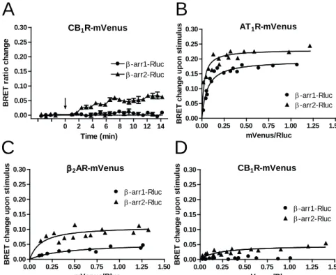
β-arrestin binding properties of CB1R were first analyzed using a bioluminescence resonance energy transfer (BRET) based approach. BRET was measured between Renilla luciferase tagged β-arr1 or β-arr2 (β-arr1-Rluc and β-arr2-Rluc) and the mVenus-tagged CB1R (CB1R-mVenus) in transiently transfected HeLa cells. Stimulation using WIN55,212-2 (WIN55, 10 μM), a potent synthetic CB1R agonist, led to an increase in the BRET signal when β-arr2-Rluc was co-expressed with CB1R-mVenus (Fig. 1A) reflecting the recruitment of the β-arr2-Rluc molecules to CB1R-mVenus following receptor activation. In contrast, the same stimulus did not cause any detectable change in
the BRET signal between β-arr1-Rluc and CB1R-mVenus (Fig. 1A), indicating that this subtype of β-arrestin does not bind to CB1R following receptor activation.
To investigate the β-arrestin binding of CB1R more thoroughly, BRET titration experiments were performed. In these experiments, BRET signal is measured and plotted as a function of the BRET acceptor/donor (mVenus/Rluc) expression ratio. In case of a specific interaction, such as the binding between a GPCR and β-arrestins upon agonist stimulus, BRET titration follows a saturation curve, the slope of which corresponds to the affinity between the two BRET partners (Marullo and Bouvier, 2007). Thus, BRET titration experiments were carried out between CB1R-mVenus and β-arr1-Rluc or β-arr2- Rluc. Furthermore, the mVenus-tagged β2-adrenoceptor and AT1 angiotensin receptor (β2AR-mVenus and AT1R-mVenus, respectively) were also tested for their β-arrestin affinities in the same system, as these two receptors are prototypical class A and class B GPCRs, respectively (Oakley et al., 2000), and therefore their β-arrestin binding properties could be used as a reference in our experiments. As expected, BRET titration curves of β2AR-mVenus and AT1R-mVenus followed saturation kinetics with both β- arrestin isoforms after agonist stimulus. While AT1R-mVenus binds both isoforms with high affinity (Fig. 1B), in case of β2AR-mVenus, substantial difference was observed between β-arr1-Rluc and β-arr2-Rluc binding, showing that this GPCR has substantially higher affinity for β-arr2 (class A GPCR, Fig. 1C). BRET titration curve of CB1R- mVenus and β-arr2-Rluc also followed saturation kinetics, however, with a very low steepness. In contrast, no detectable BRET signal change occurred with β-arr1-Rluc even at high acceptor/donor ratios (Fig. 1D).
β-arrestin binding of CB1R was also visualized using confocal microscopy. In HeLa cells transiently transfected with mCherry-tagged CB1R (CB1R-mCherry) and GFP-tagged β-arr1 or β-arr2 (β-arr1-GFP or β-arr2-GFP, respectively), CB1R-mCherry was present at the plasma membrane as well as in intracellular vesicles in non-stimulated cells. This distribution corresponds to constitutive internalization of CB1R, a well-known property of this receptor (Fig. 2B and F). On the other hand, both β-arr1-GFP and β-arr2- GFP showed diffuse cytoplasmic (and, in the case of β-arr1-GFP, also nuclear) localization in non-stimulated cells, without any detectable β-arrestin either at the plasma membrane or in intracellular vesicles (Fig. 2A and E). After stimulation with WIN55, marked redistribution of β-arr2-GFP could be observed within a few minutes, namely it appeared in punctuate structures at the plasma membrane. However, the inner regions of the cytoplasm showed no accumulation of β-arr2-GFP containing vesicles even after longer periods of stimulation (Fig. 2G). In contrast to β-arr2-GFP, β-arr1-GFP showed no change in its localization after WIN55 stimulus (Fig. 2C), indicating that this subtype of β-arrestin does not bind to CB1R. In the same system, fluorescently labeled β2AR and AT1R showed β-arrestin binding pattern, which are in good agreement with their class A and class B behavior, respectively, i.e. β2AR bound β-arr1 and β-arr2 only at the plasma membrane (Suppl. Fig. 1A-D), whereas AT1R remained bound with both isoforms in endosomal vesicles (Suppl. Fig. 1E-H).
The above data indicate that, based on its β-arrestin binding properties, CB1R belongs to the class A group of GPCRs, i.e. upon activation, β-arr2 binds to the receptor, but remains bound to it only in the proximity of the plasma membrane, whereas CB1R does not recruit β-arr1.
3.2. Dominant-negative β-arr2 impairs agonist-induced CB1R internalization
Next, the agonist-induced internalization of CB1R was investigated. We utilized the Halo labeling technique, as the analysis of the internalization of C-terminally tagged CB1R variants using confocal microscopy is difficult because of the presence of the constitutively internalized intracellular receptor population. The HaloTag protein was fused to the N-terminus of the receptor (Halo-CB1R). When expressed by the cells, HaloTag is able to rapidly and covalently bind Halo-Alexa488, its membrane-impermeant fluorescent ligand. Using this method, the receptor population residing in the plasma membrane can be selectively stained, and their internalization can be followed. The functionality of Halo-CB1R was tested using BRET assays measuring the G protein activation and the β-arr2 binding of the receptors upon WIN55 stimulus, and no significant difference was detected between the EC50 (mol/l) values of wild-type and HaloTag-labeled CB1R dose-response curves (pEC50 8.98±0.22 versus 9.02±0.12 for G protein activation and 6.48±0.14 versus 6.49±0.08 for β-arr2 binding, respectively, n=3).
To study agonist-induced internalization, cells were stained for 15 minutes, treated with vehicle or CB1R ligand for 30 minutes, and then fixed and analyzed using confocal microscopy. In vehicle-treated cells, plasma membrane staining was clearly detectable, and labeling of intracellular vesicles was minimal (Fig. 3A). WIN55 treatment resulted in the appearance of numerous intracellular vesicles, representing intensive agonist-induced internalization, which was inhibited by the CB1R inverse agonist AM251 (Fig. 3B-C). To study the role of β-arr2 in this process, first, a dominant-negative β-arr2 mutant, β-arr2- V54D (β-arr2-V54D) was applied. This mutant has reduced ability to bind to GPCRs
(Ferguson et al., 1996; Krupnick et al., 1997), and has been widely used to study the role of β-arr2 in receptor internalization. We co-expressed the RFP-tagged form of wild-type or V54D mutant β-arr2 (β-arr2-RFP and β-arr2-V54D-RFP, respectively) with Halo- CB1R. Co-expression of β-arr2-RFP did not impair the WIN55-induced CB1R internalization (Fig. 3D-G). However, in cells expressing β-arr2-V54D-RFP, agonist- induced internalization of Halo-CB1R was markedly reduced (Fig. 3H-K), suggesting a role for β-arr2 in agonist-induced CB1R internalization.
3.3. siRNA-mediated knock-down of β-arr2 impairs agonist-induced internalization of CB1R
siRNA-mediated knock-down of a protein provides an alternative approach to study its role in a given mechanism. Therefore, the effects of transfection of cells with β-arr2- specific siRNA (β-arr2 siRNA) were also investigated. Western blot analysis showed approximately 50% reduction in β-arr2 protein levels of these cells (Fig. 4A).
Internalization of Halo-CB1R in these experiments was analyzed as described above.
CB1R internalization upon WIN55 treatment was intact in cells transfected with control siRNA, but was markedly reduced in β-arr2 siRNA-transfected cells (Fig. 4B-E). As the cells that were transfected with siRNA could not be individually identified in these experiments, quantification of the samples was carried out to properly evaluate the effects of siRNA transfection. This showed a significant inhibitory effect of β-arr2 siRNA on agonist-induced CB1R internalization (Fig. 4F).
3.4. Agonist-induced internalization of CB1R
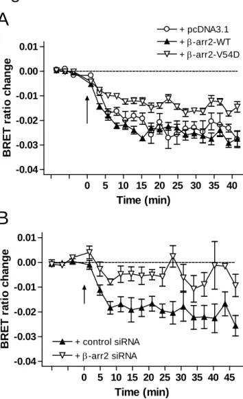
To investigate the agonist-induced internalization of CB1R in a more quantitative way, we monitored the movement of the receptor between the plasma membrane and the cytoplasm, by measuring BRET between the YFP-tagged form of ICAM-1, a plasma membrane resident protein (ICAM-YFP), and the SuperRenilla luciferase-tagged CB1R (CB1R-Sluc) in transiently transfected HeLa cells. In cells expressing CB1R-Sluc and ICAM-YFP, WIN55 stimulus led to a decrease in the BRET signal, indicating the removal of CB1R-Sluc from the plasma membrane (Fig. 5A). This decrease reached its maximum 10 to 20 minutes after stimulation, and showed then no considerable change until the end of the measurement (at approximately 40 minutes after stimulation).
Overexpression of wild-type β-arr2 had no significant effect on CB1R internalization.
However, when β-arr2-V54D was co-expressed in the cells, the extent of the BRET- decrease upon WIN55 stimulus was significantly lower than that observed in control cells (Fig. 5A). The effect of siRNA mediated β-arr2 knock-down on CB1R internalization was also studied using the same BRET assay. We found that the knock-down of β-arr2 with β- arr2 siRNA resulted in a marked reduction of BRET signal decrease after CB1R activation, reflecting the impaired agonist-induced internalization of the receptor (Fig.
5B).
These data indicate that β-arr2 binding is required for the agonist-induced internalization of CB1R, since interference with this binding following receptor activation, either caused by dominant-negative effect or through the genetic knock-down of β-arr2, results in impairment of agonist-induced CB1R internalization.
3.5. Constitutive CB1R internalization is not affected by β-arr2 impairment
Recent data suggest that constitutive internalization may occur independently of CB1R activity (McDonald et al., 2007a, 2007b; Kleyer et al., 2012), and since β-arr2 coupling is an activity-dependent process, it raises the possibility that it is a second, differently regulated endocytic process. Therefore we monitored receptor constitutive endocytosis using similar approaches with Halo-tagged CB1R to address possible differences in its regulation compared to agonist-induced internalization. In these experiments, cells were stained for 15 minutes with the Alexa488-conjugated Halo-ligand, and incubated for a total of 6 hours in medium to allow spontaneous CB1R endocytosis to occur. In control cells, a large number of vesicles could be seen intracellularly, demonstrating constitutive endocytosis of CB1R (Fig. 6A). This could be further enhanced with WIN55 treatment, whereby hardly any detectable receptor was present on the plasma membrane by the end of the 6 hours incubation period (Fig. 6B). AM251 treatment along the 6 hours had no inhibitory effect on constitutive CB1R internalization (Fig. 6C), showing that this form of internalization is not dependent on receptor activity. When studying the role of β-arr2 in this process, cells co-expressing β-arr2-RFP showed no detectable change in the extent of constitutive internalization (Fig. 6D-E). Moreover, in contrast to agonist-induced endocytosis, in cells co-expressing the dominant-negative mutant β-arr2 (β-arr2-V54D- RFP), intracellular Halo-CB1R-containing vesicles were still present, in a similar extent as in control or β-arr2-RFP expressing cells (Fig. 6F-G). When cells were co-transfected with either control or β-arr2 siRNA, no significant difference in the intracellular vs. total cell fluorescence ratio could be detected between these cells after 6 hours of incubation, showing that constitutive internalization of CB1R was not affected (Fig. 7A). The effect of siRNA was not attenuated throughout the incubation period, as in the same
experiments, agonist-induced CB1R internalization was still impaired when tested at the end of the incubation period (Fig. 7B). These data suggest that β-arr2 is not required for the spontaneous internalization of CB1R. Similar results were obtained when Halo-CB1R internalization was investigated in a Neuro-2a mouse neuroblastoma cell line, which is known to endogenously express CB1R (Graham et al., 2006). siRNA mediated knock- down of β-arr2 (resulting in an approximately 80% reduction of β-arr2 protein levels, Suppl. Fig. 1A) impaired agonist-induced internalization of Halo-CB1R significantly in these cells, but had no effect on the constitutive internalization of the receptor (Fig. 7C and D). These data indicate that the β-arr2 requirements of CB1R internalization are not specific to HeLa cells and are regulated similarly in Neuro-2a cells, which express endogenous CB1Rs.
3.6. siRNA-mediated knock-down of clathrin heavy chain impairs both agonist-induced and constitutive internalization of CB1R
Our results showed that constitutive CB1R internalization is independent of β-arr2 binding, which raises the question whether this form of CB1R endocytosis is clathrin- mediated. Therefore we applied siRNA-mediated knock-down of clathrin heavy chain in HeLa cells, and monitored constitutive endocytosis of Halo-CB1R under these conditions, as described above. Western blot analysis showed approximately 50% reduction in clathrin heavy chain levels of these cells (Suppl. Fig. 2B). We found that in cells transfected with clathrin heavy chain siRNA the spontaneous endocytosis of Halo-CB1R over 6 hours was significantly impaired compared to control siRNA-transfected cells (Fig. 8A). In the same experiments, agonist-induced CB1R internalization was also
inhibited in clathrin heavy chain siRNA-transfected cells (Fig. 8B). These results show that, although the β-arr2 requirements for agonist-induced and constitutive CB1R internalization are different, both of them occur at least partly by a clathrin-mediated process.
4. Discussion
Our data show that, based on its β-arrestin binding characteristics, CB1R can be classified as a class A GPCR. The relatively low affinity of the binding between CB1R and β-arr2 is indicated by the low steepness of slope of the BRET saturation curve between the two molecules upon CB1R stimulation, and by the lack of detectable β- arrestin2 in late endosomes after CB1R internalization in confocal experiments.
Furthermore, we found in our BRET and confocal measurements that β-arr1 recruitment to the activated CB1R is essentially absent. This was not caused by the presence of the fluorescent tag on the C-terminus of CB1R because the binding of β-arr2 could be detected using the same receptor construct. The C-terminal tagging of β-arr1 is also not likely to cause the lack of the signal, as both β-arr1-Rluc and β-arr1-GFP constructs could be successfully used to detect β-arr1 recruitment to AT1R and β2AR. In a previous study, Bakshi et al. have shown using nuclear magnetic resonance (NMR) that a C-terminal fragment of CB1R is able to bind β-arr1 (Bakshi et al., 2007). Our data suggest that such binding between the two molecules, even if present, must be very weak compared to β- arr2, when tested with native proteins in living cells.
According to Oakley and colleagues, the class A or class B behavior of a GPCR is basically determined by the absence or presence (respectively) of multiple
serine/threonine-containing clusters at the receptor C-terminus (Oakley et al., 2001). Our findings demonstrating a class A pattern for CB1R are in good agreement with this theory, as the C-terminus of CB1R contains only one such cluster (463-SVSTDTS-469) (Oakley et al., 2001).
The class A behavior of CB1R may have an important impact on the pattern of receptor internalization, de- and resensitization. Furthermore, one should be aware of this property of CB1R when studying the signal transduction of the receptor. As mentioned in the introduction, β-arrestin can by itself moderate different intracellular signaling pathways. It was also shown that the localization of β-arrestin-dependent MAPK activation is dependent on the class A/class B characteristics of the involved receptor.
After stimulation of class B receptors, β-arrestin activates a MAPK pool that remains associated with endosomes, whereas in the case of class A GPCRs, the MAPK activated by β-arrestin is able to reach the nucleus (Tohgo et al., 2003). CB1R is also known to mediate ERK phosphorylation, and early gene expression changes are associated with this pathway (Derkinderen et al., 2003; Graham et al., 2006; Valjent et al., 2001).
However, G protein-dependent and β-arrestin-dependent MAPK activation of GPCRs are known to be spatially and temporally different (Ahn et al., 2004). Although the role of β- arrestin molecules in CB1R-mediated ERK activation has been suggested (Daigle et al., 2008a), the exact role of this process in CB1R-mediated ERK activation requires additional studies. It is very likely that the class A behavior of CB1R is associated with a characteristic distribution of MAPK activation, nevertheless, direct studies that address these questions are needed to confirm them.
Agonist-induced internalization of G protein-coupled receptors is usually the direct consequence of receptor activation. Similarly, constitutive receptor internalization is also generally considered to be a result of constitutive receptor activity. This scenario also implies that the mechanisms that control the endocytosis of a given receptor after agonist activation are similarly engaged in its spontaneous endocytosis. This has also been the prevailing idea in the case of CB1R trafficking. In our study, the Halo labeling technique was utilized to analyze the mechanism of internalization of CB1R. The cellular trafficking of GPCRs is often followed by the use of fluorescent proteins fused mainly to the C-terminus of the receptor. However, the detailed analysis of the internalization of such receptor variants under microscope is often hindered by the continuously visible intracellular receptor populations (representing either maturing or endosomal receptors), and this is especially true for constitutively internalizing receptors, such as CB1R.
Receptor distribution on cell surface and in intracellular vesicles is a function of internalization and recycling rates, representing an actual equilibrium of these processes.
When cells are treated with a receptor antagonist, theoretically both processes can be altered. Receptor internalization can be inhibited by decreasing constitutive activity, or recycling may be altered by targeting the receptor to different intracellular trafficking routes. Thus, measuring C-terminally tagged receptor distribution in cells can be misleading when trafficking of constitutively internalizing receptors is measured.
Furthermore, alterations in the trafficking of C-terminally tagged CB1R have been also suggested (Rozenfeld, 2011). All these difficulties can be overcome by the use of N- terminal tags that can be covalently labeled with small, membrane-impermeable fluorescent ligands. Indeed, such receptor variants (i.e. SNAP and CLIP-tagged
receptors) have been utilized in the studying of CB1R internalization (Ward et al., 2011a, 2011b). Here we used another variant, the HaloTag labeled form of CB1R. As indicated in our study, G protein activation and β-arrestin binding of Halo-CB1R have not been altered due to the N-terminal labeling, and therefore this receptor can be used to study receptor trafficking with the same advantages as the above mentioned ones.
The main finding of our study is that constitutive CB1R internalization is not only independent of the active/inactive state of the receptor, but it occurs via a mechanism which is at least partly different from activity-driven endocytosis.
This finding is based on our observations that agonist-induced CB1R internalization requires β-arr2 binding, whereas constitutive internalization of the receptor does not. As described in the introduction, the binding between β-arr2 and CB1R as well as a role for β-arrestins in the desensitization of CB1R have been demonstrated by former studies (Bakshi et al., 2007; Daigle et al., 2008a, 2008b; Jin et al., 1999;
Kouznetsova et al., 2002; Nguyen et al., 2012; Singh et al., 2011). Therefore, the role of β-arr2 in CB1R endocytosis may seem to be a logical consequence of its recruitment to the receptor. However, in fact, none of these studies provided direct evidence at the cellular level that β-arrestin binding is required for CB1R internalization. This is important because various examples demonstrate that β-arrestin2 recruitment and receptor internalization can be independent processes, as it is the case e.g. with the PAR1 thrombin receptor (Paing et al., 2002), the CXCR2 chemokine receptor (Fan et al., 2001;
Zhao et al., 2004), the N-formyl peptide receptor (FPR) (Vines et al., 2003), or at high agonist concentration the AT1 angiotensin receptor (Gaborik et al., 2001; Hunyady and Catt, 2006; Zhang et al., 1996). Therefore, it is important to study directly the β-arrestin
dependence of CB1R internalization. In the present study we performed these experiments using a dominant-negative β-arrestin2 mutant and siRNA-mediated knock- down of β-arr2. Our data demonstrate that β-arr2 binding is required for the agonist- induced internalization of CB1R.
In contrast, using similar approaches, we have demonstrated in our study that, although both agonist-induced and constitutive CB1R endocytosis are mediated at least partly by clathrin, constitutive internalization of CB1R is a β-arr2-independent process.
Earlier, McDonald et al. raised the idea that these two forms of CB1R internalization may occur via different, or at least partly different, pathways (McDonald et al., 2007a). Our work provides experimental evidence to support their proposal. Such regulatory phenomenon is however not a unique property of CB1R, as similar differences in the mechanisms of tonic versus agonist-induced internalization have been demonstrated in the case of other GPCRs, such as the TP thromboxane A2 receptor (Parent et al., 2001), the Y1 neuropeptide Y receptor (Holliday et al., 2005) or the mGlu1 metabotropic glutamate receptor (Dale et al., 2001). Furthermore, we also show here that the spontaneous internalization of CB1R is not simply a consequence of its constitutive activity, since it also occurs in the presence of the CB1R inverse agonist AM251, i.e.
when the receptors are stabilized in their inactive state. This is in line with the data published by McDonald et al., which showed no alterations in the cellular distribution of CB1R upon inverse agonist treatment in neurons (McDonald et al., 2007a). In contrast, a previous study using inverse agonist treatment (Leterrier et al., 2006), and our study using DAGL-inhibitor treatment (Turu et al., 2007) have shown an inhibition of basal endocytosis of the receptor upon these treatments, suggesting a role for basal CB1R
activity in this process. Since active receptors internalize by β-arr2-dependent mechanism, those results may seem to be contradictory with our present results, where we now find that interfering with β-arr2 expression or function does not have effect on constitutive endocytosis. One explanation for these differences is our previous observation that CB1R has different basal activity in different cell types used in these studies (data not shown). The other possibility is that inhibition of receptor activity may interfere with intracellular trafficking, so it might also accelerate recycling, thus the rate of activity-dependent constitutive internalization may be well overestimated in those experiments. This is supported by the observation, that in resting cells, β-arr2 is not translocated to the plasma membrane, suggesting a low percent of active receptors there (Fig. 2E). The advantage of using the Halo technique is that we can now selectively follow trafficking of surface receptors, thus recycling becomes less of an issue.
Therefore, combined with previous results, the data presented here are consistent with a model in which continuous CB1R internalization in resting cells is caused to a certain extent by basal release of endocannabinoids, the rate of which may vary in different cell types, while another part of basal CB1R internalization is ‘truly’ constitutive, i.e. it is independent of CB1R activation and requires a mechanism that is different from agonist- induced, β-arrestin2-dependent internalization.
Recent work by Kleyer and colleagues demonstrated that in immune cells expressing endogenous CB1Rs, internalization of the receptor is independent of its activation (Kleyer et al., 2012), suggesting that agonist-dependent internalization may not have physiological role in primary cells. However, in hippocampal neurons, it has been demonstrated that agonist treatment leads to internalization (Coutts et al., 2001). These
data, together with differences detected in the regulation of constitutive internalization (Leterrier et al., 2006; McDonalds et al., 2007a) are in good agreement with our results showing that CB1R internalizes through at least two different mechanisms. Depending on the environment in a particular cell type, or on the methods used by different groups, one or the other mechanism might be more prevalent.
The physiological role of spontaneous CB1R endocytosis is not clear at the moment. It has been suggested that constitutive CB1R internalization in neurons have a role in the axonal distribution of the receptor (Leterrier et al., 2006; McDonald et al., 2007a). This underlines the importance of constitutive CB1R endocytosis in the central nervous system. However, CB1Rs are also expressed in peripheral cannabinoid target tissues, and we have practically no data what is the importance of such trafficking feature of the receptor in these cells. On the other hand, maintained functionality of intracellular CB1R populations has been demonstrated (Brailoiu et al., 2011; Rozenfeld and Devi, 2008), and this raises the idea that the activation of intracellular versus plasma membrane CB1R populations may differentially regulate cell functions (Rozenfeld, 2011).
Nevertheless, additional studies are needed to further clarify the importance of constitutive CB1R endocytosis in different body tissues.
Acknowledgements
This research was supported by Hungarian Science Foundation (OTKA NK-100883), TAMOP-4.2.1.B-09/1/KMR-2010-0001 and a Marie Curie International Outgoing
Fellowship within the 7th European Community Framework Programme (PIOF-GA- 2009-253628).
References
Ahn, S., Shenoy, S.K., Wei, H. and Lefkowitz, R.J., 2004. Differential kinetic and spatial patterns of beta-arrestin and G protein-mediated ERK activation by the
angiotensin II receptor. J. Biol. Chem. 279, 35518-35525
Bakshi, K., Mercier, R.W. and Pavlopoulos, S., 2007. Interaction of a fragment of the cannabinoid CB1 receptor C-terminus with arrestin-2. FEBS Lett. 581, 5009-5016 Bari, M., Oddi, S., De Simone, C., Spagnolo, P., Gasperi, V., Battista, N., Centonze, D.
and Maccarrone, M., 2008. Type-1 cannabinoid receptors colocalize with caveolin-1 in neuronal cells. Neuropharmacology 54, 45-50
Brailoiu, G.C., Oprea, T.I., Zhao, P., Abood, M.E. and Brailoiu, E., 2011. Intracellular cannabinoid type 1 (CB1) receptors are activated by anandamide. J. Biol. Chem.
286, 29166-29174
Coutts, A.A., Anavi-Goffer, S., Ross, R.A., MacEwan, D.J., Mackie, K., Pertwee, R.G.
and Irving, A.J., 2001. Agonist-induced internalization and trafficking of
cannabinoid CB1 receptors in hippocampal neurons. J. Neurosci. 21, 2425-2433 Daigle, T.L., Kearn, C.S. and Mackie, K., 2008a. Rapid CB1 cannabinoid receptor
desensitization defines the time course of ERK1/2 MAP kinase signaling.
Neuropharmacology 54, 36-44
Daigle, T.L., Kwok, M.L. and Mackie, K., 2008b. Regulation of CB1 cannabinoid
receptor internalization by a promiscuous phosphorylation-dependent mechanism.
J. Neurochem. 106, 70-82
Dale, L.B., Bhattacharya, M., Seachrist, J.L., Anborgh, P.H. and Ferguson, S.S., 2001.
Agonist-stimulated and tonic internalization of metabotropic glutamate receptor 1a in human embryonic kidney 293 cells: agonist-stimulated endocytosis is beta- arrestin1 isoform-specific. Mol. Pharmacol. 60, 1243-1253
Derkinderen, P., Valjent, E., Toutant, M., Corvol, J.C., Enslen, H., Ledent, C., Trzaskos, J., Caboche, J. and Girault, J.A., 2003. Regulation of extracellular signal-
regulated kinase by cannabinoids in hippocampus. J. Neurosci. 23, 2371-2382 DeWire, S.M., Ahn, S., Lefkowitz, R.J. and Shenoy, S.K., 2007. Beta-arrestins and cell
Fan, G.H., Yang, W., Wang, X.J., Qian, Q. and Richmond, A., 2001. Identification of a motif in the carboxyl terminus of CXCR2 that is involved in adaptin 2 binding and receptor internalization. Biochemistry 40, 791-800
Ferguson, S.S., 2001. Evolving concepts in G protein-coupled receptor endocytosis: the role in receptor desensitization and signaling. Pharmacol. Rev. 53, 1-24
Ferguson, S.S., Downey, W.E., III, Colapietro, A.M., Barak, L.S., Menard, L. and Caron, M.G., 1996. Role of beta-arrestin in mediating agonist-promoted G protein- coupled receptor internalization. Science 271, 363-366
Freund, T.F., Katona, I. and Piomelli, D., 2003. Role of endogenous cannabinoids in synaptic signaling. Physiol Rev. 83, 1017-1066
Gaborik, Z., Szaszak, M., Szidonya, L., Balla, B., Paku, S., Catt, K.J., Clark, A.J. and Hunyady, L., 2001. Beta-arrestin- and dynamin-dependent endocytosis of the AT1 angiotensin receptor. Mol. Pharmacol. 59, 239-247
Graham, E.S., Ball, N., Scotter, E.L., Narayan, P., Dragunow, M. and Glass, M., 2006.
Induction of Krox-24 by endogenous cannabinoid type 1 receptors in Neuro2A cells is mediated by the MEK-ERK MAPK pathway and is suppressed by the phosphatidylinositol 3-kinase pathway. J. Biol. Chem. 281, 29085-29095
Guan, X.M., Kobilka, T.S. and Kobilka, B.K., 1992. Enhancement of membrane insertion and function in a type IIIb membrane protein following introduction of a
cleavable signal peptide. J. Biol. Chem. 267, 21995-21998
Gyombolai, P., Pap, D., Turu, G., Catt, K.J., Bagdy, G. and Hunyady, L., 2012.
Regulation of endocannabinoid release by G proteins: a paracrine mechanism of G protein-coupled receptor action. Mol. Cell Endocrinol. 353, 29-36
Holliday, N.D., Lam, C.W., Tough, I.R. and Cox, H.M., 2005. Role of the C terminus in neuropeptide Y Y1 receptor desensitization and internalization. Mol. Pharmacol.
67, 655-664
Hsieh, C., Brown, S., Derleth, C. and Mackie, K., 1999. Internalization and recycling of the CB1 cannabinoid receptor. J. Neurochem. 73, 493-501
Hunyady, L. and Catt, K.J., 2006. Pleiotropic AT1 receptor signaling pathways mediating physiological and pathogenic actions of angiotensin II. Mol. Endocrinol. 20, 953- 970
Jin, W., Brown, S., Roche, J.P., Hsieh, C., Celver, J.P., Kovoor, A., Chavkin, C. and Mackie, K., 1999. Distinct domains of the CB1 cannabinoid receptor mediate desensitization and internalization. J. Neurosci. 19, 3773-3780
Keren, O. and Sarne, Y., 2003. Multiple mechanisms of CB1 cannabinoid receptors regulation. Brain Res. 980, 197-205
Kleyer, J., Nicolussi, S., Taylor, P., Simonelli, D., Furger, E., Anderle, P. and Gertsch, J., 2012. Cannabinoid receptor trafficking in peripheral cells is dynamically
regulated by a binary biochemical switch. Biochem. Pharmacol. 83, 1393-1412 Kouznetsova, M., Kelley, B., Shen, M. and Thayer, S.A., 2002. Desensitization of
cannabinoid-mediated presynaptic inhibition of neurotransmission between rat hippocampal neurons in culture. Mol. Pharmacol. 61, 477-485
Krupnick, J.G., Santini, F., Gagnon, A.W., Keen, J.H. and Benovic, J.L., 1997.
Modulation of the arrestin-clathrin interaction in cells. Characterization of beta- arrestin dominant-negative mutants. J. Biol. Chem. 272, 32507-32512
Leterrier, C., Bonnard, D., Carrel, D., Rossier, J. and Lenkei, Z., 2004. Constitutive endocytic cycle of the CB1 cannabinoid receptor. J. Biol. Chem. 279, 36013- 36021
Leterrier, C., Laine, J., Darmon, M., Boudin, H., Rossier, J. and Lenkei, Z., 2006.
Constitutive activation drives compartment-selective endocytosis and axonal targeting of type 1 cannabinoid receptors. J. Neurosci. 26, 3141-3153
Marullo, S. and Bouvier, M., 2007. Resonance energy transfer approaches in molecular pharmacology and beyond. Trends Pharmacol. Sci. 28, 362-365
McDonald, N.A., Henstridge, C.M., Connolly, C.N. and Irving, A.J., 2007a. An essential role for constitutive endocytosis, but not activity, in the axonal targeting of the CB1 cannabinoid receptor. Mol. Pharmacol. 71, 976-984
McDonald, N.A., Henstridge, C.M., Connolly, C.N. and Irving, A.J., 2007b. Generation and functional characterization of fluorescent, N-terminally tagged CB1 receptor chimeras for live-cell imaging. Mol. Cell Neurosci. 35, 237-248
Nguyen, P.T., Schmid, C.L., Raehal, K.M., Selley, D.E., Bohn, L.M. and Sim-Selley, L.J., 2012. beta-Arrestin2 Regulates Cannabinoid CB(1) Receptor Signaling and Adaptation in a Central Nervous System Region-Dependent Manner. Biol.
Psychiatry 71, 714-724
Oakley, R.H., Laporte, S.A., Holt, J.A., Barak, L.S. and Caron, M.G., 2001. Molecular determinants underlying the formation of stable intracellular G protein-coupled receptor-beta-arrestin complexes after receptor endocytosis. J. Biol. Chem. 276, 19452-19460
Oakley, R.H., Laporte, S.A., Holt, J.A., Caron, M.G. and Barak, L.S., 2000. Differential affinities of visual arrestin, beta arrestin1, and beta arrestin2 for G protein-coupled receptors delineate two major classes of receptors. J. Biol. Chem. 275, 17201- 17210
Pacher, P., Batkai, S. and Kunos, G., 2006. The endocannabinoid system as an emerging
Paing, M.M., Stutts, A.B., Kohout, T.A., Lefkowitz, R.J. and Trejo, J., 2002. beta - Arrestins regulate protease-activated receptor-1 desensitization but not internalization or Down-regulation. J. Biol. Chem. 277, 1292-1300
Parent, J.L., Labrecque, P., Driss, R.M. and Benovic, J.L., 2001. Role of the differentially spliced carboxyl terminus in thromboxane A2 receptor trafficking: identification of a distinct motif for tonic internalization. J. Biol. Chem. 276, 7079-7085
Rinaldi-Carmona, M., Le Duigou, A., Oustric, D., Barth, F., Bouaboula, M., Carayon, P., Casellas, P. and Le Fur, G., 1998. Modulation of CB1 cannabinoid receptor functions after a long-term exposure to agonist or inverse agonist in the Chinese hamster ovary cell expression system. J. Pharmacol. Exp. Ther. 287, 1038-1047 Roche, J.P., Bounds, S., Brown, S. and Mackie, K., 1999. A mutation in the second
transmembrane region of the CB1 receptor selectively disrupts G protein signaling and prevents receptor internalization. Mol. Pharmacol. 56, 611-618 Rozenfeld, R., 2011. Type I cannabinoid receptor trafficking: all roads lead to lysosome.
Traffic. 12, 12-18
Rozenfeld, R. and Devi, L.A., 2008. Regulation of CB1 cannabinoid receptor trafficking by the adaptor protein AP-3. FASEB J. 22, 2311-2322
Sanchez, C., Rueda, D., Segui, B., Galve-Roperh, I., Levade, T. and Guzman, M., 2001.
The CB(1) cannabinoid receptor of astrocytes is coupled to sphingomyelin hydrolysis through the adaptor protein fan. Mol. Pharmacol. 59, 955-959
Shenoy, S.K. and Lefkowitz, R.J., 2011. beta-Arrestin-mediated receptor trafficking and signal transduction. Trends Pharmacol. Sci. 32, 521-533
Singh, S.N., Bakshi, K., Mercier, R.W., Makriyannis, A. and Pavlopoulos, S., 2011.
Binding between a distal C-terminus fragment of cannabinoid receptor 1 and arrestin-2. Biochemistry 50, 2223-2234
Szekeres, M., Nadasy, G.L., Turu, G., Soltesz-Katona, E., Toth, Z.E., Balla, A., Catt, K.J.
and Hunyady, L., 2012. Angiotensin II induces vascular endocannabinoid release, which attenuates its vasoconstrictor effect via CB1 cannabinoid receptors. J. Biol.
Chem. 287, 31540-31550
Tohgo, A., Choy, E.W., Gesty-Palmer, D., Pierce, K.L., Laporte, S., Oakley, R.H., Caron, M.G., Lefkowitz, R.J. and Luttrell, L.M., 2003. The stability of the G protein- coupled receptor-beta-arrestin interaction determines the mechanism and functional consequence of ERK activation. J. Biol. Chem. 278, 6258-6267 Toth, D.J., Toth, J.T., Gulyas, G., Balla, A., Balla, T., Hunyady, L. and Varnai, P., 2012.
Acute depletion of plasma membrane phosphatidylinositol 4,5-bisphosphate impairs specific steps in endocytosis of the G-protein-coupled receptor. J. Cell
Turu, G. and Hunyady, L., 2010. Signal transduction of the CB1 cannabinoid receptor. J.
Mol. Endocrinol. 44, 75-85
Turu, G., Simon, A., Gyombolai, P., Szidonya, L., Bagdy, G., Lenkei, Z. and Hunyady, L., 2007. The role of diacylglycerol lipase in constitutive and angiotensin AT1 receptor-stimulated cannabinoid CB1 receptor activity. J. Biol. Chem. 282, 7753- 7757
Turu, G., Szidonya, L., Gaborik, Z., Buday, L., Spat, A., Clark, A.J. and Hunyady, L., 2006. Differential beta-arrestin binding of AT1 and AT2 angiotensin receptors.
FEBS Lett. 580, 41-45
Turu, G., Varnai, P., Gyombolai, P., Szidonya, L., Offertaler, L., Bagdy, G., Kunos, G.
and Hunyady, L., 2009. Paracrine transactivation of the CB1 cannabinoid receptor by AT1 angiotensin and other Gq/11 protein-coupled receptors. J. Biol. Chem.
284, 16914-16921
Valjent, E., Pages, C., Rogard, M., Besson, M.J., Maldonado, R. and Caboche, J., 2001.
Delta 9-tetrahydrocannabinol-induced MAPK/ERK and Elk-1 activation in vivo depends on dopaminergic transmission. Eur. J. Neurosci. 14, 342-352
Varnai, P. and Balla, T., 2007. Visualization and manipulation of phosphoinositide dynamics in live cells using engineered protein domains. Pflugers Arch. 455, 69- 82
Vines, C.M., Revankar, C.M., Maestas, D.C., LaRusch, L.L., Cimino, D.F., Kohout, T.A., Lefkowitz, R.J. and Prossnitz, E.R., 2003. N-formyl peptide receptors internalize but do not recycle in the absence of arrestins. J. Biol. Chem. 278, 41581-41584
Ward, R.J., Pediani, J.D. and Milligan, G., 2011a. Hetero-multimerization of the cannabinoid CB1 receptor and the orexin OX1 receptor generates a unique complex in which both protomers are regulated by orexin A. J. Biol. Chem. 286, 37414-37428
Ward, R.J., Pediani, J.D. and Milligan, G., 2011b. Ligand-induced internalization of the orexin OX(1) and cannabinoid CB(1) receptors assessed via N-terminal SNAP and CLIP-tagging. Br. J. Pharmacol. 162, 1439-1452
Wei, H., Ahn, S., Shenoy, S.K., Karnik, S.S., Hunyady, L., Luttrell, L.M. and Lefkowitz, R.J., 2003. Independent beta-arrestin 2 and G protein-mediated pathways for angiotensin II activation of extracellular signal-regulated kinases 1 and 2. Proc.
Natl. Acad. Sci. U. S. A 100, 10782-10787
Woo, J. and von Arnim, A.G., 2008. Mutational optimization of the coelenterazine- dependent luciferase from Renilla. Plant Methods 4, 23
Wu, D.F., Yang, L.Q., Goschke, A., Stumm, R., Brandenburg, L.O., Liang, Y.J., Hollt, V.
and Koch, T., 2008. Role of receptor internalization in the agonist-induced desensitization of cannabinoid type 1 receptors. J. Neurochem. 104, 1132-1143 Zhang, J., Ferguson, S.S., Barak, L.S., Menard, L. and Caron, M.G., 1996. Dynamin and
beta-arrestin reveal distinct mechanisms for G protein-coupled receptor internalization. J. Biol. Chem. 271, 18302-18305
Zhao, M., Wimmer, A., Trieu, K., Discipio, R.G. and Schraufstatter, I.U., 2004. Arrestin regulates MAPK activation and prevents NADPH oxidase-dependent death of cells expressing CXCR2. J. Biol. Chem. 279, 49259-49267
Figure legends
Fig.1 BRET measurements showing the recruitment of β-arr1 and β-arr2 to CB1R, AT1R and β2AR upon agonist stimulus
A, CB1R-mVenus was co-expressed with β-arr1-Rluc or β-arr2-Rluc in HeLa cells, and BRET was measured upon WIN55 (10 μM) stimulus. Measurements were baseline- corrected to vehicle curves (indicated by horizontal dashed line). Arrow indicates the time point of stimulation. Data are mean±SEM. B-D, BRET titration curves showing the relative affinities of AT1R (B), β2AR (C) and CB1R (D) to β-arrestin isoforms. HeLa cells were transfected with constant amounts of β-arr1-Rluc or β-arr2-Rluc, and varying amounts of the mVenus-tagged receptor, yielding different acceptor/donor ratios.
Average BRET change between 3 to 5 minutes after agonist stimulus (100 nM angiotensin II (B), 1 μM isoproterenol (C) or 10 μM WIN55 (D)) was plotted against mVenus/Rluc intensity ratios measured at the beginning of each experiment. Data resulting from at least 3 independent experiments were fitted using non-linear regression with a one-site binding equation.
Fig.2 Confocal microscopy analysis indicates class A β-arrestin binding pattern of CB1R
β-arr1-GFP (A-D) or β-arr2-GFP (E-H) and CB1R-mCherry were co-expressed in HeLa cells and analyzed by confocal microscopy. Under control conditions, β-arr1-GFP shows diffuse cytoplasmic and nuclear localization (A). After 20 minutes of WIN55 (10 μM)
stimulus, no change in β-arr1-GFP distribution can be detected (C). β-arr2-GFP shows diffuse cytoplasmic localization in control cells (E, inset). After 20 minutes of WIN55 (10 μM) stimulus, β-arr2-GFP can be detected in punctuate structures, however only in the close proximity of the plasma membrane (G, inset, arrows indicate β-arr2-GFP puncta). A large proportion of CB1R is constitutively intracellular, reflecting to spontaneous endocytosis of the receptor (B,D,F,H). Images are representative from 3 independent experiments. Scale bar 10 μm.
Fig.3 Agonist-induced internalization of CB1R is impaired by dominant-negative β- arr2
Halo-CB1R was expressed in HeLa cells alone (A,B,C) or together with wild-type β-arr2- RFP (D-G) or β-arr2-V54D-RFP (H-K), and analyzed with confocal microscopy after 15 min Halo-Alexa488 staining and 30 min vehicle (A,D,E,H,I) WIN55 (10 μM, B,F,G,J,K) or WIN55+AM251 (10 μM+30 μM, respectively, C) treatment. Vehicle treatment causes no substantial Halo-CB1R internalization (A,D,H) in either cell population, whereas WIN55 treatment leads to massive endocytosis of the receptor in control (B) or β-arr2- RFP- (F,G), but not in β-arr2-V54D-RFP- (J,K) expressing cells. WIN55-induced internalization is blocked by co-treatment with AM251 (C). Images are representative from 3 independent experiments. Scale bar 10 μm.
Fig.4 Agonist-induced internalization of CB1R is impaired by β-arr2-specific siRNA A, Western blot analysis shows an approximately 50% reduction in the β-arr2 protein levels of β-arr2 siRNA-transfected cells compared to control siRNA-transfected cells.
n=3 B-E, HeLa cells were transfected with Halo-CB1R and control (B,D) or β-arr2- specific (C,E) siRNA, and analyzed with confocal microscopy after 15 min Halo- Alexa488 staining and 30 min vehicle (B,C) or WIN55 (10 μM, D,E) treatment. Vehicle treatment causes no substantial Halo-CB1R internalization in either cell population (B,C), whereas WIN55 treatment leads to massive endocytosis of the receptor in control (D), but not in β-arr2 (E) siRNA-transfected cells. Scale bar 10 μm. F, Quantification of the data using an intracellular to total cell fluorescence ratio shows a significant increase in intracellular receptor number upon WIN55 stimulus in control siRNA-transfected cells, and this is significantly reduced in cells transfected with β-arr2 siRNA. Data are mean+SEM, n=6, *p<0.05, ns - not significant.
Fig.5 BRET measurements showing the β-arr2 dependence of agonist-induced CB1R internalization
CB1R-Sluc was co-expressed with ICAM-YFP in HeLa cells, and BRET was measured to follow the agonist-induced removal of the receptor from the plasma membrane. A, BRET signal decrease upon WIN55 (10 μM) stimulus can be detected in cells co-transfected with pcDNA3.1 (open circles). Co-expression of wild-type β-arr2 has no significant impact on BRET change (closed triangles). Co-expression of β-arr2-V54D substantially reduces BRET signal decrease (open triangles). Data are all mean±SEM, n=8. B, BRET signal decrease upon WIN55 (10 μM) stimulus can be detected in control siRNA- transfected cells (closed triangles). The BRET signal decrease is substantially reduced in cells transfected with β-arr2 siRNA (open triangles). Data are all mean±SEM, n=4.