Biomechanical analysis of human allografts for ACL reconstruction
Ph.D. Thesis
György Márk Hangody M.D.
Semmelweis University Clinical Medicine Doctoral School
Programme co-ordinator: Dr. Pap Károly
,M.D., Ph.D.
Opponents: Dr. Kádas István M.D., Ph.D.
Dr. Terebessy Tamás M.D., Ph.D.
Final Exam Committee:
Chairman: Dr. Merkely Béla M.D., Ph.D., D.Sc.
Members: Dr. Bejek Zoltán M.D., Ph.D.
Dr. Bodzay Tamás M.D., Ph.D.
Budapest
2016
1. Introduction
ACL reconstruction has become a common procedure for ACL-deficient knees. The incidence of the ACL injuries is estimated at about 250,000 case per year, and from this approximately 130,000 ACL reconstruction undertaken in the US. In our country there are 3000 surgery a year. ACL injuries have been treated with biological grafts (autograft or allografts). The demand of ACL allografts has increased in recent years, and the postoperative results are promising. It is visible in the allografts use from 2% (between 1986–1996) to 14%
(between1996–2001).
Cvetanovich et al. carried out a meta-analysis in 2014 comparing the clinical outcomes of patients undergoing ACL reconstruction with hamstring autografts and those undergoing ACL reconstruction with soft-tissue allografts and found no significant differences. The
comparison of functional outcome, reoperations, septic complications and arthrofibrosis showed no significant differences.
Usage of the allografts could decrease the operation time, postoperative bleeding and donor site pain. However, there are risks associated with the use of allografts, most notably disease transmission – both bacterial and viral, such as human immunodeficiency virus (HIV) and hepatitis, and in some cases fatal septic complications could be observed.
One of the most accepted procedures to minimize the risk of disease transmission by allograft tissue, is gamma irradiation. The pathogen inactivation is dose dependent. Lower doses of gamma irradiation (10-15 kGy) had only a bactericide effect. For complete virucidal sterilization, a radiation dosage of 30-50kGy is required. This method can damage the
structure of the tendons and can decrease their biomechanical properties and it could result an increseased laxity or early tear of the graft and these could result decrease the outcome of ACL reconstructions. Irradiation affects the biomechanical properties of allografts through two mechanisms. Firstly, gamma rays split the polypeptide chains of collagen fibers in a direct manner. Secondly, gamma rays indirectly lead to radiolysis of water molecules and then release free radicals which injure the collagen. This side effect could be decreased by using a radio-protectant solution, scavenging the free radicals.
2. Aims
The research project is aimed to evaluate the strength properties of human allografts that can be used for ACL reconstruction, and the possible changes in their strenghth caused by freezing and gamma irradiation. We wanted to determine the optimum conditions and graft preservation technique based on the measured parameters by our specimens. Our work consisted of two main parts. In the first stage the conditions for the reproducible tensional measurements were created, in the second phase the biomechanical properties of the tendografts were examined and how they change after bactericidal and virucidal doses of gamma radiation.
During my work I searched for the answers to the following questions:
1. How can we perform reproducible statically and dynamically
biomechanical measurements? What kind of clamping technique is capable to achieve the tendografts?
2. Is there any difference in the initial biomechanical properties between the different frozen samples?
3. Is there any biomechanical difference between the frozen grafts (control group) and the low dose (bactericidal range) of irradiated tendografts?
4. Is there any biomechanical difference between the frozen grafts (control group) and the high dose (virucidal range) of irradiated tendografts?
5. Is there any biomechanical difference between the low dose (bactericidal range) of irradiated- and the high dose (virucidal range) of irradiated tendografts?
6. How to respond to the different doses of gamma irradiation the same types of tendografts?
3. Materials and methods
3.1. Evaluation of different fixation methods for measuring tendons endurance properties
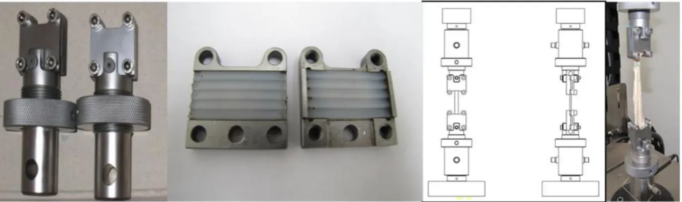
40 grafts were collected from 8 human cadavers. We removed the bone-patella tendon-bone grafts (BPTB grafts), semitendinosus and gracilis tendons (STG), quadriceps grafts, Achilles tendons and peroneus longus tendons from each lower extremity. We tested 40 tendon grafts (5 from each cadaver) with six different types of fixation devices: surgical thread (Premicron 3), wire mesh, Zwick-type universal clamp, cement fixing, Shi’s clamp and Shi’s modified, frozen clamp for an INSTRON loading machine. Int he last case we created two pockets for both sides of each clamp, and we froze the two ends of the samples into these clamps helped with dry-ice for a better fixation.
Donor tissues were removed according to the surgical protocols. All tendons were screened for degenerative changes visually and mechanically. There was no previous history evidence of injury or disease around these ligaments. To minimize the dehydration, specimens were put into physiological saline. The grafts were removed within 24 hours from death.
An Instron 8872 universal servo hydraulic computer-controlled loading testing system was used for the endurance tests. The data was evaluated by unpaired T-tests.
3.2. Biomechanical testing of frozen and gamma- irradiated tendografts
Our study included 30 human cadavers, from which 300 grafts were collected. From every donor, 5 types of grafts were harvested: Achilles, quadriceps, semitendinosus+gracilis (STG), tibialis anterior (TA) and peroneus longus (PL). The tendons were harvested from donors within 24 from death. All tendons were visually and mechanically screened for degenerative changes. There was no previous history or evidence of injury or disease of the tendons in the patient’s documentation. The grafts were soaked in a radio-protectant for 4 hours at 40 oC with agitation and 24 hours at 4 oC. Each specimen was labeled in a separate container and frozen slowly at -80 oC [15]. The grafts were divided into three groups. Group A contained
100 frozen specimens. Another 100 tendons of group B were irradiated with a target dose of 21 kGy (dose range 18-24 kGy, this is bactericidal dose) and the last 100 specimens (group C) were irradiated with a target dose of 42 kGy (dose range 38-46 kGy, this is virucidal dose).
We found no significant difference between the ages of groups. The grafts were thawed at room temperature on the day of biomechanical testing. Before testing, the cross-sectional area and the inter-clamp length of the tendons was measured. We used a micrometric caliper.
Instron 8872 servo-hydraulic loading machine was used for the endurance tests. The grafts were fixed into the frozen clamps. The freezing time was 3 minutes. Before tensile testing, pre-tensioning was applied with 50 N for 30 seconds. During cyclic loading tests, the
specimens were cycled between 50N and 250N for 1000 cycles at 2 Hz frequency, and then a load to failure test was performed.
Biomechanical parameters were calculated based on measured geometry data, crosshead displacement and force–elongation curves registered by the tensile tester. Statistical analyses were performed with Statistica 8.0. Data were presented as median with the corresponding interquartile range (25% percentile – 75% percentile). For group comparisons of variables the Kruskal-Wallis test were used. Multiple comparisons of mean ranks for all groups were applied for post hoc analyses. In all analyses, a p value of less than 0.05 was considered statistically significant.
4. Results
4.1. Results by the loading test of different type of fixations
Fixation of the grafts were achieved as done in anterior cruciate ligament reconstructions, by suturing the free ends of the quadrupled (double-folded) semitendinosus and gracilis tendons or likewise the peroneus longus tendons using Premicron 3 sutures. The mean failure loads was 381N ± 26N we measured in our study (Fig. 1).
In the second case, we tried to cement the ends of the grafts. Unfortunately, as it turned out, this was a complete failure (253N ± 18N) (Fig. 2), because of the very low friction coefficient between the tendon fibers and the cement, resulting in the relatively easy slippage of the ends of the tendons from the cement encasement. Unfortunately, in several cases, the cement mantle was damaged during the fixation into the clamp. We then tried to use the
general mounting clamp. We did not achieve sufficient results here either (527N ± 45N) (Fig.
3), since the tendons slipped out of the mounting clamp. In addition, the machines were unable to fasten the bony ends of the BPTB-grafts, because it would have resulted in severe damage of the polyethylene inlay of the clamp. In the fourth phase of our experiment, we tried a wire mesh fixation method. This could only be used for the fixation of the bony blocks of the BPTB grafts. The results were more promising (750N ± 21N), but did not reach the outcomes we desired and this method was compatible with only one type of graft, therefore further improvements were needed (Fig. 4). Then we used Shi’s clamp. The results were controversial. In cases of wider grafts – such as Achilles and quadriceps – we measured higher friction forces, but thinner grafts – such as peroneus, semitendinosus – slipped out in every case. The average loads to failure force were 997N ± 416 (Fig. 5).
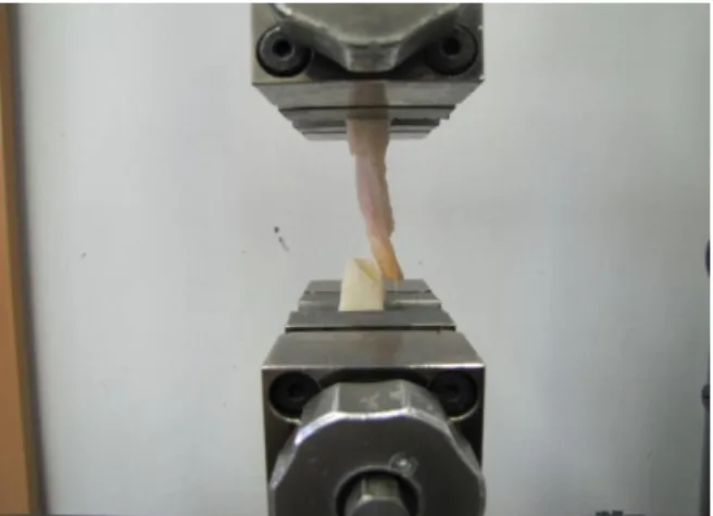
Lastly, we combined Shi’s clamp with a freezing technique. The tensile load required (2198N
± 773N) and the rupture of the tendon itself demonstrated that we found the mounting device needed for the strength testing of tendons (Fig. 6). The peak force was 3298.5 N. We
compared the results of the fifth and sixth fixation techniques. T-testing was performed and we found statistically significant differences between the two groups (p < 0.01). All grafts were torn (Fig. 7) except the patellar graft, where we were only able to reach a peak force of 1030 N in this type of graft. All of patellar grafts slid out from both Shi’s clamp and from the frozen Shi’s clamp. These grafts were only 6.375 cm long. The other grafts’ average lengths were 9.25–12 cm.
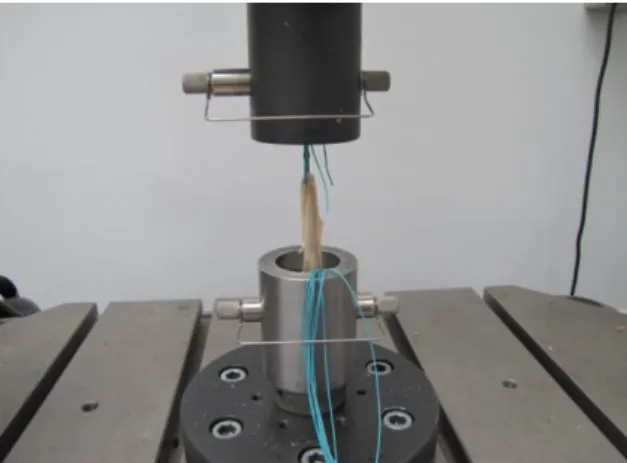
Fig. 1. The semitendinosus tendon sutured via Premicron 3 recording by the Instron loading machine
Fig. 2. Sliding out of Achilles graft from the cement mantle
Fig. 3. Tensile test with our ordinary mounting clamp
Fig. 4. Wire mesh with BPTB graft
Fig. 5. Shi’s modified clamp
Fig. 6. Shi’s modified and frozen clamp
Fig. 7. Characteristic tensile curves of the different fixation devices
4.2. Results by the biomechanical testing of frozen and gamma- irradiated tendografts
To compare the biomechanical properties of the specimens, the following four parameters were used: Young modulus of elasticity, maximum load, strain at tensile strength and strain at rupture.
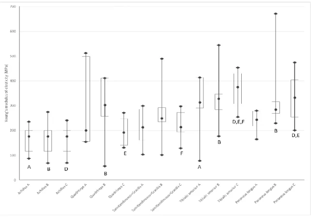
In group A, we compared the Young modulus of elasticity of the tendons, which resulted in significantly lower values of the Achilles tendon as compared to the TA (p=0.0036).
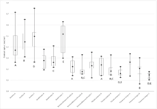
Additionally, we found no difference in maximum loads among the tendons, but the results of the STG were inferior to the other four tendons. The strain at tensile strengths of the Achilles tendons was significantly higher than that of the ST (p=0.0016), TA (p=0.042) and the quadriceps (p=0.002). The strain at rupture of the Achilles was inferior to the ST (p=0.0103) and TA (p=0.0199) (Figure 8-9-10-11).
In group B, we first compared the Young modulus of elasticity of the tendons; we found that the Achilles reached significantly lower values than the quadriceps (p=0.0042), the PL (p=0.028) and the TA (p=0.0001). After evaluating the maximum loads, the results of the STG tendons were significantly lower than those of the Achilles (p=0.002) and quadriceps (p=0.01). There was no difference between the Achilles, quadriceps and the TA. The strain at tensile strength of the Achilles were inferior compared to the quadriceps (p=0.017) and the STG tendons (p=0.000056). The strain at break of the Achilles demonstrated significantly inferior results compared to the STG (p=0.000029), PL (p=0.000032), and TA (p=0.00044).
Likewise, the quadriceps provided poorer results when compared to the STG (p=0.0215) and TA alone (p=0.0232) (Figure 8-9-10-11).
Similarly, in group C we compared the Young modulus of elasticity, and found that the Achilles reached significantly lower values than the PL (p=0.000042) and TA (p=0.00142).
The quadriceps performed worse than the TA (p=0.0037), and the STG also provided poorer values than the TA (p=0.0432). The maximum load of the Achilles showed better results than the PL (p=0.0016) and the STG (p=0.0066). The quadriceps showed also better results than PL (p=0.0018) and STG (p=0.0019). The TA had similar results like the Achilles and quadriceps. The strain at tensile strengths of Achilles were significantly less resistant to the tensile forces than the STG (p=0.0166), PL (p=0.0039) and TA (p=0.004). Also the
quadriceps were inferior compared to TA (p=0.00226) or to the PL (p=0.0006). The strains at rupture of the Achilles had significantly lower values compared to the PL (p=0.00045) and
TA (p=0.00066) and similarly, the quadriceps were significantly inferior to the PL (p=0.00045) and TA (p=0.00066) (Figure 8-9-10-11).
The measured results are represented in mediana with the corresponding interquartile range and where there was a significant difference between the groups stated in capital letters (Figure 8-11.)
Fig. 8.: Values of tendons of Young’s moduli of elasticity. Median, 25%, 75% percentile minimum and maximum values are used. The letters show the significant difference between the tendons. The exact p values are written in the text. The A-B-C letters on the X axis means the dose of gamma irradiation: A non- irradiated, B irradiated with 21 kGy, C irradiated with 42 kGy
Fig. 9. : Values of tendons of load to failure forces. Median, 25% and 75% percentile are used. Median, 25%, 75% percentile minimum and maximum values are used. The letters show the significant difference between the tendons. The exact p values are written in the text. The A-B-C letters on the X axis means the dose of gamma irradiation: A non-irradiated, B irradiated with 21 kGy, C irradiated with 42 kGy
Fig. 10. : Values of tendons of strain at tensile strength. Median, 25% and 75% percentile are used. Median, 25%, 75% percentile minimum and maximum values are used. The letters show the significant difference between the tendons. The exact p values are written in the text. The A-B-C letters on the X axis means the dose of gamma irradiation: A non-irradiated, B irradiated with 21 kGy, C irradiated with 42 kGy
Fig. 11. : Values of tendons of strain at rupture. Median, 25% and 75% percentile are used. Median, 25%, 75% percentile minimum and maximum values are used. The letters show the significant difference between the tendons. The exact p values are written in the text. The A-B-C letters on the X axis means the dose of gamma irradiation: A non-irradiated, B irradiated with 21 kGy, C irradiated with 42 kGy
Comparing the effect of gamma irradiation on each tendon the worst results were found in case of the quadriceps tendons. Young modulus of elasticity significantly decreased, however only when group B vs. C were compared (p=0.048). Both the 21 kGy and the 42 kGy gamma irradiation decreased the strain at tensile strength (group A vs. group C p=0.0021, group B vs.
group C p=0.015) and strains at rupture (group A vs. group C p=0.0108, group B vs. group C p=0.0048). The maximum load was not affected by the irradiation. In case of the other four tendons we found no difference in the examined parameters.
5. Conclusions
1. We managed to find the appropriate clamping technique for static and dynamic biomechanical testing of tendografts. This can also use for other similar research models too. The modified froozing clamp was able to fix the tendons suitable, in each event created a rupture phenomena. And on the tendon’s surface visible liquid
indicated that the section of samples examined in the freezing was not affected.
2. Our results indicate that different tendons have different initial biomechanical properties. In our opinion the most important factor is Young modulus of elasticity, which describes the elasticity of the tendons at normal loading. Our measurements showed that from the tested tendografts the TA has the best Young’s modulus of elasticity.
3. Examining the bactericidal dose of gamma irradiation’s effects it can be said that from the examined five samples the TA’s and PL’s Young's modulus of elasticity were significantly better than Achilles grafts. Compared with the control group, the quadriceps and Achilles groups’ strain at tensile strength and strain at rupture parameters showed significant attenuation.
4. Gamma irradiation of 42 kGy significantly decreased the Young’s modulus of elasticity in case of the quadriceps. However, when the comparison was carried out between the groups, TA and PL were superior to Achilles at lower doses. At higher doses, the Achilles, STG and quadriceps were all inferior to TA in this parameter. The doubled TA and PL tendons demonstrated the Young modulus of elasticity, maximum load, strain at tensile strength and strain at rupture that were equal to or better than all the other currently described ACL grafts. The Achilles and quadriceps grafts had poorer endurance properties than the others.
5. Comparig the two irradiated groups (Group-B, Group-C) we can declare the TA and PL grafts perform better than the others. In our study, both low and high doses of irradiation caused significant increases in the strain at tensile strength, and strain at
rupture parameters in case of the Achilles and the quadriceps. This could mean that the higher dose of gamma irradiation is, the lower the Achilles and quadriceps tendons’ capability to resist against elongation forces, and their capability of
restabilization to their original length is also decreased. This could lead to an increased laxity of the knee after ACL reconstruction with allografts. During these
aforementioned loading forces, the Young’s modulus of elasticity and strain at maximal forces play a critical role in joint laxity. We found no difference in case of the TA. In these force ranges, the irradiated allografts have the same elasticity
properties as the controls. We measured similar results in case of PL, however the 42 kGy irradiation significantly increased the strain at rupture parameter.
6. After low and high doses of gamma irradiation, the quadriceps and Achilles suffered the greatest decrease in biomechanical parameters. The effect of gamma irradiation on the STG and PL was slight, but the initial properties were not as good as the TA.
Additionally, semitendinosus tendons – if used alone – have to be folded into 4 strings; or when used with the gracilis, the two tendons have to be sutured together during the operation. In case of the TA and PL, only a single loop is used.
The results of this study indicate that different types of tendons react differently to gamma irradiation. Achilles and quadriceps grafts were the most sensitive to gamma irradiation. It seemed that the vulnerability of irradiation of TA and slightly of the PL were less than that of the Achilles and quadriceps tendons.
Some conclusions can be drawn from our results:
If a surgeon wants to use a fresh-frozen graft, then with the exception of the Achilles, all 4 kinds of grafts can be recommended.
If a bacteria-free graft is in focus then TA, PL and STG can be used.
If the highest level of microbiological safety is preferred, usage of TA graft can be the best choice. Certainly, additional donor screening can improves the safety of the grafts.
6. Publications
Papers connected with the Ph.D. thesis:
1) Baló E., Schandl K., Hangody Gy., Bodó L., Hangody L. (2009): Izolált elülső keresztszalag sérülés ellátásánál felhasznált különböző autológ grafttípusok
összehasonlító vizsgálata. Magyar Traumatológia Ortopédia Kézsebészet Plasztikai Sebészet, 52. (Suppl.): 8-9.
2) Baló E., Hangody Gy. M., Rahmeh H., Bodó L., Hangody L. (2010):Semitendinosus ínnal végzett egyköteges és kétköteges, négycsatornás elülső keresztszalag pótlással elért eredményeink prospektív összehasonlító vizsgálata. Biomechanica Hungarica,. 3. (2):
13–21.
3) Baló E., Schandl K., Hangody Gy., Abkarovits G., Bodó L., Hangody L. (2010): Izolált elülső keresztszalag sérülés ellátásánál felhasznált különböző autológ graft típusok összehasonlító vizsgálata. Magyar Traumatológia Ortopédia Kézsebészet Plasztikai Sebészet, 53. (1): 13-24.
4) Baló E., Bodó L., Hangody Gy., Rahmeh H., Hangody L. (2010): LCA sérülés és társsérülések ellátási lehetőségei osztályunkon. Magyar Traumatológia Ortopédia Kézsebészet Plasztikai Sebészet, 53. (Suppl.): 8.
5) Szigeti I., Hangody L., Bodó L., Kocsis K., Hangody Gy. M. (2010): Az elülső keresztszalag-sérülés ellátásának modern szemlélete. Orvosképzés, 85. (3): 225-231.
6) Gy. Hangody, G. Pánics, G. Szebényi, R. Kiss, L. Hangody, K. Pap (2016): Pitfalls during biomechanical testing - Evaluation of different fixation methods for measuring tendons endurance properties. Physiology International, 103: 86-93
7) Hangody Gy., Pap K., Abonyi B., Szebényi G., Kiss R., Hangody L. (2016): Gamma sugárzással sterilizált keresztszalag allograftok biomechanikai tulajdonságainak elemzése. Magyar Traumatológia Ortopédia Kézsebészet Plasztikai Sebészet, 59.
(Suppl.): 18-19, 97.
8) Pap K., Hangody Gy., Pánics G., Bodó L., Abonyi B., Hangody L. (2016): Allograftok – biomechanikai jellemzők és különböző sterilizálási eljárások bemutatása. Magyar
Traumatológia Ortopédia Kézsebészet Plasztikai Sebészet, 59. (Suppl.): 167.
9) Gy. Hangody, G. Szebényi, B. Abonyi, R. Kiss, L. Hangody, K. Pap (2016): Has the different dose of gamma irradiation the same effect on five different types of tendon allografts? – a biomechanical study. International Orthopaedics (accepted for publication, DOI: 10.1007/s00264-016-3336-7)
Other publications:
10) I. Szigeti, T. Koreny, G. M. Hangody, M. Wieland, L. Hangody (2013): Klinische Erfahrungen mit der autologen osteochondralen Mosaikplastik. Orthopaedie und Rheuma, 16(5): 32-34.
11) Hangody L., Vásárhelyi G., Hangody L.R., Pánics G., Hangody Gy. M. (2016):
Csontvelőből nyert, felnőtt típusú mesenchymalis őssejt koncentrátum lehetőségei a porcfelszínképzésben – korai tapasztalatok. Magyar Traumatológia Ortopédia Kézsebészet Plasztikai Sebészet, 59. (Suppl.): 29.
12) Hangody L., Abonyi B., Hangody L. R., Hangody Gy. M., Farkas J., Szabó Á., Németh Gy. (2016): Sanat Swing revíziós térdprotézis – bemutatás, fejlesztési és korai klinikai tapasztalatok. Magyar Traumatológia Ortopédia Kézsebészet Plasztikai Sebészet, 59. (Suppl.): 56, 151.