Article
Universal Properties and Specificities of the β 2 -Adrenergic Receptor-G s Protein Complex Activation Mechanism Revealed by All-Atom Molecular Dynamics Simulations
Argha Mitra1,2 , Arijit Sarkar1,2 and Attila Borics1,*
Citation: Mitra, A.; Sarkar, A.;
Borics, A. Universal Properties and Specificities of theβ2-Adrenergic Receptor-GsProtein Complex Activation Mechanism Revealed by All-Atom Molecular Dynamics Simulations.Int. J. Mol. Sci.2021,22, 10423. https://doi.org/10.3390/
ijms221910423
Academic Editor: Francisco Ciruela
Received: 30 August 2021 Accepted: 23 September 2021 Published: 27 September 2021
Publisher’s Note:MDPI stays neutral with regard to jurisdictional claims in published maps and institutional affil- iations.
Copyright: © 2021 by the authors.
Licensee MDPI, Basel, Switzerland.
This article is an open access article distributed under the terms and conditions of the Creative Commons Attribution (CC BY) license (https://
creativecommons.org/licenses/by/
4.0/).
1 Laboratory of Chemical Biology, Institute of Biochemistry, Biological Research Centre, 62. Temesvári krt., H-6726 Szeged, Hungary; argha.mitra@brc.hu (A.M.); sarkar.arajit@brc.hu (A.S.)
2 Theoretical Medicine Doctoral School, Faculty of Medicine, University of Szeged, 97. Tisza L. krt., H-6722 Szeged, Hungary
* Correspondence: borics.attila@brc.hu; Tel.: +36-62-599-600 (ext. 430)
Abstract:G protein-coupled receptors (GPCRs) are transmembrane proteins of high pharmacological relevance. It has been proposed that their activity is linked to structurally distinct, dynamically interconverting functional states and the process of activation relies on an interconnecting network of conformational switches in the transmembrane domain. However, it is yet to be uncovered how ligands with different extents of functional effect exert their actions. According to our recent hypothesis, based on indirect observations and the literature data, the transmission of the external stimulus to the intracellular surface is accompanied by the shift of macroscopic polarization in the transmembrane domain, furnished by concerted movements of highly conserved polar motifs and the rearrangement of polar species. In this follow-up study, we have examined theβ2-adrenergic receptor (β2AR) to see if our hypothesis drawn from an extensive study of theµ-opioid receptor (MOP) is fundamental and directly transferable to other class A GPCRs. We have found that there are some general similarities between the two receptors, in agreement with previous studies, and there are some receptor-specific differences that could be associated with different signaling pathways.
Keywords:GPCR; adrenergic; activation mechanism; signal transduction; molecular dynamics
1. Introduction
G protein-coupled transmembrane receptors (GPCRs) constitute one of the largest and most important protein superfamilies of the human genome. Their importance mainly derives from their remarkably high pharmacological relevance [1–3]. GPCRs are classified into six sub-families (A-F) based on their sequence and function. From the structural perspective GPCRs share similar architecture in their transmembrane domains and possess high sequential and structural diversity of their extra- and intracellular loops and the extracellular (N-terminal) and cytosolic (C-terminal) domains. These variable domains are proposed to be responsible for ligand and G protein/arrestin specificity, whereas the transmembrane (TM) domain controls the transmission of external signals to the intracellular surface of the protein. The variety of G proteins that mediate GPCR signaling is very low relative to the diversity of GPCRs and their external activators. Therefore, the activation of GPCRs is suggested to follow a general structural mechanism.
Adrenergic receptors, responsible for the homeostasis between stressful and resting conditions of the body, belong to the most populated class (A) of GPCRs. Theβ2-adrenergic receptor (β2AR) is linked to respiratory, as well as other smooth muscle relaxation. These receptors are stimulated by endogenous neurotransmitters, such as epinephrine and nore- pinephrine and the signal transduction of β2AR is mediated predominantly by the Gs
(stimulatory) protein complex [4]. The agonist-β2AR-Gs signaling triad is an important target of drug development activities to treat severe respiratory conditions, such as chronic
Int. J. Mol. Sci.2021,22, 10423. https://doi.org/10.3390/ijms221910423 https://www.mdpi.com/journal/ijms
obstructive pulmonary disease (COPD) or asthma. Remarkable efforts have been invested in the optimization of GPCR targeting drugs to reduce their undesired side effects. Such efforts necessitated the in-depth structural analysis of target GPCR structures [2].
Theβ2AR was among the first GPCRs of which a three-dimensional structure was solved at atomic resolution [5] and numerous experimental studies have been conducted to elucidate the structural details of activation of this TM receptor [6–15]. To date, the β2AR is the most widely investigated class A GPCR and is frequently used as a universal model for the study of the structural mechanism of activation for class A GPCRs [16].
Recent developments in the field of experimental structural biology led to the rapid ac- cumulation of experimental data of GPCR structures which are now readily available in the Brookhaven Protein Data Bank as well as in GPCRdb, a specific, comprehen- sive collection of all GPCR structures published to date (http://gpcrdb.org, accessed on 23 August 2021) [15]. A general structural difference between active and inactive state class A GPCRs, includingβ2AR, is the position of the sixth helix of the TM domain (TM6) [5,7,8].
The capabilities of conventional experimental techniques to provide information about the mechanism of transition between these structural states are limited. The current theory of the structural mechanism of class A GPCR activation was framed on the basis of results acquired through the application of state-of-the-art molecular dynamics (MD) simulation hardware and techniques [17–19]. According to this theory, GPCRs exist in a dynamic ensemble of multiple active, inactive, and intermediate states, even in the absence of ligands. The populations of these states are shifted upon ligand binding, depending on the functional properties of the bound ligand. The fifth, sixth, and seventh TM helices (TM5, TM6, TM7, respectively) have been emphasized for their foremost interplay in signal transduction [16,20].
As well as the rearrangement of TM helices, highly conserved polar functional mo- tifs, E/DRY, NPxxY, and CWxP, have been appointed as participants of the activation mechanism [21,22]. Specific rearrangements of intramolecular interactions involving these motifs, the conserved allosteric Na+ binding site [23,24], and the extended network of water molecules in the internal cavities connecting the orthosteric ligand-binding pocket to the cytosolic domains have been proposed recently as general machinery of signal transmission in GPCRs [25,26]. In order to respond to the most recent challenge of rational drug design and to develop high-affinity, high-efficacy, and functionally selective GPCR ligands a quantitative model of the activation mechanism is necessary. Models built exclu- sively on a structural basis have limited capabilities to differentiate between ligands with similar structure, physico-chemical properties, and binding affinity, but different efficacy.
Consequently, the creation of a directly transferable model would need the introduction of new perspectives.
Our recent results of extensive MD simulations of theµ-opioid receptor (MOP) indi- cated that the dynamic motions of polar amino acid side chains of conserved motifs are highly correlated [27]. Such concerted motions were only observed during the simulations of the agonist- and Gi protein-bound active state MOP, suggesting that this phenomenon could be associated with the signal transduction process, corroborating the above-cited pro- posal [25,26]. These polar amino acid side chains of the orthosteric and allosteric binding pockets, the NPxxY and E/DRY motifs and the cytosolic helix (H8), form a polar signaling channel connecting the binding pocket to the intracellular G protein-binding surface. Fre- quent transitions between rotameric states, however, were not observed for these specific side chains, which casts doubt on the channel’s operation as a sequential conformational switch. According to our recent proposal, receptor activation is accompanied by a shift of macroscopic polarization in a shielded central duct of the TM domain, initiated by ligand binding and propagated by the minuscule rearrangements of polar amino acid side chains along TM7. TM7 was further implied in the activation mechanism as a potential conduc- tor owing to its inherent dipole moment. Evidently, MD simulations employing fixed point charge force fields cannot reveal exact or quantitative details of processes involving
charge shift. Nevertheless, independent mutation data provided convincing support for the interplay of the above-mentioned polar species [27].
Here we present an extensive, unbiased, atomistic MD simulation study of the full sequenceβ2AR, embedded in a native-like caveolar membrane bilayer, in the presence of an endogenous agonist and the Gs protein complex orβ-arrestin-2. Simulations were started from the active and inactive structural form of the receptor, revealed by previous X-ray crystallographic studies [5,7]. Analyses were conducted according to the above- mentioned novel perspective, to examine that our previously proposed indirect hypothesis could be extended to other class A GPCRs.
2. Results and Discussion 2.1. Simulation System Integrity
On the grounds that the disposition of TM helices was proposed to have a pivotal role in the activation mechanism [16,20,25,26], an important specific aim of this current study was to study the full sequenceβ2AR in order to take account of the pull of theN- and C-terminal domains posed on the TM helices and to see if that affects the internal dynamics of the TM domain. These highly variable and flexible domains are generally omitted in the experimental structures of GPCRs and, consequently, from the corresponding MD simula- tion studies. Here, approximate structures of the terminal domains were generated through folding simulations (see Section3). Even if parallel folding simulations provide convergent results, the correct and complete folding of these domains in the available time frame could not be guaranteed. Nevertheless, their effect on TM dynamics, primarily exerted by their mass is satisfactorily taken into account by using these approximate structures.
Unfolding ofN- andC-terminal domains during simulations could result in contacts formed between the neighboring periodic images of these unfolded domains which could lead to artifacts. The evolution of the radii of gyration ofN- andC-terminal domains indicated partial unfolding during some of the production simulations (Figure S1), but the minimum distance between theN- andC-terminal domains, was never below 1.4 nm (Figure S2). Therefore, the possibility of artificial contact between these domains could be excluded.
With regard to the stability of simulation systems, no dissociation or notable rela- tive displacement of macromolecular components were observed during the simulations.
Epinephrine, however, dissociated from the orthosteric binding pocket on two occasions.
First, it was ejected from the binding pocket during the first 100 ns of one of the three replica simulations of the active Gs protein-boundβ2AR. The second time it was observed for the inactive Gs protein-boundβ2AR, when epinephrine left the orthosteric binding site after approximately 600 ns of simulation time.
In the second replica simulation of the active Gsprotein-boundβ2AR, the ligand took an opposite orientation in the binding pocket, compared to the X-ray crystallographic structure [8] of theβ2AR-epinephrine complex (Figure1). These simulations were not excluded from analysis but the results were interpreted accordingly. A reference simulation of the active G protein-boundβ2AR in which epinephrine was restrained to the binding pocket was performed in order to explain discrepancies emerging from ligand dissociation.
The instability ofβ2AR–epinephrine complexes observed during some of the simulations may be explained by the smaller size and remarkably lower affinity of epinephrine rela- tive to the ligands used in previous simulations of theβ2AR (µM vs. pM range affinity, respectively) [17,28].
Figure 1. Ligand position in the orthosteric binding pocket in the 1st (b) and 2nd (c) replica simulations of the active β2AR–Gsprotein–epinephrine complex in comparison with the crystallographic structure ((a), PDB code: 4LDO).
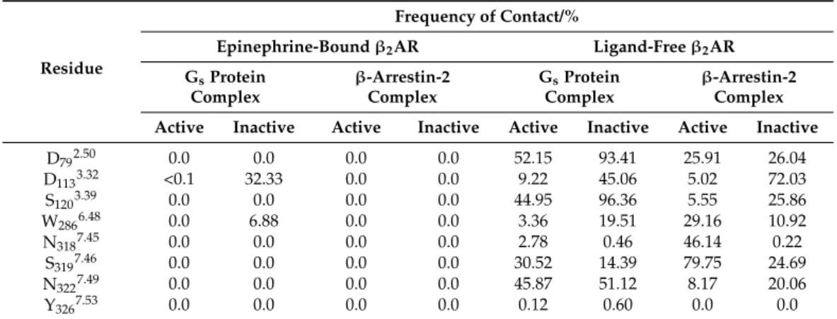
2.2. Allosteric Na+Binding
Na+penetration to the allosteric Na+binding site (D792.50) (Figure2, Table1) was not observed for the epinephrine-boundβ2AR, regardless of the state of the receptor, except when epinephrine dissociated from the orthosteric binding site during the course of the simulation. Furthermore, Na+entrance from the cytosolic side did not occur in any of the systems. In the absence of epinephrine, however, localization of Na+at the ortho- (D1133.32) (Figure S3) and allosteric sites took place. Frequent contacts were formed between Na+ and the ortho- and allosteric sites as well as residues of the conserved CWxP and NPxxY motifs, but no relevant trend of contact frequencies was identified which could be directly associated with the modulation of receptor activation by Na+ions (Table1). Interestingly, Na+ was occasionally present in the orthosteric binding pocket of the Gsprotein- and epinephrine-bound, active stateβ2AR. This phenomenon may be less relevant since it was only noticeable in one of the simulation replicas (Figure S3) and the frequency of contact between Na+and D1133.32was negligible (Table1). The presence of Na+in the orthosteric pocket and the proximal CWxP motif was more prominent for the Gsprotein- and epinephrine-bound inactive stateβ2AR. However, this was a clear consequence of ligand dissociation after approximately 600 ns of simulation time. No Na+insertion to the ortho- or allosteric sites was observed in any of the ligand- andβ-arrestin-2-bound states.
Figure 2.Minimum distance between Na+ions and the allosteric Na+binding site, D792.50of the active (a) and inactive state (b)β2AR during simulations. Black:β2AR–Gs protein–epinephrine complex, 1st replica; Red: β2AR–Gsprotein–epinephrine complex, 2ndreplica; Green: β2AR–Gs
protein–epinephrine complex, 3rdreplica; Blue:β2AR–Gs protein–epinephrine complex, with re- strained epinephrine and GDP; Yellow:β2AR-β-arrestin-2–epinephrine complex; Orange: ligand-free β2AR–Gsprotein complex; Magenta: ligand-freeβ2AR-β-arrestin-2 complex.
Table 1.The frequency of contact (d≤0.4 nm) between Na+ions and polar amino acid side chains of the allosteric and orthosteric binding pockets and nearby conserved motifs.
Residue
Frequency of Contact/%
Epinephrine-Boundβ2AR Ligand-Freeβ2AR GsProtein
Complex
β-Arrestin-2 Complex
GsProtein Complex
β-Arrestin-2 Complex Active Inactive Active Inactive Active Inactive Active Inactive
D792.50 0.0 0.0 0.0 0.0 52.15 93.41 25.91 26.04
D1133.32 <0.1 32.33 0.0 0.0 9.22 45.06 5.02 72.03
S1203.39 0.0 0.0 0.0 0.0 44.95 96.36 5.55 25.86
W2866.48 0.0 6.88 0.0 0.0 3.36 19.51 29.16 10.92
N3187.45 0.0 0.0 0.0 0.0 2.78 0.46 46.14 0.22
S3197.46 0.0 0.0 0.0 0.0 30.52 14.39 79.75 24.69
N3227.49 0.0 0.0 0.0 0.0 45.87 51.12 8.17 20.06
Y3267.53 0.0 0.0 0.0 0.0 0.12 0.60 0.0 0.0
These observations are in complete agreement with previous MD simulation data of this receptor [29], and other class A GPCRs [27,30–32], and corroborate that the al- losteric Na+binding site is only accessible through the orthosteric binding pocket and the bound orthosteric ligand blocks the entrance of Na+to the allosteric site. Intracellular access of Na+ions through the TM domain is closed by the bound Gsprotein complex or β-arrestin-2. Translocation of Na+ions through the TM region was observed in previous MD simulations of the active state MOP in the absence of bound ligands and intracellular proteins, suggesting that it takes place during the process of receptor deactivation [33].
2.3. Transmembrane Helix and Loop Dynamics
In our previous study of the MOP, TM7 was found to be the most ordered among the TM helices of the active state Gi protein-bound receptor. Furthermore, the helical conformation of TM7 was indicated to be the closest to ideal when the receptor was bound to the Giprotein complex. Conversely, the lowest degree of order of TM7 was observed in theβ-arrestin-2-bound state. Based on these geometric features it was assumed that TM7 possesses the highest dipole moment in the Giprotein-bound state, which could facilitate electron, proton, or ion conduction along the helix axis [34]. Such conduction capacity could be relevant for the proposed model, which involves the shift of charge balance between the orthosteric binding pocket and the intracellular G protein-binding interface during class A GPCR activation [27]. Such a proposed role of TM7 was not corroborated by the results obtained here forβ2AR (Figure S4). As opposed to the MOP, TM7 was among the least ordered TM helices of theβ2AR. Furthermore, the relatively high order of TM7 in the active, epinephrine- and Gsprotein-bound state was not reproduced in the reference simulation of that system and was also matched by theβ-arrestin-2 and epinephrine-bound receptor (Figure S4). This suggests that the above-described trend is a specific property of MOP and/or class A GPCRs signaling through the Giprotein complex.
Atomic displacement analysis of the TM6 of epinephrine-boundβ2AR indicated moderate dispositions from the corresponding starting structures (Figures S5 and S6), similar to our previous simulation results for the MOP [27]. Remarkable rearrangements of TM helices have occurred during previous simulations of theβ2AR, but at longer timescales and in the absence of bound intracellular proteins [17]. Slightly larger dispositions of TM6 were observed in the ligand-free systems, confirming the stabilizing effect of the bound agonist, again, in agreement with our previous results [27]. One exception was the β-arrestin-2-bound inactive receptor in which, similar to the ligand-bound systems, no significant TM6 disposition took place. This, in contrast with our previous results, suggests the preference ofβ-arrestin-2 for the inactive structure of this receptor.
The unexpected disorder of TM7, discussed above, prompted us to analyze the dispo- sition of this helical segment to see if the dynamics of the NPxxY motif show any correlation with the activation state or with the presence of agonist and/or intracellular signaling
proteins. Surprisingly, large dispositions (~0.4 nm RMSD) of the NPxxY motif were found, relative to the active state structure and to the corresponding TM7 dispositions in several simulations (Figures3and S7).
Figure 3.Disposition of the NPxxY motif during simulations with respect to the active (green) and inactive (red) crystallo- graphic structures of theβ2AR. (a) activeβ2AR–Gsprotein–epinephrine complex, 1st replica; (b) inactiveβ2AR–Gsprotein–
epinephrine complex; (c) activeβ2AR–β-arrestin-2–epinephrine complex; (d) inactiveβ2AR–β-arrestin-2–epinephrine complex. (e) active, ligand-freeβ2AR–Gsprotein complex; (f) inactive, ligand-freeβ2AR–Gsprotein complex; (g) active, ligand-freeβ2AR–β-arrestin-2 complex; (h) inactive, ligand-freeβ2AR–β-arrestin-2 complex.
Most interestingly, this large disposition of the NPxxY motif coincides with the in- tensive concerted dynamics of the second segment of the polar signaling channel, which could be associated either with receptor activation or constitutional activity (see data and discussion below). It should be noted, that the results of the epinephrine and G protein- bound inactive state receptor and the ligand-free G protein-bound inactiveβ2AR are very
similar, due to epinephrine dissociation after 600 ns. The results of secondary structure analysis indicated that ICL1, ICL3, and H8 maintain their secondary structures in all re- ceptor states and only minor, reversible changes occur, resulting from internal dynamics (Figures S8–S12). ICL2, on the other hand, adopted anα-helical structure in active states and got partially unfolded in inactive states (Figure4, Figure S13). ICL2 of the active β2AR was recently shown to beα-helical when the receptor is bound by Gsand partially unfolded when the Giprotein complex is attached [35]. Similar signaling protein selectivity was observed previously for the MOP receptor [27]. However, in contrast with the present results, the structure of ICL2 did not demonstrate any dependence on the activation state of the MOP. Nevertheless, the present simulation results are in agreement with extensive experimental data reporting anα-helical structure of ICL2 in the active [7,8,36,37] and unfolded structure in the inactive state ofβ2AR [5,11].
Figure 4.Evolution of the secondary structure of ICL2 during simulations. (a) activeβ2AR–Gsprotein–epinephrine complex, 1st replica; (b) inactiveβ2AR–Gs protein – epinephrine complex; (c) active β2AR–β-arrestin-2–epinephrine complex;
(d) inactiveβ2AR–β-arrestin-2–epinephrine complex. (e) active, ligand-freeβ2AR–Gsprotein complex; (f) inactive, ligand- freeβ2AR–Gsprotein complex; (g) active, ligand-freeβ2AR–β-arrestin-2 complex; (h) inactive, ligand-freeβ2AR–β-arrestin- 2 complex.
2.4. Correlated Side-Chain Motions in the Transmembrane Domain
Dynamic cross-correlation matrix (DCCM) analysis (Figures5and S14) of the trans- membrane domain and the extra- and intracellular loops indicated, that similar to that observed for the MOP receptor previously [27], the orthosteric binding pocket and the G protein-binding interface of theβ2AR are connected through a channel of polar amino acids which are engaged in concerted motions in the active, or constitutively active states of the receptor (Figures6and S15). However, there are several differences between the channel residues of the MOP and theβ2AR. The first difference is that, unlike in the case of
MOP, the residues of the DRY motif are not involved in correlated motions. In the active Gi protein-bound MOP, R1653.50of the DRY and D3408.47of H8 were frequently connected through a salt bridge and, consequently, their motions were in intense correlation. Inβ2AR no such salt bridge could be formed by the analogous S3298.47of H8 and the occurrence of H-bonds was also very low, which may provide an explanation for the missing involvement of the DRY motif (See further discussion below). Nevertheless, S3298.47was found to move in accord with the NPxxY motif inβ2AR, but the occurrence of that connection was also not as pronounced as in the case of MOP. Instead, D3318.49and R3338.51residues of the H8 showed a high degree of correlation. R3338.51is conserved (Figure6) and was suggested previously to be important for the G protein-coupling of the adenosine A2Breceptor [38]
While such occasional similarities with other class A GPCRs may support the importance of H8 residues, their increased variability compared to the other residues of the channel could also be associated with G protein specificity. A further difference observed between the MOP andβ2AR is that the allosteric Na+binding site (D792.50) is less intensely involved in the correlated motions of channel residues ofβ2AR. However, this correlation was present during the reference simulation when epinephrine was mildly restrained to the orthosteric binding pocket, suggesting that the presence of a strongly bound, correctly oriented ligand initiates coupling of D792.50to the signaling cascade (Figure S15). An interesting, specific feature of the polar signaling channel ofβ2AR is that it could be subdivided into two segments, suggested by the results of NPxxY disposition and DCCM analysis. The first segment spans the orthosteric (Y3167.43) and allosteric (D792.50) binding pockets, N3187.45 and the NPxxY motif (N3227.49and Y3267.53), while the second segment shares the last residue of the NPxxY motif (Y3267.53) and includes the tip of TM7 (R3287.55) and the three H8 residues (S3298.47, D3318.49, and R3338.51) (Figures6and S15). The rationale behind this subdivision is provided by NPxxY disposition data as was already mentioned earlier in this report. The relatively large, approximately 0.4 nm (RMSD) disposition of the NPxxY motif resulted in intense concerted motions in the above-mentioned second segment of the polar signaling channel. Such movements were observed in inactive states and in the absence of ligand, whereas the full sequence of correlated motions was incomplete in those systems. This suggests that the elevated concerted dynamics of the second segment could also be associated with the constitutive activity ofβ2AR. This presumption was sup- ported by that correlated motions of this second segment were decoupled uponβ-arrestin-2 binding and/or if epinephrine was bound in the wrong relative orientation, stabilizing a conformational state which is inappropriate for signaling. Further support is provided by results of a previous extensive study, where an intermediate structure was identified in the absence of ligands, which may represent a receptor conformation that facilitates Gsprotein insertion and suggests that the activation process, in terms of structural changes, starts at the intracellular side of the receptor. The role of the NPxxY motif in the formation of such intermediate was also emphasized [17].
2.5. Intramolecular Interactions
The frequencies of intramolecular salt bridges and H-bonds, previously proposed to be relevant for class A GPCR activation, are summarized in Table2. As opposed to observations taken previously for the MOP [27], intramolecular interactions between the DRY motif and H8 were missing from allβ2AR systems examined here. Salt bridge formation is not facilitated between R1313.50and S3298.47and no other potential, proximal partners were found in H8 that could participate in the formation of a salt bridge analogous to that between R1653.50of the DRY and D3408.47of H8 in the active, Giprotein-bound MOP.
No stable H-bond formation was indicated between the DRY motif and H8 either. This specific interaction was first described in our previous report [27] as it was not present in the high-resolution experimental structures of the MOP. Since this interaction is not facilitated inβ2AR, it is most likely a specific property of the MOP. Considerable frequencies of salt bridges and H-bonds were observed between the neighboring D1303.49and R1313.50of the DRY motif in the active states, although high-resolution experimental structures indicated
coincidentally that this interaction is only present in inactive states and absent in active receptors [5,7,8,11–13]. Formation of the “ionic lock” between the DRY motif (R1313.50) and TM6 (E2686.30) was also not observed, neither in the active nor in the inactive states.
According to earlier proposals, this interaction acts as a constraint in the inactive state and gets disrupted upon receptor activation, followed by the release and disposition of TM6 [21]. Mutations affecting the participants of this ionic lock resulted in the elevated constitutional activity of theβ2AR [39], however, the presence of this ionic lock was not corroborated by the crystallographic structures of this receptor [5,12]. The absence of ionic lock interactions in these structures was attributed to residual basal activity present in the crystalline state [12]. Formation of this ionic lock was observed previously in MD simulations of receptor deactivation, but in the absence of intracellular signaling proteins and at significantly longer timescales [17].
Figure 5.Dynamic cross-correlation matrices of the Gsprotein-boundβ2AR in active and inactive states. Panels (a–c) are magnified views of regions of amino acid residues of interest. Black and white panels show correlations above the threshold of 0.65 MI.
Figure 6.The polar signaling channel of theβ2AR identified by dynamic cross-correlation analysis. (a) Polar amino acids of which motions are correlated in the Gsprotein-bound active state. (b) Polar amino acids of which motions are correlated and connecting the orthosteric binding pocket to the G protein-binding interface. Non-polar hydrogens are omitted for clarity. (c) Activeβ2AR–Gsprotein–epinephrine complex, 1st replica; (d) inactiveβ2AR–Gsprotein–epinephrine complex;
(e) activeβ2AR–β-arrestin-2–epinephrine complex; (f) inactiveβ2AR–β-arrestin-2–epinephrine complex. Red arrows indicate correlated motions of the respective amino acids. (g) Degree of conservation of polar signaling channel residues of human class A GPCRs.
In agreement with the experimental structure [7], the systematic presence of H-bonds between D1303.49 of the DRY motif and Y141 of ICL2 was indicated in simulations of the active stateβ2AR. In inactive states, D1303.49of the DRY motif was found to interact with S143 and L144. This is in agreement with the results of the secondary structure analysis of ICL2 and supports the discussion above, regarding the role of this loop in the activation mechanism. No considerable trends were observed between the different receptor states and the frequencies of DRY-TM5 [40], CWxP-TM7 [41], and D1133.32-Y3167.43 interactions [40] within the time frame of simulations.
Table 2.Frequency of intramolecular salt bridges and H-bonds expressed as percentages of the total conformational ensemble, generated by MD simulations.
Interactions Residues Involved
Epinephrine-Bound Ligand-Free
Active State Inactive State Active State Inactive State
GsProtein
Complex β-Arrestin-2 GsProtein
Complex β-Arrestin-2 GsProtein
Complex β-Arrestin-2 GsProtein
Complex β-Arrestin-2
1 2 3 Restrained
Salt Bridge
Intra-DRY D1303.49;
R1313.50 26.5 5.7 6.0 0 65.1 40.4 35.5 40.5 10.7 51.2 41.3
H-bonds
DRY-H8 R1313.50;
S3298.47 0.1 0 0.3 0 0.1 0 0 0.1 0.1 0.0 0.0
BP D1133.32;
Y3167.43 14.0 80.6 82.1 95.3 28.3 65.9 69.2 96.1 9.9 0.1 14.6
intra-DRY D1303.49;
R1313.50 65.9 64.5 17.9 16.8 35.7 80.0 72.9 82.3 30.5 99.8 83.5
DRY-ICL2
D1303.49;
Y141ICL2 99.3 97.5 92.4 98.0 99.8 0 0 98.1 86.2 0 0
D1303.49; S143ICL2; L144ICL2
0 0 0 0 0 99.5 90.8 0 0 0 37.3
DRY-TM5 R1313.50;
Y2195.58 3.9 10.5 4.6 45.2 30.9 0 0 5.7 3.3 0 0
DRY-TM6 R1313.50;
E2686.30 0 0 0 0 0 0 0 0 0 0 0
CWxP-TM7
C2856.47; W2866.48; N3187.45
1.8 3.4 45.1 4.1 5.3 11.7 4.3 17.6 0.5 53.7 3.8
BP = orthosteric binding pocket of theβ2AR; Ballesteros–Weinstein numbering of residues is indicated in superscript.
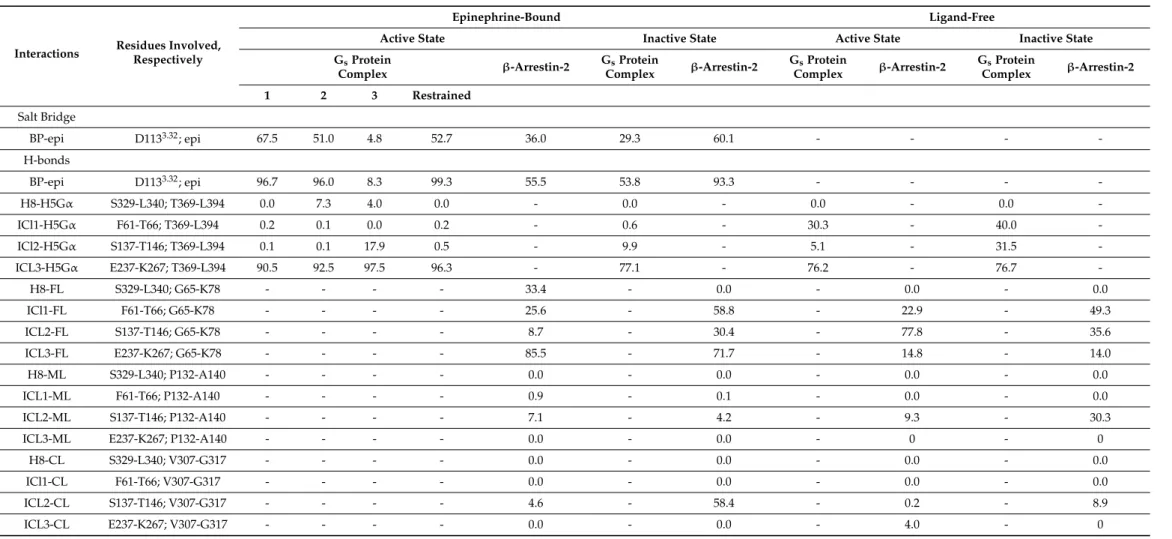
2.6. Intermolecular Interactions
The results of analyses of intermolecular interactions are summarized in Table3.
Similar to that observed previously for the MOP receptor [27], the interaction between the ligand and the anchor residue of the binding pocket (D1133.32) was strongest in the active, Gsprotein-boundβ2AR. The difference between active, inactive, Gsprotein, andβ-arrestin- 2 bound states was not as outstanding as in the case of MOP. This observed trend is also in slight contradiction with the fact that ligand disposition also occurred in Gsprotein-bound active states. Analysis ofβ2AR–Gsprotein interactions demonstrated that contacts between ICL1 and helix 5 of theαsubunit of the Gsprotein (H5Gα) are negligible in ligand-bound receptors, regardless of the activation state. Medium frequency was, however, observed in ligand-free states. The ICL2-H5Gαcontact was expected to be the most specific among the contacts between the Gsprotein and theβ2AR [7], based on previous results [27] and the above presented secondary structure and intramolecular H-bond analysis. Even so, this contact was found to be very weak during simulations of the active state, epinephrine and Gsprotein-boundβ2AR. Higher frequencies of H-bonds between ICL2 and H5Gα were observed in inactive states and the absence of epinephrine. This may suggest that the loss of interaction between ICL2 and H5Gαin agonist-bound active states indicates the initiation of Gsprotein dissociation, although longer simulations would be needed to confirm this assumption. The frequency of ICL3-H5GαH-bonds was found to be high in all simulation setups, therefore, a specific role of this contact cannot be deduced from the results presented here. Differences were found in the interactions betweenβ-arrestin-2 andβ2AR, depending on the presence of epinephrine. In the active ligand-bound state the finger loop (FL) ofβ-arrestin-2 was found to be in frequent contact with H8, ICL1, and ICL3, whereas in the active ligand-free state FL was in stronger contact with ICL2 at the expense of contacts with ICL3. In the inactive states, a higher preference of FL towards ICL1 was observed, regardless of the presence of epinephrine. No contacts were found between ICL3 of the agonist-boundβ1adrenergic receptor (β1AR) and the finger loop ofβ-arrestin-1 in a recent cryo-electronmicroscopic (cryo-EM) structure of this molecular complex [42].
However, a parallel study of the neurotensin receptor 1 (NTSR1)–β-arrestin-1 complex revealed, that the interface betweenβ-arrestin-1 and NTSR1, including the finger loop, is highly dynamic and the relative orientations captured by the cryo-EM structure are likely to represent one of many conformational states [43]. A specific contact between the C-loop ofβ-arrestin-2 (CL) and the unfolded ICL2 was indicated in the inactive ligand-bound receptor. In the inactive ligand-free receptor ICL2 was rather in contact with the middle loop ofβ-arrestin-2 (ML), but with significantly lower frequency. The overall frequency of interactions was highest for inactive, epinephrine-boundβ2AR suggesting that it is the most preferred forβ-arrestin-2 binding. However, taking into account that the ligand dissociated during the corresponding simulation, such an assumption cannot be taken.
The second highest H-bond frequency betweenβ2AR andβ-arrestin-2 was observed for the active, epinephrine-bound state. This latter apparent preference is corroborated by experimental data reporting the visual arrestin-bound [44] and active, GTprotein-bound structures of rhoposhin [45], which were almost identical. On the other hand, the cryo-EM structure ofβ1AR andβ-arrestin-1 demonstrated that this receptor adopts an intermediate state with regard to the disposition of TM6 when bound byβ-arrestin-1 [42].
Table 3.Frequency of intermolecular salt bridges and H-bonds expressed as percentages of the total conformational ensemble, generated by MD simulations.
Interactions Residues Involved, Respectively
Epinephrine-Bound Ligand-Free
Active State Inactive State Active State Inactive State
GsProtein
Complex β-Arrestin-2 GsProtein
Complex β-Arrestin-2 GsProtein
Complex β-Arrestin-2 GsProtein
Complex β-Arrestin-2
1 2 3 Restrained
Salt Bridge
BP-epi D1133.32; epi 67.5 51.0 4.8 52.7 36.0 29.3 60.1 - - - -
H-bonds
BP-epi D1133.32; epi 96.7 96.0 8.3 99.3 55.5 53.8 93.3 - - - -
H8-H5Gα S329-L340; T369-L394 0.0 7.3 4.0 0.0 - 0.0 - 0.0 - 0.0 -
ICl1-H5Gα F61-T66; T369-L394 0.2 0.1 0.0 0.2 - 0.6 - 30.3 - 40.0 -
ICl2-H5Gα S137-T146; T369-L394 0.1 0.1 17.9 0.5 - 9.9 - 5.1 - 31.5 -
ICL3-H5Gα E237-K267; T369-L394 90.5 92.5 97.5 96.3 - 77.1 - 76.2 - 76.7 -
H8-FL S329-L340; G65-K78 - - - - 33.4 - 0.0 - 0.0 - 0.0
ICl1-FL F61-T66; G65-K78 - - - - 25.6 - 58.8 - 22.9 - 49.3
ICL2-FL S137-T146; G65-K78 - - - - 8.7 - 30.4 - 77.8 - 35.6
ICL3-FL E237-K267; G65-K78 - - - - 85.5 - 71.7 - 14.8 - 14.0
H8-ML S329-L340; P132-A140 - - - - 0.0 - 0.0 - 0.0 - 0.0
ICL1-ML F61-T66; P132-A140 - - - - 0.9 - 0.1 - 0.0 - 0.0
ICL2-ML S137-T146; P132-A140 - - - - 7.1 - 4.2 - 9.3 - 30.3
ICL3-ML E237-K267; P132-A140 - - - - 0.0 - 0.0 - 0 - 0
H8-CL S329-L340; V307-G317 - - - - 0.0 - 0.0 - 0.0 - 0.0
ICl1-CL F61-T66; V307-G317 - - - - 0.0 - 0.0 - 0.0 - 0.0
ICL2-CL S137-T146; V307-G317 - - - - 4.6 - 58.4 - 0.2 - 8.9
ICL3-CL E237-K267; V307-G317 - - - - 0.0 - 0.0 - 4.0 - 0
BP = orthosteric binding pocket of theβ2AR; epi = epinenphrine; H5Gα= helix 5 of the Gsproteinαsubunit; FL/ML/CL = finger loop/middle loop/C loop ofβ-arrestin-2. Ballesteros–Weinstein numbering of residues is indicated in superscript.
3. Methods
3.1. System Building
The sequence of the humanβ2AR (UniProtKB-P07550-ADRB2) was obtained from the UniProt database (http://www.uniprot.org, accessed on 23 August 2021). All X-ray crystallographic structures used in this study were downloaded from the Brookhaven Protein Data Bank (http://www.rcsb.org, accessed on 23 August 2021). The active and inactive stateβ2AR (pdb codes: 3SN6 and 2RH1, respectively) [5,7],β-arrestin-2 (pdb code:
3P2D) [44], the Gsαprotein (pdb code: 1AZT) [46] and theβ2AR-bound epinephrine (pdb code: 4LDO) [8] were used as starting structures for MD simulations in this study. All crystallographic chaperones and fusion proteins were removed from the corresponding structures. Theαsubunit of the Gsprotein complex was missing from the crystallographic structure of the activeβ2AR (pdb code: 3SN6) [7], therefore, it was supplemented from an independent crystallographic structure of that subunit (pdb code: 1AZT) [46], together with the bound GDP. Epinephrine was inserted in the binding pocket of the receptors in a protonated form. The third intracellular loop (ICL3, E237-K267) ofβ2AR, missing from the crystallographic structures was modeled using the Modeller ver. 9.20 software [47] and the missing residues in the second extracellular loop (ECL2, A176-H178) were retrieved using the Swiss-PdbViewer ver. 4.10 program [48]. The missingN- andC-terminal domains (M1-D29 and C341-L413, respectively) ofβ2AR were modeled by performing 10 ns folding simulations using the GROMACS ver. 2018.3 program package [49], following a previously described protocol [27] and attached to the TM domain of the receptor manually.
The CHARMM-GUI [50] web-based platform was used to include post-translational modifications ofβ2AR and to insert the receptor in a solvated membrane bilayer. Residues N6, N15, and N187 were glycosylated [51] and residue C341 was palmitoylated in both the active and inactive state receptors [52], whereas phosphorylations at theC-terminal domain (S355, S356, and S364) [53,54] were included only forβ-arrestin-2 bound systems.
Complex type glycans were used for glycosylation of theN-terminal domain, consisting of a common core (Manα1–3 (Manα1–6) Manβ1–4GlcNAcβ1–4GlcNAcβ1–N) and sialic acid (N-acetylneuraminic acid).
The receptor complexes were inserted in a previously introduced and examined explicit caveolar membrane bilayer [27] using the membrane builder tool of CHARMM-GUI [50]. The bilayer consisted of the following components: cholesterol (CHL-32.8%), 1-palmitoyl-2-oleoyl- glycero-3-phosphocholine (POPC-14.9%), 1-palmitoyl-2-oleoyl-sn-glycero-3-phosphoethanola- mine (POPE-27.8%), 1-palmitoyl-2-oleoyl-sn-glycero-3-phospho-L-serine (POPS-3.6%), 1- palmitoyl-2-oleoyl-sn-glycero-3-phosphoinositol (POPI2–6.0%), palmitoyl-sphingomyelin (PSM-9.9%), and monosialodihexosylganglioside (GM3–5.0%) [55]. The asymmetric lower and upper leaflet compositions of the membrane were set according to the recent literature data [56]. Membrane-insertedβ2AR complexes were then solvated with explicit TIP3P wa- ter molecules in a hexagonal-shaped periodic box, and Na+and Cl− ions (0.15 M) were added to neutralize the system and to attain physiological ionic strength. System coordinates and topologies were generated in GROMACS format and CHARMM36 all-atom force field parameters were assigned to all system components [57].
3.2. MD Simulations
All energy minimizations and MD simulations were performed using the GROMACS 2018.3 program package [49]. Initially, all complex systems were subjected to 5000 steps steepest descent, and then 5000 steps conjugate gradient energy minimization. The con- vergence criteria were set to 1000 kJ mol−1nm−1for both minimization steps. Minimized systems were then thoroughly equilibrated following a six-step protocol, supplied by CHARMM-GUI. According to the protocol, two consecutive MD simulations were executed at 303.15 K temperature in the canonical (NVT) ensemble, then four further simulations in the isobaric (NPT) ensemble at 303.15 K temperature and 1 bar pressure. Positional restraints were applied on the heavy atoms of the proteins and membrane constituents which were decreased gradually throughout the steps of the equilibration protocol. The
first three equilibration MD runs were 25 ps long and were performed in 1 fs time steps.
The following two were continued for 100 ps in 2 fs time steps. The last equilibration step was extended to 50 ns and was executed in 2 fs time steps. The chemical bonds were constrained to their correct lengths using the LINCS algorithm. The v-rescale algorithm [58]
with a coupling constant of 1 ps was applied for temperature control. The pressure was regulated using Berendsen (semi-isotropic) pressure coupling [59] with a 5 ps coupling constant and 4.5×10⁻⁵bar−1isothermal compressibility. The Particle Mesh Ewald (PME) method was used to calculate energy contributions from electrostatic interactions. Van der Waals interactions were calculated using a twin-range cutoff. All cut-off values were set to 1.2 nm.
Eleven independent production simulations were performed at 310 K in the NPT ensemble, with other parameters similar as above. Each simulation was 1µs long and included the active and inactive stateβ2AR, complexed either with the heterotrimeric Gs
protein orβ-arrestin-2, in the presence or the absence of orthosterically-bound epinephrine.
The simulation of the active β2AR bound to the Gs protein and epinephrine was per- formed in three replicates. An additional reference simulation was performed for this system in which a mild positional restraint (200 kJ mol−1on heavy atoms) was applied for epinephrine to prevent spontaneous ejection from the binding pocket. None of the other production simulations have included any restraints. System coordinates were saved after every 5000 steps providing trajectories with 100,000 snapshots.
3.3. MD Trajectory Analysis
MD trajectories were examined using the analysis suite of the GROMACS 2018.3 pack- age [49] to check the integrity of simulation systems and to observe protein conformational changes and minute details of different activation states of theβ2AR. Specific analyses were performed to compare results to those obtained for the MOP in a recent study [27].
Root mean square deviation (RMSD) of protein backbone atoms were calculated and compared between different systems to assess the structural stability of the macromolecular complexes and to identify significant displacements of key structural components as a function of time. The gmx rms program was used for RMSD calculation. The gmx helix utility was used to calculate properties of TM helices and to measure their deviation from the idealα-helical structure. Secondary structures of intracellular loops (ICL1: F61-T66, ICL2: S137-T146, ICL3: E237-K267), and the cytosolic helix (H8: S329-L340) was assigned using the DSSP method [60]. The frequency of intra- and intermolecular H-bonds was calculated using the gmx hbond program. H-bonds were assigned within 0.35 nm donor- acceptor distance and below 30.0 degrees of donor-hydrogen-acceptor angle. The presence of salt bridges between acidic and basic functional groups was assigned with 0.40 nm distance and 90.0 degrees angle cutoff values. Distances and angles between these groups were calculated with the gmx distance and gmx gangle programs, respectively. The gmx mindist program was used to observe Na+penetration into the TM domain and to calculate the minimum distance between periodic replicas ofN- andC-terminal domains. Dynamic cross-correlation matrix (DCCM) analysis, available in an earlier version of the GROMACS suite (g_correlation, ver. 3.3) [61], was performed to examine the dynamic motions of amino acid side chains in the TM domain and connecting loops. DCCM matrices were converted to heat map images using the gmx xpm2ps utility and analyzed using the Gimp ver. 2.8 software. Color intensities corresponding to 0.65 MI (mutual information) and the participation of at least four atoms from each amino acid side chain were set as the threshold of correlation assignment. The Pymol ver. 2.1.0 and VMD 1.9.4a12 software were used for molecular visualization. The Xmgrace ver. 5.1.25. program was used to prepare graphs.
3.4. Sequence Alignment and Conservation Analysis
Downloaded from the UniProt database in FASTA format were 267 sequences of class A human GPCRs (without orphan and olfactory receptors). Multiple-sequence alignment was carried out using the Clustal Omega program [62] and analyzed with the Jalview 2.10.5 software [63]. The ADRB2_HUMAN (P07550) was set as the reference sequence for conservation analysis.
4. Conclusions
Results presented here for theβ2AR provide support for our previously proposed hypothesis [27] and justify its extension to other class A GPCRs. The above results also suggest that the previously proposed potential contribution of the electrostatic balance in the TM domain is warranted for detailed, quantitative examination. While several previous assumptions drawn from results gathered for the MOP receptor were reinforced, some had to be adjusted to account for the differences observed in the case ofβ2AR. The general features of GPCR activation proposed here and previously by others [16,20,25,26] and the receptor-specific, characteristic details may provide alternative opportunities for the discov- ery of a new class of GPCR drugs. The extended perspective of the activation mechanism, if further pursued, may provide a more in-depth explanation for ligand-induced effects in multiple functional states and could help to identify and quantitatively assess specific physico-chemical properties of GPCR ligands that furnish different functional properties.
Supplementary Materials:The Supplementary Materials are available online athttps://www.mdpi.
com/article/10.3390/ijms221910423/s1.
Author Contributions:Conceptualization, A.B.; methodology, A.M. and A.B.; formal analysis, A.M., A.S.; investigation, A.M. and A.S.; writing—original draft preparation, A.M. and A.B.;
writing—review and editing, A.M., A.S. and A.B.; visualization, A.M. and A.B.; supervision, A.B.; project administration, A.B. All authors have read and agreed to the published version of the manuscript.
Funding: Scholarship for A.S. was provided by the ‘Stipendium Hungaricum’ program of the Hungarian Ministry of Foreign Affairs and Trade and the Tempus Public Foundation.
Institutional Review Board Statement:Not applicable.
Informed Consent Statement:Not applicable.
Data Availability Statement:All data contained within the article or Supplementary Materials are available upon request.
Acknowledgments:Computing resources were provided by the Government Agency of Information Technology Development, Hungary.
Conflicts of Interest:The authors declare no conflict of interest.
References
1. Venter, J.C.; Adams, M.D.; Myers, E.W.; Li, P.W.; Mural, R.J.; Sutton, G.G.; Smith, H.O.; Yandell, M.; Evans, C.A.; Holt, R.A.; et al.
The sequence of the human genome.Science2001,291, 1304–1351. [CrossRef] [PubMed]
2. Hauser, A.; Attwood, M.M.; Rask-Andersen, M.; Schiöth, H.B.; Gloriam, D. Trends in GPCR drug discovery: New agents, targets and indications.Nat. Rev. Drug Discov.2017,16, 829–842. [CrossRef] [PubMed]
3. Congreve, M.; de Graaf, C.; Swain, N.A.; Tate, C.G. Impact of GPCR Structures on Drug Discovery. Cell2020, 181, 81–91.
[CrossRef] [PubMed]
4. Brandt, D.R.; Asano, T.; Pedersen, S.E.; Ross, E.M. Reconstitution of catecholamine-stimulated guanosinetriphosphatase activity.
Biochemistry1983,22, 4357–4362. [CrossRef]
5. Cherezov, V.; Rosenbaum, D.M.; Hanson, M.A.; Rasmussen, S.G.; Thian, F.S.; Kobilka, T.S.; Choi, H.J.; Kuhn, P.; Weis, W.I.;
Kobilka, B.K.; et al. High-resolution crystal structure of an engineered humanβ2-adrenergic G protein-coupled receptor.Science 2007,318, 1258–1265. [CrossRef] [PubMed]
6. Hanson, M.A.; Cherezov, V.; Griffith, M.T.; Roth, C.B.; Jaakola, V.P.; Chien, E.Y.; Velasquez, J.; Kuhn, P.; Stevens, R.C. A specific cholesterol binding site is established by the 2.8 A structure of the human beta2-adrenergic receptor.Structure2008,16, 897–905.
[CrossRef]
7. Rasmussen, S.G.F.; DeVree, B.T.; Zou, Y.; Kruse, A.C.; Chung, K.Y.; Kobilka, T.S.; Thian, F.S.; Chae, P.S.; Pardon, E.; Calinski, D.;
et al. Crystal structure of the beta2-adrenergic receptor-Gs protein complex.Nature2011,477, 549–555. [CrossRef] [PubMed]
8. Ring, A.; Manglik, A.; Kruse, A.C.; Enos, M.D.; Weis, W.; Garcia, K.C.; Kobilka, B.K. Adrenaline-activated structure ofβ2- adrenoceptor stabilized by an engineered nanobody.Nature2013,502, 575–579. [CrossRef]
9. Weichert, D.; Kruse, A.C.; Manglik, A.; Hiller, C.; Zhang, C.; Hübner, H.; Kobilka, B.K.; Gmeiner, P. Covalent agonists for studying G protein-coupled receptor activation.Proc. Natl. Acad. Sci. USA2014,111, 10744–10748. [CrossRef]
10. Zou, Y.; Weis, W.I.; Kobilka, B.K. N-Terminal T4 Lysozyme Fusion Facilitates Crystallization of a G Protein Coupled Receptor.
PLoS ONE2012,7, e46039. [CrossRef] [PubMed]
11. Wacker, D.; Fenalti, G.; Brown, M.A.; Katritch, V.; Abagyan, R.; Cherezov, V.; Stevens, R.C. Conserved binding mode of hu-man beta2-adrenergic receptor inverse agonists and antagonist revealed by X-ray crystallography. J. Am. Chem. Soc. 2010, 132, 11443–11445. [CrossRef] [PubMed]
12. Rasmussen, S.G.F.; Choi, H.J.; Rosenbaum, D.M.; Kobilka, T.S.; Thian, F.S.; Edwards, P.C.; Burghammer, M.; Ratnala, V.R.;
Sanishvili, R.; Fischetti, R.F.; et al. Crystal structure of the human beta2-adrenergic G-protein-coupled receptor.Nature2007,450, 383–387. [CrossRef]
13. Rasmussen, S.; Choi, H.-J.; Fung, J.J.; Pardon, E.; Casarosa, P.; Chae, P.S.; DeVree, B.; Rosenbaum, D.M.; Thian, F.S.; Kobilka, T.S.;
et al. Structure of a nanobody-stabilized active state of theβ2 adrenoceptor.Nature2011,469, 175–180. [CrossRef]
14. Rosenbaum, D.M.; Zhang, C.; Lyons, J.; Holl, R.; Aragao, D.; Arlow, D.H.; Rasmussen, S.; Choi, H.-J.; DeVree, B.; Sunahara, R.K.;
et al. Structure and function of an irreversible agonist-β2 adrenoceptor complex.Nature2011,469, 236–240. [CrossRef] [PubMed]
15. Munk, C.; Mutt, E.; Isberg, V.; Nikolajsen, L.F.; Bibbe, J.M.; Flock, T.; Hanson, M.A.; Stevens, R.C.; Deupi, X.; Gloriam, D.E. An online resource for GPCR structure determination and analysis.Nat. Methods2019,16, 151–162. [CrossRef] [PubMed]
16. Latorraca, N.R.; Venkatakrishnan, A.J.; Dror, R.O. GPCR Dynamics: Structures in Motion. Chem. Rev. 2016, 117, 139–155.
[CrossRef]
17. Dror, R.O.; Arlow, D.H.; Maragakis, P.; Mildorf, T.J.; Pan, A.C.; Xu, H.; Borhani, D.W.; Shaw, D.E. Activation mechanism of the beta2-adrenergic receptor.Proc. Natl. Acad. Sci. USA2011,108, 18684–18689. [CrossRef] [PubMed]
18. Nygaard, R.; Zou, Y.; Dror, R.O.; Mildorf, T.J.; Arlow, D.H.; Manglik, A.; Pan, A.C.; Liu, C.W.; Fung, J.J.; Bokoch, M.P.; et al. The dynamic process ofβ(2)-adrenergic receptor activation.Cell2013,152, 532–542. [CrossRef] [PubMed]
19. Dror, R.O.; Arlow, D.H.; Borhani, D.W.; Jensen, M.Ø.; Piana, S.; Shaw, D.E. Identification of two distinct inactive confor-mations of the beta2-adrenergic receptor reconciles structural and biochemical observations.Proc. Natl. Acad. Sci. USA2009,106, 4689–4694.
[CrossRef] [PubMed]
20. Marino, K.A.; Shang, Y.; Filizola, M. Insights into the function of opioid receptors from molecular dynamics simulations of available crystal structures.Br. J. Pharmacol.2017,175, 2834–2845. [CrossRef] [PubMed]
21. Palczewski, K.; Kumasaka, T.; Hori, T.; Behnke, C.; Motoshima, H.; Fox, B.; Le Trong, I.; Teller, D.C.; Okada, T.; Stenkamp, R.E.;
et al. Crystal Structure of Rhodopsin: A G Protein-Coupled Receptor.Science2000,289, 739–745. [CrossRef] [PubMed]
22. Stoddart, L.A.; Kellam, B.; Briddon, S.J.; Hill, S.J. Effect of a toggle switch mutation in TM6 of the human adenosine A3 receptor on Gi protein-dependent signalling and Gi-independent receptor internalization.Br. J. Pharmacol.2014,171, 3827–3844. [CrossRef]
23. Pert, C.B.; Pasternak, G.; Snyder, S.H. Opiate Agonists and Antagonists Discriminated by Receptor Binding in Brain.Science1973, 182, 1359–1361. [CrossRef] [PubMed]
24. Liu, W.; Chun, E.; Thompson, A.A.; Chubukov, P.; Xu, F.; Katritch, V.; Han, G.W.; Roth, C.B.; Heitman, L.; Ijzerman, A.; et al.
Structural Basis for Allosteric Regulation of GPCRs by Sodium Ions.Science2012,337, 232–236. [CrossRef]
25. Zhou, Q.; Yang, D.; Wu, M.; Guo, Y.; Guo, W.; Zhong, L.; Cai, X.; Dai, A.; Jang, W.; I Shakhnovich, E.; et al. Common activation mechanism of class A GPCRs.eLife2019,8, e50279. [CrossRef] [PubMed]
26. Filipek, S. Molecular switches in GPCRs.Curr. Opin. Struct. Biol.2019,55, 114–120. [CrossRef] [PubMed]
27. Mitra, A.; Sarkar, A.; Szabó, M.; Borics, A. Correlated Motions of Conserved Polar Motifs Lay out a Plausible Mechanism of G Protein-Coupled Receptor Activation.Biomolecules2021,11, 670. [CrossRef]
28. Gregorio, G.G.; Masureel, M.; Hilger, D.; Terry, D.S.; Juette, M.; Zhao, H.; Zhou, Z.; Perez-Aguilar, J.M.; Hauge, M.; Mathiasen, S.;
et al. Single-molecule analysis of ligand efficacy inβ2AR-G-protein activation.Nature2017,547, 68–73. [CrossRef] [PubMed]
29. Fleetwood, O.; Matricon, P.; Carlsson, J.; Delemotte, L. Energy Landscapes Reveal Agonist Control of G Protein-Coupled Receptor Activation via Microswitches.Biochemistry2020,59, 880–891. [CrossRef] [PubMed]
30. Yuan, S.; Vogel, H.; Filipek, S. The Role of Water and Sodium Ions in the Activation of theµ-Opioid Receptor.Angew. Chem. Int.
Ed.2013,52, 10112–10115. [CrossRef]
31. Shang, Y.; Lerouzic, V.; Schneider, S.; Bisignano, P.; Pasternak, G.W.; Filizola, M. Mechanistic Insights into the Allosteric Modulation of Opioid Receptors by Sodium Ions.Biochemistry2014,53, 5140–5149. [CrossRef]
32. Selent, J.; Sanz, F.; Pastor, M.; De Fabritiis, G. Induced Effects of Sodium Ions on Dopaminergic G-Protein Coupled Receptors.
PLoS Comput. Biol.2010,6, e1000884. [CrossRef] [PubMed]
33. Hu, X.; Wang, Y.; Hunkele, A.; Provasi, D.; Pasternak, G.W.; Filizola, M. Kinetic and thermodynamic insights into sodium ion translocation through theµ-opioid receptor from molecular dynamics and machine learning analysis.PLoS Comput. Biol.2019, 15, e1006689. [CrossRef] [PubMed]
34. Hol, W.G. Effects of theα-helix dipole upon the functioning and structure of proteins and peptides. Adv. Biophys. 1985,19, 133–165. [CrossRef]
35. Ma, X.; Hu, Y.; Batebi, H.; Heng, J.; Xu, J.; Liu, X.; Niu, X.; Li, H.; Hildebrand, P.W.; Jin, C.; et al. Analysis ofβ2AR-Gs and β2AR-Gi complex formation by NMR spectroscopy.Proc. Natl. Acad. Sci. USA2020,117, 23096–23105. [CrossRef] [PubMed]
36. Zhang, Y.; Yang, F.; Ling, S.; Lv, P.; Zhou, Y.; Fang, W.; Sun, W.; Zhang, L.; Shi, P.; Tian, C. Single-particle cryo-EM structural studies of theβ2AR–Gs complex bound with a full agonist formoterol.Cell Discov.2020,6, 1–5. [CrossRef] [PubMed]
37. Yang, F.; Ling, S.; Zhou, Y.; Zhang, Y.; Lv, P.; Liu, S.; Fang, W.; Sun, W.; Hu, L.A.; Zhang, L.; et al. Different conformational responses of theβ2-adrenergic receptor-Gs complex upon binding of the partial agonist salbutamol or the full agonist isoprenaline.
Natl. Sci. Rev.2020,8, nwaa284. [CrossRef]
38. Liu, R.; Nahon, D.; le Roy, B.; Lenselink, E.B.; Ijzerman, A.P. Scanning Mutagenesis in a Yeast System Delineates the Role of the NPxxY(x)5,6 F Motif and Helix 8 of the Adenosine A2B Receptor in G Protein Coupling.Biochem. Pharmacol.2015,95, 290–300.
[CrossRef]
39. Ballesteros, J.A.; Jensen, A.D.; Liapakis, G.; Rasmussen, S.; Shi, L.; Gether, U.; Javitch, J. Activation of theβ2-Adrenergic Receptor Involves Disruption of an Ionic Lock between the Cytoplasmic Ends of Transmembrane Segments 3 and 6.J. Biol. Chem.2001, 276, 29171–29177. [CrossRef] [PubMed]
40. Huang, W.; Manglik, A.; Venkatakrishnan, A.J.; Laeremans, T.; Feinberg, E.; Sanborn, A.L.; Kato, H.; Livingston, K.E.; Thorsen, T.S.;
Kling, R.C.; et al. Structural insights intoµ-opioid receptor activation.Nature2015,524, 315–321. [CrossRef] [PubMed]
41. Jongejan, A.; Bruysters, M.; Ballesteros, J.A.; Haaksma, E.; Bakker, R.A.; Pardo, L.; Leurs, R. Linking Agonist Binding to Histamine H1 Receptor Activation.Nat. Chem. Biol.2005,1, 98–103. [CrossRef] [PubMed]
42. Lee, Y.; Warne, T.; Nehmé, R.; Pandey, S.; Dwivedi-Agnihotri, H.; Chaturvedi, M.; Edwards, P.C.; García-Nafría, J.; Leslie, A.G.W.;
Shukla, A.K.; et al. Molecular basis ofβ-arrestin coupling to formoterol-boundβ1-adrenoceptor. Nature2020,583, 862–866.
[CrossRef] [PubMed]
43. Yin, W.; Li, Z.; Jin, M.; Yin, Y.-L.; de Waal, P.W.; Pal, K.; Yin, Y.; Gao, X.; He, Y.; Gao, J.; et al. A complex structure of arrestin-2 bound to a G protein-coupled receptor.Cell Res.2019,29, 971–983. [CrossRef]
44. Zhan, X.; Gimenez, L.E.D.; Gurevich, V.V.; Spiller, B.W. Crystal Structure of Arrestin-3 Reveals the Basis of the Difference in Receptor Binding Between Two Non-visual Subtypes.J. Mol. Biol.2011,406, 467–478. [CrossRef] [PubMed]
45. Gao, Y.; Hu, H.; Ramachandran, S.; Erickson, J.W.; Cerione, R.A.; Skiniotis, G. Structures of the Rhodopsin-Transducin Complex:
Insights into G-Protein Activation.Mol. Cell2019,75, 781–790. [CrossRef]
46. Sunahara, R.K.; Tesmer, J.J.G.; Gilman, A.G.; Sprang, S.R. Crystal Structure of the Adenylyl Cyclase Activator Gs.Science1997, 278, 1943–1947. [CrossRef]
47. Fiser, A.; Do, R.K.G.; Šali, A. Modeling of loops in protein structures.Protein Sci.2000,9, 1753–1773. [CrossRef]
48. Johansson, M.U.; Zoete, V.; Michielin, O.; Guex, N. Defining and searching for structural motifs using DeepView/Swiss- PdbViewer.BMC Bioinform.2012,13, 173. [CrossRef]
49. Abraham, M.J.; Murtola, T.; Schulz, R.; Páll, S.; Smith, J.; Hess, B.; Lindahl, E. GROMACS: High performance molecular simulations through multi-level parallelism from laptops to supercomputers.SoftwareX2015,1–2, 19–25. [CrossRef]
50. Jo, S.; Kim, T.; Iyer, V.G.; Im, W. CHARMM-GUI: A web-based graphical user interface for CHARMM.J. Comput. Chem.2008,29, 1859–1865. [CrossRef]
51. Mialet-Perez, J.; Green, S.A.; Miller, W.E.; Liggett, S.B. A primate-dominant third glycosylation site of theβ2-adrenergic receptor routes receptors to degradation during agonist regulation.J. Biol. Chem.2004,279, 38603–38607. [CrossRef]
52. O’Dowd, B.F.; Hnatowich, M.; Caron, M.G.; Lefkowitz, R.J.; Bouvier, M. Palmitoylation of the human beta 2-adrenergic receptor.
Mutation of Cys341 in the carboxyl tail leads to an uncoupled nonpalmitoylated form of the receptor.J. Biol. Chem.1989,264, 7564–7569. [CrossRef]
53. Zamah, A.M.; Delahunty, M.; Luttrell, L.; Lefkowitz, R.J. Protein Kinase A-mediated Phosphorylation of theβ2-Adrenergic Receptor Regulates Its Coupling to Gs and Gi.J. Biol. Chem.2002,277, 31249–31256. [CrossRef] [PubMed]
54. Hausdorff, W.P.; Bouvier, M.; O’Dowd, B.F.; Irons, G.P.; Caron, M.G.; Lefkowitz, R.J. Phosphorylation Sites on Two Domains of theβ2-Adrenergic Receptor Are Involved in Distinct Pathways of Receptor Desensitization.J. Biol. Chem.1989,264, 12657–12665.
[CrossRef]
55. Pike, L.J.; Han, X.; Chung, K.N.; Gross, R.W. Lipid rafts are enriched in arachidonic acid and plasmenylethanolamine and their composition is independent of caveolin-1 expression: A quantitative electrospray ionization/mass spectrometric analysis.
Biochemistry2002,41, 2075–2088. [CrossRef] [PubMed]
56. Ingólfsson, H.I.; Melo, M.N.; van Eerden, F.J.; Arnarez, C.; Lopez, C.A.; Wassenaar, T.A.; Periole, X.; de Vries, A.H.; Tieleman, D.P.;
Marrink, S.J. Lipid Organization of the Plasma Membrane.J. Am. Chem. Soc.2014,136, 14554–14559. [CrossRef]
57. Huang, J.; MacKerell, A.D., Jr. CHARMM36 all-atom additive protein force field: Validation based on comparison to NMR data.J.
Comput. Chem.2013,34, 2135–2145. [CrossRef] [PubMed]
58. Bussi, G.; Donadio, D.; Parrinello, M. Canonical sampling through velocity rescaling.J. Chem. Phys.2007,126, 014101. [CrossRef]
59. Berendsen, H.J.C.; Postma, J.P.M.; Van Gunsteren, W.F.; DiNola, A.; Haak, J.R. Molecular dynamics with coupling to an external bath.J. Chem. Phys.1984,81, 3684–3690. [CrossRef]
60. Kabsch, W.; Sander, C. Dictionary of protein secondary structure: Pattern recognition of hydrogen-bonded and geometrical features.Biopolymers1983,22, 2577–2637. [CrossRef]
61. Lange, O.F.; Grubmüller, H. Generalized correlation for biomolecular dynamics. Proteins Struct. Funct. Bioinform. 2005,62, 1053–1061. [CrossRef] [PubMed]
62. Sievers, F.; Wilm, A.; Dineen, D.; Gibson, T.J.; Karplus, K.; Li, W.; López, R.; McWilliam, H.; Remmert, M.; Söding, J.; et al. Fast, scalable generation of high-quality protein multiple sequence alignments using Clustal Omega. Mol. Syst. Biol.2011,7, 539.
[CrossRef] [PubMed]
63. Waterhouse, A.M.; Procter, J.; Martin, D.; Clamp, M.; Barton, G.J. Jalview Version 2—A multiple sequence alignment editor and analysis workbench.Bioinformatics2009,25, 1189–1191. [CrossRef] [PubMed]