VALUE OF THE EAZYPLEX
®CSF DIRECT ASSAY IN RAPID DIAGNOSIS OF INVASIVE
MENINGOCOCCAL DISEASE – CASE REPORT
MATTHIAS KARRASCH1,2, SVENEISENACH3, ULRICH VOGEL4, JANZINKE3, OTTOW. WITTE3, ALBRECHTGÜNTHER3, BERND ROMEIKE5and
JÜRGENRÖDEL1*
1Institute of Medical Microbiology, Jena University Hospital, Jena, Germany
2Present address: Institute of Clinical Chemistry and Laboratory Medicine, Jena University Hospital, Jena, Germany
3Hans Berger Department of Neurology, Jena University Hospital, Jena, Germany
4Institute of Hygiene and Microbiology, German Reference Laboratory for Meningococci and Haemophilus influenzae (NRZMHi), University of Würzburg, Würzburg, Germany
5Institute of Pathology, Jena University Hospital, Jena, Germany
(Received: 21 December 2017; accepted: 19 February 2018)
There is a need for easy-to-use molecular assays for diagnosis of invasive meningococcal disease. Here, we report the rapid identification ofNeisseria menin- gitidisin a cerebrospinalfluid sample from a patient with purulent meningitis using a commercially available loop-mediated isothermal amplification assay, resulting in a prompt de-escalation of the initial empiric antibiotic therapy.
Keywords: Neisseria meningitidis, LAMP, invasive meningococcal disease Introduction
Neisseria meningitidis is the second most common cause of community- acquired adult bacterial meningitis [1]. Invasive meningococcal disease (IMD) has an overall mortality of 10% that can increase to 40% when septicemia occurs [2].
Waterhouse–Friderichsen syndrome is a severe complication of IMD, affecting mainly young and previously well individuals [3]. It is characterized by clinical adrenal insufficiency due to acute and massive hemorrhagic necrosis of the adrenal glands as well as disseminated intravascular coagulation (DIC) [3]. In developed countries, the use of serogroup C conjugate vaccines resulted in a significant decline of serogroup C-caused invasive diseases, and several years ago, a protein
*Corresponding author; E-mail:juergen.roedel@med.uni‑jena.de
vaccine against serogroup B meningococci has been introduced [4]. Fast micro- biological diagnosis and immediate treatment are of vital importance for the clinical outcome in patients with IMD [5]. The microbiological diagnosis of N.
meningitidis cerebrospinal fluid (CSF) infection has several limitations. The sensitivity of CSF Gram-staining ranges from 60% to 90% and accuracy depends on the skills and experience of the operator [6]. Direct antigen detection assays based on latex agglutination have a low sensitivity and specificity [5]. CSF culture takes 1–2 days, but it can remain negative when antibiotic treatment has already been started prior lumbar puncture [6]. Polymerase chain reaction (PCR) repre- sents a sensitive and rapid method for identification of bacteria in culture-negative samples. However, multiplex PCR assays are often too laborious to be applied in emergency diagnostics and fully automated rapid PCR tests are expensive [7].
Loop-mediated isothermal amplification (LAMP) may serve as an alternative rapid molecular technique that is easy to perform and does not require nucleic acid purification [8]. Recently, a Conformité Européenne-labeled IVD LAMP assay for the detection of bacterial pathogens in CSF samples that cause relatively moderate costs for consumables and equipment has been introduced (eazyplex®CSF direct, Amplex Diagnostic, Gars Bahnhof, Germany). However, its usefulness under real performance conditions needs to be evaluated.
Case Presentation
A 21-year-old female patient was hospitalized with massive cephalgia, stiffness of the neck, fever, rapid disease progression, and a petechial rash predominantly on the lower abdomen and legs, the latter having developed within a few hours. High fever and chills had also occurred the day before admittance.
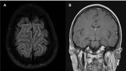
Several weeks ago, the patient had been treated with penicillin because of tonsillitis. During clinical neurological examination, a native cerebral computer tomography was conducted without clinical signs of cerebral edema. Axial magnetic resonance images (MRI) depicted diffuse sulcal hyperintensities indi- cating protein leakage due to the disturbance of the blood–brain barrier (Figure1A). MRI T1-weighted, contrastfluid (gadolinium) imaging demonstrated meningeal contrast enhancement (Figure1B). CSF revealed a purulent appearance with unexpectedly low pleocytosis of 837 cells/μl, a high protein content of 3.5 g/dl, elevated lactate (6.2 mmol/L), and a glucose level of 3 mmol/L resulting in a low glucose quotient of 38.5% (blood glucose level of 7.8 mmol/L). Serum laboratory results showed elevated leukocytes (19.6 Gpt/L), C-reactive protein (221.7 mg/L), procalcitonin (1.62 ng/ml), and bilirubin (32 μmol/L). Other laboratory findings showed a mild DIC with a low thrombocyte count on day 2 (132 Gpt/L), decreased
antithrombin III of 77%, and a decreased thromboplastin time (Quick) of 58%. Chest X ray demonstrated a right-sided pleural effusion as well as an atelectasis of the right- sided lower, middle, and partly upper lobe.
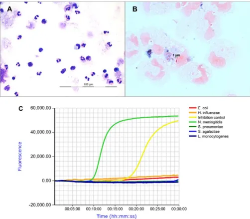
In the routine Pappenheim stain of the CSF sample, neutrophil granulocytes were predominantly observed (>90%) (Figure2A). In the Gram-stained prepara- tion, Gram-negative diplococci with typical shape of meningococci could be identified (Figure2B). To rapidly verify microscopic results, an aliquot of the CSF sample was tested using the eazyplex® CSF direct LAMP assay that targets the most relevant meningitis-causing bacterial species (N. meningitidis, Streptococcus pneumoniae, Haemophilus influenzae, Listeria monocytogenes, Streptococcus agalactiae, andEscherichia coli). An amount of 125μl of CSF was mixed with 25μl of lysis solution and boiled for 3 min. Then, 125μl of the lysed suspension were mixed with 125μl of resuspension solution buffer. An amount of 25μl of the suspension was added to each of the tubes of the eazyplex®test strip containing the lyophilized master mixes for specific species and an inhibition control. The strip was gently knocked to remove air bubbles and loaded into the Genie II machine (Optigene Ltd., Horsham, UK; purchased from Amplex Biosystems).
The tests were carried out at 65 °C for 30 min. Amplification was measured by real-timefluorescence detection using a DNA intercalating dye. Threshold times
Figure 1.Magnetic resonance imaging. (A) Fluid attenuated inversion recovery shows sulcal hyperintensities, indicating protein leakage due to the disturbance of the blood–brain barrier. (B) Slight intense meningeal contrast enhancement in coronal T1-weighted imaging after i.v. gadolinium
administration, demonstrating inflammation in this region
for fluorescence intensity values and result interpretation were automatically determined by the eazyplex® software. N. meningitidis was detected with a threshold time of 9 min (Figure 2C). After reporting the result, initial empiric combination treatment with ceftriaxone, ampicillin, and aciclovir was de-escalated to ceftriaxone monotherapy.N. meningitidisserotype B was isolated from blood culture (BACTEC Plus Aerobic/F, BD Diagnostics, Heidelberg, Germany) but not from CSF samples inoculated onto Columbia sheep blood agar, chocolate agar (Oxoid, Thermo Fisher Scientific, Wesel, Germany), and brain heart infusion (BD Diagnostics). A blood culture bottle signaled bacterial growth at 18 h following its arrival at the laboratory and species identification was performed from subcultures
Figure 2.Microscopy and LAMP assay of the CSF sample. (A) Pappenheim stain demonstrates a moderate cell number with predominant neutrophil granulocytes and few eosinophils and basophils (original magnification 600×). (B) Gram stain shows granulocytes and Gram-negative diplococci (original magnification 1,000×). (C) eazyplex®CSF direct amplification curves with a positive signal forN. meningitidisat a threshold time of 9 min (left curve). The inhibition control was positive
after 19 min (right curve)
about 24 h later. Finetyping of the isolate was conducted according to protocols described previously [9]. The strain had the molecular finetype B:P (porA)1.7– 2.4:F (fetA)1–5, which is the most prevalent type in Germany [10]. Sensitivity of the isolate against ceftriaxone was confirmed using Etest (minimum inhibitory concentration: 0.004 mg/L), referred to the EUCAST breakpoint.
Follow-up blood cultures did not grow meningococci anymore. The patient remained stable and vital signs were always in normal ranges. The decline of thrombocytes on days 2 and 3 to a minimum of 132 Gpt/L improved without intervention. Transcranial doppler ultrasonography revealed no brain vessel spasm at different time points. Ceftriaxone was given at 4 g/day for 14 days and the patient improved very fast, despite initial DIC with petechial rash. Breathing training and balancedfluid therapy attained a profound therapeutic effect on the lung atelectasis. After mobilization and physiotherapy, patient could be dismissed home on day 18 for further physical out-patient rehabilitation in fair clinical condition.
Discussion
IMD belongs to the most serious infectious diseases. It can quickly become life-threatening and surviving patients can develop long-term sequelae [2]. Rapid initiation of appropriate antimicrobial therapy and intensive care is essential to achieve the best outcome for the patient. In the classical workflow,N. meningitidis infection is diagnosed by microscopy and culture [5]. However, culture is time- consuming and often remains falsely negative when the patient has been pretreated with antibiotics. Fast identification of the bacterial species causing meningitis is required to target the therapy and avoid an unnecessary broad antimicrobial coverage as well as to decide whether prophylactic treatment of contact persons has to be started [5, 6]. This case highlights the usefulness of LAMP for rapid molecular diagnosis of bacterial meningitis. In comparison with PCR, a set of 4–6 primers is used for LAMP, and because of the high amplification capacity under isothermal conditions, the assays have a short running time [8, 11]. Moreover, DNA purification is not required, because the Bst DNA polymerase used for LAMP tolerates PCR-inhibiting substances [8]. The eazyplex® CSF direct test cannot differentiateN. meningitidisserotypes, but in a recent paper, the reliability of a serotype-specific LAMP assay for identification and typing of meningococci has been demonstrated [11]. In this case report, the preliminary CSF Gram stain result could immediately be verified using the eazyplex®CSF direct test, whereas subsequent culture was negative, and bacteria were isolated only from blood culture. Based on the eazyplex® result, the empirical treatment regime could be
adapted very soon. LAMP can be suggested as a diagnostic tool that facilitates the diagnosis of bacterial meningitis and helps to overcome diagnostic challenges for identifyingN. meningitidis in CSF, in particular, in cases where the CSF Gram stain result is uncertain or culture remains negative. Because of a low hands-on time of less than 10 min, the eazyplex®CSF direct assay can easily be integrated into a diagnostic routine workflow.
Acknowledgements
This work was supported by a grant from the German Federal Ministry of Education and Research (BMBF, 13N13890). The NRZMHi is supported by the Robert Koch Institute with funds of the German Federal Ministry of Health (funding code: 1369-237). The authors would like to thank Martin Bokemeyer (Institute of Neuroradiology, Jena University Hospital) for the medical judgement and provision of the brain MRT pictures. Informed consent was obtained from the subject to publish this report.
Conflict of Interest The authors declare no conflict of interest.
References
1. van de Beek, B., Brouwer, M., Hasbun, R., Koedel, U., Whitney, C. G., Wijdicks, E.:
Community-acquired bacterial meningitis. Nat Rev Dis Primers2, 16074 (2016).
2. Chang, Q., Tzeng, Y.-L., Stephens, D. S.: Meningococcal disease: Changes in epidemiolo- gy and prevention. Clin Epidemiol4, 237–245 (2012).
3. Varon, J., Chen, K., Sternbach, G. L.: Rupert Waterhouse and Carl Friderichsen: Adrenal apoplexy. J Emerg Med16, 643–647 (1998).
4. Parikh, S. R., Andrews, N. J., Beebeejaun, K., Campbell, H., Ribeiro, S., Ward, C., White, J. M., Borrow, R., Ramsay, M. E., Ladhani, S. N.: Effectiveness and impact of a reduced infant schedule of 4CMenB vaccine against group B meningococcal disease in England: A national observational cohort study. Lancet388, 2775–2782 (2016).
5. Vázquez, J. A., Taha, M. K., Findlow, J., Gupta, S., Borrow, R.: Global meningococcal initiative: Guidelines for diagnosis and confirmation of invasive meningococcal disease.
Epidemiol Infect144, 3052–3057 (2016).
6. Wu, H. M., Cordeiro, S. M., Harcourt, B. H., Carvalho, M., Azevedo, J., Oliveira, T. Q., Leite, M. C., Salgado, K., Reis, M. G., Plikaytis, B. D., Clark, T. A., Mayer, L. W., Ko, A. I., Martin, S. W., Reis, J. N.: Accuracy of real-time PCR, Gram stain and culture for Streptococcus pneumoniae,Neisseria meningitidisandHaemophilus influenzaemeningitis diagnosis. BMC Infect Dis 13, 26 (2013).
7. Leber, A. L., Everhart, K., Balada-Llasat, J. M., Cullison, J., Daly, J., Holt, S., Lephart, P., Salimnia, H., Schreckenberger, P. C., DesJarlais, S., Reed, S. L., Chapin, K. C., LeBlanc, L., Johnson, J. K., Soliven, N. L., Carroll, K. C., Miller, J. A., Dien Bard, J., Mestas, J., Bankowski, M., Enomoto, T., Hemmert, A. C., Bourzac, K. M.: Multicenter evaluation of BioFire FilmArray meningitis/encephalitis panel for detection of bacteria, viruses, and yeast in cerebrospinalfluid specimens. J Clin Microbiol54, 2251–2261 (2016).
8. Rödel, J., Bohnert, J. A., Stoll, S., Wassill, L., Edel, B., Karrasch, M., Löffler, B., Pfister, W.: Evaluation of loop-mediated isothermal amplification for the rapid identification of bacteria and resistance determinants in positive blood cultures. Eur J Clin Microbiol Infect Dis36, 1033–1040 (2017).
9. Elias, J., Harmsen, D., Claus, H., Hellenbrand, W., Frosch, M., Vogel, U.: Spatiotemporal analysis of invasive meningococcal disease, Germany. Emerg Infect Dis12, 1689–1695 (2006).
10. Karch, A., Vogel, U., Claus, H.: Role of penA polymorphisms for penicillin susceptibility in Neisseria lactamica and Neisseria meningitidis. Int J Med Microbiol 305, 729–735 (2015).
11. Lee, D., Kim, E. J., Kilgore, P. E., Takahashi, H., Ohnishi, M., Tomono, J., Miyamoto, S., Omagari, D., Kim, D. W., Seki, M.: A novel loop-mediated isothermal amplification assay for serogroup identification ofNeisseria meningitidesin cerebrospinalfluid. Front Micro- biol6, 1548 (2016).