Signaling through Syk or CARD9 Mediates Species-Speci fi c Anti- Candida Protection in Bone Marrow Chimeric Mice
Erik Zajta,aKatalin Csonka,aAdél Tóth,aLaszló Tiszlavicz,bTamás Németh,c,eAnita Orosz,cÁdám Novák,aMáté Csikós,a Csaba Vágvölgyi,aAttila Mócsai,cAttila Gácsera,d
aHCEMM-USZ Fungal Pathogens Research Group, Department of Microbiology, Faculty of Science and Informatics, University of Szeged, Szeged, Hungary
bDepartment of Pathology, University of Szeged, Szeged, Hungary
cDepartment of Physiology, Semmelweis University School of Medicine, Budapest, Hungary
dMTA-SZTE“Lendület”Mycobiome Research Group, University of Szeged, Szeged, Hungary
eDepartment of Rheumatology and Clinical Immunology, Semmelweis University, Budapest, Hungary
Erik Zajta and Katalin Csonka contributed equally to this work. Author order was determined based on who started the project.
ABSTRACT
The spleen tyrosine kinase (Syk) and the downstream adaptor protein CARD9 are crucial signaling molecules in antimicrobial immunity. Candida parapsilosis is an emerging fungal pathogen with a high incidence in neonates, while Candida albi- cans is the most common agent of candidiasis. While signaling through Syk/CARD9 promotes protective host mechanisms in response to C. albicans, its function in immu- nity against C. parapsilosis remains unclear. Here, we generated Syk
2/2and CARD9
2/2bone marrow chimeric mice to study the role of Syk/CARD9 signaling in immune responses to C. parapsilosis compared to C. albicans. We demonstrate various functions of this pathway (e.g., phagocytosis, phagosome acidi
fication, and killing) in Candida- challenged, bone marrow-derived macrophages with differential involvement of Syk and CARD9 along with species-speci
fic differences in cytokine production. We report that Syk
2/2or CARD9
2/2chimeras rapidly display high susceptibility to C. albicans, while C. parapsilosis infection exacerbates over a prolonged period in these animals.
Thus, our results establish that Syk and CARD9 contribute to systemic resistance to C.
parapsilosis and C. albicans differently. Additionally, we con
firm prior studies but also detail new insights into the fundamental roles of both proteins in immunity against C.
albicans. Our data further suggest that Syk has a more prominent in
fluence on anti- Candida immunity than CARD9. Therefore, this study reinforces the Syk/CARD9 path- way as a potential target for anti-Candida immune therapy.
IMPORTANCE
While C. albicans remains the most clinically signi
ficant Candida species, C. parapsilosis is an emerging pathogen with increased af
finity to neonates. Syk/CARD9 signaling is crucial in immunity to C. albicans, but its role in in vivo responses to other pathogenic Candida species is largely unexplored. We used mice with hematopoietic systems de
ficient in Syk or CARD9 to comparatively study the function of these proteins in anti-Candida immunity. We demonstrate that Syk/CARD9 signaling has a protec- tive role against C. parapsilosis differently than against C. albicans. Thus, this study is the
first to reveal that Syk can exert immune responses during systemic Candida infections species speci
fically. Additionally, Syk-dependent immunity to a nonalbicans Candida species in an in vivo murine model has not been reported previously. We highlight that the contribution of Syk and CARD9 to fungal infections are not identical and underline this pathway as a promising immune-therapeutic target to
fight Candida infections.
KEYWORDS
Candida parapsilosis, Candida albicans, Syk, CARD9, antifungal immunity, antifungal immune therapy, bone marrow chimera
CitationZajta E, Csonka K, Tóth A, Tiszlavicz L, Németh T, Orosz A, Novák Á, Csikós M, Vágvölgyi C, Mócsai A, Gácser A. 2021.
Signaling through Syk or CARD9 mediates species-specific anti-Candidaprotection in bone marrow chimeric mice. mBio 12:e01608- 21.https://doi.org/10.1128/mBio.01608-21.
EditorGustavo H. Goldman, Universidade de Sao Paulo
Copyright© 2021 Zajta et al. This is an open- access article distributed under the terms of theCreative Commons Attribution 4.0 International license.
Address correspondence to Attila Gácser, gacsera@bio.u-szeged.hu.
Received4 June 2021 Accepted5 August 2021 Published
® 31 August 2021
A dvancements in the study of antifungal immunity have revealed various approaches to promote recovery from fungal diseases through the modulation of immune responses. Although adjunctive antifungal immune therapies signify a promising direction in aiding the efficiency of antimycotic drugs, several challenges hinder their de- velopment (1
–3). One dif
ficulty is that taxonomically close species may induce dissimilar immune responses.
Invasive Candida infections have a worldwide annual incidence of
;700.000 (4), and they are associated with a mortality of
;30 to 75% (5, 6). Although C. albicans is the most frequent agent, C. parapsilosis has a record of globally increasing incidence, and it poses a particular threat to neonates and patients on parenteral nutrition (6
–9).
The two species may induce different host responses. For instance, C. albicans favors the polarization of Th cells to the Th17 phenotype more ef
ficiently than C. parapsilosis in cell culture (10). Also, unlike C. albicans, C. parapsilosis triggers prominent interleukin 27 (IL-27) production to suppress protective in
flammatory processes in mice. Therefore, the notion of therapeutically blocking IL-27 signaling during C. parapsilosis infections has been raised, underlining the need to identify species-speci
fic immune reactions (11). The differences between the immunogenicity of the two species arise partially from their nonidentical mor- phology, cell wall composition, and interaction with pattern recognition receptors (PRRs) (12).
A major function of the spleen tyrosine kinase (Syk) is to mediate signaling initiated by PRRs (e.g., Dectin-1) binding microbial structures (13, 14), especially fungal pathogen-associ- ated molecular patterns (15
–17). A major pathway proceeding through PKCd and Vav pro- teins (18, 19) relies on the downstream adaptor caspase recruitment domain-containing pro- tein 9 (CARD9), which functions as a component of supramolecular structures such as the CARD9-BCL10-MALT-1 or the CARD9-H-Ras-Ras-GRF1 complexes (20
–22). Nevertheless, Syk and CARD9 can operate independently (23, 24). Upon fungal stimuli, the Syk/CARD9 path- way results in the activation of innate antifungal mechanisms that may later shape adaptive immunity (23, 25
–33). Consequently, de
ficiency in Syk/CARD9 signaling increases susceptibil- ity to invasive fungal infections in both humans and mice (21, 25, 26, 28, 33
–36). For exam- ple, CARD9 mutations have been associated with chronic mucocutaneous candidiasis, meningoencephalitis, and colitis caused by C. albicans (26, 34, 35). Although multiple studies have underlined the pivotal role of Syk and CARD9 in immunity against C. albicans (21, 25, 26, 28, 33, 37), several
findings are conferred only from chemical inhibition of Syk activity or the use of arti
ficial yeast mimics (e.g., zymosan or heat-killed cells). In contrast, the only direct link between this pathway and C. parapsilosis-induced immune responses is the compro- mised IL-1b production of Syk-blocked THP-1 cells after fungal challenge (38).
In this study, we aimed to ascertain if Syk/CARD9 signaling is a prominent compo- nent of immunity to C. parapsilosis. We demonstrate that this pathway is involved in various immune responses evoked by C. parapsilosis in bone marrow-derived macrophages (BMDMs). Utilizing Syk- or CARD9-de
ficient bone marrow chimeric mice (referred to as Syk
2/2or CARD9
2/2chimeras and the respective wild-type controls as Wt
Sykand Wt
CARD9chimeras), we also provide evidence that this pathway confers protection in the setting of invasive C. parapsilosis infection, which becomes especially apparent in the late phase of the infection.
As most experiments were carried out in comparison with live C. albicans, we con
firm previous studies and provide new insights into the essential roles of Syk and CARD9 in immunity against this pathogen. Finally, we demonstrate that this signaling pathway is differentially involved in mediating protection against the two Candida species in vivo.
RESULTS
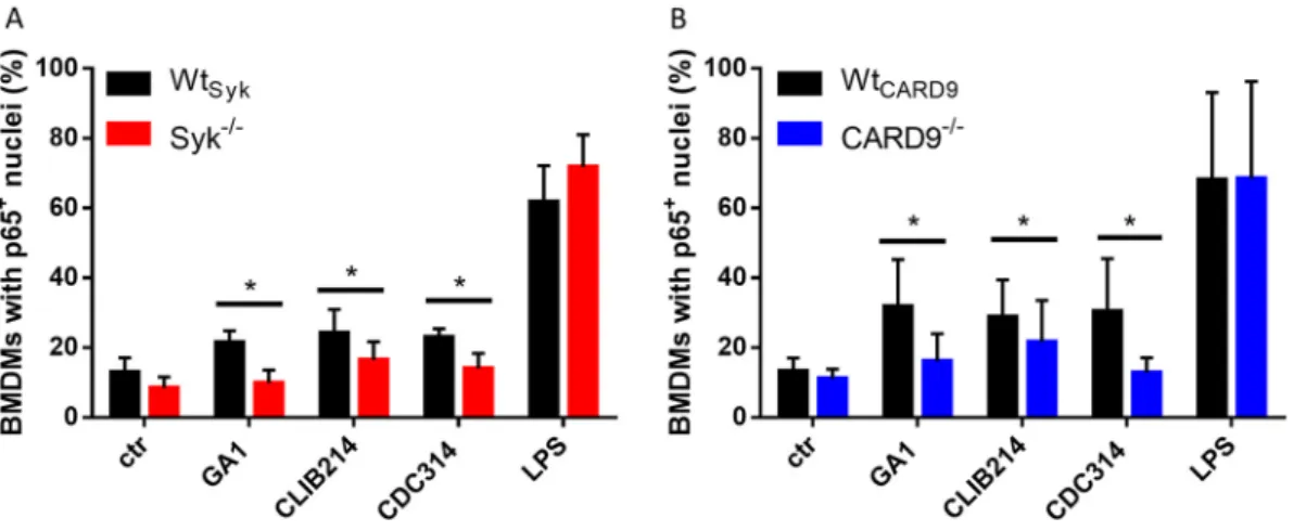
Syk and CARD9 mediate nuclear translocation of NF-jB p65 inC. parapsilosis- treated BMDMs.
The classical activation of the NF-k B pathway through nuclear trans- location of the p65 subunit is a major component of antifungal immunity (39
–41).
Recently, C. parapsilosis was shown to trigger this translocation in THP-1 cells (42). However, it is unclear whether Syk or CARD9 controls this process. Therefore, we implemented immune staining of p65 to monitor its translocation in C. parapsilosis-infected (strains GA1, CLIB214, and CDC317) BMDMs cultured from Wt
Syk, Syk
2/2, Wt
CARD9, and CARD9
2/2chimeras. We found a decreased proportion of Syk
2/2or CARD9
2/2macrophages with p65
1nuclei compared to
Zajta et al. ®
Wt
Sykor Wt
CARD9cells, respectively, in case of all strains (mean
6SD of percent decrease com- pared to wild-type [WT] control cells; Syk
2/2, GA1, 51.06
623.40; CLIB214, 31.89
611.35;
CDC314, 39.33
615.40; CARD9
2/2, GA1, 44.75
626.56; CLIB214, 26.58
622.29; CDC314, 49.89
626.03) (Fig. 1). Lipopolysaccharide (LPS)-induced NF-k B activation served as positive control, with over 60% of BMDMs displaying p65 translocation regardless of genetic back- ground (Fig. 1). These
findings suggest that activation of NF-k B in C. parapsilosis-infected BMDMs is under the in
fluence of the Syk/CARD9 pathway.
The Syk/CARD9 pathway differentially controls cytokine production of BMDMs in response toC. parapsilosisandC. albicans.
The involvement of Syk and CARD9 in NF-k B regulation suggested that the consequent cytokine production would also depend on these proteins during C. parapsilosis infection. Therefore, we assessed the cytokine response of Wt
Syk, Syk
2/2, Wt
CARD9,and CARD9
2/2BMDMs following fungal stimuli.
First, we used proteome pro
filer membranes to identify cytokines in cell culture superna- tants collected from BMDMs challenged with the GA1 strain. In the case of Wt
Sykand Wt
CARD9BMDMs, tumor necrosis factor alpha (TNF-a), and chemokines KC (CXCL1), MIP-1a (CCL3), and MIP-2 (CXCL2) were detected (Fig. S1 in the supplemental material). Compared to the WT BMDMs, the TNF-a yield from Syk
2/2or CARD9
2/2cells was diminished after the fungal challenge. While the chemokine secretion of Syk
2/2BMDMs appeared intact (Fig. S1A), CARD9
2/2cells secreted hardly any or no detectable KC, MIP-1a, and MIP-2 compared to the corresponding WT cells (Fig. S1B). These cytokines were not detected in the supernatants of uninfected control BMDMs from any genetic background (data not shown).
Next, we sought to test these observations with enzyme-linked immunosorbent assay (ELISA), including multiple C. parapsilosis strains (GA1, CLIB214, and CDC317) and the SC5314 C. albicans strain as reference (Fig. 2 and Table S1). The TNF-a production of C. parapsilosis- or C. albicans-treated Syk
2/2or CARD9
2/2macrophages was lower than that of Wt
Sykand Wt
CARD9cells by
.42% (Fig. 2A and B). While the chemokine yield of C. albicans-stimulated Syk
2/2macrophages dropped by
;90% in comparison to Wt
SykBMDMs, the KC, MIP-1a, and MIP-2 expression of C. parapsilosis-treated Syk
2/2BMDMs did not decrease (Fig. 2C, D, and E). In contrast, we found a decrease of at least 25% in the chemokine production of Candida-infected CARD9
2/2BMDMs (with the exception of CLIB214 for MIP-1a production) (Fig. 2F, G, and H). Notably, the dependence of the production of any studied cytokines on either Syk or CARD9 was greatest in the case of the C. albicans strain (Fig. 2A to H).
Taken together, we concluded that both Syk and CARD9 participate in the regulation of cytokine production of BMDMs upon both C. parapsilosis and C. albicans infection, but the involvement of Syk differs between the two Candida species.
FIG 1 Nuclear translocation of NF-kB p65 in BMDMs uponC. parapsilosisinfection. (A and B) BMDMs were treated withC.
parapsilosis(strains GA1, CLIB214, and CDC317; MOI of 5:1) or LPS (1mg/ml) for 90 min, or untreated control (ctr) cells were used. Nuclei were stained with DRAQ5 and NF-kB p65 with an Alexa Fluor 488-conjugated antibody. Cells were analyzed by imagingflow cytometry. Percentage of WtSyk, Syk2/2(A), WtCARD9, and CARD92/2(B) BMDMs with p651nuclei are shown. Data represent the mean6SD. Data are pooled from a minimum of 4 independent experiments. The paired Student'sttest was applied.*,P,0.05.
FIG 2 Cytokine production of BMDMs uponC. parapsilosisorC. albicansinfection. (A to H) WtSyk, Syk2/2(A, C, D, and E), WtCARD9, and CARD92/2(B, F, G, and H) BMDMs were treated withC. parapsilosis(strains GA1, CLIB214 and CDC317; MOI of 5:1) orC. albicans(strain SC5314; MOI of 1:25) for 24 h. Cell culture supernatants were analyzed for TNF-a(A and B), KC (C and F), MIP-1a(D and G), and MIP-2 (E and H) by ELISA. Percentages of WT control values are shown here, while absolute concentrations are listed in Table S1 in the supplemental material. Data represent the mean6SD. Data are pooled from a minimum of 4 independent experiments. The paired Student'sttest was applied.*,P,0.05;**,P,0.01;***,P,0.001;****,P,0.0001. Cytokine production measured by proteome profiler membranes is shown in Fig. S1.
Zajta et al. ®
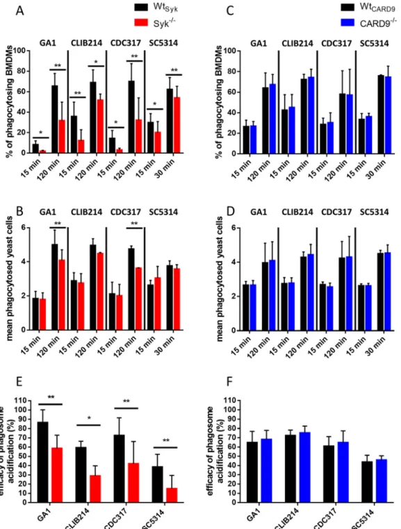
Syk, but not CARD9, promotes phagocytosis ofC. parapsilosisandC. albicansby BMDMs and subsequent phagosome acidification.
PRRs exploiting the Syk/CARD9 pathway have been indicated as mediators of phagocytosing fungal elements (15, 43
–45). Additionally, heat-killed Candida cells were shown to be ingested in a Syk-depend- ent manner by neutrophils (46). As C. parapsilosis and C. albicans cells contain motives recognized by Syk/CARD9-dependent receptors, we hypothesized that internalization of the live form of these yeasts by macrophages was also regulated by this pathway.
Therefore, we infected BMDMs with
fluorescently labeled (Alexa Fluor 488 or green
fluorescent protein [GFP]) C. parapsilosis or C. albicans cells to monitor their uptake using imaging
flow cytometry. Early (15 min) and late (120 and 30 min for C. parapsilosis and C.
albicans, respectively) experimental time points were selected. The proportion of phago- cytosis-positive Wt
Sykmacrophages signi
ficantly exceeded that of Syk
2/2cells regardless of Candida species or incubation time (Fig. 3A). Furthermore, the average number of ingested yeast cells/BMDM within the phagocytosing population was signi
ficantly higher in Wt
Sykcells than in Syk
2/2macrophages in the case of the GA1 and CDC317 C. parapsilosis strains at the late time point. A trend of nonsigni
ficant decrease of this parameter was also detectable in CLIB214- or SC5314-treated Syk
2/2BMDMs at this time point (Fig. 3B). In contrast, CARD9
2/2cells phagocytosed both C. parapsilosis and C. albicans as effectively as their Wt
CARD9counterparts (Fig. 3C and D). Thus, we concluded that Syk, but not CARD9, contributes to the ingestion of both live C. parapsilosis and C. albicans by BMDMs.
As Syk was involved in the acidi
fication of phagosomes containing heat-killed C.
albicans in RAW cells (31, 47), our next step was testing if phagosome acidi
fication fol- lowing the phagocytosis of live C. parapsilosis and C. albicans also relies on this path- way in BMDMs. To this end, we double labeled C. parapsilosis and C. albicans cells with Alexa Fluor 488 or GFP together with pHrodo Red, a pH-sensitive dye that gains
fluo- rescence upon acidi
fication of phagosomes. Following coincubation with BMDMs, the Alexa Fluor 488
1/GFP
1and pHrodo Red
1macrophage populations were examined to determine the ef
ficiency of phagosome acidi
fication. Relative to Wt
Sykcells, phago- some acidi
fication was less ef
ficient in Syk
2/2cells after coincubation with any of the applied strains (Fig. 3E). In contrast, phagosome acidi
fication was unaltered in CARD9
2/2BMDMs (Fig. 3F). Therefore, the acidi
fication of phagosomes containing C.
parapsilosis or C. albicans is mediated by Syk, but not CARD9, in BMDMs.
Syk, but not CARD9, plays a role in the killing ofC. parapsilosisby BMDMs.
Previous studies revealed that the Syk/CARD9 signal transduction may modulate the candidaci- dal activity of phagocytes or intracellular replication of Candida cells within innate immune cells (15, 26, 36, 48
–50). Also, our current
findings show that the phagocytosis of C. parapsilosis by murine macrophages and the subsequent phagosome acidi
fication is regulated by Syk. Thus, we speculated that Syk
2/2, but not CARD9
2/2, macrophages would be defective in the elimination of C. parapsilosis cells.
Therefore, we compared the killing ef
ficacy of WT and mutant macrophages. Truly, Syk
2/2BMDMs displayed diminished killing ef
ficiency against C. parapsilosis, although the reduction only followed a nonsigni
ficant (P = 0.0726) trend in the case of the CDC317 strain (Fig. 4A). As expected, CARD9
2/2BMDMs eliminated C. parapsilosis similarly to Wt
CARD9BMDMs (Fig. 4B).
Therefore, killing of C. parapsilosis is regulated by Syk, but not CARD9, in BMDMs.
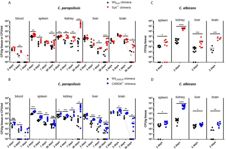
Susceptibility of Syk2/2or CARD92/2bone marrow chimeric mice toC. parapsilosis andC. albicansfollows dissimilar patterns.
As our in vitro studies revealed the participa-
tion of Syk/CARD9 signaling in the immune recognition of C. parapsilosis, we hypothesized
that the absence of either Syk or CARD9 in hematopoietic cells would compromise host im-
munity upon systemic C. parapsilosis infections. To test this, we challenged Syk
2/2, CARD9
2/2,
and their corresponding WT control bone marrow chimeric mice with the C. parapsilosis GA1
strain via intravenous (i.v.) injection. C. albicans SC5314 was used as a reference strain. Fungal
burden in C. parapsilosis-treated animals was evaluated 2, 5, 7, and 30 days postinfection
(dpi). As injection of Syk
2/2or CARD9
2/2chimeras with C. albicans was lethal by 4 dpi (data
not shown), fungal burden was determined at 2 dpi. At this time, no signi
ficant difference was
detected between the C. parapsilosis-challenged Wt
Sykand Syk
2/2or Wt
CARD9and CARD9
2/2chimeras in terms of blood, liver, and brain tissue colonization (Fig. 5A and B). However, fungal burden in the spleen and kidneys of Syk
2/2chimeras was
;2.2-fold and
;1.7-fold higher, respectively, than in Wt
Sykanimals (Fig. 5A). Likewise,
;2.7-fold and
;2-fold higher colonization was detected in the spleen and kidneys of C. parapsilosis-infected CARD9
2/2FIG 3 BMDM phagocytosis efficiency and phagosome acidification uponC. parapsilosisorC. albicansinfection. (A to D) WtSyk, Syk2/2 (A and B), WtCARD9, and CARD92/2(C and D) BMDMs were treated withfluorescent (Alexa Fluor 488/GFP)C.
parapsilosis(strains GA1, CLIB214, and CDC317; MOI of 5:1) for 15 min and 2 h or withC. albicans(strain SC5314; MOI of 5:1) for 15 and 30 min. Phagocytosis was assessed by imagingflow cytometry. Percentages of phagocytosis-positive macrophages (A and C) and the mean number of ingested yeasts per macrophage (B and D) within the phagocytosing population are shown. (E and F) WtSyk, Syk2/2(E), WtCARD9, and CARD92/2(F) BMDMs were treated with double-labeled (Alexa Fluor 488/GFP plus pHrodo Red)C. parapsilosisorC. albicans(MOI of 5:1) for 15 min. Efficacy of phagosome acidification was determined by imagingflow cytometry. Data represent the mean6SD. Data are pooled from a minimum of 3 independent experiments.
The paired Student'sttest was applied.*,P,0.05;**,P,0.01.
Zajta et al. ®
chimeras compared to their WT counterparts (Fig. 5B). At later time intervals, C. parapsilosis colonization of all examined organs of the Syk
2/2or CARD9
2/2chimeras exceeded that of the respective WT chimeras in most cases (Fig. 5A and B). The difference was most appa- rent at 30 dpi where fungal burden was over 58-fold (brain), 310-fold (liver), and 3,100-fold
FIG 5 Susceptibility to systemicC. parapsilosisandC. albicansinfection. (A to D) WtSyk, Syk2/2(A and C), WtCARD9, and CARD92/2(B and D) bone marrow chimeric mice were infected i.v. withC. parapsilosisGA1 (2107yeast cells/mouse) (A and B) orC. albicansSC5314 (105yeast cells/mouse) (C and D).
Animals were euthanized at 2, 5, 7, or 30 (A and B) dpi. Blood or spleen, kidney, liver, and brain homogenates were plated on YPD plates, and CFU were determined 2 days later. Data are pooled from at least 2 independent experiments. The Mann-Whitney test was applied.*,P,0.05;**,P,0.01;***, P,0.001;****,P,0.0001; ns, not significant.
FIG 4 Killing efficacy of macrophages uponC. parapsilosisinfection. (A and B) WtSyk, Syk2/2(A), WtCARD9, and CARD92/2(B) BMDMs were treated withC. parapsilosis(strains GA1, CLIB214, and CDC317; MOI of 5:1) for 3 h.
Killing efficacy was determined by CFU counting. Data represent the mean6 SD. Data are pooled from a minimum of 3 independent experiments. The paired Student'sttest was applied.*,P,0.05;**,P,0.01.
(kidneys) higher in Syk
2/2chimeras than in Wt
Sykchimeras (Fig. 5A). At this time, over 270 times higher colonization was determined in the kidneys of CARD9
2/2chimeric mice than Wt
CARD9controls (Fig. 5B). Also, while C. parapsilosis was cleared from the brain of all Wt
CARD9chimeras, the mean fungal burden in this organ was 2,700 CFU/g in the CARD9
2/2genotype (Fig. 5B). Additionally, kinetic patterns suggested that the tendency toward an initial clearance was reversed by 30 dpi in the spleen, liver, and kidneys of Syk
2/2and the kidneys of CARD9
2/2chimeras (Fig. 5A and B). Moreover, 2 of the C. parapsilosis-infected Syk
2/2chimeras were euthanized before day 30 due to deteriorated health conditions. The kidneys of these animals were severely deformed by sizable abscesses full of yeast cells (Fig. S2). However, macroscopic renal abscesses were not seen on the kidneys of any other C. parapsilosis-infected mice.
In contrast to the C. parapsilosis challenge at 2 dpi, signi
ficantly higher fungal burdens were detected in all inspected organs of C. albicans-treated Syk
2/2or CARD9
2/2chimeras than the corresponding WT mice (Fig. 5C and D). The difference was most prominent in the kidneys (
.200-fold), which also manifested in small abscesses in both the Syk
2/2and CARD9
2/2backgrounds (Fig. S3A and B). Nonetheless, no C. albicans colonies were recov- ered from the blood in any genetic background (data not shown).
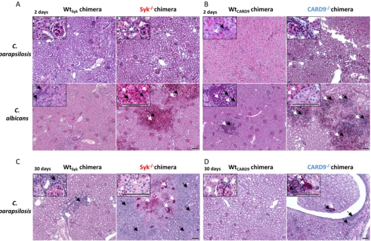
As kidneys are the main target organs of systemic candidiasis in mice (51, 52) and our CFU data indicated marked differences in this organ between the Syk
2/2or CARD9
2/2and WT chimeras, we examined the presence of fungal elements and associated leukocyte in
fil- trates in the kidneys using periodic acid-Schiff (PAS)-stained histological tissue sections at 2 and 30 dpi (Fig. 6 and Fig. S4A). At 2 dpi, yeast cells or leukocytes were only sporadically de- tectable in C. parapsilosis-treated mice of any genetic background (Fig. 6A and B). While C.
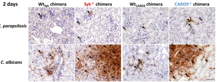
FIG 6 Histopathology of kidneys duringC. parapsilosisandC. albicansinfection. WtSyk, Syk2/2(A and C) WtCARD9, and CARD92/2(B and D) bone marrow chimeric mice were infected i.v. withC. parapsilosisGA1 (2107yeast cells/mouse) orC. albicansSC5314 (105yeast cells/mouse) or mock infected with PBS (see Fig. S4A) and euthanized at 2 (A and B) or 30 (C and D) dpi. PAS-stained histological sections were prepared to monitor fungal elements (white arrows) and leukocyte infiltrates (black arrows). Scale, 100mm. Kidney morphology ofC. parapsilosis- orC. albicans-infected mice at 2 dpi are depicted in Fig. S2, while pathological features of aC. parapsilosisSyk2/2chimera euthanized at 26 dpi due to severe health conditions are shown in Fig. S3.
Zajta et al. ®
albicans hyphae/yeast cells and immunocyte clusters were sometimes observed in the kid- neys of WT chimeras, extensive patches occupied by fungal structures mingling with in
filtrat- ing leukocytes and necrotic tissues were conspicuous in the kidneys of Syk
2/2or CARD9
2/2chimeras at 2 dpi (Fig. 6A and B). At day 30, yeast cells or frequent leukocyte in
filtrates were not characteristic of C. parapsilosis-infected WT chimeras but were observed in Syk
2/2and CARD9
2/2chimeras with detectable tissue necrosis in Syk
2/2chimeras (Fig. 6C and D).
Nevertheless, while immune cells and C. parapsilosis yeast clusters were always present in the kidneys of Syk
2/2chimeras, these traits were not always detected in CARD9
2/2chimeras.
Therefore, the absence of Syk or CARD9 in the hematopoietic cell population led to increased susceptibility to C. parapsilosis invasive infection. However, unlike in the case of C. albicans, massive fungal growth occurred only at a later stage of infection.
Rapid inflammation inC. albicans-infected, but not inC. parapsilosis-infected, kidneys of Syk2/2or CARD92/2bone marrow chimeric mice.
As leukocyte in
filtrates were plentiful in the kidneys of C. albicans-infected, but not C. parapsilosis-infected, Syk
2/2or CARD9
2/2chimeras at 2 dpi, we aimed to further characterize this in
flammatory response in this organ. We found that mainly neutrophilic granulocytes occupied these areas as implied by expansive myeloperoxidase (MPO)-positive staining localized to infected sites (Fig. 7 and Fig. S4A). As noted, however, leukocyte in
filtrates were present in the kidneys of C. parapsilosis-infected Syk
2/2or CARD9
2/2chimeras at 30 dpi. At this time, we found that neutrophils localized to infected sites were abundant in the kidneys of both mutant chime- ras with CD68
1macrophages and CD3
1T cells also recruited to the tissues. However, these cell populations were not as consistent in CARD9
2/2chimeras as in Syk
2/2chimeras (Fig. 8B and Fig. S4B). At 2 dpi, in
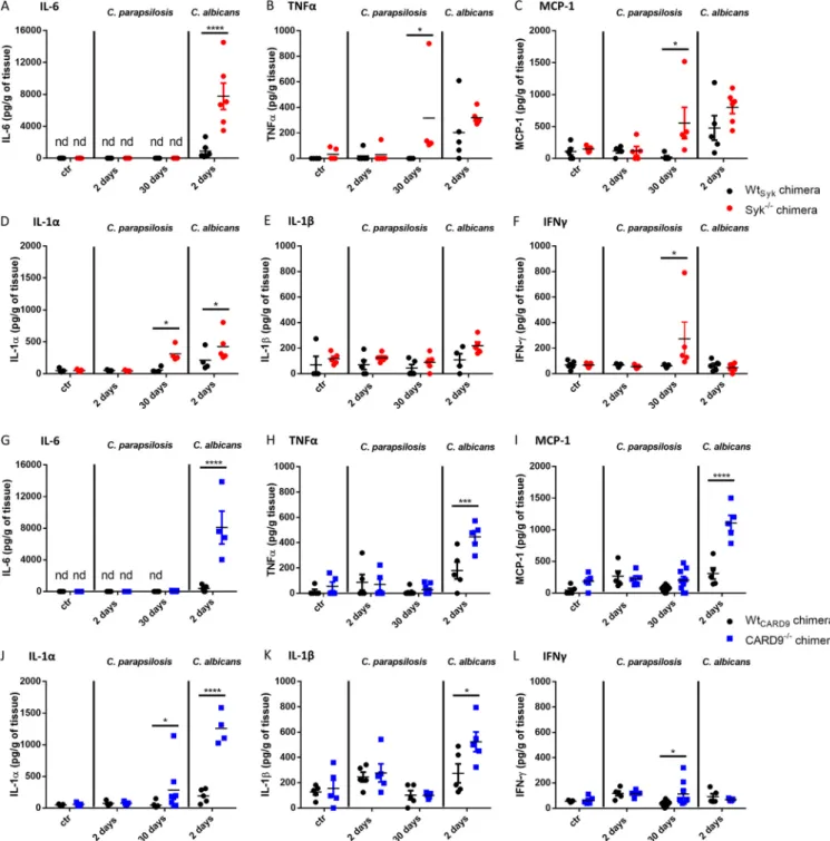
flammation was also detected by ELISA in the kidneys of C. albi- cans-infected Syk
2/2or CARD9
2/2chimeras, as kidney homogenates demonstrated an induction of IL-6, IL-1a, IL-1-b, TNF-a, and monocyte chemoattractant protein 1 (MCP-1) production, although only IL-6 and IL-1-a concentrations were signi
ficantly higher in the Syk
2/2than in the Wt
Sykbackground (Fig. 9A, D, G, H, I, J, and K). In contrast, C. parapsilo- sis did not induce prominent production of these proin
flammatory cytokines in any genetic background at 2 dpi (Fig. 9). At 30 dpi, however, in
flammation in the kidneys of C. parapsilosis-challenged Syk
2/2or CARD9
2/2chimeras was also con
firmed by ELISA.
While the amounts of TNF-a, MCP-1, IL-1a, and IFN-
gwere higher in Syk
2/2than in Wt
Sykchimeric animals, only increased IL-1a and IFN-
gresponses were observed in C. parapsilo- sis-infected CARD9
2/2chimeras (Fig. 9B, C, D, F, J, L).
Taken together, these data indicate that, while the absence of Syk or CARD9 in the he- matopoietic cell population leads to an early, intense in
flammatory response to C. albicans
FIG 7 Presence of MPO in histological kidney sections at 2 dpi. WtSyk, Syk2/2, WtCARD9, and CARD92/2bone marrow chimeric mice were infected i.v. withC.
parapsilosis(2107yeast cells/mouse) orC. albicans(105yeast cells/mouse) or mock infected with PBS (see Fig. S4), and animals were euthanized at 2 dpi. Immune staining for MPO (brown) was implemented to detect neutrophil granulocytes. Black arrows indicate positivity for MPO staining. Scale, 100mm.
infection, C. parapsilosis triggers a mostly delayed Syk-dependent and moderately CARD9- dependent response in the kidneys of chimeric mice.
DISCUSSION
In this study, we used bone marrow chimeric mice to de
fine the involvement of Syk and CARD9 in immune responses to C. parapsilosis with comparisons to C. albicans. As was previously reported about C. albicans (23), we now demonstrated that both signal- ing proteins regulate the activation of NF-k B in C. parapsilosis-infected BMDMs.
Intriguingly, C. glabrata led to Syk phosphorylation without NF-k B in macrophage-like cells (53). Therefore, C. parapsilosis resembles C. albicans rather than C. glabrata in reg- ulating this transcriptional factor via the Syk/CARD9 pathway. Fitting with the role of NF-k B in governing cytokine production (17, 39, 40, 54), we established that the cyto- kine response of BMDMs to C. parapsilosis is in
fluenced by Syk/CARD9 signaling.
Furthermore, we showed that Syk controls cytokine production of C. albicans-chal- lenged BMDMs as already described in Syk
2/2or SHP-2
2/2dendritic cells (DCs) (25, 33) and various CARD9
2/2cell types (21, 26, 28, 49). Notably, we found intact chemokine secretion in C. parapsilosis-stimulated, but not in C. albicans-stimulated, Syk
2/2BMDMs, suggesting species-speci
fic differences in terms of Syk activation. Another interesting
finding of our study was the CARD9-dependent, but Syk-independent,
FIG 8 Presence of MPO and CD681or CD31cells in histological kidney sections at 30 dpi. WtSyk, Syk2/2, WtCARD9, and CARD92/2bone marrow chimeric mice were infected i.v. withC. parapsilosis(2107yeast cells/mouse) orC. albicans (105yeast cells/mouse) or mock infected with PBS (see Fig. S4B), and animals were euthanized at 30 dpi. Immune staining for MPO, CD68, and CD3 (brown) was implemented to detect neutrophil granulocytes, macrophages, and T cells, respectively. Note that 2 sets of microphotographs are shown for two CARD92/2chimeras. Black arrows indicate positivity for MPO, CD68, or CD3 staining. Scale, 100mm.Zajta et al. ®
chemokine synthesis of BMDMs stimulated with C. parapsilosis. Similarly, a CARD9-de- pendent, but Syk-independent, mechanism was previously described in BMDMs chal- lenged with C. albicans hyphae (23). Although Toll-like receptors (TLRs) (55, 56) and nu- cleotide-binding oligomerization domain 2 (NOD2) (22, 57) were proposed to signal through CARD9, future investigations are required to elucidate Syk-independent sig- naling through CARD9 in response to fungal stimuli.
Although multiple studies have covered the potential function of Syk and CARD9 in the phagocytosis of zymosan (25, 28, 29, 32, 58) or
“yeast particles
”(30), the direct role
FIG 9 Cytokine production in kidneys following systemic infection withC. parapsilosisandC. albicans.(A to F) WtSykand Syk2/2bone marrow chimeras were infected i.v. withC. parapsilosis(2107yeast cells/mouse) orC. albicans(105yeast cells/mouse) or mock infected with PBS (ctr), and animals were euthanized at 2 or 30 dpi. Cytokines were measured from kidney homogenates with ELISA. (G to L) WtCARD9and CARD92/2 bone marrow chimeras were infected, and kidney homogenates were analyzed for cytokines similarly to panels A to F. Data are pooled from at least 2 independent experiments. Two- way ANOVA was applied.*,P,0.05;***,P,0.001;****,P,0.0001.
of Syk in leukocytes or CARD9 in macrophages in ingesting live C. parapsilosis or C. albi- cans cells has not been experimentally addressed to our knowledge. Our present results revealed that BMDMs phagocytose both species in a Syk-dependent, yet CARD9-independent, manner. Inhibition of Syk also hindered the phagocytosis of heat- killed C. albicans, C. glabrata, and C. auris by neutrophils (46). Our data are in line with the CARD9-independent internalization of C. albicans by human neutrophils and monocytes (26, 49). Similarly, we found that phagosome acidi
fication in BMDMs chal- lenged with either C. parapsilosis or C. albicans was regulated by Syk, but not CARD9.
This supports the Syk-dependent phagosome acidi
fication observed in RAW cells treated with heat-killed C. albicans by reference 31 and that Syk, but not CARD9, was required for proper maturation of C. albicans-containing phagosomes in macrophages (47). Previous research also showed that neutrophils kill C. albicans in a Syk-dependent manner (15, 46), while CARD9 was dispensable in this activity in murine neutrophils (50). Likewise, elimination of C. parapsilosis by BMDMs relied on Syk, but not CARD9, in our setting. Interestingly, human cells seem to differ from murine cells in that they can exploit CARD9 to eliminate C. albicans (15, 26, 49).
Several studies reported that de
ficiency in Syk (33) or CARD9 (21, 26, 28, 36) in mice leads to severe susceptibility to C. albicans (21, 26, 28, 33) or C. tropicalis (36) infections.
Our invasive candidiasis experiments now revealed that Syk and CARD9 in hematopoi- etic cells contribute to systemic resistance to C. parapsilosis. Furthermore, we reaf-
firmed the crucial and almost immediate role of this pathway in the case of C. albicans.
Although excessive in
flammation (cytokines, immunocyte in
filtrates) was evident in the kidneys of C. albicans-infected and C. parapsilosis-infected Syk- or CARD9-de
ficient mice at 2 and 30 dpi, respectively, it likely does not contradict the decreased proin
flam- matory cytokine production of Syk
2/2or CARD9
2/2myeloid cells but is, rather, the conse- quence of unrestricted fungal presence triggering Syk/CARD9-independent signaling (e.g., TLR-MyD88 pathways). In the case of the C. parapsilosis challenge, immunocyte in
filtrates and elevated amounts of proin
flammatory cytokines were more characteristic in the kidneys of Syk
2/2animals than in those of CARD9
2/2mice at 30 dpi. This may be the consequence of the CARD9-dependent, but Syk-independent, chemokine production observed in C. para- psilosis-treated macrophages that could allow for more ef
ficacious leukocyte recruitment to the site of infection in Syk
2/2chimeras than in CARD9
2/2chimeras.
The mechanism through which Syk and CARD9 counteract C. albicans infections is grounded in signaling initialized by Dectin-1, Dectin-2, Dectin-3, Mincle, and CR3 (12, 59
–63).
However, it is unclear which receptors utilize the Syk/CARD9 pathway to provide the observed in vivo protection against C. parapsilosis. Cell culture experiments with C. parapsilo- sis revealed that Dectin-1 could regulate immune responses in vitro (10, 12, 42). In contrast, phagocytosis of C. parapsilosis by human neutrophils was independent of Dectin-1 (64).
Furthermore, no dependence on Dectin-1 in long-term (21 dpi) clearance of C. parapsilosis was reported (12), and our previous results demonstrated that the fungal load was unaf- fected by Dectin-1 using several time points (1, 3, 7, 14, and 20 dpi) and multiple C. parapsilo- sis strains (65). Therefore, it is tempting to hypothesize the involvement of Syk/CARD9-bound PRRs other than Dectin-1 in systemic resistance against C. parapsilosis. Fitting with the crucial role of Syk in B-cell development (66, 67) and that Syk was involved in more cellular processes in response to Candida stimuli than CARD9 in our in vitro experiments, fungal burdens in Syk
2/2chimeras tended to surpass those of CARD9
2/2chimeras. Therefore, we propose that Syk has a more prominent function in antifungal immunity than CARD9 against both Candida species.
Notably, the excess of fungal burden in some organs (kidneys, livers, brain) of C.
parapsilosis-challenged Syk
2/2or CARD9
2/2chimeras only surpassed that of WT chime- ras dramatically at 30 dpi and, by this time, leukocyte in
filtrates containing macrophages, neu- trophils, and T cells and proin
flammatory cytokine production were also evident in the kidneys of mostly Syk
2/2chimeras. In contrast, the kidneys and brain of Syk
2/2and the kidneys of CARD9
2/2chimeras treated with C. albicans were marked by more than 100-fold higher colo- nization and massive in
flammation by 2 dpi. This is well before adaptive immunity could
Zajta et al. ®
develop. Regarding systemic resistance, this may suggest that the Syk/CARD9 pathway plays a greater role in innate immunity against C. albicans than C. parapsilosis. In our setting, it appears that Syk- and CARD9-independent innate mechanisms initially exert some control over the growth of C. parapsilosis in host tissues. However, it is possible that the absence of Syk and CARD9 is suf
ficient for a failure in the development of adaptive immunity, and innate mecha- nisms alone are unable to prevent the proliferation of host-adapted C. parapsilosis cells over an extended period of infection. The notion that both proper innate and adaptive immunity are required for the total elimination of this yeast is corroborated by our observation that severe combined immunode
ficient mice
—infected according to the same methods as this study
—were unable to clear C. parapsilosis as ef
ficiently as WT mice (unpublished data). The species-speci
fic kinetic patterns of fungal clearance by the Syk
2/2or CARD9
2/2chimeras may also arise from inherent differences between C. parapsilosis and C. albicans. For example, a study found less beta-glucan in the cell wall of C. parapsilosis than that of C. albicans (12).
Accordingly, while the beta-glucan receptor Dectin-1 can play a crucial role in the resistance against C. albicans (12, 59, 68), it seems less important against C. parapsilosis (12, 65). In the ab- sence of Syk or CARD9, Dectin-1 signaling is damaged, promoting rapid initial expansion of C.
albicans yeast cells in the host. Subsequently, the formation of more virulent hyphae faces only limited control without signaling from Dectin-2 and Dectin-3 (23, 62), allowing for fast overgrowth as demonstrated in the kidneys of Syk
2/2or CARD9
2/2chimeras at 2 dpi.
Extensive hyphal colonies of C. albicans comprise a potent inducer of in
flammation through mechanisms such as the production of the candidalysin toxin that induces host cell lysis (69).
In contrast, C. parapsilosis does not synthesize candidalysin nor form hyphae (7), which leaves it prone to phagocytosis throughout the whole duration of infection. Additionally, this yeast is less capable of triggering danger signals or in
flammation (7, 38) and can actively hinder in
flammatory responses by enhancing IL-27 signaling (11). For example, IL-27 may downregu- late IL-17 signaling and therefore attenuate neutrophil functions (11, 70, 71). All of these may contribute to the gradual overgrowth of C. parapsilosis that eventually induces in
flammatory responses as observed at 30 dpi in the Syk
2/2or CARD9
2/2chimeras. Delayed in
flammation in an immunocompromised host, such as the Syk
2/2or CARD9
2/2chimeras, may serve to grant C. parapsilosis a prolonged window for proliferation rather than leading to swift death.
As, unlike C. albicans, C. parapsilosis is horizontally transmissible (7, 72); this strategy may allow for exploiting the host as a reservoir for propagation.
Before this study, directly Syk-dependent immunity to a nonalbicans Candida species in an in vivo model was not reported. Recent investigations demonstrated that Syk mediates key neutrophil responses to C. albicans, C. glabrata, and C. auris in vitro. Based on their data, the authors suggested that
“Syk may have differential roles depending on the fungal species
”(46).
In accordance, our study is the
first to show that Syk may regulate protective immune responses to Candida infections species-speci
fically in vivo. CARD9 is also differentially involved in immunity to Candida species (36), which our data con
firm. Additionally, our study offers a methodological novelty through the use of chimeras fully devoid of Syk or CARD9 selectively in their hematopoietic systems to examine anti-Candida immune responses. The use of chime- ras was necessitated by the perinatal mortality of fully Syk knockout (KO) mice (66, 67); there- fore, we opted for CARD9
2/2bone marrow chimeras instead of fully CARD9 KO animals for better comparison. Using this model, phenotypes are better attributable to immunocytes than a fully KO model would allow, as the expression of Syk/CARD9-dependent receptors is not re- stricted to immunocytes (73, 74).
Our work prompts further investigations on exactly which PRRs and cell types are
responsible for Syk/CARD9-mediated functions in response to C. parapsilosis. While
Syk/CARD9 signaling confers protection against C. albicans largely via neutrophils (26,
33, 46), our ongoing research challenges the assumption that the susceptible pheno-
type of Syk
2/2or CARD9
2/2chimeras infected with C. parapsilosis is a result of compro-
mised neutrophil functions (data not shown). Dissection of how this pathway regulates
adaptive immunity to C. parapsilosis and understanding on a molecular level of how
CARD9 may operate independently of Syk in myeloid cells must also be achieved in
the future. Finally, testing Syk and CARD9 agonists in the setting of experimental
infections by common pathogenic Candida species will provide valuable information on the curative prospects of Syk/CARD9 signaling on a species-speci
fic scale.
Over the last decade, the modulation of the Syk/CARD9-dependent mechanisms has been proposed as an approach to combat microbial infections (75), and the regula- tion of Syk activity for therapeutic purposes has become reality (76). Our
findings sup- port ongoing efforts to target this pathway for anti-Candida immune therapy.
MATERIALS AND METHODS
Ethics statement.Animal experiments were carried out in accordance with the Hungarian national (1998.XXVIII; 40/2013) and European (2010/63/EU) animal ethics guidelines. Procedures were approved by the Animal Welfare Committees of the University of Szeged and the Semmelweis University as well as the Hungarian National Animal Experimentation and Ethics Board. The license numbers for animal experiments performed in this work were XIV-I-001/2150-4/2012 for the generation of bone marrow chimeras and XVI./
3646/2016 with the modification CSI/01/3646-6/2016 for thein vivocandidiasis experiments.
Mice.Syk-deficient, CARD9-deficient, and the respective wild-type bone marrow chimeric mice (referred to as Syk2/2, WtSyk, CARD92/2, and WtCARD9bone marrow chimeras) were used in this study for the cultivation of macrophages and forin vivoexperiments.
Syk2/2and WtSykbone marrow chimeric mice were generated by fetal liver transplantation as described previously (77) with minor modifications. Briefly, heterozygous mice on the C57BL/6 genetic background har- boring a deleted Syk allele (Syktm1Tyb) (66) were mated. Syk2/2fetuses were distinguished based on their pete- chiae morphology along with PCR (78) and used to obtain Syk2/2bone marrow chimeras. Fetuses with normal morphology (Syk1/1and the Syk1/2genotypes) were utilized for the generation of WtSykchimeras. Embryos of 17 to 18 days were used to isolate fetal liver cells. The recipient mice (;8 to 10 weeks old) on the C57BL/6 genetic background were lethally irradiated by 11 Gy from a137Cs source and subsequently injected intrave- nously with fetal liver cell suspensions. Syk deficiency was checked 4 weeks after the transplantation based on the defective B-cell differentiation in Syk-deficient hematopoietic systems (66). Blood was sampled and stained with anti-B220 (clone RA3-6B2), anti-Ly6G (clone 1A8), and anti-CD45.2 (clone 104) (all from BD Biosciences) antibodies forflow cytometric analysis. Bone marrow chimeras were considered Syk2/2if, during the time of detecting 500-neutrophil granulocytes (CD45.21Ly6G1cells), the proportion of B cells (CD45.21B2201cells) was no more than 8% within the total population consisting of B cells and neutrophils together.
Bone marrow transplantation was applied in order to generate CARD92/2and WtCARD9bone marrow chimeras as described (79). Wild-type and CARD92/2C57BL/6 mice [Card9tm1a(EUCOMM)Hmgu] homozygous for the CD45.2 allele served as bone marrow donors, and a congenic strain carrying the CD45.1 allele on the C57BL/6 genetic background (B6.SJL-Ptprca) was used as recipient. Bone marrow cells were intravenously injected into previously lethally irradiated recipients (;8 to 20 weeks old). Four weeks later, peripheral blood was stained with anti-Ly6G and anti-CD45.2 antibodies (both from BD Biosciences) and assessed byflow cytometry. The transplantation was considered sufficient if over 98% of Ly6G1neutrophils were CD45.21.
Cell cultures.The macrophage colony-stimulating factor (M-CSF)-producing L929fibroblast cell line (a kind gift from Csaba Vizler, Biological Research Centre, Szeged) was used to obtain L929-conditioned medium. To this end, confluent cultures were incubated with nonsupplemented Dulbecco’s modified Eagle’s medium (DMEM; Lonza) in 75-ml tissue cultureflasks for 10 days. Cell culture supernatants were subsequently sterilefiltered, aliquoted, and kept at220°C until utilization.
Based on previous studies (25, 80), primary bone marrow-derived macrophages (BMDMs) were cul- tured from the bone marrows of 8- to 15-week-old female and male bone marrow chimeric mice in BMDM medium (80% [vol/vol] DMEM supplemented with 10% heat-inactivated fetal bovine serum [FBS;
Lonza] and 1% penicillin-streptomycin mixture [Lonza]; 20% [vol/vol] L929-conditioned medium) for 7 to 9 days in 96-, 24-, or 12-well plates, according to the experiment. Fresh medium was added every other day.
We checked the functionality of the macrophage culturing method by immunostaining with anti- CD11b (Sony) and anti-F4/80 antibodies (BioLegend) or isotype controls (Sony and BioLegend) followed byflow cytometric analysis where CD11b1F4/801double-positive cells were considered macrophages (81). As the proportion of these cells was over 80% in the case of all genotypes (data not shown), we regarded the method as functional.
Fungal strains and preparation for experiments.C. parapsilosisGA1 (SZMC 8110), CLIB214 (SZMC 1560), CDC317, andC. albicansSC5314 (SZMC 1523) clinical isolates and a GFP-expressing derivative of CLIB214 (genotype, CpNEUT5L/CpNEUT5L::pECpOE-GFP-N-N5L) were used in this study. All strains were main- tained on YPD agar plates (1% yeast extract, 2% bactopeptone, 2% glucose, and 2% agar) at 4°C and were refreshed monthly by streaking onto fresh medium followed by 2 days of incubation at 30°C. Before experi- ments,Candidacells were grown overnight at 30°C in 2 ml liquid YPD medium (no agar), and 200ml of the sus- pension was added to another 2 ml of liquid YPD for a second round of overnight incubation at 30°C. Yeast cells were harvested by centrifugation, washed three times with phosphate-buffered saline (PBS), and counted using a hemocytometer. Adequate cell concentrations determined by the multiplicity of infection (MOI) for the individual experiments were set in PBS forin vivoinfection experiments or in DMEM supplemented with 10%
heat-inactivated FBS and 1% penicillin-streptomycin mixture forin vitrocoincubation experiments.
Nuclear translocation of NF-jB p65.Macrophages were infected withC. parapsilosis(MOI of 5:1) or treated with LPS (1mg/ml, positive control) in 12-well plates for 90 min. Cell culture medium was refreshed on untreated control cells. At 75 min, DRAQ5 (Thermo Fisher Scientific) nuclear stain (2.5mM) was added to each well, and the incubation was continued for 15 min. Supernatants were discarded, and wells were washed twice with PBS. Macrophages were trypsinized with TrypLE Express enzyme
Zajta et al. ®
(Gibco) for 5 min and were suspended with 10% FBS-PBS. Staining of NF-kB p65 was carried out based on an R&D Systems protocol (https://www.rndsystems.com/resources/protocols/flow-cytometry-protocol -staining-intracellular-molecules-using-detergents). Briefly, centrifuged macrophages werefixed in 4% PFA- PBS and washed twice. Cells were suspended in 100ml 0.3% Triton X-100-PBS and were stained with 5ml Alexa Fluor 488-NF-kB p65 antibody (R&D Systems) for 30 min in the dark. After two further washing steps, macrophages were loaded in the Amnis FlowSight imagingflow cytometer in 100ml PBS. Brightfield andfluores- cence microscopic images of single macrophages were captured using laser excitation at 488 and 642 nm.
Nuclear Localization Wizard of the IDEAS 6.2 software was utilized to determine cell populations with NF-kB p65 translocated into nuclei.
Detection of cytokines by Proteome Profiler.BMDMs were infected withC. parapsilosis(GA1 strain;
MOI of 5:1) for 24 h in 24-well plates, and cell culture supernatants were pooled from at least 3 inde- pendent experiments. The Proteome Profiler mouse cytokine array panel A kit (R&D Systems) was applied for multiplex detection of cytokines according to the manufacturer’s instructions (https://
resources.rndsystems.com/pdfs/datasheets/ary006.pdf). A total of 700ml pooled supernatant was used for a single membrane. Chemiluminescence was visualized by Image Studio Digits 3.1.
Measurement ofin vitrocytokine production by ELISA.BMDMs were cultured in 24-well plates and infected withC. parapsilosisstrains (MOI of 5:1) or theC. albicansstrain (MOI of 1:25) for 24 h. The concentrations of TNF-a, KC, MIP1-a, and MIP-2 in cell culture supernatants were determined by com- mercial ELISA kits (R&D Systems; catalog nos. DY410, DY453, DY450, and DY452, respectively) in accord- ance with the manufacturer’s instructions. OD measurement was carried out using the SPECTROstar Nano microplate reader (BMG Labtech), and data were analyzed with the MARS data analysis software.
Phagocytosis assay.The experiment was carried out as described (38, 82). In short, yeast cells stained with Alexa Fluor 488 succinimidyl ester (Invitrogen) or a GFP-expressing transformant derived from the CLIB214C. parapsilosisisolate were cocultured with macrophages (MOI of 5:1) in 12-well cell culture plates. In the case ofC. parapsilosisstrains, coincubation times were 15 min and 120 min. As hyphae formation may interfere with phagocytosis and hinder the quantification of ingested yeast cells, 15-min and 30-min time points were applied forC. albicansinfections to avoid hyphenation. Macrophages were washed twice with PBS, trypsinized, and suspended in FBS-PBS. Pelleted cells were resuspended in 100ml PBS and loaded into the Amnis FlowSight imagingflow cytometer. Singlet macrophages were gated and monitored with white light and 488 nm laser illumination. The IDEAS 6.2 software was used for data analysis. Alexa Fluor 4881or GFP1macro- phages were defined as the phagocytosing cell population. Microphotographs of individual macrophages were submitted to the Spot Count Wizard to determine the average number of ingested yeast cells per macrophage within the phagocytosing population.
Phagosome acidification.The method used by Papp et al. (83) was modified in this experiment.
Candidacells were dually labeled with Alexa Fluor 488 succinimidyl ester and the pH-sensitivefluorescent dye pHrodo Red succinimidyl ester (Invitrogen) in Hanks’balanced salt solution. The GFP-expressing CLIB214 strain was only stained with pHrodo Red succinimidyl ester. Macrophages were infected with the labeled yeast cells (MOI of 5:1) in 12-well cell culture plates for 15 min. Cocultures were then treated and loaded into the Amnis FlowSight imagingflow cytometer as in the phagocytosis assay. Singlet macrophages were examined with white light and 488 nm laser illumination. A compensation matrix was applied to eliminate the partially overlapping emission of pHrodo Red and Alexa Fluor 488 or GFP. We used the IDEAS 6.2 software for data analysis. While pHrodo Red gains brightfluorescence in acidified phagosomes, the emission of Alexa Fluor 488 is independent of cellular localization. Thus, phagosome acidification efficacy was calculated as follows:
proportion of pHrodo Red1macrophages
proportion of Alexa Fluor 4881or GFP1macrophages100%
Elimination ofC. parapsilosisby macrophages.Macrophages were coincubated with differentC.
parapsilosisstrains (MOI of 5:1) in 24-well plates in triplicates for 3 h. Macrophage-free control wells with identical numbers of yeast cells were also used. After incubation, macrophages were disrupted by add- ing distilled water and forcibly pulling the culture through a 26-gauge needle 5 times. Serially diluted lysates were plated on YPD plates and incubated at 30°C for 2 days. The number of CFU was determined, the average values of replicates were calculated, and the killing efficiency was determined as follows:
CFUcontrol well–CFUcoculture well
CFUcontrol well 100%
In vivoinfection of bone marrow chimeras and fungal burden of tissues.Mice were injected with C. parapsilosis(2107yeast cells/100ml PBS per mouse) orC. albicans(2105yeast cells/100ml PBS per mouse) via the lateral tail vein.C. parapsilosis-infected animals were euthanized 2, 5, 7, and 30 dpi, whileC. albicans-infected ones were sacrificed on day 2. Blood was sampled from the retro-orbital plexus or the caudalvena cavaand was plated on YPD agar plates. Spleens, kidneys, livers, and brains were sur- gically collected, weighed, and homogenized in 2 ml PBS using a TT-30K digital handheld homogenizer (Hercuvan). Homogenates were plated on YPD agar plates. CFU were counted after 2 days of incubation at 30°C, and fungal burden was calculated as CFU/ml blood or CFU/g tissue for the assessed organs.
Cytokine content in kidneys.Mice were infected withC. parapsilosisorC. albicansas described in the previous paragraph or mock infected with 100ml PBS. Kidneys were harvested 2 and 30 (in the case ofC. parapsilosis) dpi and were homogenized. The LEGENDplex bead-based immunoassay approach (BioLegend) was used for multiplex identification of cytokines in the kidney homogenates using a BD FACSAria Fusion device (BD Biosciences) and the BD FACSDiva software.
Histopathology of kidneys.Mice were infected with C. parapsilosisorC. albicansas described above. Kidneys were collected andfixed in 4% paraformaldehyde (PFA)-PBS 2 and 30 (in the case ofC.
parapsilosis) dpi, and periodic acid-Schiff (PAS)-stained sections were prepared to detect fungal elements and leukocyte infiltrates. Preparations labeled with rabbit anti-MPO (Abcam), anti-CD68, and anti-CD3 (Boster Biological) primary antibodies and stained with kits implementing anti-rabbit secondary antibod- ies (Abcam, catalog no. ab209101; Boster Biological, catalog no. SV0002-1) were also made to visualize the presence of neutrophil granulocytes, macrophages, and T cells, respectively.
Quantification and statistical analysis.Significance was determined atP,0.05. Allin vitroassays were analyzed by paired Student'sttest. Mann-Whitney tests were used for the evaluation of fungal bur- den, and two-way analysis of variance (ANOVA) was applied for the assessment of the kidney cytokine content. Tests were performed and diagrams were created with the GraphPad Prism 6.0 software.
Statistical details and tests are shown in thefigure legends.
SUPPLEMENTAL MATERIAL
Supplemental material is available online only.
FIG S1, TIFfi
le, 0.2 MB.
FIG S2, EPSfi
le, 1.4 MB.
FIG S3, TIFfi
le, 1.9 MB.
FIG S4, EPSfi
le, 1.8 MB.
TABLE S1, PDFfi
le, 0.3 MB.
ACKNOWLEDGMENTS
We thank Janka Zs. Csepregi and Nikolett Szénási for their practical assistance in generating bone marrow chimeras and Annamária Marton for advice on animal handling and experimentation. We owe gratitude to Renáta Tóth and to Joshua D. Nosanchuk for improving the manuscript. Csaba Papp helped with microphotographs, while he, Flóra Bohner, and Tamás Takács were of great assistance in tissue homogenization for CFU determination.
A.G. was supported by grants 20391 3/2018/FEKUSTRAT, NKFIH K 123952, and GINOP- 2.3.2.-15-2016-00035. A.G., E.Z., K.Cs., and Á.N. were additionally funded by LP2018-15/2018.
This work was funded and supported by the Hungarian National Research, Development and Innovation Of
fice (NKFIH-OTKA grant no. KKP129954 to A.M. and NKFIH-OTKA grant no. FK 132251 to T.N.), the Hungarian Ministry of National Economy VEKOP-2.3.2-16-2016-00002 (to A.M.), the János Bolyai Research Scholarship of the Hungarian Academy of Sciences (no. BO/
00520/20/5 to T.N.), and the New National Excellence Program of the Ministry for Innovation and Technology from the source of the National Research, Development and Innovation Fund (No. UNKP 20-5-SE-4 to T.N.). The project received funding from the EU's Horizon 2020 research and innovation program under grant agreement no. 739593.
REFERENCES
1. Carvalho A, Duarte-Oliveira C, Gonçalves SM, Campos A, Lacerda JF, Cunha C. 2017. Fungal vaccines and immunotherapeutics: current con- cepts and future challenges. Curr Fungal Infect Rep 11:16–24.https://doi .org/10.1007/s12281-017-0272-y.
2. Gellin B, Modlin JF, Casadevall A, Pirofski L. 2001. Adjunctive immune therapy for fungal infections. Clin Infect Dis 33:1048–1056.https://doi.org/10.1086/
322710.
3. Ravikumar S, Win MS, Chai LYA. 2015. Optimizing outcomes in immuno- compromised hosts: understanding the role of immunotherapy in invasive fungal diseases. Front Microbiol 6:1322.https://doi.org/10.3389/fmicb.2015 .01322.
4. Bongomin F, Gago S, Oladele RO, Denning DW. 2017. Global and multi- national prevalence of fungal diseases—estimate precision. J Fungi 3:57.
https://doi.org/10.3390/jof3040057.
5. Brown GD, Denning DW, Gow NAR, Levitz SM, Netea MG, White TC. 2012.
Hidden killers: human fungal infections. Sci Transl Med 4:165rv13.https://
doi.org/10.1126/scitranslmed.3004404.
6. Quindós G. 2014. Epidemiology of candidaemia and invasive candidiasis.
A changing face. Rev Iberoam Micol 31:42–48.https://doi.org/10.1016/j .riam.2013.10.001.
7. Tóth R, Nosek J, Mora-Montes HM, Gabaldon T, Bliss JM, Nosanchuk JD, Turner SA, Butler G, Vágvölgyi C, Gácser A. 2019.Candida parapsilosis:
from genes to the bedside. Clin Microbiol Rev 32:e00111-18.https://doi .org/10.1128/CMR.00111-18.
8. Trofa D, Gácser A, Nosanchuk JD. 2008.Candida parapsilosis, an emerging fungal pathogen. Clin Microbiol Rev 21:606–625.https://doi.org/10.1128/
CMR.00013-08.
9. Yapar N. 2014. Epidemiology and risk factors for invasive candidiasis. Ther Clin Risk Manag 10:95–105.https://doi.org/10.2147/TCRM.S40160.
10. Tóth A, Csonka K, Jacobs C, Vágvölgyi C, Nosanchuk JD, Netea MG, Gácser A. 2013.Candida albicansandCandida parapsilosisinduce different T-Cell responses in human peripheral blood mononuclear cells. J Infect Dis 208:
690–698.https://doi.org/10.1093/infdis/jit188.
11. Patin EC, Jones AV, Thompson A, Clement M, Liao C-T, Griffiths JS, Wallace LE, Bryant CE, Lang R, Rosenstiel P, Humphreys IR, Taylor PR, Jones GW, Orr SJ. 2016. IL-27 induced by SelectCandidaspp. via TLR7/NOD2 signal- ing and IFN-b production inhibits fungal clearance. J Immunol 197:
208–221.https://doi.org/10.4049/jimmunol.1501204.
12. Thompson A, Griffiths JS, Walker L, da Fonseca DM, Lee KK, Taylor PR, Gow NAR, Orr SJ. 2019. Dependence on Dectin-1 varies with multipleCandidaspe- cies. Front Microbiol 10:1800.https://doi.org/10.3389/fmicb.2019.01800.
13. Heinrich A, Heyl KA, Klaile E, Müller MM, Klassert TE, Wiessner A, Fischer K, Schumann RR, Seifert U, Riesbeck K, Moter A, Singer BB, Bachmann S, Slevogt H. 2016.Moraxella catarrhalisinduces CEACAM3-Syk-CARD9-de- pendent activation of human granulocytes. Cell Microbiol 18:1570–1582.
https://doi.org/10.1111/cmi.12597.
14. Ostrop J, Jozefowski K, Zimmermann S, Hofmann K, Strasser E, Lepenies B, Lang R. 2015. Contribution of MINCLE–SYK signaling to activation of
Zajta et al. ®