MURINE MONONUCLEAR PHAGOCYTES FROM BONE MARROW Carleton C. Stewart
I. INTRODUCTION
When bone marrow from mammals, including humans, is cul- tured in a soft agar medium supplemented with appropriate colony-stimulating factors, discrete colonies of both granu- locytes and mononuclear phagocytes will form. Recent studies have suggested the existence of different classes of colony- stimulating factors (1, 2) , and one of them, macrophage growth factor (MGF), causes only colonies of mononuclear phagocytes to form (3).
Goud and Van Furth (4, 5) were the first to use exten- sively medium without agar to grow, in the presence of colony-stimulating factors, bone marrow colonies of mono- nuclear phagocytes. Buhles (6) has subsequently defined many of these growth kinetics. Liquid culture of bone marrow has several advantages over the agar method: the growth kinetics, functional characteristics, cytochemistry, and receptor analy- sis can all be easily measured without agar interfering with
METHODS FOR STUDYING Copyright © 1981 by Academic Press, Inc.
MONONUCLEAR PHAGOCYTES 5 All rights of reproduction in any form reserved.
ISBN 0-12-044220-5
the cells· Furthermore, macrophages will continuously phago- cytose the agar causing potential functional alteration. On the other hand, the agar system has a certain advantage: in- dividual colonies can be isolated and removed frcm culture for subsequent cloning, either in agar or liquid medium. Since the least-differentiated progenitor cells are nonadherent, there is also the possibility that in liquid culture cells can float to new locations on the dish due to incubator vibration causing additional colonies other than those produced by the original progenitor cells to form. This will not occur when cells are held in the agar matrix.
We shall describe the methodology for obtaining murine bone marrow and for measuring the growth kinetics and colony- forming ability of progenitor cells for mononuclear phago- cytes- While there are many sources of colony-stimulating ac- tivity (1), only that produced by L-929 cells will be con- sidered. The conditioned medium from these cells has predomi- nantly MGF activity, and granulocytes are rarely found after the third or fourth day of culture. MGF purified from this conditioned medium results in the formation of only mononuclear phagocyte colonies in either the liquid or agar culture
systems (3).
II. REAGENTS
A. Animals and L-929 Cells
Mice of any strain may be used. L-929 cells are used to prepare conditioned medium (LCM) rich in MGF. These cells may be obtained from the American Type Culture Collection, 12301 Parklawn Drive, Rockville, Maryland 20852. The quality of LCM varies among different sources of L cells; it may be neces- sary to test several L-cell lines.
B. Chemicals
The following chemicals are required: Noble agar (Difco, Detroit, Michigan); pronase Grade B (Calbiochem); hexadecyl- trimethy1ammonium bromide (cetrimide) (Fisher Scientific);
méthylène blue (Fisher Scientific); formaldehyde (Fisher Scientific); 0.25% trypsin (GIBCO, Grand Island, New York);
a-MEM (GIBCO, Grand Island, New York); 100 x penicillin streptomycin solution (GIBCO, Grand Island, New York); 100 glutamine solution (GIBCO, Grand Island, New York); 7.5 sodium bicarbonate solution (GIBCO, Grand Island, New York);
guinea pig complement (lypholized) (GIBCO, Grand Island, New York).
C. Medium Preparation
We generally use a-MEM without glutamine, sodium bicar- bonate, penicillin, streptomycin, ribonucleosides, and dexoxy- ribonucleosides. Other media of similar composition can also be used. We recommend using either powdered or 10x liquid medium because double-strength medium (2x) is used for the preparation of agar medium· Stock medium should be stored at 4°C and used within 30 days because glutamine, some vitamins, and penicillin will lose activity with time.
Prepare 300 ml single-strength, stock medium containing 3 ml lOOx glutamine (200 irff) , 3 ml lOOx penicillin (104 U/ml) streptomycin (10 yg/ml) solution, and 9 ml of 7.5% sodium bicarbonate solution. We designate this serum-free medium a-O.
Prepare 50 ml double-strength medium (2x α-Ο), and add 1 ml lOOx glutamine, 1 ml lOOx penicillin-streptomycin solu- tion and 3 ml 7.5% sodium bicarbonate solution.
D. L Cell Conditioned Medium (LCM) Preparation
a-0 supplemented with 10% fetal bovine serum (FBS)1 a-10 is used to produce the L-929 cell conditioned medium
(LCM), which is the source of MGF. Cells, in 20-ml a-10, are plated in 75-cm2 T flasks at a concentration of 5000 cells/ml (1x10^ cells total). After cultures become confluent following 7 days incubation, the conditioned medium is poured off, centrifuged at 2000 g for 10 min, and stored frozen
(-20°C). No detectable loss of MGF activity occurs for at least 1 year.
To passage cells, the monolayer is first rinsed twice with a-0 to remove all traces of serum that will inactivate the trypsin. Five milliliters of 0.25% trypsin is added, the flask is swirled, and the trypsin decanted. The monolayer with a film of trypsin is incubated 5 min at room temperature, after which time 10 ml of a-10 medium is added. The monolayer is suspended in the medium by gentle shaking. A suspension of 5000 cells/ml is prepared in a-10 and 20 ml added to each 75-cm2 T flask.
Iprescreened batches of newborn or calf bovine serum may be substituted. Serum may require heat inactivation at a temperature of 56°C for a full 30 min.
E. Growth Medium for Liquid Cultures
Mix together 80 ml a-0 with 10 ml fetal bovine serum, and 10 ml LCM. After mixing, the medium must be filtered through a 0.22 ym Micropore filter to remove debris found in rethawed sera and LCM.
F. Agar Medium Preparation
Weigh out 600 mg Nobel agar and suspend it in 50 ml dis- tilled water in a glass medium bottle. Autoclave 20 min.
This solution may be stored at 4°C. To use, melt the agar in boiling water and place the liquefied agar in a 43°C water bath. Allow it to come to temperature. Warm 2x a-0 medium to 43°C and mix one part of 2x medium with 1 part liquefied agar
(at 43°C) to produce the single-strength agar medium without serum.
G. Growth Medium for Agar Culture
Mix together 60 ml a-0, 20 ml fetal bovine serum, and 20 ml L cell conditioned medium. Filter the medium through a 0.22 ym Micropore filter.
H. Pronase and Cetrimide
The pronase - cetrimide procedure is used to count cells.
Pronase is used to digest dead cells. The detergent cetrimide lyses cells, liberating nuclei that are counted. In this way, the nuclei from adherent macrophages can be counted. A stock solution of 10x pronase is prepared by dissolving 2.5 gm pro- nase and 0.85 gm sodium chloride in 100 ml distilled water.
Do not use phosphate-buffered saline because a precipitate of calcium phosphate will form. Aliquot the solution and freeze.
Just prior to using, thaw and filter it through a 0.45 ym Micropore filter. Since this is an autolytic enzyme, it should be used within 2 hr of thawing.
Cetrimide is prepared by mixing 30 gm of cetrimide with 8.5 gm sodium chloride and 370 mg disodium ethylene diamine- tetraacetic acid (EDTA) with 1 liter of warm (30°-40°C) dis- tilled water. Disodium EDTA is used to minimize aggregation and to maintain a pH of 5 - 5.5. The solution must be filtered through a 0.45 ym Micropore filter prior to its use with an electronic particle counter.
J. Yeast and Méthylène Blue
Bakers1 yeast (can be obtained from many sources, in- cluding Sigma Chemical) may be used as a convenient particle for phagocytosis to identify objectively mononuclear phago- cytes. To prepare yeast, suspend one package of dried Bakers' yeast in 250 ml of saline. Autoclave 15 min. Do not use live yeast as the cells are a contamination hazard. Dead yeast cells are stored at 4°C, and they are stable for at least 18 months.
To fix and stain cells, we use a méthylène blue solution.
To prepare, mix together 0.39 gm méthylène blue, 0.1 gm po- tassium hydroxide, 0.85 gm sodium chloride, 25 ml formaldehyde
(39%), and 75 ml distilled water. When all constituents have dissolved filter through Whatman No. 4 filter paper.
III. PROCEDURES
A. Preparation of Humidity Chambers
These chambers are prepared to minimize evaporation from the cultures. Place a 35-mm lidless culture dish containing 2 ml distilled water into a 100-mm Petri dish.
£. Obtaining Bone Marrow Cells
First, read the procedures, then do each step in the or- der defined in Scheme 1. This will minimize the manipulations required. Fill with 3 ml of a-0 as many 3-ml syringes with 27-gauge needles attached as there will be femurs to be flushed. Using forceps and scissors, dissect down to the fe- mur and clean away the muscle. Separate the femur at the proximal and distal ends and cut the tips at both ends to ex- pose the marrow cavity. Be careful not to cut too much of the apetheses away as this will reduce the yield of bone marrow.
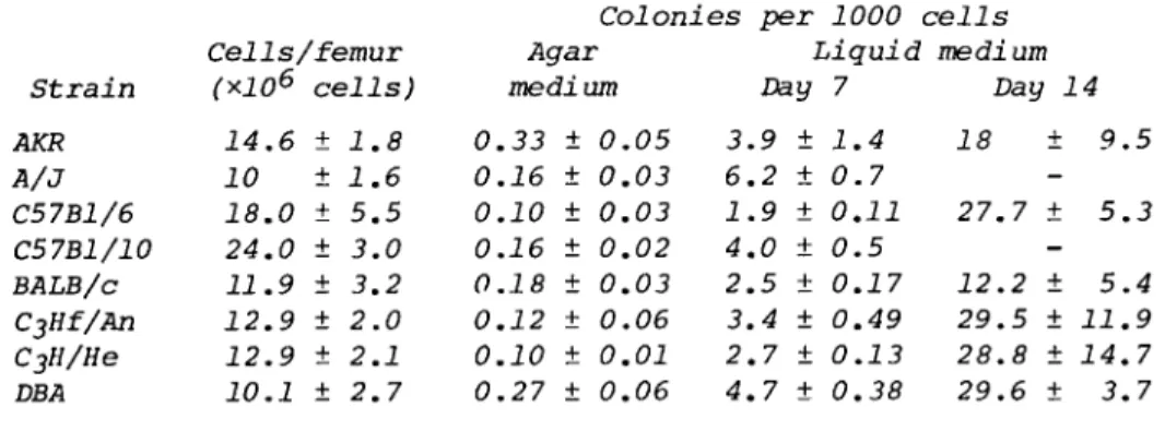
Insert the 27-gauge needle into the proximal end of the femur and inject 1.5 ml medium holding the femur over a Falcon plas- tic tube (No. 2051). Repeat, injecting 1.5 ml through the distal end of the femur. Repeat for as many femurs as are required. The approximate yield of cells per femur for some commonly used mice are shown in Table I. Count the cells.
Count cells in suspension with pronase and cetrimide;
make an initial dilution of the cell suspension to give about 106 cells/ml. Remove 0.5 ml and place this in a 7-dram vial.
SCHEME
A Guide to Setting up Bone Marrow Cultures
Prior to obtaining cells After obtaining cells Put 20-35 mm culture dishes
in 10-100 mm petri dishes, add 1 lidless 35-mm culture dish containing 2 ml water Fill 3-ml syringes with
OL-MOPS 16 x 100-mm tube
Put 10 ml agar medium in 16 x 100-mm tube
Put 2.5 ml QL-0 in 50-ml coni- cal centrifuge tube with 43 ml growth medium for liquid culture
Put 6.3 ml growth medium for liquid culture in 10 x 10 mm tube
Put 3.5 ml -O in 16 x 100 mm tube and two 35-mm dishes
Obtain BM3- with 3-ml syringe and put in empty 16 x 100 mm tube
Count cells and add 2 x 10⁶ to 10 ml agar medium, mix
Add 2.5 ml cells to 50-ml cen- trifuge tube and pipette 3 ml into fourteen 35-mm culture dishes to count adherent and nonadherent cells daily
Add 3.5 ml stock cells to tube and plate 3 ml in two 35-mm dishes, count adherent and nonadherent cells one hour later
Put 3 ml agar culature medium in 16 x 100 mm tube, keep in 43°C water bath
Mix 3 ml cells with agar culture medium and pipette 1 ml in four 35-mm culture dishes; in- cubate 7 days
Add 0.7 ml cells to 10 x 100 mm tube and pipette 3 ml into two 35-mm dishes, stain colonies on day 7
aBM, bone marrow.
TABLE
Strain AKR A/3 C57B1/6 C57B1/10 BALB/c C^Hf/An C3H/He DBA
I. Colony For:
Cells/femur
(*106 cells) 14.6 ± 1.8 10 ± 1.6 18.0 ± 5.5 24.0 ± 3.0 11.9 ± 3.2 12.9 ± 2.0 12.9 ± 2.1 10.1 ± 2.7
mation by Bone Marrow Cells Colonies per 1000 c Agar
medium 0.33 ± 0.05 0.16 ± 0.03 0.10 ± 0.03 0.16 ± 0.02 0.18 ± 0.03 0.12 ± 0.06 0.10 ± 0.01 0.27 ± 0.06
Liquid Day 7 3.9 ± 1.4 6.2 ± 0.7 1.9 ± 0.11 4.0 ± 0.5 2.5 ± 0.17 3.4 ± 0.49 2.7 ± 0.13 4.7 ± 0.38
zells medium
Day 14 18 ± 9.5
-
27.7 ± 5.3 12.2 ± 5.4 - 29.5 ± 11.9 28.8 ± 14.7 29.6 ± 3.7
Then add 0.5 ml freshly filtered pronase diluted 1:5 with saline.
Mix and incubate the suspension 15 min at 37 C. Add 10 ml of cetrimide counting solution directly to the vial and count the cell suspension using an electronic particle counter. The same procedure is used for hemocytometer counting except that the cells are added to only 1 ml of cetrimide.
While the counter settings used to count erythrocytes can be used as approximate settings for counting nuclei from hemato- poietic cells, it is best to determine the exact settings for nuclei. Instructions for this procedure are found in the coun- ter manual.
C. Colony Formation in Agar Culture
Prepare cells in growth medium for agar culture at a con- centration of about 2 x 10⁵ cells/ml by adding 2 x 10⁶ bone marrow cells to 10 ml agar medium in a 16 x 100 mm plastic culture tube. These cells will also be used in Section D. Add 3 ml of this cell suspension to 3 ml of agar culture medium in a second 16 x 100 mm plastic culture tube, mix, and immediately pipette 1 ml into four 35-mm culture dishes. Place these dishes in two humidity chambers and incubate them for 7 days.
Count the number of colonies that have formed, using a dissect- ing microscope.
It is possible to fix the colonies in situ by adding 1.0 ml of 1.5% glutaraldehyde to the cultures. Refrigerate cultures and read them as soon as possible before they become dehydrated.
Individual colonies can be removed for cytology using a Pasteur transfer pipette and dissecting microscope. We disperse the colony in 0.3 ml of a-10 by repeated passage through a 27-gauge needle attached to a tuberculin syringe and then prepare slides using a cytocentrifuge (Shandon Southern Instrument, Inc.,
Sewickley, Pennsylvania 15143). These preparations can then be stained with Wright or Giemsa blood stains.
D. Growth Curve and Colony Formation in Liquid Culture Add 2.5 ml of the cell suspension prepared above to 2.5 ml of a-0 in a 50-ml conical centrifuge tube. Then add 45 ml of growth medium for liquid culture. This will result in 50 ml of bone marrow cells at a concentration of 10,000 cells/ml. Pipette 3 ml (30,000 cells) into 14 35-mm culture dishes. Place them into seven humidity chambers prepared above and place these in the CO2 incubator.
To prepare cultures for colonies, add 0.7 ml of the sus- pension to 6.3 ml growth medium for liquid culture (1000
cells/ml) and plate 3 ml into two 35-mm culture dishes. Place these dishes into the humidity chambers and incubate them for 7 days. (Plates may be stained directly with méthylène blue or, just prior to staining, the adherent cells can be pulsed with yeast. First, rinse the cultures to remove nonadherent cells. Cytocentrifuge preparations may be made of the nonad- herent cells. Add 1 ml of medium containing 10% fetal bovine serum and 1% freshly reconstituted (with distilled water, do not use restoring solution as it contains sodium azide) guinea pig complement to the adherent cells. Add 50 yl of yeast and mix. Incubate cells for 30 min at 37°C. Remove plates, rinse plate three times with 3 ml saline and add 2 ml méthylène blue solution. After 20 min, discard stain, rinse with run- ning tap water, and air dry.
It was noted above that extra cells were prepared in growth medium for agar culture for the purpose of determining the initial number of adherent and nonadherent cells. Add 3.5 ml of these cells to 3.5 ml of a-O. Plate 3 ml (3 x 10⁵ cells) of this suspension directly in two 35-ml culture dishes.
Incubate these cells for 1 hr.
Determine the number of adherent and nonadherent cells on two cultures at 1 hr and every day for 7 days. For the 1-hr determination, use the culture prepared above containing 3 x 10^ cells and, for the subsequent days, use the cultures prepared at 3 x 10⁴ cells. To count cells, resuspend the non- adherent cells in the growth medium and transfer it to a 7-dram vial. Rinse the plates with 2 ml of a-0 and pool it with non- adherent cells. Immediately add (so it will not dry) 1 ml of pronase diluted 1:10 with a-0 to the culture dish containing the adherent cells. Then add 0.5 ml undiluted pronase to the vial containing the nonadherent cells. (A portion of the non- adherent fraction may be processed for cytology as described
for agar colonies.)
Incubate vials and dishes at 37°C for 15 min. Add 10 ml of cetrimide to the vial of nonadherent cells and count them
(or count directly with a hemocytometer). Rinse off adherent cells as follows: Transfer diluted pronase in culture dish to a 7-dram counting vial. Add 2 ml of cetrimide solution and systematically rinse the plate using a transfer pipette. We generally rinse the dish in a clockwise fashion, beginning at 1:00, proceeding around the plate to 12:00, and then ending up in the center of the dish. Be careful not to create air bubbles by keeping the pipette tip submerged. This systematic
rinse is important, because macrophages are extremely adherent and they will not be quantitatively removed unless this pro- cedure is strictly followed. It takes about 1 min per plate.
If a large number of plates are going to be treated, they
should be processed in batches of ten so that none of the plates are incubated longer than 25 min with pronase. For electronic particle counting, transfer the 2 ml of nuclei to 8 ml of cet- rimide. For hemocytometer counting, count directly without further dilution. On days 1 - 3 counts will be low due to cell death.
Extra cultures can be prepared for specific mononuclear phagocyte marker or functional studies. Bone marrow-derived mononuclear phagocyte colonies contain all forms of mononuclear phagocytes from the monoblast to the mature macrophage in vary- ing proportions.
IV. CONCLUDING REMARKS
The expected yield of cells and the frequency of colony- forming cells for bone marrow obtained from several mouse strains is shown in Table I. When cells are grown in liquid medium, large colonies with an average diameter of 2.7 ± 0.7 mm have formed by day 7; there is, however, a tenfold increase
(29 ± 1.2) in the number of colonies on the culture dish by day 14. These latter colonies are nearly all small colonies whose average diameter is 1.1 ± 0.1 mm. Thus, two populations of bone marrow progenitor cells for mononuclear phagocytes are found: One is nonadherent and characterized by a short lag period and extensive proliferation. The other population of cells is adherent and is characterized by a longer lag period before proliferation begins ( 4 - 6 days). This offers a con- venient means of separating the less mature nonadherent pro- genitor cells from the more mature adherent cells of the mono- nuclear phagocyte series for studying proliferative and func- tional potential. Colonies of bone marrow cells that have formed on day 7 are shown in Fig. 1.
plated in 35-mm culture dishes at 1CT cells/culture in 3 ml of growth medium for liquid culture.
Cells were fixed and stained with méthylène blue. The gross appearance of the colonies is shown on the left, and a photomicrograph (*100) of the edge of a colony is shown on the right.
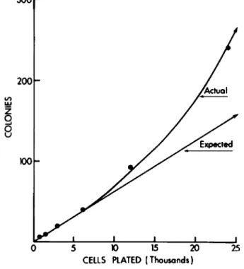
When the number of colonies formed in liquid culture is used as an index of progenitor cells within the population, it is possible that some of the colonies formed are derived from nonadherent progenitor cells that have floated to a distant location where a new colony is produced. Thus, a single pro- genitor cell could produce two or more colonies. One way to determine if this problem exists is to culture serially diluted cells. As shown in Fig. 2, a greater deviation from linearity occurs as the cell number is increased, when colony formation is plotted against cells plated. While this problem does not occur when agar is used because the cells are held in a matrix,
300 r
5 K) 15 20 CELLS PLATED (Thousands)
25
Fig. 2. Nonlinear colony formation in liquid culture:
Bone marrow cells from CjHf/An mice were serially diluted 1:2 in growth medium for liquid culture and 3 ml was plated on
35-mm culture dishes. After 7 days incubation, the cultures were stained with méthylène blue and colonies were counted.
Colony formation was linear with plated cells up to 6000 ce 11 s/culture. As cell number was further increased, a con- tinuing deviation from linearity occurred. Cultures es- tablished at 48,000 cells were completely confluent.
the number of colonies formed in agar is one-third to one-tenth as many as in liquid culture medium (Table I). This difference is not due to the above described progenitor cell flotation.
The reasons for the difference, however, are not known. Indeed, the difference between the two culture systems is generally not recognized as most investigators only use the agar system and have never compared the two.
It has been shown that the addition of hemolysate to agar medium (7-9) will result in a two- to fivefold increase in the number of colonies which form. Similar increases have been re- ported when cells are cultured in low oxygen tension (10). In both situations, however, the number of colonies that form do not exceed the number that normally form in liquid medium. We have never been able to find an increase in plating efficiency
due to oxygen or hemolysate when cells are cultured in liquid medium. It would appear, therefore, that the presence of agar compromises the colony-forming ability of some potential colony-forming cells and that colony formation in the agar sys- tem reflects only a portion of them.
The single most important variable is medium pH. For good growth, it is absolutely essential that cells not be subjected to a fluctuating pH as might be encountered if the incubator door is frequently opened. We cannot emphasize enough the im- portance of this variable. If an incubator must be used that is frequently opened so air mixes with the internal atmosphere, then cultures should be placed in a closed container inside the incubator, which can be independently supplied with the humidi- fied CO2 and air atmosphere. (These containers are available from Streck Laboratories, Inc., Box 6036, Omaha, Nebraska 68106 or Bellco Glass, Inc., 340 Edrudo Road, Vineland, New Jersey 98360.)
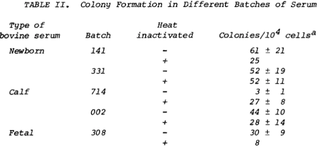
It is advisable to prescreen fetal bovine serum to select a batch that is not toxic to cells and that will promote good growth. For most of our experience in culturing bone marrow cells, we have used fetal bovine serum as the supplement. We recently found that bovine newborn or calf serum can also be used. Results from one series of tests are shown in Table II.
It may be necessary to heat inactivate the sera at 56°C for a full 30 min to detoxify the serum before it is used.
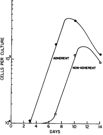
The growth of kinetics bone marrow mononuclear phagocytes is shown in Fig. 3. Cells (3 x 10⁴) were initially plated and, as a function of time, the nonadherent and adherent cells were separately counted. Adherent cells proliferated exponentially with a doubling time of about 24 hr, while a relatively constant recovery of nonadherent cells was obtained during the first week of culture. Thereafter, the nonadherent cells also exponen- tially increased. Note that cells stop proliferating exponen- tially about day 10, when the cultures have been depleted of es- sential factors. Proliferation can be maintained for longer
TABLE II. Colony Formation in Different Batches of Serum Type
bovine of serum Newborn
Calf
Fetal
Batch 141 331 714 002 308
Heat inactivated
- + + + + +
Colonies/104 <
61 ± 21 25 52 ± 19 52 ± 11 3 ± 1 27 ± 8 44 ± 10 28 ± 14 30 ± 9
8 Results from three experiments.
periods if more growth medium is added. We find that each milliliter of growth medium will support the growth of bone marrow cells to a maximum number of 2 x 10^ cells. If 2 x 10^ cells are desired, 10 ml of growth medium would be required. There is a simple calculation to determine how long it will take an initial number of cells to reach a de- sired number of cells. The time (t) to reach this number
(e.g., 2 x 10⁶ cells) from any starting number (e.g., 104) of bone marrow cells (NQ) with a known fraction (f = 0.1) of progenitor cells and a known doubling time (e.g., T^ = 24 hr) is given by
t = (TD/0.693)In(N/fN0) For the example above,
24 2 x 10
t = An = 343 hr or 14 days
u#byj
o.oi x io
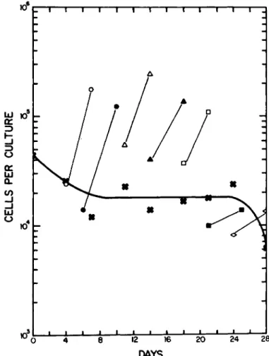
4It is possible to passage these nonadherent progenitor cells and establish new cultures. As shown in Fig. 4, when the nonadherent cells are sequentially passed every 3 or 4 days (usually Mondays and Fridays), they will give rise to new cultures of adherent cells. The adherent cells will prolifer- ate exponentially, and the nonadherent cells will renew them- selves for the next passage. The simplest interpretation of this data is that, on the average, each nonadherent cell gives rise to two daughters, one of which remains nonadherent and one of which becomes adherent.
10V
Fig. 3. Proliferation of bone marrow cells. Bone marrow cells from CjHf/Άη mice were plated in 35-mm culture dishes at 3 x 10⁴ cells/culture in 3 ml of growth medium for liquid cul- ture. On dags 3, 7, 10, and 14, the nonadherent cells were removed from two plates and put in separate 7 dram vials.
Both the adherent (* ) and nonadherent cells (o ) were treated with pronase for 15 min and then counted with cetrimide using an electronic particle counter as described in Section II. Ά.
The doubling time of each population during exponential growth is 24 hr.
The passages cannot be maintained indefinitely, however, and by day 30 no nonadherent cells can be recovered from the cultures. We calculate that a single progenitor cell can give rise to approximately 10 progeny. This can provide a suffi- cient number of cells for functional studies. In fact, if every progenitor cell from a single mouse femur were cultured,
10-rr-r
Kf
T 1 1 1 1—D
12 20 24 28
DAYS
Fig. 4. Maintenance of nonadherent cell proliferation.
Bone marrow cells from CjHf/An mice were plated in 35-mm cul- ture dishes at 3 x 10⁴ cells/culture in 3 ml of growth medium for liquid culture. Every 3 or 4 days the nonadherent cells
(x ) were removed, pooled, and counted. These cells were then replated (3 ml) into new culture dishes. Fresh growth medium was added to the adherent cells ( ο , Φ , Δ , Α , ϋ , β ; and they were counted a week later.
one could obtain between 1 01 3 and 1 01 4 cells. Since these cells are absolutely dependent on MGF for growth, one would require 104 liters of growth medium to accomplish this. Only
1 liter would be required, however, to obtain 10^ (^1 gm) mononuclear phagocytes, and this would take about 14 days if all the cells from a single femur were cultured.
REFERENCES
1. C. C. Stewart and H-s. Lin. Macrophage growth factor and its relationship to colony stimulating factor. J. Re- ticuloendothel. Soc. 23: 269-285, 1978.
2. E. R. Stanley. Colony-stimulating factor (CSF) radioim- munoassay: Detection of CSF subclass stimulating macro- phage production. Proc. Nat. Acad. Sei. 76: 2969-2973, 1979.
3. E. R. Stanley and C. J. Guilbert. Regulation of macro- phage production by a colony-stimulating factor. In
"Mononuclear Phagocytes - Functional Aspects" (R. van Furth, ed.), pp. 417-433. Nijhoff, Boston, 1980.
4. Th. J. L. M. Goud and R. van Furth. Identification and characterization of the monoblast in mononuclear phago- cytes colonies grown in vitro. J. Exp. Medm 142: 1180- 1199, 1975.
5. Th. J. L. M. Goud and R. van Furth. Proliferative charac- teristics of monoblasts grown in vitro. J. Exp. Med. 142:
1200-1217, 1975.
6. W. C. Buhles. Studies on mononuclear phagocyte progenitor cells: Morphology of cells and colonies in liquid culture of mouse bone marrow. J. Reticuloendothel. Soc. 25: 363- 378, 1979.
7. T. R. Bradley, P. A. Telfer, and P. Fry. The effect of Erythrocytes on mouse bone marrow colony development in vitro. Blood 38: 353-360, 1971.
8. J. Rothmann, C. F. Hertogs, and 0. H. Pluznik. Replace- ment of serum by hemolysate as groth promoter for murine leukemic and normal hemopoietic progenitor cells in cul- ture. Exp. Hematol. 5: 117-124, 1977.
9. K. Tsuneoka, Y. Takagi, K. Hirashima, and M. Shihita.
Enhancement of the action of colony stimulating factor (CSF) by soluble components of erythrocytes in mouse bone marrow cell cultures. Exp. Hematol. 6: 445-450, 1978.
10. T. R. Bradley, G. S. Hodgson, and M. Rosendal. The effect of oxygen tension on hemapoietic and fibroblast cell pro- liferation in vitro. J. Cell. Physiol. 97: 411-522, 1978.