International Journal of
Molecular Sciences
Article
Symbiotic NCR Peptide Fragments Affect the Viability, Morphology and Biofilm Formation of Candida Species
Bettina Szerencsés1, Attila Gácser1 , Gabriella Endre2, IldikóDomonkos2, Hilda Tiricz2, Csaba Vágvölgyi1 , János Szolomajer3, Dian H. O. Howan3, Gábor K. Tóth3,4, Ilona Pfeiffer1,* andÉva Kondorosi2,*
Citation: Szerencsés, B.; Gácser, A.;
Endre, G.; Domonkos, I.; Tiricz, H.;
Vágvölgyi, C.; Szolomajer, J.; Howan, D.H.O.; Tóth, G.K.; Pfeiffer, I.; et al.
Symbiotic NCR Peptide Fragments Affect the Viability, Morphology and Biofilm Formation ofCandidaSpecies.
Int. J. Mol. Sci.2021,22, 3666.
https://doi.org/10.3390/ijms22073666
Academic Editor: Loredana Frasca
Received: 13 January 2021 Accepted: 28 March 2021 Published: 1 April 2021
Publisher’s Note:MDPI stays neutral with regard to jurisdictional claims in published maps and institutional affil- iations.
Copyright: © 2021 by the authors.
Licensee MDPI, Basel, Switzerland.
This article is an open access article distributed under the terms and conditions of the Creative Commons Attribution (CC BY) license (https://
creativecommons.org/licenses/by/
4.0/).
1 Department of Microbiology, Faculty of Science and Informatics, University of Szeged, H-6726 Szeged, Hungary; betti414@gmail.com (B.S.); gacsera@bio.u-szeged.hu (A.G.);
csaba@bio.u-szeged.hu (C.V.)
2 Biological Research Centre, Institute of Plant Biology, H-6726 Szeged, Hungary; endre.gabriella@brc.hu (G.E.);
domonkos.ildiko@brc.hu (I.D.); tiriczh@brc.hu (H.T.)
3 Department of Medical Chemistry, University of Szeged, H-6720 Szeged, Hungary;
szolomajer.janos@med.u-szeged.hu (J.S.); dian.howan@med.u-szeged.hu (D.H.O.H.);
toth.gabor@med.u-szeged.hu (G.K.T.)
4 MTA-SZTE Biomimetic Systems Research Group, University of Szeged, H-6720 Szeged, Hungary
* Correspondence: pfeiffer@bio.u-szeged.hu (I.P.); eva.kondorosi@gmail.com (É.K.)
Abstract:The increasing rate of fungal infections causes global problems not only in human health- care but agriculture as well. To combat fungal pathogens limited numbers of antifungal agents are available therefore alternative drugs are needed. Antimicrobial peptides are potent candidates be- cause of their broad activity spectrum and their diverse mode of actions. The model legumeMedicago truncatulaproduces >700 nodule specific cysteine-rich (NCR) peptides in symbiosis and many of them have in vitro antimicrobial activities without considerable toxicity on human cells. In this work we demonstrate the anticandidal activity of the NCR335 and NCR169 peptide derivatives against fiveCandidaspecies by using the micro-dilution method, measuring inhibition of biofilm formation with the XTT (2,3-Bis-(2-Methoxy-4-Nitro-5-Sulfophenyl)-2H-Tetrazolium-5-Carboxanilide) assay, and assessing the morphological change of dimorphicCandidaspecies by microscopy. We show that both the N- and C-terminal regions of NCR335 possess anticandidal activity as well as the C-terminal sequence of NCR169. The active peptides inhibit biofilm formation and the yeast-hypha transforma- tion. Combined treatment ofC. auriswith peptides and fluconazole revealed synergistic interactions and reduced 2-8-fold the minimal inhibitory concentrations. Our results demonstrate that shortening NCR peptides can even enhance and broaden their anticandidal activity and therapeutic potential.
Keywords:NCR peptide; antifungal activity;Candida; morphological switch; biofilm
1. Introduction
Fungal infections are increasing and represent serious health threat particularly in the increasing population of immunosuppressed patients. Invasive fungal infections are generally associated with high mortality [1] and the use of effective antifungal drugs is crucial for the outcome of the patient’s disease. Unfortunately, the repertoire of antifungal agents is rather limited and for systemic therapy only three classes of drugs are available:
polyenes, triazoles and echinocandins. In addition to the limited spectrum of antifungals, similarly to bacterial resistance against the antibiotics, development of drug resistance against these agents has been reported inCandidaandCryptococcusspecies and in some moulds [2]. The problem to fight fungal infections also exists in agriculture leading to significant loss in agricultural productivity. Therefore, there is a great and urgent need for novel types of antifungal agents, and alternative antifungal strategies both in healthcare therapies and in the agriculture [3].
Antimicrobial peptides (AMPs) have a great potential as an untapped source of novel biologically active agents to fight against bacteria, fungi, parasites, and viruses. AMPs are
Int. J. Mol. Sci.2021,22, 3666. https://doi.org/10.3390/ijms22073666 https://www.mdpi.com/journal/ijms
Int. J. Mol. Sci.2021,22, 3666 2 of 20
produced in all domains of life and are part of the host innate immune response protecting the host against microbial infections. AMPs show extraordinary diversity in nature, their length varies between 10 and 60 amino acids and almost all AMPs are cationic. Most prevalently AMPs act directly on the microbial membranes leading to the loss of membrane potential, leakage and finally death of cells. However, a remarkable quality of many AMPs is that they act by multiple mechanisms and can have numerous intracellular biological targets that are distinct from targets of the traditional antibiotics/drugs. The antimicrobial peptide database lists 3081 peptides mainly from natural sources with an average peptide length of 33 amino acids and a net charge of +3.3 [4]. Plant AMPs are poorly represented in the AMP databases, although even a single plant can produce close to 1000 different AMPs.
Defensins, secreted peptides having 8 or 10 conserved cysteine residues, represent the major class of AMPs in plants. In addition, certain legumes, belonging to the Inverted Repeat Lacking Clade (IRLC) such as alfalfa and otherMedicagospecies or pea, clover, lentil, vetch, and other plants in this clade, have evolved a defensin-related but unique gene family which codes for nodule-specific cysteine rich (NCR) peptides produced only inRhizobium- legume symbiosis.Medicago truncatulaforms nitrogen fixing symbiosis withSinorhizobium melilotisoil bacteria. The interaction between the host plant and the bacterium leads to the formation of a symbiotic organ, the root nodule where the bacteria live inside the nodule cells. In these infected cells the plant manipulates the fate of bacteria, by transforming them to non-cultivable, huge polyploid bacteria with the help of NCR peptides [5,6]. In Medicago truncatula, more than 700 genes code for NCR peptides [7,8]. NCRs have 4 or 6 cysteines in conserved position and otherwise high divergence in their amino acid composition and sequence resulting in a great diversity in their physicochemical properties that is also reflected by the wide range of isoelectric point (pI) of peptides from 3.2 to 11.2.
Many of these symbiotic NCR peptides have in vitro strong antimicrobial activities [9].
Cationic NCRs such as NCR247 (pI: 10.15) and NCR335 (pI: 11.22) show broad range and partially overlapping activities, indicating that in addition to the positive net charge, the amino acid sequence contributes to the antimicrobial properties [10]. NCR247 acts via multiple mechanisms, interacting with the bacterial membranes and by entering the cells with many bacterial proteins and affecting both transcription and translation and inhibiting cell division by interacting with the conserved bacterial FtsZ protein required for septum formation and cell division [11,12]. Shorter chimeric derivatives of NCR247 gained even higher activities and were also able to kill the most resistant and problematic ESKAPE bacteria at low concentrations making them comparable or even superior to third generation antibiotics with the advantage of lack of cytotoxicity on human cells [13].
In addition to bacteria, NCRs can also kill fungi. Previously we investigated the anti- fungal effect of 19 NCR peptides with pI from 3.6 to 11.2 againstCandida albicansand found that peptides with pI > 9.5 inhibited the growth ofC. albicans[14]. The minimal inhibitory concentrations (MIC) of these cationic NCR peptides (NCR192, NCR137, NCR147, NCR 280, NCR183, NCR247, NCR044, NCR030, NCR335) were in the range of 1.4–10µM. For ex- ample, the MIC of NCR335 (pI: 11.22) and NCR044 (pI: 10.32) was 1.4–2.5µM. Importantly, these concentrations of cationic NCR peptides, which efficiently eliminatedC. albicansdid not affect the viability of vaginal epithelial cells while they inhibited theC. albicans-induced killing of epithelial cells. NCR044 exhibited potent antifungal activity against plant fungal pathogens as well, such as variousFusariumspecies andBotrytis cinereaand was shown to act by multifaceted mechanisms [15]. Thus, cationic NCRs, due to their potent antifungal activity, negligible host toxicity and low resistance rates emerge as potential candidates for developing new antifungal therapies.
Weakly cationic peptides such as NCR169 (pI: 8.45) or neutral and anionic NCRs were inactive againstC. albicansin the studied concentration range [14]. However, we noticed that, unlike the full sequence of NCR169, its C-terminal half was highly cationic (pI: 10.1) leading us to assume that this part may have antimicrobial activity.
In this study, the growth inhibitory and fungicide effects of eleven NCR335 derivatives, and the C-terminal region of NCR169 were tested against 7 strains of 5 human pathogenic
Int. J. Mol. Sci.2021,22, 3666 3 of 20
Candidaspecies. We also investigated how the most effective peptides act on the biofilm formation and on the yeast to hypha morphological switch ofC. albicansandC. tropicalisas hyphal growth represents an important virulence factor in these dimorphic species [16,17].
Moreover, synergism was demonstrated between peptides and between peptides and the antifungal drug, fluconazole againstC. auriswhich significantly reduced the MIC values and helps to overcome the mild toxicity of these peptides and antifungal drugs.
2. Results
2.1. Design and Synthesis of NCR335 and NCR169 Peptide Derivatives
NCR335 is not only one of the most cationic NCR peptides but it is exceptional due to its 64 amino acid (RLNTTFRPLNFKMLRFWGQNRNIMKHRGQKVHFSLILSDCKTNKD- CPKLRRANVRCRKSYCVPI) long sequence compared to the average length of 33 amino acids of NCRs. NCR335 is composed of an NCR-specific sequence with four conserved cysteine residues on the C-terminal 33 amino acid long sequence. The 31 amino acid long N-terminal sequence (RLNTTFRPLNFKMLRFWGQNRNIMKHRGQKV) is unrelated to the NCR sequences and lacks cysteine residues but has +8.1 net charge and pI: 12.72, which raised the possibility that this sequence may also have antimicrobial activity. Therefore, we investigated separately the activity of the N- and C-terminal regions of NCR335. To identify the regions responsible for antifungal activities peptide fragments were synthetized from the N-terminal sequence corresponding to the 1–19, 1–15, 7–21, 16–29 amino acid sequences, while from the 33 amino acid long NCR335C peptide the 13–33, 17–33, 17–27, 1–8, 9–16, 1–16 sequences were synthetized (Table1). Except for NCR335C1–8and NCR335C9–16, all peptides have significant + net charge and high pI.
Table 1.Physicochemical properties of NCR peptides synthetized with C-terminal amidation.
Peptide Sequence of the Peptide No. AA pI Net Charge
NCR335N1–19 RLNTTFRPLNFKMLRFWGQ 19 14 +5
NCR335N1–15 RLNTTFRPLNFKMLR 15 14 +5
NCR335N7–21 RPLNFKMLRFWGQNR 15 14 +5
NCR335N16–29 FWGQNRNIMKHRGQ 14 14 +4.1
NCR335C1–33 HFSLILSDCKTNKDCPKLRRANVRCRKSYCVPI 33 10.37 +6.8
NCR335C13–33 KDCPKLRRANVRCRKSYCVPI 21 10.91 +6.8
NCR335C17–33 KLRRANVRCRKSYCVPI 17 11.73 +6.9
NCR335C17–27 KLRRANVRCRK 11 12.59 +6.9
NCR335C1–8 HFSLILSD 8 7.57 +0.1
NCR335C9–16 CKTNKDCP 8 9.23 +1.9
NCR335C1–16 HFSLILSDCKTNKDCP 16 8.07 +1.0
NCR169C17–38 KSKKPLFKIWKCVENVCVLWYK 22 10.48 +5.9
NCR169C17–38ox KSKKPLFKIWK ˆCVENV ˆCVLWYK 22 11.01 +6
NCR169C17–38W10,20/A KSKKPLFKIAKCVENVCVLAYK 22 10.48 +6
C indicates two cysteines joined by disulphide bond. Alanin residues replacing Wˆ 10and W20are underlined.
In the case of NCR169 (EDIGHIKYCGIVDDCYKSKKPLFKIWKCVENVCVLWYK), we investigated the C-terminal 22 amino acid long sequence (KSKKPLFKIWKCVENVCVL- WYK), NCR169C17–38, with a net charge of +5.9 and a pI of 10.48 in linear and a net charge of +6 and a pI of 11.01 in oxidized form, and by replacing the two tryptophan residues (W10
and W20) with alanine (Table1). This latter substitution was made because tryptophan residues could be required for interaction with microbial membranes and can play a critical role in the antimicrobial activity [18].
The 3D folding of peptides longer than 8 amino acids was predicted with the PEP- FOLD3 tool (FigureA1). The folding of NCR335N1–29indicates a relatively disordered structure, with a relatively short alpha helical segment in the N- and C-terminal half of the peptide. The two halves of the molecule (NCR335N1–19and NCR335N16–29) retain the original steric structure, a short alpha helical region on each fragment. Interestingly the predicted 3D structure of NCR335N7–21appears to be almost exclusively alpha helical
Int. J. Mol. Sci.2021,22, 3666 4 of 20
and different from the other NCR335N derivatives. In NCR335C1–33a helical segment is predicted at the N terminus in the otherwise disordered structure. Similar folding is pre- dicted for NCR335C17–27and NCR335C13–33. In contrast, the 3D structure of NCR335C17–33
appears to be dramatically different: the helical part is absent while two short antiparallel βstrands are formed. In NCR169C17–38, a helical region is predicted in the middle, while the ends are disordered. Replacement of tryptophan residues to alanine does not change the 3D structure.
2.2. NCR Based Peptides Affect the Growth and Survival of Candida Strains
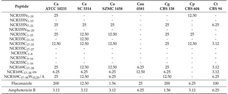
The antifungal activity of all the 14 peptides was tested against the following op- portunistic human pathogenCandidaspecies and strains:C. albicansstrains ATCC 10231, SC 5314 and SZMC 1458,C. auris0381,C. glabrataCBS 138,C. parapsilosisCBS 604 and C. tropicalisCBS 94. To determine the minimal inhibitory concentration of the peptides, the strains were incubated with two-fold serial dilutions of the peptides from 25µM to 1.56µM (Table2).
Table 2.Minimal inhibitory concentration (µM) of the NCR peptide fragments againstCandidastrains.
Peptide Ca
ATCC 10231
Ca SC 5314
Ca SZMC 1458
Cau 0381
Cg CBS 138
Cp CBS 604
Ct CBS 94
NCR335N1–19 25 - - - - 12.50 -
NCR335N1–15 - - - -
NCR335N7–21 25 25 25 - 25 - 6.25
NCR335N16–29 - - - -
NCR335C1–33 25 12.50 12.50 - 25 25 -
NCR335C13–33 - 12.50 - - - - -
NCR335C17–33 12.50 12.50 12.50 - 25 12.50 3.12
NCR335C17–27 - - - -
NCR335C1–8 - - - -
NCR335C9–16 - - - -
NCR335C1–16 - - - -
NCR169C17–38 25 12.50 12.50 6.25 25 - 3.12
NCR169C17–38ox 6.25 6.25 6.25 12.50 6.25 - 3.12
NCR169C17–38W10,20/A 25 12.50 6.25 - 12.50 - 6.25
Fluconazole 200 12.50 1.56 25 200 6.25 100
Amphotericin B 3.12 3.12 3.12 6.25 1.56 3.12 6.25
-: no growth inhibition was observed. Ca: Candida albicans, Cau: Candida auris, Cg: Candida glabrata, Cp: Candida parapsilosis, Ct:
Candida tropicalis.
Both the N- and the C-terminal regions of NCR335 exhibited anti-Candidaactivity. Of the NCR335N peptides, NCR335N7–21displayed the broadest activity; exceptC. parapsilosis andC. auris.It inhibited the growth ofC. albicansstrains ATCC 10231, SC 5314 and SZMC 1458 andC. glabrataCBS 138 at 25µM andC. tropicalisCBS 94 at 6.25µM. Compared to NCR335N7–21, the overlapping NCR335N1–19peptide, which lacks the last two amino acids, and has an N-terminal extension of seven amino acids, remained active only against C. albicans ATCC 10231 (MIC 25µM), but became effective againstC. parapsilosis(MIC 12.50µM). NCR335N1–15and NCR335N16–29did not affect the growth of the studied strains in this concentration range.
The C-terminal part of NCR335, NCR 335C1–33, with the characteristic pattern of cysteine residues, was effective against all strains exceptC. aurisandC. tropicaliswith MICs of 12.50 and 25µM. The shorter peptide, NCR335C17–33corresponding to the last 17 amino acids of NCR 335C1–33 showed even higher activity and became remarkably effective againstC. tropicalis(MIC 3.12µM). The four amino acid longer NCR335C13–33peptide was only effective againstC. albicansSC 5314. NCR335C17–27, NCR335C1–8, NCR335C9–16and NCR335C1–16were ineffective, having no effect on the growth of any of the tested strains.
Int. J. Mol. Sci.2021,22, 3666 5 of 20
The NCR169 derivatives NCR169C17–38 and NCR169C17–38ox proved to be active against all the examined strains includingC. aurisbut were ineffective againstC. parap- silosis.The MIC values of both peptides ranged from 3.12µM to 25µM, and the oxidized form NCR169C17–38ox was more efficient against theC. albicansstrains and C. glabrata.
NCR169C17–38W10,20/A lost the activity againstC. aurisbut maintained activity against the other strains.
The effectiveness of these peptides was compared to that of two antifungal agents:
fluconazole and amphotericin B (Table2). Fluconazole is used to treat various fungal and yeast infections including candidiasis. Fluconazole was tested in a concentration range from 200 to 0.78µM and was highly effective againstC. albicansSZMC 1458 (MIC 1.56µM), followed byC. parapsilosis(MIC 6.25µM),C. albicansSC 5314 (MIC 12.50µM),C. auris (MIC 25µM),C. tropicalis(MIC 100µM),C. glabrata(MIC 200µM) andC. albicansATCC 10231 (MIC 200µM). The MIC values of amphotericin B were between 1.56 and 6.25µM:
C. glabratawas the most sensitive (MIC 1.56µM), followed by the threeC. albicansstrains andC. parapsilosis(MIC 3.12µM) and then byC. auris(MIC 6.25µM). While amphotericin B seems to be highly effective, it is only used for life-threatening fungal infections due to its serious side effects.
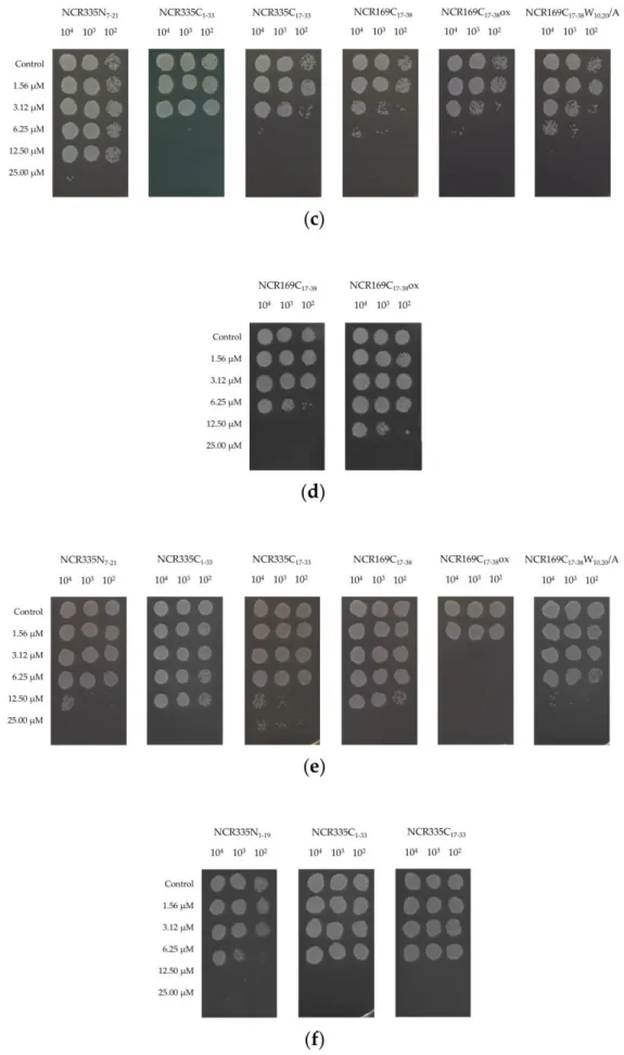
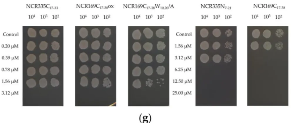
To determine whether the inhibitory effect of the peptides is generated by fungistatic or fungicide action, theCandidacells were treated with the peptides in the same con- centration range (1.56–25µM) as in the growth inhibition assay. After 24 h 5µL sam- ple from the untreated control cells and from the peptide treated cells was added to 95 µL water and diluted further 10- and 100-fold. From each dilution, 5 µL was placed on solid medium and the growth of the control and peptide treated cells was com- pared after 48 h incubation at 30◦C. This assay confirmed that all the effective peptides, NCR335N1–19, NCR335N7–21, NCR335C1–33, NCR335C13–33,NCR335C17–33,NCR169C17–38, NCR169C17–38ox, NCR169C17–38W10,20/A provoked growth inhibition through fungicidal actions (FigureA2). The fungicide concentrations, required for the complete elimination of theCandidacells, were either equal to or twice higher than the minimum inhibitory concentrations presented in Table2.
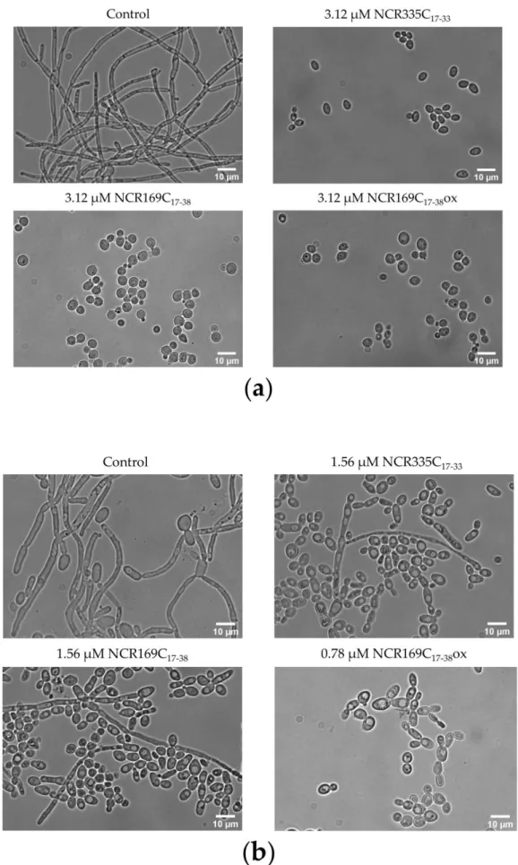
2.3. NCR335C17–33and NCR169C17–38Reduce Hyphae Formation
As hyphae formation is an important virulence factor, we investigated how the broad range activity peptides, NCR335C17–33, NCR169C17–38 and NCR169C17–38ox affect the morphological switch ofC. albicansATCC 10231 andC. tropicalisbetween the hyphal and yeast growth forms at sub-lethal concentrations (Figure1). Observing the cells after 48 h of cultivation with bright field microscopy revealed that treatment ofC. albicansATCC 10231 with 3.12 µM peptides completely inhibited the hyphal growth, which was the characteristic growth form of the control culture. In the untreated control culture ofC.
tropicalisthe hyphal growth was predominant but shifted to the yeast form in the cultures treated with NCR335C17–33and NCR169C17–38at 1.56µM and was the only form of the culture treated with NCR169C17–38ox at 0.78µM. However, not only the morphology changed, but the cell number also declined, particularly in the latter case.
Int. J. Mol. Sci.2021,22, 3666 6 of 20
Int. J. Mol. Sci. 2021, 22, x FOR PEER REVIEW 6 of 20
Figure 1. Morphology of the untreated control and the NCR335C17–33-, NCR169C17–38- or NCR169C17–38ox-treatedC.
albicansATCC 10231 (a) andC. tropicalisCBS 94 (b) cells. Scale bars represent 10µm.
Int. J. Mol. Sci.2021,22, 3666 7 of 20
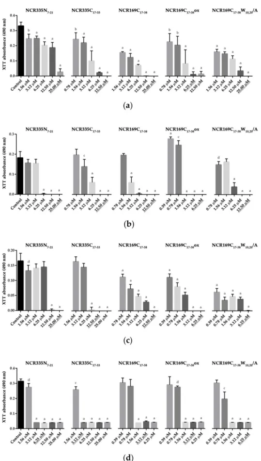
2.4. The Anticandidal NCR Peptide Derivatives Inhibit Biofilm Formation
Biofilm formation is another important virulence factor forC. albicansandC. trop- icalis, as cells located in the biofilm are more resistant to the antifungal agents due to the encompassing matrix that protects against the penetration of the antifungal drugs.
Consequently, biofilm is a major source of persistent or recurrent infections [19]. The biofilm inhibitory ability of NCR335N7–21, NCR335C17–33, NCR169C17–38, NCR169C17–38ox and NCR169C17–38W10,20/A was tested on the three strains ofC. albicansand C. tropi- calis,by cultivating the strains in the presence of increasing concentrations of peptides for 72 h (Figure2). The viability of the biofilm located cells was measured by the XTT (2,3-Bis-(2-Methoxy-4-Nitro-5-Sulfophenyl)-2H-Tetrazolium-5-Carboxanilide) assay. All five peptides significantly inhibited biofilm formation at the MICs or lower concentra- tions (p ≤0.0001). However, theCandidastrains exhibited certain differences in their sensitivity to the peptides. NCR335N7–21was effective at MIC inC.albicansATCC 10231, at half MIC inC. albicansSZMC 1458 andC. tropicalisand at one-fourth of the MIC in C. albicansSC 5314. NCR335C17–33acted at the MIC inC. tropicalisand at half MIC in the other strains. NCR169C17–38was required at MIC inC. albicansSZMC 1458, at half MIC inC.albicansATCC 10231 andC. tropicalisand at one-fourth of the MIC inC. albicans SC 5314. NCR169C17–38ox acted at the MIC inC.albicansATCC 10231, at half MIC in C. albicansSZMC 1458 andC. tropicalisand at one-fourth of the MIC inC. albicansSC 5314.
NCR169C17–38W10,20/A was required at MIC in C. albicansSZMC 1458, at half MIC in C. albicansATCC 10231 andC. albicansSC 5314 and at one-fourth of the MIC inC. tropicalis.
Thus, biofilm inhibition was the most effectively achieved by NCR335N7–21, NCR169C17–38 and NCR169C17–38ox inC. albicansSC 5314 and by NCR169C17–38W10,20/A inC. tropicalis.
2.5. NCR335C17–33and NCR169C17–38Drastically Reduce the Hyphal Form in Biofilms
Candidabiofilms are complex structures that may comprise different cell types [20], therefore the morphology of the cells was also investigated in the biofilms ofC. albicans ATCC 10231 andC. tropicalisafter the treatment with NCR335C17–33and NCR169C17–38at sub-lethal concentrations. Both peptides caused significant biofilm inhibition inC. albicans ATCC 10231 andC. tropicalis(Figure2). Accordingly, the scanning electron microscopy (SEM) images showed lower structural complexity of the biofilms in both species after the treatment with NCR335C17–33or NCR169C17–38compared to the control biofilms (Figure3).
Both peptides reduced the abundance of the hyphal form and fostered more the yeast form resulting in decreased ratios of the hyphae/yeast form.C. tropicaliswas particularly sensitive to the peptides; at 1.56µM NCR335C17–33 practically only the yeast form was present, while at 1.56µM NCR169C17–38the cell number was drastically reduced, and the yeast form was also dominant in the biofilm.
Int. J. Mol. Sci.2021,22, 3666 8 of 20
Int. J. Mol. Sci. 2021, 22, x FOR PEER REVIEW 8 of 20
Figure 2. Effect of the peptides on biofilm formation of C. albicans and C. tropicalis. C. albicans ATCC 10231 (a); C. albicans SC 5314 (b); C. albicans SZMC 1458 (c) and C. tropicalis CBS 94 (d). Un- Figure 2.Effect of the peptides on biofilm formation ofC. albicansandC. tropicalis.C. albicansATCC 10231 (a);C. albicans SC 5314 (b);C. albicansSZMC 1458 (c) andC. tropicalisCBS 94 (d). Underlined concentrations correspond to the minimal inhibitory concentration (MIC) of a given peptide. The XTT absorbance values represent the mean±standard deviation calculated from three independent experiments (a,p≤0.0001, b,p≤0.001, c,p≤0.01, d,p≤0.05, unpairedttest).
Int. J. Mol. Sci.2021,22, 3666 9 of 20
Int. J. Mol. Sci. 2021, 22, x FOR PEER REVIEW 9 of 20
derlined concentrations correspond to the minimal inhibitory concentration (MIC) of a given pep- tide. The XTT absorbance values represent the mean ± standard deviation calculated from three independent experiments (a, p ≤ 0.0001, b, p ≤ 0.001, c, p ≤ 0.01, d, p ≤ 0.05, unpaired t test).
Figure 3. Morphology of the biofilm-located C. albicans ATCC 10231 (a) and C. tropicalis CBS 94 (b).
The strains were cultivated without (control) or with NCR335C17–33 or NCR169C17–38 for 72 h and the cells were visualized by scanning electron microscopy. Scale bars represent 10 µ m.
Figure 3.Morphology of the biofilm-locatedC. albicansATCC 10231 (a) andC. tropicalisCBS 94 (b).
The strains were cultivated without (control) or with NCR335C17–33or NCR169C17–38for 72 h and the cells were visualized by scanning electron microscopy. Scale bars represent 10µm.
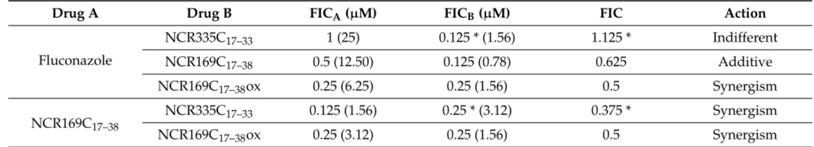
2.6. Combined Treatment of C. auris with NCR Peptide Derivatives and Fluconazole Reveals Synergism
In therapy, application of two or more antibiotics simultaneously can be more ef- fective than each alone. C. aurisis one of the least treatableCandidaspecies, and in this study only NCR169C17–38(MIC 6.25µM) and NCR169C17–38ox (MIC 12.50µM) were ac- tive against this species as well as fluconazole (MIC 25µM) and amphotericin B (MIC
Int. J. Mol. Sci.2021,22, 3666 10 of 20
6.25µM). Therefore, we investigated whether combined application of these peptides with fluconazole or NCR335C17–33, which was active against otherCandidaspecies but not againstC. auris, could reduce MIC values and reveal synergism by checkerboard titra- tion assay (Table3). Amphotericin B, due to its high toxicity was not included in this assay. Synergism was found in three combinations: (i) fluconazole and NCR169C17–38ox, (ii) NCR169C17–38 and NCR169C17–38ox and (iii) NCR169C17–38 and NCR335C17–33. In these cases, the growth inhibition was achieved when 6.25µM fluconazole was combined with 1.56µM NCR169C17–38ox, 3.12µM NCR169C17–38with 1.56µM NCR169C17–38ox or 1.56µM NCR169C17–38with 3.12µM NCR335C17–33. In the case of fluconazole (12.50µM) and NCR169C17–38 (0.78µM) additivity was observed. The action of fluconazole and NCR335C17–33was indifferent.
Table 3.Combined activity of NCR peptide derivatives and fluconazole.
Drug A Drug B FICA(µM) FICB(µM) FIC Action
Fluconazole
NCR335C17–33 1 (25) 0.125 * (1.56) 1.125 * Indifferent
NCR169C17–38 0.5 (12.50) 0.125 (0.78) 0.625 Additive
NCR169C17–38ox 0.25 (6.25) 0.25 (1.56) 0.5 Synergism
NCR169C17–38 NCR335C17–33 0.125 (1.56) 0.25 * (3.12) 0.375 * Synergism
NCR169C17–38ox 0.25 (3.12) 0.25 (1.56) 0.5 Synergism
Fractional inhibitory concentration (FIC) index values: ≤0.5: synergism; 0.5 < FIC≤1.0: additive; 1.0 < FIC≤2.0: indifferent; >2:
antagonism. (µM) corresponds to the concentration of drug A and Drug B for the indicated action. * represents a fictitious FIC value which could be lower as NCR335C17–33was inactive againstC. aurisat 25µM nevertheless 25µM was used as MIC for the calculation of FIC.
2.7. Anti-Candida NCR Peptides Exhibit No or Only Moderate Cytotoxicity on Human Keratinocytes
Cytotoxicity of antifungal agents, such as amphotericin B, may limit or hamper their therapeutic use. TheMedicagoNCR peptides tested to date did not show any or only minor toxicity, however, we could not exclude the possibility that the increased antifungal activity of these truncated NCR derivatives provokes cytotoxicity. Thus, human keratinocyte HaCaT cells were treated with NCR335N1–19, NCR335N7–21, NCR 335C1–33, NCR335C13–33, NCR169C17–38, NCR169C17–38ox and NCR169C17–38W10,20/A in a concentration range from 0.78 to 25µM for 48 hrs and the viability of cells was determined by the MTT assay and compared to the viability of the control cultures (FigureA3). None of the peptides displayed notable cytotoxicity, although a slight decrease in viability was observed at 25µM of all peptides except for NCR169C17–38W10,20/A, which had no effect on cell viability.
3. Discussion
The scarce resource and the side effect of antifungal compounds and the increas- ing number of resistant strains compel the search for new effective antifungals [21,22].
Antimicrobial peptides are promising candidates [23,24] as they have several beneficial characteristics i.e., broad-spectrum activity, low toxicity [25], moderate immunogenicity, good penetration capability, low propensity for development of resistance, distinct mode of actions and lack of cross resistance with the commonly used antifungals [26]. Natural antifungal peptides have been isolated from bacteria, fungi, plants, insects, amphibians, birds, and mammals and generally they possess a small size, overall positive charge, and amphipathic nature [27]. The majority of antifungal AMPs act via their interaction with the cell envelop provoking membrane permeabilization and leakage of cells. However, it is becoming increasingly apparent that peptides can have multifaceted mechanisms in- hibiting various cell functions, such as nucleic acid and protein synthesis, and metabolism or cause production of reactive oxygen species and apoptosis [26]. Legume plants repre- sent countless sources of symbiotic NCR peptides, many of which have broad ranges of antimicrobial and antifungal activities without notable toxicity on human cells [9]. Isola- tion of NCRs from root nodules is not feasible due to the small 1–3 mm size of nodules.
Int. J. Mol. Sci.2021,22, 3666 11 of 20
Moreover, production of several hundreds of NCRs in the nodule with similar sizes and many with similar physicochemical properties would allow isolation of a mixture and not individual NCRs. Their production in heterologous expression systems is also challenging due to their strong antimicrobial activities. Thus, chemical synthesis of NCRs is the most straightforward way for their production, which, however, can relatively be costly for larger peptides.
In this study, the starting natural antifungal peptide for designing new, shorter drug candidates was the unusually long NCR335 peptide. The other peptide, NCR169 had no antifungal activity [14] but based on the high positive charge of the C-terminal half, we presumed that this sequence might have antifungal activity and was tested as a proof of concept. The antifungal activities of peptides were assessed against fiveCandidaspecies and altogether 7 strains including intrinsically resistant ones. Our work demonstrated that truncated derivatives of NCR335 from both the N- and C-terminal halves as well as the C-terminal half of NCR169 have anticandidal activity.
All the NCR335N derivatives have a high positive charge (NCR335N1–19, NCR335N1–15
and NCR335N7–21: +5 NCR335N16–29: +4.1) but only NCR335N7–21exhibited a broad spec- trum activity, inhibiting the growth of all tested strains ofC. albicans, C. glabrata and C. tropicalisbut were ineffective againstC. aurisandC. parapsilosis. NCR335N1–19was only effective againstC. albicansATCC 10231 andC. parapsilosiswhile NCR335N1–15and NCR335N16–29despite their ‘+’ net charge had no anticandidal activity in the tested con- centration range. These results indicate that the ‘+’ net charge itself is not sufficient for the antifungal activity. Interestingly the predicted 3D structure of NCR335N7–21 pep- tide exhibiting the broadest spectrum appears to be alpha helical and different from the other NCR335N derivatives, indicating that the helical region might be important for the antifungal activity.
Of the C-terminal fragments of NCR335: NCR335C1–33/13–33/17–33/17–27had a +6.8-6.9 net charge. NCR335C1–33 was effective againstC. albicans,C. glabrataandC. parapsilo- siswhile it did not affect the growth ofC. aurisandC. tropicalis. Its shorter derivative, NCR335C17–33was even more active and effective against all strains includingC. tropicalis but not againstC. auris. NCR335C13–33 was only effective againstC. albicansSC 5314, which together with the restricted efficacy of NCR335N1–19againstC. albicansATCC 10231 indicates significant differences in the susceptibility of the studied strains ofC. albicans.
Truncating further the sequence of NCR335C17–33, resulting in NCR335C17–27and the loss of activity suggests that the C-terminal sequence (C18–33) is indispensable for the antifun- gal action. The two 8 amino acid long peptides, NCR335C1–8and NCR335C9–16having +0.1 and +1.9 charge, respectively were inactive. The predicted folding of NCR335C1–33, NCR335C17–27and NCR335C13–33sequences indicates a helical segment at the N-terminus in the otherwise unordered structure. In contrast, the 3D structure of the most active NCR335C17–33peptide revealed two shortβstrands but no helical region.
Testing C-terminal part of the NCR169 sequence has confirmed our assumption as NCR169C17–38has indeed anticandidal activity and was effective against allCandidaspecies and strains except forC. parapsilosis.There was some difference between the linear and oxidized forms as the latter was somewhat more effective on theC. albicansstrains and significantly more effective againstC. glabrata. The reason for the differential activity of the linear and oxidized forms can only be speculated, though it is known for NCR247 that its reduced and oxidized forms affect distinctly transcription and translation in bacteria [28,29].
Thus, likewise the cellular responses to linear and oxidized forms of NCR169C17–38may be different. Tryptophan in antifungal peptides can play a critical role [18]. Replacement of W to A in NCR169C17–38W10,20/A had only mild effect on the anticandidal activity except for the lost activity againstC. aurisindicating that tryptophan is essential in NCR169C17–38
againstC. aurisbut not for the otherCandidaspecies. Cationic NCR peptides can provoke membrane permeabilization on a concentration dependent manner and by entering bac- teria or fungi can have multiple intracellular targets [9]. NCR335, NCR247 and NCR192 were shown to provoke membrane permeabilization ofC. albicanscells but at sublethal
Int. J. Mol. Sci.2021,22, 3666 12 of 20
doses NCR247 was detected both in the fungal membrane and in the cytosol [14]. NCR044 was shown to bind to fungal cell wall and multiple membrane phospholipids, and by penetrating the membrane it accumulated in the cytoplasm and localized to the nuclear region [15]. Thus, most likely the antifungal, anticandidal NCRs exert multifaceted mecha- nisms like in bacteria where NCRs affect various cellular functions and provoke death of bacteria within minutes. Likewise, the active NCR peptide derivatives in this study caused not only growth inhibition but also killing ofCandidacells. The yeast to hypha transition is critical to the pathogenesis ofC. albicans[30]. Treatment of the dimorphicC. albicans andC. tropicaliswith NCR335C17–33, NCR169C17–38and NCR169C17–38ox resulted in the inhibition of filamentation and thus decreased pathogenesis of these species.
Biofilms are much more resistant to antimicrobials than planktonic cells. We show that all the active peptides inhibited biofilm formation ofC. albicansandC. tropicaliseither at the MIC or below, at12MIC and 14MIC.
Combined application of antifungal peptides with each other or with other antifungal drugs can be beneficial resulting in significant reduction of the MIC values compared to their MICs in single use. In a recent study combined action of two antifungal pep- tides from venom glands, ToAP2 and NDBP-5.7 with each other and with fluconazole or amphothericin B againstC. albicansresulted in up to 4×reduced MICs compared to the compounds alone [31]. In our work, we assessed the interaction of NCR169C17–38 with NCR169C17–38ox and with NCR335C17–33 as well as the interaction of fluconazole with these peptides againstC. auris. Interestingly synergistic interactions were detected between NCR169C17–38and NCR169C17–38ox at 2×and 8×lower MICs, respectively, supporting the possibility that the linear and the oxidized form affect different cell functions. Synergism was also found between fluconazole and NCR169C17–38ox at 4×and 8×reduced MICs, respectively, and between NCR169C17–38at 4×reduced MIC and NCR335C17–33at 3.12µM.
This latter interaction is particularly interesting since NCR335C17–33itself had no activity againstC. auris.
The cytotoxicity of antifungal agents can largely limit their therapeutic potential.
Testing the cytotoxicity of all the active NCR peptide derivatives up to 25µM on human keratinocyte HaCaT cells showed no or only negligible toxicity and 25% reduction of cell viability was only observed at 25µM. This cytotoxicity can be, however overcome with combined application of peptides with antifungal drugs necessitating much lower MICs of the peptides.
Our work thus demonstrates that the advantageous characteristics of these NCR peptide derivatives such as their broad-spectrum fungicidal activity, inhibition of the yeast to hypha transition and biofilm formation, their synergistic interactions with antifungals and their low toxicity on human cells make them promising anticandidal therapeutic drug candidates.
4. Materials and Methods 4.1. Peptide Synthesis
Peptides were synthesized according to the standard procedure of the solid-phase peptide synthesis (SPPS) by using an automatic peptide synthesizer (CEM Liberty Blue, Matthews, NC, USA) on TentaGel S RAM resin (the loading of the amino groups was 0.23 mmol/g). The applied chemistry utilized the Fmoc amino protecting group and diisopropylcarbodiimide/oxyma coupling with a fivefold excess of reagents. Removal of the fluorenyl-9-methoxycarbonyl group was carried out with 10% piperazine and 0.1 mol 1-hydroxy-benzotriazole dissolved in 10% ethanol and 90% DMF in 2 cycles (75◦C, 15 s and 90◦C, 50 s). After completion of the synthesis, the peptides were detached from the resin with a 95:5 (v/v) trifluoroacetic acid (TFA)/water mixture containing 3% (w/v) dithiothreitol and 3% (w/v) triisopropylsilane at room temperature for 3 h. The resin was removed by filtration and the peptides were precipitated by the addition of ice cold diethyl ether. Next, the precipitate was filtered, dissolved in water and lyophilized. The crude peptides were analyzed and purified by reverse-phase high-performance liquid
Int. J. Mol. Sci.2021,22, 3666 13 of 20
chromatography (RP-HPLC). Peptides were purified using a C18 column; Perfectsil™
100 ODS-3, 5µ 20× 250 mm, flow 4 mL/min for preparative separation and Perfect- sil™ 100 ODS-3, 5µ 4.6×250 mm, flow 1 mL/min for analytical investigations (MZ- Analysentechnik, Mainz, Germany) with a solvent system of (A) 0.1% (v/v) TFA in water and (B) 80% (v/v) acetonitrile and 0.1% TFA (v/v) in water at a flow rate of 4.0 mL min−1. The absorbance was detected at 220 nm. The appropriate fractions were pooled and lyophilized. Purity of the final products was characterized by analytical RP-HPLC at a flow rate of 1.0 mL min−1. The identity of the peptides was proved by ESI-MS spectrom- etry using Waters SQ detector (Milford, MA, USA). Peptide structures from amino acid sequences were predicted with the PEP-FOLD3 approach of Lamiable et al. available at https://bioserv.rpbs.univ-paris-diderot.fr/services/PEP-FOLD3/[32–34].
4.2. Strains and Growth Conditions
All theCandidastrains (Table4) were grown overnight in YPD medium (1% pepton, 1% dextrose, 0.5% yeast extract) at 30◦C in water bath shaker prior to each experiments.
The cells were harvested by centrifugation (5 min, 3000×g) washed twice with sterile distilled water and suspended in 5-fold diluted Difco Yeast Nitrogen Basew/oAmino Acids medium (Becton, Dickinson and Company, Sparks, MD, USA) supplemented with 1% dextrose, referred to as YNB medium in the text. Cells were counted in Bürker chamber and diluted to the proper concentration.
Table 4.List of the tested strains.
Species Strain Number
Candida albicans ATCC 10231
Candida albicans SC 5314
Candida albicans SZMC 1458
Candida auris 0381
Candida glabrata CBS 138
Candida parapsilosis CBS 604
Candida tropicalis CBS 94
ATCC: American Type Culture Collection; CBS: Centraalbureau voor Schimmelcultures; SC: Squibb Institute for Medical Research, New Brunswick, NJ, US; SZMC: Szeged Microbiological Collection.
To induce morphological change ofC. albicansandC. tropicalisYNB medium was changed for eight-fold diluted AIM-V+AlbuMAX (BSA) medium (Gibco, Thermo Fisher Scientific, Dublin, Ireland) referred to as 1/8AIM in the text.
4.3. Antifungal Activity Assays
The growth inhibition of the peptides listed in Table1was tested in 96-well microtiter plates onCandidastrains. The minimal inhibitory concentration (MIC) was determined with the micro-dilution method by adding 5µL serially two-fold-diluted peptide solution to 95µL of cell suspension (4×10⁴cell/mL) in YNB medium. After 48 h of incubation at 30◦C the optical density of the cultures was measured at 620 nm in SPECTROstar Nano plate reader (BMG LabTech, Offenburg, Germany). Minimal inhibitory concentration was defined as growth inhibition≥90% compared to 100% growth of the untreated control.
The experiments were carried out in three biological repeats always in triplicates.
The same initial experimental setting was used to determine the fungicide effect of the peptides. FiveµL samples were taken from the cultures after 24 h and were added to 95µL sterile distilled water, and diluted to 10- and 100-fold (indicated as 104, 103and 102 based on the cell number of the control culture in FigureA2). FiveµL from each dilution were placed on solid YPD medium and the growth of the strains was detected after 48 h incubation at 30◦C.
Int. J. Mol. Sci.2021,22, 3666 14 of 20
4.4. Biofilm Formation Assay of C. albicans and C. tropicalis Strains
A total of 95µL of cell suspensions at 4×10⁴ cell/mL in 1/8AIM medium were added into the wells of microplates and supplemented with 5µL of two-fold dilution series of the peptides or 5µL of medium as control. Plates were incubated at 37◦C in 5 % CO2level for 72 h and the formed biofilms were washed twice with phosphate buffered saline (PBS) to remove the slightly attached cells. The viability of the biofilm-embedded cells was measured with the XTT reduction assay. XTT was solved in PBS at 0.5 mg/mL concentration and supplemented with 1µM menadion. After adding 100µL XTT solution to each well the plates were incubated for 2 h at 37◦C in dark. Subsequently 80µL of each supernatant was transferred to new 96-well plates and the absorbance was measured at 490 nm using SPECTROstar Nano plate reader (BMG LabTech, Offenburg, Germany). The experiments were carried in 5 biological replicates in duplicates.
4.5. Morphological Analysis of C. albicans and C. tropicalis Cells
Bright-field microscopy. The morphology ofC. albicansATCC 10231 andC. tropicalis cells was examined after 48 h incubation with the selected peptides (NCR335C17–33and NCR169C17–38) by Zeiss Axio Observer inverted microscope (Carl Zeiss AG, Jena, Ger- many). The concentration of the NCR335C17–33was 1.56 and 3.12µM, NCR169C17–381.56 or 3.12µM while NCR169C17–38ox0.78 or 3.12µM, the initial cell concentration was set to 4×10⁴cell/mL. Cells cultivated without peptides were used as control.
Scanning electron microscopy. Polyethylene terephtalate, glycol-modified (PET-G) cover slips (Sarstedt, Nümbrecht, Germany) were placed into the wells of 12-well microtiter plate. 600µL ofC. albicansATCC 10231 andC. tropicalissuspensions at 4×10⁴cells/mL were loaded into the wells and treated with the selected peptides for 72 h at 37◦C in 5% CO2 level the cultivation medium was removed and the samples were washed with PBS. Cells were fixed with 2.5% glutaraldehyde in PBS for 2 h at room temperature. After the fixation, C. tropicaliscells were filtered on poly-L-lysine-coated polycarbonate filters. Then both C. albicansandC. tropicalissamples were dehydrated in aqueous solutions of increasing ethanol concentrations, critical point dried, covered with 15 nm gold by a Quorum Q150T ES sputter (Quorum, Laughton, UK) and observed in a JEOL JSM-7100F/LV scanning electron microscope (Jeol Ltd., Tokyo, Japan).
4.6. Combined Treatment of C. auris with Fluconazole and Selected Peptides
The effects of the different combinations of fluconazole and NCR335C17–33, NCR169C17–38or NCR169C17–38ox were determined by standard checkerboard titration method [35]. The fluconazole was tested in a concentration range from 6.25 to 75µM, NCR169C17–38 and NCR169C17–38ox from 0.78 to 12.50µM, NCR335C17–33from 1.56 to 25µM. The initial cell concentration in each well was 4×10⁴cell/mL. After the incu- bation for 72 h at 30◦C, the optical density of the cultures was detected at 620 nm in SPECTROstar Nano plate reader (BMG LabTech, Offenburg, Germany). The inhibitory concentrations were determined for each compound alone and in combinations. The ex- periments were carried out at least three times. The effect of combinations was evaluated by fractional inhibitory concentration (FIC) index. FIC = FICA+ FICB. FICA= (MICAin combination)/(MICAalone); FICB= (MICBin combination)/(MICBalone)
4.7. Assessment of the Viability of Human Keratinocytes
The viability of human keratinocytes (HaCaT) was detected after peptide-treatments and compared to the untreated control. Briefly, HaCaT cells were seeded into 96-well micro-plates (10,000 cells/well) then cultured in a 37◦C incubator at 5% CO2 in 95%
humidity. On the following day, the cells were treated with increasing concentrations of NCR335 and NCR169C derivatives. After 48 h treatments, HaCaT cells were washed with PBS (phosphate buffered saline) and incubated for an hour at 37◦C with MTT reagent (Sigma-Aldrich, St. Louis, MO, USA) at 0.5 mg/mL concentration diluted in the culture medium. Formazan crystals were solubilized in DMSO (Sigma-Aldrich, St. Louis, MO,
Int. J. Mol. Sci.2021,22, 3666 15 of 20
USA) and the absorption was measured at 570 nm in SPECTROstar Nano plate reader (BMG LabTech, Offenburg, Germany). The experiments were performed at least three times using four independent biological replicates.
4.8. Statistical Analysis
Data represent the mean±standard deviation (SD) calculated from at least three independent experiments. Statistical analysis was performed by using GraphPad Prism v6.07 (GraphPad Software, Inc., La Jolla, CA, USA). The unpairedttest was used and results were considered statistically significant whenp≤0.05.
5. Conclusions
Natural NCR peptides represent an exceptionally rich source of antimicrobial activities.
Here we provided evidence on the example of two peptides, NCR335 and NCR169, that their synthetic truncated derivatives can maintain and even gain anticandidal activities.
The anticandidal peptides are able to inhibit the yeast to hypha morphological switch and biofilm formation ofC. albicansandC. tropicalisat low concentrations. Importantly, NCR169C17–38 proved to be effective against the multidrug resistant species, C. auris.
Synergistic interactions of the peptides with each other or with antifungal drugs make them even more attractive and increase their therapeutic potential. The few known examples of antifungal peptides and the need for novel antibacterial and antifungal agents urge further exploitation of the NCRs’ activities and advantageous properties.
Author Contributions:Conceptualization,É.K., G.E., A.G. and I.P.; methodology, A.G., I.P., I.D. and G.K.T.; software, B.S. and G.K.T.; validation, A.G., I.P., H.T., G.E. andÉ.K.; investigation, B.S., H.T., J.S., D.H.O.H. and I.D. writing, I.P. andÉ.K.; visualization, B.S. and G.K.T.; funding acquisition, C.V., A.G. andÉ.K. All authors have read and agreed to the published version of the manuscript.
Funding:This work was funded by the Hungarian National Office for Research, Development and Innovation (NKFIH) through the grants NKFIH K 123952 and GINOP 2.3.2-15-2016-00014 Evomer, and GINOP 2.3.2-15-2016-00015 I-KOM, and by the NKFIH Frontline Research project KKP129924, and the Balzan research grant toÉK.
Institutional Review Board Statement: Not applicable as this study does not involve humans or animals.
Informed Consent Statement:Not applicable.
Data Availability Statement:The supporting data of these findings are available on request from the corresponding author, [I.P.].
Conflicts of Interest:Authors declare that there is no conflict of interest.
Int. J. Mol. Sci.2021,22, 3666 16 of 20
Appendix A
Int. J. Mol. Sci. 2021, 22, x FOR PEER REVIEW 16 of 20
Appendix A
Figure A1. Predicted structure of the NCR peptide derivatives. Folding of the active NCR335N7–21 and NCR335C17–33 pep- tides is highlighted in the frames.
Figure A1.Predicted structure of the NCR peptide derivatives. Folding of the active NCR335N7–21and NCR335C17–33 peptides is highlighted in the frames.
Int. J. Mol. Sci. 2021, 22, x FOR PEER REVIEW 16 of 20
Appendix A
Figure A1. Predicted structure of the NCR peptide derivatives. Folding of the active NCR335N7–21 and NCR335C17–33 pep- tides is highlighted in the frames.
Figure A2.Cont.
Int. J. Mol. Sci.2021,22, 3666 17 of 20
Int. J. Mol. Sci. 2021, 22, x FOR PEER REVIEW 17 of 20
Figure A2.Cont.
Int. J. Mol. Sci.2021,22, 3666 18 of 20
Int. J. Mol. Sci. 2021, 22, x FOR PEER REVIEW 18 of 20
Figure A2. Growth of Candida species after AMP-treatment. (a) Candida albicans ATCC 10231, (b) C. albicans SC 5314, (c) C.
albicans SZMC 1458, (d) C. auris 0381, (e) C. glabrata CBS 138, (f) C. parapsilosis CBS 604, (g) C. tropicalis CBS 94. Peptides and their applied concentration are indicated in the panels.
Figure A3. Cytotoxicity of the peptides on HaCaT cells. The viability of human keratinocytes was checked by MTT assay after 48 h of peptide treatment.
Figure A2.Growth ofCandidaspecies after AMP-treatment. (a)Candida albicansATCC 10231, (b)C. albicansSC 5314, (c)C.
albicansSZMC 1458, (d)C. auris0381, (e)C. glabrataCBS 138, (f)C. parapsilosisCBS 604, (g)C. tropicalisCBS 94. Peptides and their applied concentration are indicated in the panels.
Int. J. Mol. Sci. 2021, 22, x FOR PEER REVIEW 18 of 20
Figure A2. Growth of Candida species after AMP-treatment. (a) Candida albicans ATCC 10231, (b) C. albicans SC 5314, (c) C.
albicans SZMC 1458, (d) C. auris 0381, (e) C. glabrata CBS 138, (f) C. parapsilosis CBS 604, (g) C. tropicalis CBS 94. Peptides and their applied concentration are indicated in the panels.
Figure A3. Cytotoxicity of the peptides on HaCaT cells. The viability of human keratinocytes was checked by MTT assay after 48 h of peptide treatment.
Figure A3.Cytotoxicity of the peptides on HaCaT cells. The viability of human keratinocytes was checked by MTT assay after 48 h of peptide treatment.
References
1. Low, C.Y.; Rotstein, C. Emerging fungal infections in immunocompromised patients.F1000 Med. Rep.2011,3, 14. [CrossRef]
[PubMed]
2. Vandeputte, P.; Ferrari, S.; Coste, A.T. Antifungal Resistance and New Strategies to Control Fungal Infections.Int. J. Microbiol.
2012,2012, 713687. [CrossRef]
Int. J. Mol. Sci.2021,22, 3666 19 of 20
3. Brauer, V.S.; Rezende, C.P.; Pessoni, A.M.; De Paula, R.G.; Rangappa, K.S.; Nayaka, S.C.; Gupta, V.K.; Almeida, F. Antifungal Agents in Agriculture: Friends and Foes of Public Health.Biomolecules2019,9, 521. [CrossRef]
4. Wang, G. The antimicrobial peptide database provides a platform for decoding the design principles of naturally occurring antimicrobial peptides.Protein Sci.2020,29, 8–18. [CrossRef]
5. Mergaert, P.; Uchiumi, T.; Alunni, B.; Evanno, G.; Cheron, A.; Catrice, O.; Mausset, A.E.; Barloy-Hubler, F.; Galibert, F.;
Kondorosi, A.; et al. Eukaryotic control on bacterial cell cycle and differentiation in theRhizobium-legume symbiosis.Proc. Natl.
Acad. Sci. USA2006,103, 5230–5235. [CrossRef]
6. Van de Velde, W.; Zehirov, G.; Szatmari, A.; Debreczeny, M.; Ishihara, H.; Kevei, Z.; Farkas, A.; Mikulass, K.; Nagy, A.;
Tiricz, H.; et al. Plant peptides govern terminal differentiation of bacteria in symbiosis.Science2010,327, 1122–1126. [CrossRef]
[PubMed]
7. Montiel, J.; Downie, J.A.; Farkas, A.; Bihari, P.; Herczeg, R.; Balint, B.; Mergaert, P.; Kereszt, A.; Kondorosi, E. Morphotype of bacteroids in different legumes correlates with the number and type of symbiotic NCR peptides.Proc. Natl. Acad. Sci. USA2017, 114, 5041–5046. [CrossRef] [PubMed]
8. Nallu, S.; Silverstein, K.A.; Zhou, P.; Young, N.D.; Vandenbosch, K.A. Patterns of divergence of a large family of nodule cysteine-rich peptides in accessions ofMedicago truncatula.Plant. J.2014,78, 697–705. [CrossRef] [PubMed]
9. Lima, R.M.; Kylarova, S.; Mergaert, P.; Kondorosi, E. Unexplored Arsenals of Legume Peptides With Potential for Their Applications in Medicine and Agriculture.Front. Microbiol.2020,11, 1307. [CrossRef]
10. Tiricz, H.; Szucs, A.; Farkas, A.; Pap, B.; Lima, R.M.; Maroti, G.; Kondorosi, E.; Kereszt, A. Antimicrobial nodule-specific cysteine-rich peptides induce membrane depolarization-associated changes in the transcriptome ofSinorhizobium meliloti.Appl.
Environ. Microbiol.2013,79, 6737–6746. [CrossRef] [PubMed]
11. Farkas, A.; Maroti, G.; Durgo, H.; Gyorgypal, Z.; Lima, R.M.; Medzihradszky, K.F.; Kereszt, A.; Mergaert, P.; Kondorosi, E.
Medicago truncatulasymbiotic peptide NCR247 contributes to bacteroid differentiation through multiple mechanisms.Proc. Natl.
Acad. Sci. USA2014,111, 5183–5188. [CrossRef] [PubMed]
12. Penterman, J.; Abo, R.P.; De Nisco, N.J.; Arnold, M.F.; Longhi, R.; Zanda, M.; Walker, G.C. Host plant peptides elicit a transcriptional response to control theSinorhizobium meliloticell cycle during symbiosis.Proc. Natl. Acad. Sci. USA2014,111, 3561–3566. [CrossRef] [PubMed]
13. Jenei, S.; Tiricz, H.; Szolomajer, J.; Timar, E.; Klement, E.; Al Bouni, M.A.; Lima, R.M.; Kata, D.; Harmati, M.; Buzas, K.; et al.
Potent Chimeric Antimicrobial Derivatives of theMedicago truncatulaNCR247 Symbiotic Peptide.Front. Microbiol.2020,11, 270.
[CrossRef]
14. Ordogh, L.; Voros, A.; Nagy, I.; Kondorosi, E.; Kereszt, A. Symbiotic plant peptides eliminateCandida albicansboth in vitro and in an epithelial infection model and inhibit the proliferation of immortalized human cells.Biomed. Res. Int.2014,2014, 320796.
[CrossRef] [PubMed]
15. Velivelli, S.L.S.; Czymmek, K.J.; Li, H.; Shaw, J.B.; Buchko, G.W.; Shah, D.M. Antifungal symbiotic peptide NCR044 exhibits unique structure and multifaceted mechanisms of action that confer plant protection. Proc. Natl. Acad. Sci. USA2020,117, 16043–16054. [CrossRef]
16. Deorukhkar, S.C.; Saini, S.; Mathew, S. Virulence Factors Contributing to Pathogenicity ofCandida tropicalisand Its Antifungal Susceptibility Profile.Int. J. Microbiol.2014,2014, 456878. [CrossRef]
17. Mayer, F.L.; Wilson, D.; Hube, B.Candida albicanspathogenicity mechanisms.Virulence2013,4, 119–128. [CrossRef]
18. Feng, X.; Jin, S.; Wang, M.; Pang, Q.; Liu, C.; Liu, R.; Wang, Y.; Yang, H.; Liu, F.; Liu, Y. The Critical Role of Tryptophan in the Antimicrobial Activity and Cell Toxicity of the Duck Antimicrobial Peptide DCATH.Front. Microbiol.2020,11, 1146. [CrossRef]
19. Taff, H.T.; Mitchell, K.F.; Edward, J.A.; Andes, D.R. Mechanisms ofCandidabiofilm drug resistance.Future Microbiol.2013,8, 1325–1337. [CrossRef]
20. Gulati, M.; Ennis, C.L.; Rodriguez, D.L.; Nobile, C.J. Visualization of Biofilm Formation inCandida albicansUsing an Automated Microfluidic Device.J. Vis. Exp.2017, e56743. [CrossRef]
21. Fisher, M.C.; Hawkins, N.J.; Sanglard, D.; Gurr, S.J. Worldwide emergence of resistance to antifungal drugs challenges human health and food security.Science2018,360, 739–742. [CrossRef] [PubMed]
22. Revie, N.M.; Iyer, K.R.; Robbins, N.; Cowen, L.E. Antifungal drug resistance: Evolution, mechanisms and impact.Curr. Opin.
Microbiol.2018,45, 70–76. [CrossRef] [PubMed]
23. Lei, J.; Sun, L.; Huang, S.; Zhu, C.; Li, P.; He, J.; Mackey, V.; Coy, D.H.; He, Q. The antimicrobial peptides and their potential clinical applications.Am. J. Transl. Res.2019,11, 3919–3931. [PubMed]
24. Fernandez de Ullivarri, M.; Arbulu, S.; Garcia-Gutierrez, E.; Cotter, P.D. Antifungal Peptides as Therapeutic Agents.Front. Cell Infect. Microbiol.2020,10, 105. [CrossRef] [PubMed]
25. Rathinakumar, R.; Walkenhorst, W.F.; Wimley, W.C. Broad-spectrum antimicrobial peptides by rational combinatorial design and high-throughput screening: The importance of interfacial activity.J. Am. Chem. Soc.2009,131, 7609–7617. [CrossRef] [PubMed]
26. Buda De Cesare, G.; Cristy, S.A.; Garsin, D.A.; Lorenz, M.C. Antimicrobial Peptides: A New Frontier in Antifungal Therapy.mBio 2020,11. [CrossRef]
27. Bondaryk, M.; Staniszewska, M.; Zielinska, P.; Urbanczyk-Lipkowska, Z. Natural Antimicrobial Peptides as Inspiration for Design of a New Generation Antifungal Compounds.J. Fungi2017,3, 46. [CrossRef]
Int. J. Mol. Sci.2021,22, 3666 20 of 20
28. Haag, A.F.; Kerscher, B.; Dall’Angelo, S.; Sani, M.; Longhi, R.; Baloban, M.; Wilson, H.M.; Mergaert, P.; Zanda, M.; Ferguson, G.P.
Role of cysteine residues and disulfide bonds in the activity of a legume root nodule-specific, cysteine-rich peptide.J. Biol. Chem.
2012,287, 10791–10798. [CrossRef]
29. Shabab, M.; Arnold, M.F.; Penterman, J.; Wommack, A.J.; Bocker, H.T.; Price, P.A.; Griffitts, J.S.; Nolan, E.M.; Walker, G.C. Disulfide cross-linking influences symbiotic activities of nodule peptide NCR247. Proc. Natl. Acad. Sci. USA2016,113, 10157–10162.
[CrossRef]
30. Gow, N.A.; van de Veerdonk, F.L.; Brown, A.J.; Netea, M.G.Candida albicansmorphogenesis and host defence: Discriminating invasion from colonization.Nat. Rev. Microbiol.2011,10, 112–122. [CrossRef]
31. Do Nascimento Dias, J.; de Souza Silva, C.; de Araujo, A.R.; Souza, J.M.T.; de Holanda Veloso, P.H., Jr.; Cabral, W.F.;
da Gloria da Silva, M.; Eaton, P.; de Souza de Almeida Leite, J.R.; Nicola, A.M.; et al. Mechanisms of action of antimicro- bial peptides ToAP2 and NDBP-5.7 againstCandida albicansplanktonic and biofilm cells.Sci. Rep.2020,10, 10327. [CrossRef]
[PubMed]
32. Lamiable, A.; Thevenet, P.; Rey, J.; Vavrusa, M.; Derreumaux, P.; Tuffery, P. PEP-FOLD3: Faster de novo structure prediction for linear peptides in solution and in complex.Nucleic Acids Res.2016,44, W449–W454. [CrossRef] [PubMed]
33. Shen, Y.; Maupetit, J.; Derreumaux, P.; Tuffery, P. Improved PEP-FOLD Approach for Peptide and Miniprotein Structure Prediction.
J. Chem. Theory Comput.2014,10, 4745–4758. [CrossRef]
34. Thevenet, P.; Shen, Y.; Maupetit, J.; Guyon, F.; Derreumaux, P.; Tuffery, P. PEP-FOLD: An updated de novo structure prediction server for both linear and disulfide bonded cyclic peptides.Nucleic Acids Res.2012,40, W288–W293. [CrossRef] [PubMed]
35. Leber, A.L. (Ed.)Clinical Microbiology Procedures Handbook, 4th ed.; ASM Press: Washington, DC, USA, 2016.