Thyroid hormone action in the
central nervous system and peripheral tissues
Ph.D. Thesis
Petra Tímea Mohácsik
Semmelweis University
János Szentágothai Ph.D. School of Neuroscience
Institute of Experimental Medicine Hungarian Academy of Sciences
Tutor: Balázs Gereben D.V.M., Ph.D., D.Sc.
Opponents: István Ábrahám M.D., Ph.D., D.Sc Orsolya Dohán M.D., Ph.D.
Chairman of committee: Alán Alpár M.D., Ph.D., D.Sc.
Members of committee: Veronika Jancsik Ph.D.
Zita Puskár Ph.D.
Budapest
2016
2 1. Table of contents
1. Table of contents ... 2
2. Abbreviations ... 7
3. Introduction ... 11
3.1 Thyroid hormones ... 11
3.2 Structure and function of the HPT axis ... 12
3.3 Development of hypothalamic TH-mediated negative feedback ... 16
3.4 Regulation of tissue thyroid hormone levels ... 17
3.4.1 Cellular thyroid hormone uptake is mediated by thyroid hormone transporters ... 18
3.4.2 Regulation of thyroid hormone metabolism by the deiodinase enzyme family ... 19
3.4.3 Compartmentalization of TH activation and inactivation in the CNS... 24
3.4.4 TR-s regulate transcription of TH targeted genes ... 26
3.4.5 Regulation of tissue-specific TH action ... 27
3.5 Regulation of the HPT axis under pathophysiological conditions ... 28
3.6 The role of TH in iBAT-mediated non-shivering thermogenesis ... 29
4. Aims ... 32
5. Materials and methods ... 33
5.1 Experimental animals ... 33
5.1.1 Chicken ... 33
5.1.2 Rat and Mouse ... 33
5.1.3 Human tissue sample ... 33
3
5.1.4 Generation of transgenic THAI-Mouse by transposon-mediated
technology ... 33
5.2 Recombinant DNA technology ... 35
5.3 Animal treatments and sample collection ... 35
5.3.1 TH administration to chicken embryos and posthatch chickens, to study the formation of negative feedback on TRH neurons. ... 35
5.3.2 T3 administration to adult male rats, and ME microdissection for in vitro D3 activity measurement ... 36
5.3.3 Modulation of serum TH level in THAI-Mouse to study TH responsiveness of different tissues ... 36
5.3.4 LPS treatment and microdissection of hypothalamic subdivisions of THAI-Mice ... 36
5.3.5 LPS treatment on Wistar rats and sample preparation for double-labelling immunohistochemistry for D3 and parvocellular releasing hormones ... 37
5.3.6 Sympathetic denervation of iBAT and cold stress in THAI-Mice ... 37
5.3.7 GC24-treatment: Testing selectivity of GC24, a TRβ-specific compound in THAI-Mice ... 37
5.4 Immunohistochemistry, and in situ hybridization on embryonic and posthatch chicken brain ... 38
5.4.1 In situ hybridization and cRNA probe labelling ... 38
5.4.2 Pretreatment for light microscopic immunohistochemistry ... 39
5.4.3 Pretreatment for immunofluorescent double and triple-labelling... 40
5.4.4 Immunofluorescent double-labelling for D3, and parvocellular releasing- or release inhibiting hormones in control and LPS treated rat hypothalamus ... 41
5.4.5 Immunofluorescent triple-labelling for D3, MCT8, and parvocellular releasing- or release inhibiting hormones ... 41
5.5 Subcellular localization of D3 with Superresolution microscopy (N-STORM) ... 42
4
5.6 Confocal imaging in double and triple-labelling studies and quantification of
colocalization of D3 with the different hypophysiotropic peptides ... 43
5.7 Specificity of antibodies ... 44
5.7.1 Determination of staining specificity of D3 antiserum by preabsorption ... 44
5.7.2 Determination of D3 antiserum specificity by Western blot ... 44
5.8 Deiodination assay ... 44
5.8.1 Measurement of D2 activity ... 44
5.8.2 Measuring D2 activity in chicken hypothalamic samples ... 45
5.8.3 Measuring D2 activity in intact and denervated iBAT tissue in THAI-Mice exposed to cold stress ... 45
5.8.4 Measurement of D3 activity ... 46
5.9 Detection of luciferase activity in vitro and in vivo ... 46
5.9.1 In vitro luciferase activity measurement ... 46
5.9.2 Detection and quantification of luciferase activity in live animals ... 47
5.10Studies in cell cultures ... 47
5.10.1 Determination of chicken Dio2 promoter responsiveness to Nkx2.1 in U87 glioma cell line ... 47
5.10.2 Generation of the THAIC-HEK293T cell line ... 48
5.11Taqman real-time quantitative PCR ... 48
5.12Analysis of norepinephrine content of iBAT ... 50
5.13Indirect calorimetric measurements ... 50
5.14Serum thyroid hormone measurement ... 50
5.15Statistics ... 51
6. Results ... 52
6.1 Regulation of the onset of negative feedback in the developing chicken hypothalamus ... 52
5
6.1.1 T4 induced negative feedback of the chicken HPT axis forms between
E19 and P2 ... 52
6.1.2 D2 mRNA is expressed in E13 tanycytes ... 54
6.1.3 D2 activity markedly increased in the developing chicken hypothalamus between E15 and P2 ... 57
6.1.4 Nkx2.1 mRNA was expressed in the chicken tanycytes but not in perivascular elements ... 57
6.1.5 D3 is expressed in chicken tanycytes ... 58
6.1.6 Nkx2.1 transcriptionally activates the chicken Dio2 promoter ... 59
6.2 Regulation of thyroid hormone availability in parvocellular neurosecretory neurons of the rodent hypothalamus ... 61
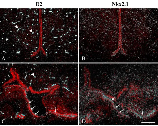
6.2.1 Distribution of D3 protein in the ME of the rat ... 61
6.2.2 D3 distribution at the subcellular level ... 62
6.2.3 Phenotype of D3-immunoreactive hypophysiotropic terminals in the rat ME ... 64
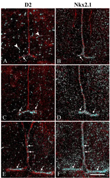
6.2.4 Colocalization of MCT8 and D3 in a subpopulation of hypophysiotropic axons in the ME... 66
6.2.5 Distribution of D3 in axon varicosities of parvocellular neurons in LPS induced non-thyroidal illness syndrome ... 67
6.3 Generation and characterization of Thyroid hormone action indicator mouse model ... 69
6.3.1 Transgenic construc ... 69
6.3.2 The thyroid hormone action indicator HEK-293T cell line ... 70
6.3.3 Homozygote THAI-Mouse harbours two copies of transgene and represents systemic euthyroidism ... 71
6.3.4 The THAI-Mouse model is an “indicator” of TH signalling ... 73
6.3.5 The THAI-Mouse model detects tissue hypothyroidism ... 76
6.3.6 TH signalling can be assessed in the live THAI-Mouse ... 77
6
6.3.7 Assessment of tissue-specific TH action in discrete brain regions:
implications for pathogenesis of NTIS ... 78
6.3.8 THAI-Mouse reveals strong activation of TH signalling in cold-stimulated iBAT ... 79
6.3.9 THAI-Mouse can test the performance of TR isoform-specific TH analogues ... 82
7. Discussion ... 84
7.1 Mechanisms contributing to formation of negative feedback during the development of HPT axis ... 84
7.2 D3-mediated TH inactivation represents a novel mechanism in regulation of local TH levels in hypophysiotropic parvocellular neurons ... 87
7.3 Generation and characterization of thyroid hormone action indicator mouse model ... 93
8. Conclusions ... 98
9. Summary ... 100
10. Összefoglalás... 101
11. References ... 102
12. List of publications ... 130
12.1List of publications the thesis is based on ... 130
12.2Other publications ... 130
13. Acknowledgements ... 132
7 2. Abbreviations
3’ FR - 3’ flanking region 5’ FR – 5’ flanking region
ABC - Avidin-biotin-peroxidase complex AMPK - AMP activated protein kinase ARC - Arcuate nucleus
ATA – American Thyroid Association BAT - Brown adipose tissue
BBB - Blood-brain barrier BSA - Bovine serum albumin BT - Biotinylated tyramide BW – Bodyweight
cAMP – Cyclic adenosine monophosphate cDNA – complementary DNA
ChP – Choroid plexus CMV – Cytomegalovirus CNS - Central nervous system CRE - cAMP-response-element
CREB - cAMP-response-element binding protein CRH - Corticotropin-releasing hormone
D1 - Type 1 deiodinase D2 - Type 2 deiodinase D3 - Type 3 deiodinase DAB - Diaminobenzidine DCV – Dense core vesicle
DMEM - Dulbecco's Modified Eagle's Medium DMN - Dorsomedial nucleus
DTT - DL-dithiothreitol DR – Direct repeats
EDTA - Ethylenediaminetetraacetic acid EEC – Expression enhancer casette EGTA - Ethylene glycol tetraacetic acid
8 ER – Endoplasmic reticulum
FBS –Fetal Bovine Serum FFA – Free fatty acids
FISH – Fluorescence in situ hybridization
GAPDH – Glyceraldehyde-3-phosphate dehydrogenase GHRH - Growth hormone-releasing hormone
GnRH - Gonadotropin-releasing hormone gw – gestational week
HPLC – High pressure liquid chromatography HPT axis - Hypothalamus-pituitary-thyroid axis HT - Hypothalamus
iBAT - Interscapular brown adipose tissue i.p. - Intraperitoneal
IR - Immunoreactive
IRD – Inner ring deiodination KO - Knock out
LAT - L-type amino acid transporter LPS – Bacterial lipopolysaccharide Luc – Firefly Luciferase
MBH – Mediobasal hypothalamus
MCT family – Monocarboxylate transporter family MCT8 - Monocarboxylate transporter type 8 MCT10 - Monocarboxylate transporter type 10 ME - Median eminence
MPOA – Medial preoptic area NE – Norepinephrine
NFκB - Nuclear factor-kappa B NiDAB – Nickel-Diaminobenzidine NHS - Normal horse serum
NST- Non-shivering thermogenesis NTIS – Non-thyroidal illness syndrome
OATP - Organic anion transporting polypeptide
9
OATP1C1 - Organic anion transporting polypeptide 1c1 ORD – Outer ring deiodination
PB – Phosphate buffer
PBS - Phosphate buffered saline PCR – Polymerase chain reaction PFA - Paraformaldehyde
PKA – Protein kinase A POA – Preoptic area
PVN - Paraventricular nucleus PTU – 6-n-propyl-2-thiouracil RLU – Relative light unit ROI – Region of interest RT – Room temperature
RTH – Resistance to thyroid hormone syndrome rT3 - Reverse T3, 3,3’,5’ triiodothyronine RXR - Retinoid X receptor
SCN - Suprachiasmatic nucleus SNS – Sympathetic nervous system SST - Somatostatin
STORM – Stochastic Optical Reconstruction Microscopy T2 - Diiodothyronine
T3 - 3,5,3’ triiodothyronine T4 - Thyroxine
TH, THs - Thyroid hormone, thyroid hormones
THAI-Mouse – Thyroid hormone action indicator mouse THAIC – Thyroid hormone action indicator construct TK promoter – Thymidine kinase promoter
TR, TRs - Thyroid hormone receptor, thyroid hormone receptors TRα - Thyroid hormone receptor alpha
TRα1 - Thyroid hormone receptor alpha type 1 TRα2 - Thyroid hormone receptor alpha type 2 TRβ - Thyroid hormone receptor beta
10 TRβ1 - Thyroid hormone receptor beta type 1 TRβ2 - Thyroid hormone receptor beta type 2 TRE - Thyroid hormone response element TRH - Thyrotropin-releasing hormone TSH - Thyroid stimulating hormone
TSHβ - Thyroid stimulating hormone beta subunit UCP-1 - Uncoupling protein-1
VMH – Ventromedial hypothalamus WT - Wild type
Abbreviations used only once were explained in the text.
11 3. Introduction
3.1 Thyroid hormones
Thyroid hormones (TH) are crucial regulators of many fundamental biological processes e.g. metabolism (Lechan and Fekete, 2006), neuronal development (Bernal et al., 2003; Williams, 2008), reproduction (Dawson, 1993; Nakao et al., 2008), stress (Baumgartner et al., 1998; Araki et al., 2003), and growth (Giustina and Wehrenberg, 1995). It has been well established, that untreated congenital hypothyroidism has devastating consequences on newborns represented by growth retardation and impaired neuronal development manifested in mental retardation, loss of hearing, speech and coordination problems and low muscle tone. Millions of people are suffering from TH deficiencies around the word and require TH supplementation to correct hypothyroidism and get rid of fatigue and mental dysfunctions while in contrast, need medications to lower TH levels and normalize cardiac function and neurological symptoms caused by hyperthyroidism. The high prevalence and high impact of TH-related symptoms on medical conditions and life quality and the essential role played by TH in the proper function of the brain and various organ systems calls for intense investigations addressing the regulation of TH economy.
The human thyroid gland produces mainly thyroxin (T4) and in a lesser extent triiodothyronine (T3) (Larsen et al., 1998). Both molecules are derived from the amino acid tyrosine and contain an inner tyrosyl ring and an outer phenolic ring linker via an ester bond, covalently bound to iodine molecules. To exert it’s biological effect the T4 prohormone needs to be activated to T3 that can bind the thyroid hormone receptor (TR). TH are stable molecules, T4 and T3 half-life in humans are ~ 1 week and 1 day, respectively, while in rats is 11, and 4 hours, respectively. TH are hydrophobic molecules and their vast majority (~99%) is bound to carrier proteins in the blood. The daily TH production is tightly controlled by the hypothalamus-pituitary-thyroid (HPT) axis keeping serum T3 levels in the physiological range via negative feedback regulation at the different levels of the HPT axis. Stability of serum TH levels is important but TH action occurs predominantly at the tissue level and the HPT axis itself is not capable to achieve tissue-specific and tightly regulated TH availability. Thus tissue/cell-type specific regulation of TH levels are achieved by a complex regulatory system that allows to customize TH availability in a cell-type specific manner. This
12
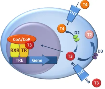
system involves the transport of TH by various TH transporters, the activation and inactivation by deiodinase enzyme family and the regulation of TH action by TH receptors and it’s co-regulators (Figure 1). This tissue-specific system is functionally interlinked with the HPT axis in the hypothalamus.
Figure 1. TH action and its regulation by TH transporters and deiodinases. T3 ligands the TR/RXR(Retinoid X receptor) heterodimer binding to a thyroid hormone response element (TRE) in the regulatory region of TH responsive genes. The ability of TR/RXR receptor to affect gene transcription is modulated by co-activators/co-repressors (CoA/CoR) and factors regulating intracellular TH availability; TH transporters (THT) and deiodinases. As the first step in the regulation of TH action, type 2 deiodinase (D2) activates T4 by converting it to T3. Type 3 deiodinase (D3) catalyzes the inactivating pathway by degrading T3 and converting T4 to the transcriptionally inactive reverse T3.
The expression of the two main deiodinases, D2 and D3, varies according to cell-type.
3.2 Structure and function of the HPT axis
The hypothalamus is the integrator of hormonal and sensory information reflecting the internal and external environment and regulates homeostasis by responding to changing conditions. The hypothalamus represents the center of the endocrine brain and works as the central regulator of neuroendocrine systems. Consequently, it controls growth, metabolism, reproduction, stress response, water balance, and contraction of smooth muscle cells during delivery and nursing (Reichlin, 1967). The hypothalamus is
13
bordered from anterior-posterior direction by the stria terminalis and the region located posterior to the corpus mamillare while the thalamus and the ventral surface of the brain represent its dorsal and ventral borders, respectively (Flament-Durand, 1980). Central regulators of different endocrine axes are located in specific regions of the hypothalamus. Parvocellular neurosecretory neurons, like thyrotropin releasing hormone (TRH) or corticotrophin releasing hormone (CRH) expressing neurons are located in the paraventricular nucleus (PVN)(Lennard et al., 1993; Merchenthaler and Liposits, 1994; Fekete et al., 2000) while gonadotropin releasing hormone (GnRH) expressing neurons are in the medial preoptic area (MPOA) (Merchenthaler et al., 1980;
Rance et al., 1994) and GnRH and growth hormone releasing hormone (GHRH) neurons are restricted to arcuate nucleus (ARC) (Sawchenko et al., 1985). Despite the distinct location of the neuronal cell bodies, axons of all hypophysiotropic neurons expand to external zone of median eminence (ME) where they terminate in close proximity of fenestrated portal capillaries of the anterior pituitary (Wiegand and Price, 1980; Merchenthaler et al., 1984) (Figure 2). Their peptide hormones are secreted to the ME and travels via the fenestrated capillaries in blood to target cells of the anterior pituitary to evoke the release of pituitary hormones to the circulation (Sawyer, 1978).
Magnocellular neurons like angiotensin-vasopressin, and oxytocin neurons are located in PVN and supraoptic nucleus and send their axons directly to the posterior pituitary via the ME and release neuropeptides in peripheral blood (Vandesande and Dierickx, 1975).
The ME belongs to the circumventricular organs by sharing a unique feature of these brain regions to be located outside of the blood brain barrier (BBB) (Ganong, 2000).
This way numerous blood derived factors can gain access to the dense network of axon terminals of the ME. Therefore this region allows hypophysiotropic TRH neurons to get into direct contact with serum TH levels. TRH expressing neurons can be also found outside the hypothalamus (e.g. in the thalamus, brain stem or the spinal cord) but hypophysiotropic TRH neurons, the central regulators of HPT axis (Fekete and Lechan, 2014), are confined to the medial parvocellular and periventricular subdivision of PVN in rat (Merchenthaler and Liposits, 1994; Fekete et al., 2000). TRH axon terminals expand to the external zone of the ME in the mediobasal hypothalamus (MBH) where they secrete the TRH peptide into fenestrated, portal capillaries that reaches the
14
thyrotropes and evokes thyroid stimulating hormone (TSH) release into peripheral blood to allow TSH-induced stimulation of follicular cells of the thyroid gland. TH regulate their own production by negative feedback both at the level of the pituitary and the hypothalamus (Segerson et al., 1987; Dahl et al., 1994). The reference level of serum TH-s in blood are set by set point formation during development and as a consequence the axis is programmed to keep circulation T3 levels in the desired range (Figure 2).
Figure 2. The hypothalamo-pituitary-thyroid axis regulation and localization of hypophysiotropic parvocellular neurons in the hypothalamus. Hypophysiotropic parvocellular neurons are restricted to distinct regions of hypothalamus while all neurons send their axons to the external zone (EZ) of median eminence (ME), where neuropeptides are secreted from axon terminals into fenestrated portal capillaries (FC).
Neuropeptides reach the anterior pituitary and evoke the release of the appropriate hormones into the peripheral blood. TRH cells, the main regulators of HPT axis are located in paraventricular nucleus (PVN) and evoke TSH release from pituitary thyrotropes followed by the induction of TH secretion from the thyroid gland. TH regulate their own production by negative feedback both at the level of the hypothalamus and the pituitary. Tanycytes (Tan) are specialized glial cells lining the
15
floor and wall of the third ventricle (III.) This cell-type forms the exclusive type 2 deiodinase (D2) expressing cellular compartment in the hypothalamus and represents the main source of T3 in this region. IZ: internal zone; MPOA: medial preoptic area;
ARC: arcuate nucleus.
The pituitary responds within minutes to altered plasma TH and regulate TSH secretion.
Minor increase in TH levels inhibits TRH action on thyrotropes and decreases TSH secretion (Snyder and Utiger, 1972) and in contrary, a modest decrease in TH levels is sufficient to sensitize the anterior pituitary to TRH (Vagenakis et al.). Both T3 and T4 can directly inhibit TSH synthesis (Weiss et al., 1997). TH feedback on TRH neurons works via nuclear TR-mediated transcriptional actions that makes this pathway more time consuming compared to non-transcriptional events. Negative feedback control on TRH neurons is mediated by nuclear thyroid hormone receptor beta 2 (TRβ2) (Abel et al., 2001) and results in decreased proTRH mRNA levels. In hypothyroid mice TH supplementation requires ~5 hours to normalize TRH expression in PVN (Sugrue et al., 2010). In the first step TH-s have to gain access to brain across BBB, and needs to be transported via specific TH transporters. T4 is the TH form that preferentially transported into the brain thus local activation to T3 is especially important, to exert TH action at specific brain regions. In the brain, D2 is the dominant activating enzyme which is mostly expressed in glial compartment and which serves as a T3 source for neurons (Crantz et al., 1982; Guadano-Ferraz et al., 1997). It is well known that D2- mediated T3 conversion is essential in feedback regulation of hypophysiotropic TRH neurons (Gereben et al., 2008; Fonseca et al., 2013). In contrast to D2, D3 is involved in the inactivation of T3 in the neuronal compartment (Tu et al., 1999; Verhoelst et al., 2002). Therefore, in addition to the peripheral TH levels, the hypothalamic activity of deiodinase enzymes determines the local level of T3 that regulates the hypophysiotropic TRH neurons in the hypothalamus. In the hypothalamus, the main T3 generating, D2 expressing cells are tanycytes, the specialized glial cells, lining the floor and wall of the third ventricle (Tu et al., 1997; Guadano-Ferraz et al., 1999; Gereben et al., 2004).
While the cell bodies of TRH neurons are located in some distance to tanycytes (Wiegand and Price, 1980), the axon terminals and tanycyte processes are in close proximity in the ME, representing a locus where tanycyte derived T3 can act on TRH
16
neurons (Figure 2). This is supported by studies in astrocyte-specific D2 knock out (KO) mice demonstrating that preserved tanycytal D2 activity is sufficient to maintain normal T4 dependent negative feedback and unchanged TH economy (Fonseca et al., 2013).
3.3 Development of hypothalamic TH-mediated negative feedback
The timing and mechanism of hypothalamic set point formation is poorly understood.
Studies in human fetuses and infants are limited by ethical considerations while rodents provide a poor model for these studies due to the different developmental kinetics of the HPT axis. Pituitary development starts early in humans, at gestational week (gw) 4 with the formation of Rathke pouch from the oral ectoderm, which will form later the anterior pituitary. In mouse this event occurs at embryonic day (E)9 during midgestation and results in fully differentiated thyrothrop cells only few days before birth at E17 (Japon et al., 1994). In humans, the anterior pituitary is fully differentiated at gw16, however biologically active TSH is released after gw17 (Pope et al., 2006). The neural lobe, and the infundibulum develops from neural tube ectoderm. The rodent hypothalamus is formed before E16 and the differentiation of nuclei occurs between E19 and day 10 of postnatal life (Szentágothai, 1968; Hyyppä, 1969). Compared to the slow hypothalamic development of rodents, human newborns have a well-developed hypothalamic axis at birth. Hypothalamic nuclei start to differentiate and compartmentalize during gw15-17, the ME is also evolved at gw18 and all nuclei, including the parvocellular subdivision of PVN, reach adult-like structure at gw24-33 (Koutcherov et al., 2002). In the last trimester of gestation all anatomical structures are present for the matured and functional regulation of the HPT axis in the human fetus.
In rodents, serum T4 levels start to increase exponentially at E18-22 several days prior to birth, which phenomenon occurs in human fetus much earlier, at the beginning of the third trimester. TRH is expressed in the rat hypothalamic PVN from E16 (Burgunder and Taylor, 1989), but TH inhibits the hypophysiotropic TRH expression only from day 7 after birth (P7) in this species (Taylor et al., 1990). In humans the HPT axis is largely maturated at gw28-34. Fetal pituitary is already sensitive to TRH in gw25 and responds with elevated TSH when mother is injected with TRH (ROTI et al., 1983). In preterm (gw28) hypo/hyperthyroid infant, the pituitary can respond with elevated or decreased serum TSH (Fisher and Klein, 1981). However, in E18 rat thyroid function is largely
17
dependent on pituitary TSH secretion and negative feedback on thyrotropes but is not affected by the HT (Jost et al., 1974). It can be concluded that the kinetics of HPT axis development is strikingly different in rodents and humans that make rodent a poor models for these studies.
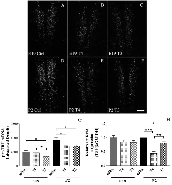
However, in contrast to the relatively low developmental state reached by rats and mice at the time of prenatal-postnatal transition, chickens have a well-developed HPT axis at hatching (Debonne et al., 2008). Similarly to mammals, a time gap between the onset of HPT function and TH-mediated negative feedback can be also observed during the ontogeny of the chicken embryo. This is supported by the observation that the chicken anterior pituitary starts promoting thyroidal secretion already at E10 (Thommes et al., 1984) followed by the increase of T4 and T3 levels in the serum and brain tissue (Prati et al., 1992; Grommen et al., 2008). However, despite the increasing TH levels, the thyroid stimulating hormone beta subunit (TSHβ) mRNA level increases until E19 (Gregory et al., 1998) indicating the lack of negative feedback before this developmental stage. Hypothalamic TRH peptide level also increases continuously during chicken development although no data are available specifically on the level of TRH in the hypophysiotropic neurons (Geris et al., 1998). Despite the non-mammalian nature of the chickens models the mentioned arguments make chicken a useful model for studies aimed to better understand the development of the human HPT axis(De Groef et al., 2008).
3.4 Regulation of tissue thyroid hormone levels
While serum TH levels are generally kept stable, tissue-specific TH availability can be largely independent from serum levels due to the function of a complex regulatory mechanism which is represented in many tissues and is characteristic for a given tissue/cell-type. Members of this machinery are the TH transporters, the TH activating/inactivating deiodinase enzymes and TH receptors along with their co- regulators.
18
3.4.1 Cellular thyroid hormone uptake is mediated by thyroid hormone transporters
TH need to enter the cell to exert their biological effects. It was earlier speculated that TH as an apolar molecule penetrates the cells by diffusion (Hillier, 1970) and forms a gradient from the plasma membrane to the nucleus. However, it has become clear that TH uptake is mediated by active transport through different TH transporters.
Monocarboxylate transporter 8 (MCT8) is a member of the MCT family and is the best characterized TH transporter. The MCT8 gene is located on the X chromosome the protein involves 12 transmembrane domains and forms homodimers in the lipid layer of the plasma membrane. It is expressed in different species in various tissues like liver, heart, thyroid, brown adipose tissue and brain (Visser, 2007). MCT8 is the predominant neuronal T3 transporter (Dumitrescu et al., 2004; Friesema et al., 2004) but it can also transport T4 (Friesema et al., 2003). MCT8 is widely expressed in the brain, including choroid plexus (Chp), amygdala, hippocampus, hypothalamus, different cortical areas and in the microvessels of anterior pituitary (Müller and Heuer, 2014). Mutations and the resulting impaired function of MCT8 is manifested in severe phsychomotoric retardation, delayed myelinilation, axial hypotonia, muscle hypoplasia and as a consequence poor head control and almost completely absent of speech in infants (Schwartz and Stevenson, 2007). TH homeostasis is highly affected, and patients have markedly low T4 and high T3 levels in serum, but normal TSH. This deficiency was identified as the genetic background of the previously described Allan-Herndon-Dudley syndrome (AHDS) (Schwartz et al., 2005).The MCT8 KO mice have the same endocrine phenotype, however they lack neuronal abnormalities (Trajkovic et al., 2007).
MCT10 is another member of MCT family, and has a 49% identity to MCT8. It also contains 12 transmembrane domains, and transports T3 preferably according to in vitro experiments in COS-1 cells. (Friesema et al., 2008) MCT10 is ubiquitously expressed but available information on physiological function is limited (Ramadan et al., 2006).
Transporters of organic anion transporting polypeptide (OATP) family have affinity for a broad range of ligands, but one member, the organic anion transporting polypeptide 1c1 (OATP1c1) prefers mostly iodothyronines (T4, T3, reverse T3 (rT3)) making this protein a potent TH transporter. Its affinity for T3 is 6-fold lower than that for T4.
OATP1c1 has been reported to be expressed at the BBB, mostly in endothelial cells of
19
rodent brain capillaries, astrocytes (Schnell et al., 2015), and also at brain-cerebrospinal fluid barrier, in ChP, and Leydig cells of the testicle (Sugiyama et al., 2003; Tohyama et al., 2004).
In the MBH glial derived tanycytes also express both MCT8 (Alkemade et al., 2005) and OATP1c1 (Roberts et al., 2008) representing a focal TH metabolizing locus, where T4 can be accumulated, converted to T3 by D2, and released to the surrounding region.
Lack of neurological phenotype of MCT8 KO mice predicted that beyond MCT8 other TH transporter(s) also contribute significantly to the TH homeostasis of the mouse brain. Indeed, it has been demonstrated that the deletion of both MCT8 and OATP1c1 are required to severely impact TH homeostasis in the mouse brain (Mayerl et al., 2014).
L-type amino acid transporters (LAT-s) were also identified as TH transporters. Due to wide ligand binding affinity LAT1 and LAT2 are transporting large neutral amino acids like tryptophane, and leucine , and facilitate TH transport in mouse and human (Ritchie et al., 1999; Friesema et al., 2001; Zevenbergen et al., 2015). In vitro experiments on Xenopus laevis oocytes revealed, that large excess of amino acids or high dose iodothyronines inhibited LAT-1 transporter-mediated uptake conversely in a concentration dependent manner (Friesema et al., 2001).
3.4.2 Regulation of thyroid hormone metabolism by the deiodinase enzyme family
TH metabolism is regulated by members of the selenodeiodinase enzyme family.
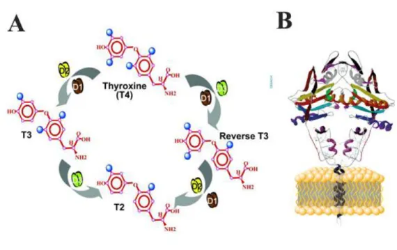
Biochemically the activation of T4 can be catalyzed both by type 1 and type 2 deiodinase enzymes by removing one iodine from the 5’ phenolic ring of the T4 molecule. While D2 removes iodine exclusively from the outer ring (outer ring deiodination (ORD)), D1 can also perform inner ring deiodination (IRD) that converts T4 to rT3. Importantly, under in vivo euthyroid conditions TH activation is catalyzed almost exclusively by D2 since due to the low substrate affinity D1 cannot generate T3 only in hyperthyroidism when T3 is elevated (Maia et al., 2005). Furthermore, both T4 and T3 can be inactivated by D3 by IRD (Figure 3A) (Bianco et al., 2002).
Only 20% of human daily T3 production is related to the intrathyroidal deiodination while the rest is produced in extrathyroidal tissues by D2-mediated T3 generation.
20
Compared to humans, the contribution of the rodent thyroid gland to T3 production is higher, since in rodents ~40% of daily T3 is born in the gland (Bianco et al., 2002;
Bianco et al., 2014). Still, even in these species D2 mediated T3 generation is highly significant. Thus D2-mediated TH activation is a critical process to meet the T3 demand of the body.
Figure 3. Schematic representation of deiodinase-mediated TH metabolism and deiodinase protein structure (A) Type 2 deiodinase (D2) and type 1 deiodinase (D1) can activate T4 to T3 by removing one iodine from outer ring, while D1 can also deiodinate the inner ring and convert T4 to reverse T3 (rT3). Type 3 deiodinase (D3) inactivates TH-s and produces rT3 or diiodothyronine (T2). (B) Deiodinases are one transmembrane domain containing proteins that are anchored to a membrane bilayer and need to form homodimers to achieve catalytic activity.
A (Gereben et al., 2008); B http://deiodinase.org/wp- content/uploads/2015/10/Deiodinase-Structure1.jpg
3.4.2.1 Structure and subcellular localization of deiodinase enzymes Iodothyronine converting enzymes are responsible for regulation of active TH levels in the serum and locally in tissues. Deiodinases are selenoproteins and contain
21
selenocysteine, a rare amino acid, in their active center. Selenocysteine is encoded by an in-frame UGA that normally serves as a stop codon. However, in selenoproteins UGA is subjected to read through and dictates the incorporation of the rare amino acid selenocysteine. The underlying complex co-translational machinery consists of a cis- acting mRNA stem-loop structure located in the 3’ untranslated region of the selenoprotein encoding mRNA called as the selenocysteine insertion sequence (SECIS) along with several trans-acting factors e.g. the the SECIS binding protein-2 (SBP-2) and a specific elongation factor, EFsec. Insertion of selenocysteine is a low efficiency process that results in a low translation rate of selenoproteins that also limits the production of deiodinases. However, importantly selenocysteine increases substrate affinity of D2 to T4 with three orders of magnitude compared to cysteine along with its effect to decrease translational activity by several hundred fold (Steinsapir et al., 2000).
When the selenocysteine residue of D1 was
mutated to cysteine, an ~ 400-fold higher amount of D1 could be produced (Berry et al., 1992) but this mutated protein had decreased deiodination capacity (Berry et al., 1991; Bianco et al., 2002). Thus the presence of selenocysteine in selenoproteins allows that a low amount of deiodinase enzyme can perform the oxido-reductive deiodination in a highly effective manner. This scenario clearly represent a massive biological advantage due to the highly controllable nature allowed by the low amount of deiodinase enzymes.
D2 is targeted to the endoplasmic reticulum (ER) (Maciel et al., 1979; Baqui et al., 2000), while D1 and D3 are located in the plasma membrane (Baqui et al., 2000; Baqui et al., 2003). Due to this specific location deiodinases have a distinct role in regulation of TH availability at the cellular level. ER resident D2 produces T3 in the cytoplasm in close vicinity of the cell nucleus making D2 the main source of nuclear T3, while D1- mediated conversion on the cytoplasm membrane rapidly equilibrates with plasma pool, and has minor contribution to intranuclear T3 availability (Silva and Larsen, 1978).
Deiodinase enzymes contain a single transmembrane domain, and a ~15 amino acid long active center, that is relatively well conserved among the three enzymes.
Deiodinase enzymes form homodimers and this process is a prerequisite of allowing the globular domains to reach the conformation required by the active center for efficient deiodination (Curcio-Morelli et al., 2003a) (Figure 3B). One functional D1 subunit has
22
50% activity of native homodimer(Leonard et al., 2001), while D2 lost 90% of its activity if no homodimers are formed (Leonard et al., 2005). A highly conserved amino acid sequence in the C-terminal portion of the globular domains is responsible for homodimer binding, and mutation or deletion of this domain results in decreased enzyme activity (Leonard et al., 2005; Simpson et al., 2006). Fluorescence resonance energy transfer (FRET) studies in HEK-293 cells transiently overexpressing truncated D2 lacking transmembrane domain, revealed that this domain plays also an important role in homodimerization (Sagar et al., 2007). Both domains contribute to formation of dimers, however it has been shown, that binding site in the globular domain is sufficient to form catalytically active enzyme if the dimerizing partner has an intact transmembrane domain (Leonard et al., 2001; Sagar et al., 2007). Heterodimerization was also detected among deiodinases in a lesser extent, but biological significance of this phenomenon needs to be elucidated (Sagar et al., 2008).
3.4.2.2 Tissue distribution of deiodinases
In humans D1 enzyme is expressed in liver, kidney, pituitary and thyroid but it is absent from the central nervous system (CNS)(St Germain and Galton, 1997; Kester et al., 2004). In the rat D1 is also expressed in CNS, placenta, and intestine (Bates et al., 1999). Due to its highly T3 sensitive promoter, D1 can rapidly react to elevating serum T3 levels, while the contribution of D1 to extra thyroidal T3 production is not dominant in euthyroid humans (LoPresti et al., 1989).
D2 is widely expressed in the human CNS, pituitary, thyroid, heart, skeletal muscle, placenta, however in rats D2 is expressed also in brown adipose tissue (BAT), but is absent form heart, skeletal muscle and thyroid (Croteau et al., 1996). D2 is expressed in embryonic and newborn mouse liver with a peak expression at P1 that is followed by a rapid decline during development (Fonseca et al., 2015). While D2 is absent from the adult rat and human liver, it is expressed in this organ of chickens (Gereben et al., 1999). Both in the rodent and chicken hypothalamus D2 is predominantly expressed in tanycytes (Gereben et al., 2004).
D3 is expressed in human CNS (Kallo et al., 2012), fetal liver (Richard et al., 1998), and it is the predominant deiodinase in placenta (Huang et al., 2003). This protects the fetus from high maternal TH levels. More is known about D3 distribution in rat and mouse tissues. D3 is expressed widely in adult rat CNS (Tu et al., 1999), skin (Huang et al.,
23
1985) and placenta (Galton et al., 1999), and D3 has been found in skeletal muscle, intestine and liver of newborn rats (Bates et al., 1999).
3.4.2.3 Enzymatic properties
Selenocysteine is essential in catalysis of deiodination in all three enzymes (see 3.4.2.1
“Structure and subcellular localization of deiodinase enzymes”). Deiodinases have distinct enzymatic properties and kinetics. D1 was identified as the only deiodinase sensitive to 6-n-propyl-2-thiouracil (PTU) (Oppenheimer et al., 1972), an anti-thyroid drug used in hyperthyroid patients to inhibit TH synthesis in thyroid. D1 can catalyze both IRD and ORD, compared to exclusive ORD capacity of D2 an IRD deiodination of D3 (Fekkes et al., 1982) (Figure 3.) D1 enzyme activity follows ping-pong kinetics, between two substrates: iodothyronine and endogenous thiol cofactor (Visser et al., 1976).
According to in vitro measurements D2 has three orders of magnitude lower Michaelis- Menten constant (KM) for T4, than D1. In addition, compared to T4, rT3 is just a slightly worse substrate of D2. D2 catalyzes deiodination with sequential kinetics, which means that binding of both the iodothyronine and the thiol cofactor are required for catalysis (Visser et al., 1982). In contrast to D1, D2 is insensitive to PTU. Not only D2, but D1 and D3 can be inhibited by iopanoic acid (Wu et al., 2005), and amiodarone (Rosene et al., 2010).
D3 is an obligate inner ring deiodinase and catalyzes the T3 to rT3 and T3 to T2 conversion, producing transcriptionally inactive hormones. It plays a crucial role in prevention of tissues from toxic levels of T3. D3 is insensitive to PTU, and follows sequential kinetics like D2 (Kaplan et al., 1983).
3.4.2.4 Complex regulation of D2-mediated TH activation
D2 activity is tightly regulated by different transcriptional and post-translational mechanism, to fine tune T3 availability rapidly and effectively by altered activation capacity. Human and rat Dio2 gene has been shown to be positively regulated in promoter studies by nuclear factor-kappa B (NfκB), a second messenger molecule, involved in bacterial lipopolysaccharide (LPS) and cytokine signalling (Fekete et al., 2004). Computer analysis and promoter studies in cell culture revealed that the Dio2 gene promoter contains functional cyclic-AMP-response elements (CRE) which make
24
this gene cyclic adenosine monophosphate (cAMP) responsive in human, rat and mouse tissues (Bartha et al., 2000; Canettieri et al., 2000; Song et al., 2000). Norepinephrine induced cAMP signalling trans-activates D2 transcription, that is essential in non- shivering thermogenesis in cold stressed interscapular BAT (iBAT). Nkx2.1, a homeodomain containing transcription factor has regulatory effect on the expression of D2 in the human thyroid while the DIO2 promoter contains two functional Nkx2.1 binding sites none of them is presented in the case of rat, resulting in lack of induction in promoter studies (Gereben et al., 2001).
The T3-mediated transcriptional suppression of D2 is well known, but the mechanism of the action remains to be clarified. A negative thyroid hormone response element (TRE) is predicted in the promoter but it has not been identified (Kim et al., 1998). At the post-translational level, D2 activity is regulated by its substrates T4 and rT3. D2 protein is degraded rapidly (activity half-life decreases under 1h) by ubiquitination- mediated selective proteolysis in the presence of T4 (Gereben et al., 2000). Importantly, the von Hippel-Lindau protein-interacting deubiquitinating enzyme-1 (VDU-1 also called as USP33) serves as deubiquitinase of D2 (Curcio-Morelli et al., 2003b) and makes the process reversible by removing ubiquitin.
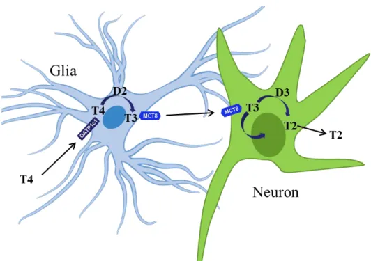
3.4.3 Compartmentalization of TH activation and inactivation in the CNS TH activation and inactivation is highly compartmentalized in the CNS. Astrocytes and tanycytes of the neuroglial compartment are the predominant source of T3 present in the brain while TR in neurons represents a major target of TH (Mohácsik et al., 2011).
Astrocytes, distributed throughout the brain, and tanycytes, lining the floor and the wall of the third ventricle, take up T4 and convert it to T3 via D2. Neurons cannot generate T3 but they can take it up from glial cells with MCT8 transporter, and they can further regulate intracellular TH levels by inactivating deiodinase, D3 (Figure 4). Direct evidence of neuroglia derived T3 - neuron interaction could be obtained in a two- dimensional H4-SK-N-AS glioma-neuron cell system where physiological amount of T4 in the D2 expressing glial compartment modulated TH-sensitive gene expression in the neuronal cell layer (Freitas et al., 2010).
Glial cells are not only source of T3 but also represent a target of this hormone (Mohácsik et al., 2011). TH affects the differentiation and maturation of different glial
25
subtypes including astrocytes, oligodendrocytes, and microglia (Almazan et al., 1985;
Lima et al., 2001; Trentin, 2006). Impaired maturation of oligodendrocytes in hypothyroidism results in decreased number of, myelin producing oligodendrocyte cell bodies in the main white matter tracts (Schoonover et al., 2004) and along with delayed expression of myelin encoding genes, the myelination of axons are insufficient (Berbel et al., 1994; Ibarrola and Rodríguez-Peńa, 1997). Astrocytes secrete epidermal growth factor (EGF), basic fibroblast growth factor (bFGF), and nerve growth factor (NGF), in a T3-dependent manner regulating extracellular matrix formation(Trentin et al., 2003), neuronal migration (Martinez and Gomes, 2002), and neurite growth (Lindsay, 1979;
Alvarez-Dolado et al., 1994; Hashimoto et al., 1994).
D3 protects neurons from T3 excess. The absence of this enzyme results in uncontrolled TH action that can trigger serious damage in TH sensitive tissues, especially in developing animal. Studies in (Hernandez et al., 2006) mice revealed, that D3 is essential in formation of negative feedback in the HPT axis. General D3 KO mice suffer from thyrotoxicosis during embryonic and neonatal life, at the time period when HPT axis set points are set, and this results in the development of central hypothyroidism in adulthood. From age P15 peripheral tissues are also hypothyroid, animals are smaller than wild type mice, and suffer from low fertility (Hernandez et al., 2006).
26
Figure 4. Compartmentalization of TH activation and inactivation in the CNS. Glial cells are the main source of activated TH in the CNS by taking up T4 by organic anion transporting polypeptide 1c1 (OATP1c1) transporter and converting it to T3 via type 2 deiodinase (D2). Neurons depend on glia released T3 that gets into neurons via monocarboxylate transporter 8 (MCT8). T3 impacts neuronal gene expression and metabolism and can be inactivated by type 3 deiodinase (D3). (Depiction of the glial
cell was obtained from
https://commons.wikimedia.org/wiki/File:Diagram_of_an_astrocyte_- _a_type_of_glial_cell_CRUK_029.svg was subjected to modifications.)
3.4.4 TR-s regulate transcription of TH targeted genes
TR represents a prerequisite of TH action. TR is a ligand-induced transcription factor and belongs to the nuclear receptor superfamily.TRα and TRβ are encoded by two separate genes. TRα has 4 isoforms that differ in the length and amino acid sequence of their C-terminals. Only the primary transcript, TRα1 is capable to bind T3, and initiate transcriptional activation. TRα2 is an alternative splice variant of the TRα1, while two other forms the TRdα1 and TRdα2 are alternative transcripts of TRα gene transcribed from an intronic internal promoter (Chassande et al., 1997). TRβ1 and 2 differ in length and amino acid composition of N terminus and are transcribed from TRβ gene using
27
two different promoters and alternative splicing. The TRβ3 isoform is reported only in rats (Williams, 2000). The ligand binding and DNA binding domain of the four transcriptionally active isoforms is highly conserved and later recognizes the TRE in the promoter region of target genes. All isoforms form heterodimers with retinoid X receptors (RXR). The α/RXR heterodimers are currently hold responsible for TH action (Brent, 2012). TR homodimers also exist but their role remains to be better understood.
TRα1 and β1 are universal receptors and are reported in all studied issue. However, TRα1 is dominant in heart, intestine and brain, while TRβ1 expression is more pronounced in kidney, liver, and skeletal muscle. TRβ2 is essential in negative feedback regulation of HPT axis, and in correct development of retina, and inner ear (Forrest et al., 1996a).
Mutations in TR have several consequences. The Resistance to thyroid hormone (RTH) syndrome is caused by mutations/deletions of TRβ gene (Refetoff et al., 1993). The unfunctional receptor dramatically lowers the effect of T3 on TRβ dominant tissues, resulting in growth and mental retardation, retinal degeneration, impaired hearing, and HPT axis regulation hallmarked by elevated serum free TH and TSH levels (Forrest et al., 1996c; Ferrara et al., 2012). Mutation in TRα gene results in growth retardation, gastrointestinal abnormalities; however the HPT axis is minimally affected. Patients have normal serum TH levels and TSH, but TRα dominant tissues are resistant to TH action (Bochukova et al., 2012) (Moran et al., 2014). There are several TRβ knock-out and knock-in mouse models available to investigate the RTH syndrome showing a similar phenotype found in human patients (Forrest et al., 1996b).
3.4.5 Regulation of tissue-specific TH action
Cellular transport is the first rate limiting step in TH action followed by deiodinase- mediated customization of T3 availability in the cell prior to TR binding, that induces conformational changes allowing co-activators/repressors to associate with the receptor (Figure 1). TH responsive genes can be regulated by TH either in a positive or negative manner. A complex combination of different promoter elements, DNA methylation pattern, co-action of TR isoforms, TR homo/heterodimerization and activity of co- activators and co-repressors can determine whether T3 will upregulate or downregulate transcription of TH responsive target gene. In the promoter of a gene positively
28
regulated by TH, TRE-s should be present that are unique consensus sequences serving as a binding site for heterodimeric TR. The most widely characterized TRE-s consist of two hexamer half sites, called direct repeats (DR), separated by 4 base pairs (DR+4) (Harbers et al., 1996) that can bind both TRα and TRβ. However, other TRE-s are also know, e.g. ones with different orientation and spacing of half-sites, like inverted repeat 0 (IR0), in rat growth hormone promoter (Brent et al., 1989), and everted repeat 6 (ER6), in rat and mouse myelin basic protein promoter (Farsetti et al., 1997). Still in the absence of T3 TR/RXR heterodimers are bound to TRE and recruit repressor molecules like N-CoR, that form a complex with histone deacetylases and change chromatin structure to suppress transcriptional activity (Darimont et al., 1998; Astapova et al., 2008). In case the receptor is liganded by T3, co-repressor complex dissociates and chromatin structure changes in a way that AF-2 activator domain of TR-s get available for nuclear receptor coactivator-1 (SRC-1), and transcription activity increases (Darimont et al., 1998; Alonso et al., 2009). Hepatic D1 mRNA is highly sensitive to T3, and shows a more robust increase to T3, than other well characterized TH sensitive genes in liver like Spot-14 and α-glicerophosphate dehidrogenase (Zavacki et al., 2005).
The human DIO1 gene promoter has been found to be the most responsive to T3 (Toyoda et al., 1995). Promoter of human DIO1 gene contains two functional TRE-s, but canonical TRE-s in rat and mice Dio1 5’ FR has not been identified until now. T3 effect on rat and mouse D1 mRNA is transcriptional and cannot be blocked by cycloheximide indicating no need for intermediate protein synthesis (Maia et al., 1995;
Kim et al., 1998). There are also genes which are negatively regulated by T3. These are proteins e.g. involved in negative feedback regulation of HPT axis, like TRH (Lezoualc'h et al., 1992; Abel et al., 2001), TSHβ (Weiss et al., 1997), and D2 (Kim et al., 1998). TRβ is critical in negative trans-repression of these genes, and DNA binding is essential in negative regulation (Shibusawa et al., 2003), but the mechanism of this action is still under extensive characterization. Negative TREs are heterogenous and poorly understood.
3.5 Regulation of the HPT axis under pathophysiological conditions
Humoral and neuronal inputs to TRH neurons can overwrite normal feedback regulation in specific conditions and challenges like fasting, cold exposure, infection. The non-
29
thyroidal illness syndrome (NTIS) is a striking example of altered regulation of the HPT axis and occurs typically in critically ill patients. It is characterized by falling serum TSH and TH levels that is not followed by the upregulation of the HPT axis (Fekete et al., 2004; Gereben et al., 2008; Boelen et al., 2011). Although severe NTIS is strongly related to high mortality rate, it is still debated whether it should be considered as a metabolic adaptation or calls for therapeutic intervention (De Groot, 2015; Gereben Balázs, 2016).
A frequently used model of NTIS in rodents is the systemic inflammation induced by LPS (bacterial lipopolysaccharide) injection. After LPS administration circulating TH levels are falling but both TRH and TSH are decreased (Fekete et al., 2004; Fekete et al., 2005a) Importantly, in this model D2 is upregulated in hypothalamic tanycytes supposedly with the involvement of NfκB-mediated signalling pathways (Fekete et al., 2004; Zeold et al., 2006a). Previous studies in mice demonstrated, that LPS induced a~50% decrease in TRH expression in the PVN of wild type C57BL/6 mice while it did not occur in D2-KO animals (Freitas et al., 2010) indicating that D2 plays an important role in this process. Unilateral transection of ascending brain stem did not inhibit LPS induced fall in TRH expression, indicating that these inputs are not involved in this phenomenon (Fekete et al., 2005b). Dissection of altered regulation of TH action in NTIS would be required to better understand the nature and potential treatment of this syndrome.
3.6 The role of TH in iBAT-mediated non-shivering thermogenesis
The iBAT plays an instrumental role in energy dissipation via non-shivering thermogenesis (NST) (Lowell and Spiegelman, 2000) both in rodents and in human newborns (Hull, 1977; Cannon and Nedergaard, 2004). Recent imaging techniques also allowed to establish the presence of functional BAT islands in different regions of the body of human adults (Nedergaard et al., 2007; Virtanen et al., 2009), that put this tissue into the frontline of obesity research. The activation of this tissue in cold is crucial for heat generation in cold. The process is regulated by the central and peripheral sympathetic nervous system (SNS). Cold sensitive receptors at the periphery send signal to ventromedial hypothalamus (VMH), through the preoptic area (POA), and activate sympathetic afferent fibers to the BAT (Streckfuß et al., 2005).
30
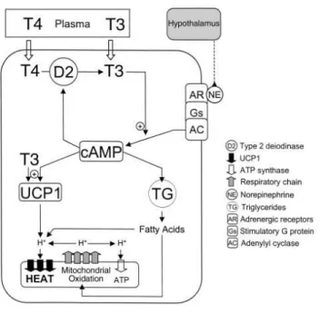
Brown adipocytes have abundant mitochondria supply and express uncoupling protein-1 (UCP-1) or thermogenin. This protein is located in the inner mitochondrial membrane (Aquila et al., 1985) and uncouples the respiratory chain that results in the promotion of heat production instead of making ATP. The mechanism of NST has been intensively studied. Cold induced hypothalamic response facilitates norepinephrine (NE) release from sympathetic nerve endings (Schonbaum et al., 1963) that evokes cAMP response through β3-adrenergic receptors in brown adipocytes (Rubio et al., 1995). cAMP signalling increases UCP-1 and D2 expression rapidly and the increased T3 generation further accelerates cAMP production and UCP-1 transcription (Bianco et al., 1988).
Free fatty acids (FFA) are released from lipid stores due to active lipases phosphorylated by protein kinase A (PKA), to serve as a substrate for the uncoupling (Fedorenko et al., 2012). UCP-1 uncouples the mitochondrial respiratory chain, by dissipating the proton gradient between the two mitochondrial membranes and facilitates FFA oxidation and heat production instead of making ATP (Figure 5).
TH-s have a key role in this process (Lowell and Spiegelman, 2000). Hypothyroid rats cannot maintain normal body temperature and die several hours after acute cold exposure (Bianco and Silva, 1987). T3 is responsible for sufficient lipogenesis (Christoffolete et al., 2004), and adequate thermogenesis (de Jesus et al., 2001) during cold exposure. Local T3 excess saturates TRs rapidly and TRβ1 induces UCP-1 transcription while TRα1 mediates the thermogenic response (Martinez de Mena et al., 2010; Ribeiro et al., 2010). D2 has been shown to be a crucial component of BAT induced thermogenesis (Silva and Larsen, 1983; de Jesus et al., 2001). T3 also affects the central regulation of thermogenic response, and energy consumption. T3 evoked decreased AMP activated protein kinase (AMPK) phosphorylation and modulated lipid metabolism selectively in VMH of rats, resulting in elevated sympathetic activation, increased amount of thermogenic markers in the BAT and lower body weight (Lopez et al., 2010).
31
Figure 5. Cold induced sympathetic activation of iBAT is regulated by the hypothalamus and local mechanisms. Norepinephrine (NE) released from sympathetic nerve terminals in interscapular brown adipose tissue (iBAT) acts on β3-adrenergic receptors and increases uncoupling protein-1 (UCP-1) and type 2 deiodinase (D2) expression via cAMP signalling, furthermore facilitates the release of triglycerides from cell stores. D2-mediated T3 production accelerates cAMP response and UCP-1 expression. UCP-1 uncouples the mitochondrial respiratory chain and produces heat instead of ATP using fatty acids as a substrate.(de Jesus et al., 2001)
32 4. Aims
Our studies were performed to better understand the regulation of tissue-specific TH action. Specifically, we aimed to
1. study how onset of negative feedback regulation of the HPT axis occurs
- determine the period of the onset of TH-mediated negative feedback in the developing chicken hypothalamus
- identify underlying mechanisms governing set point formation of the HPT axis
2. study the regulation of thyroid hormone availability in hypothalamic neuroendocrine axes
- address TH inactivation in parvocellular neurosecretory neurons of rats
- investigate how regulation of TH availability in neurosecretory neurons responds to challenges in a model of non-thyroidal illness syndrome
3. generate a novel transgenic mouse model allowing the detection of tissue-specific thyroid hormone action in live animals and tissue samples
- characterize the Thyroid hormone action indicator mouse model
- study the hypothalamic pathogenesis of the non-thyroid illness syndrome and interscapular BAT regulation using this model
33 5. Materials and methods
5.1 Experimental animals
All animal experiments were conducted in compliance with the European Communities Council Directive (2010/63/EU) and (Decree 86/609/EEC) and approved by the Animal Care and Use Committee of the Institute of Experimental Medicine (Hungarian Academy of Sciences, Budapest). Experiments on chicken embryos and posthatch chickens were also conducted in accordance with legal requirements of Ethical Committee for animal experiments of the KU Leuven (Leuven, Belgium).
5.1.1 Chicken
White Leghorn chicken embryos and posthatch chickens were obtained from Ceva- Phylaxia (Budapest, Hungary) and Wyverkens (Halle, Belgium). The incubation was started at E0. Posthatch chickens were kept in a room with a 14L/10D photoperiod and water and food available ad libitum.
5.1.2 Rat and Mouse
Adult, male Wistar rats (N=20, b.w. 220–250 g, Toxi-Coop Ltd., Budapest) and Thyroid Hormone Action Indicator (THAI) mice (FVB/Ant background; new transgenic mouse line) were kept under standard laboratory conditions with food and water ad libitum.
5.1.3 Human tissue sample
For testing antibody specificity with Western blot, fresh-frozen hypothalamic samples were obtained from the Human Brain Tissue Bank, Semmelweis University from Prof.
Miklós Palkovits.
5.1.4 Generation of transgenic THAI-Mouse by transposon-mediated technology
To generate transgenic founders, the pronucleus of fertilized FVB/Ant (FVB.129P2- Pde6b+ Tyrc-ch/AntJ) (Errijgers et al., 2007) oocytes was injected with a mixture containing the plasmid harbouring the targeting transposon cassette (Figure 20) (1
34
ng/µl) along with in vitro transcribed mRNA encoding the Sleeping Beauty transposase (Mates, 2011) (5 ng/μl). Four females were identified as founders using TaqMan assay on tail DNA with a luciferase probe and crossed with wild/type FVB/Ant (FVB.129P2- Pde6b+ Tyrc-ch/AntJ) male mice followed with inbred coupling of F1 generation.
Indirect evidence obtained with luciferase TaqMan polymerase chain reaction (PCR) on different lines and generations suggest that a single copy of the transgenic cassette was inserted into the genome of all lines. Pronuclear injection, breeding and genotyping was made by Ferenc Erdélyi and Gábor Szabó, Medical Gene Technology Unit, Institute of Experimental Medicine, Budapest, Hungary.
5.1.4.1 Determination of copy number of transgenic THAI-Mouse lines
Copy number of the transgenic cassette was determined with fluorescence in situ hybridization (FISH). FISH was carried out on lymphocytes, isolated from spleen under aseptic conditions. Chromosomal preparations were performed according to standard techniques using 0.067 M KCl followed by fixation in methanol/acetic acid. We labelled an 1045bp fragment of the dCpG Luciferase coding sequence (nt 599-1644) with digoxigenin using Dig-Nick translation mix (Roche, Germany) and precipitated with mouse COT-1 DNA (Invitrogen) according to the manufacturer’s instructions.
Precipitated probe was dissolved in hybridization buffer. FISH was performed on methanol/acetic acid-fixed cells suspensions. Slides/DNA preparation and hybridization were carried out according to standard techniques. Hybridized sections were incubated overnight at room temperature with anti-digoxigenin-POD (1:100) antibody (Roche, Germany) in 1% BSA solution. Signal was biotin:tyramide (1:1000) amplified for 10 min in 0.05M Tris-HCl buffer pH 7.6 containing 0.003% H2O2). The deposited reaction product was detected with Fluorescein-(DTAF)-streptavidin antibody (Jackson Immunoresearch, USA). The analysis was done in at least 100 interphase nuclei and in a few metaphases if present on the hybridization area. FISH was performed by Irén Haltrich and Zsuzsa Tóth; 2nd Department of Pediatrics, Faculty of Medicine, Semmelweis University, Budapest, Hungary
35
5.1.4.2 Insertion site mapping in #4 THAI-Mouse with Splinkerette PCR
Splinkerette PCR was performed, as described (Potter and Luo, 2010). In brief, genomic DNA was digested with BstYI restriction enzyme. A universal splinkerette oligonucleotide was used to amplify the region between the 5’ end of the cassette and the first BstYI site. The cassette-specific oligonucleotide was specific for the multiple cloning site of the PT/2BH vector located 3’ to the 5’ inverted terminal repeat (ITR) sequence. Nested PCR was performed to reamplify the PCR product followed by sequencing.
5.2 Recombinant DNA technology
The recombinant DNA constructs, e.g. the thyroid hormone action indicator construct (THAIC), chicken deiodinase promoter constructs and vectors containing inserts to generate RNA probes for in situ hybridization were prepared with standard techniques.
DNA fragments were amplified with Taq or Vent polymerase driven PCR and cloned to plasmid vectors by ligating the overhanging restriction sites cleaved by various restriction enzymes with T4 ligase. The newly prepared recombinant DNA was transformed to bacteria, and the correct clone was amplified in bacterial culture in the presence of antibiotics (Gereben et al., 1999; Zeold et al., 2006a).
5.3 Animal treatments and sample collection
5.3.1 TH administration to chicken embryos and posthatch chickens, to study the formation of negative feedback on TRH neurons.
Animals were injected intravenously with either 1µg T4, 200 ng T3 or vehicle (saline with 5mM NaOH) as control 8h before sampling at stage E19 and P2. The applied 5:1 ratio of injected T4 and T3 mirrored the average ratio of T4:T3 present in the yolk (Prati et al., 1992; Van Herck et al., 2013). Injections were performed in embryos into a chorioallantoic blood vessel just beneath the egg shell or into the leg vein in hatched animals. Animals were euthanized by decapitation. Brains were removed quickly from the skull, immediately frozen in isopentane at -35 °C, covered with powdered dry ice and stored at -80 ºC until further use to in situ hybridization.
36
Pituitaries were removed at the same time and snap frozen on dry ice, and subsequently used for Taqman qPCR. Injection was performed by Pieter Vancamp and Veerle M.
Darras, Department of Biology, Division of Animal Physiology and Neurobiology, KU Leuven, Belgium.
5.3.2 T3 administration to adult male rats, and ME microdissection for in vitro D3 activity measurement
Adult male Wistar rats were injected i.p. with 50 µg/of T3/100 g body weight (N=9) or vehicle (N=9) every second days in 8 days. After decapitation the ME was dissected under a Zeiss Semi DV4 stereomicroscope (Carl Zeiss GMBH, Hamburg, Germany) and immediately frozen on dry ice. Three ME samples were pooled from nine, while five cortex samples were collected from five animals.
5.3.3 Modulation of serum TH level in THAI-Mouse to study TH responsiveness of different tissues
The F2 generation was screened for tissue luciferase activity. To characterize basal and TH-induced luciferase activity of peripheral tissues and brain regions, 60-70 day old males were injected i.p. with 5µg/day/mouse of L-T4 or vehicle control for 3 days or 1 μg/g bw of L-T3 for one day. Hypothyroid mice were generated by adding 0.1% sodium perchlorate and 0.5% methimazole in drinking water for 3 weeks combined with low iodine diet. After decapitation tissues were removed, snap frozen on dry ice and stored at -80°C for luciferase activity measurement.
5.3.4 LPS treatment and microdissection of hypothalamic subdivisions of THAI-Mice
70 day-old male THAI line #23 mice were treated i.p. with LPS (150 µg/animal; E. coli 0127:B8, Sigma) and decapitated 6, 8 and 10h after injection. Brains were removed from skull, snap frozen in -25°C isopentane, and 1 mm coronal sections were prepared with blades in a pre-cooled mouse brain cast. Brain areas (ARC + ME and PVN) were microdissected with punch needle from frozen sections placed on pre-cooled glass
37
slides (Palkovits, 1986). Punch samples from two animals were pooled, and subsequently were assayed with real-time quantitative PCR after RNA isolation.
5.3.5 LPS treatment on Wistar rats and sample preparation for double- labelling immunohistochemistry for D3 and parvocellular releasing hormones
200g male Wistar rats were treated i.p. with LPS (250 µg/ 100g bw; E. coli 0127:B8, Sigma) for 12 hours, and brains were transcardially fixed with 4% paraformaldehyde (PFA) (for detection of GnRH, CRH, GHRH and somatostatin) or 4% acrolein/2% PFA (for detection of TRH).
5.3.6 Sympathetic denervation of iBAT and cold stress in THAI-Mice
iBAT was unilaterally denervated with surgical transection of its sympathetic nerves (Pulinilkunnil et al., 2011). Cold stress was applied three days after surgery. During cold stress, single housed animals were kept under standard light conditions at 4°C;
water and food were available ad libitum. Control animals were kept at room temperature (22°C) under same conditions. After decapitation, samples from the iBAT were collected on dry ice and stored at -80°C for NE measurement, deiodination assay (see 5.8.3) and Taqman analysis.
5.3.7 GC24-treatment: Testing selectivity of GC24, a TRβ-specific compound in THAI-Mice
70-day old, male THAI #4 mice were made hypothyroid by adding 0.1% sodium perchlorate and 0.5% methimazole to the drinking water and feeding animals with iodine-deficient diet (Research Diets Inc., New Brunswik, NJ) for three weeks to inhibit endogenous TH production. Hypothyroid mice were treated with a single, i.p. injection of 1.53 nM/g bw GC24 (a kind gift of Dr. Tom Scanlan), (equimolar to 1 μg/g BW T3), or dimethyl sulfoxide as vehicle 24 h before tissue sampling. After decapitation, liver (TRβ-dominant tissue) and heart (TRα-dominant tissue) were removed and snap frozen on dry ice.