INSECT PHYSIOLOGY
Module of Applied Entomology
• Basic insect morphology
• Integument of insects
• Basic neuronal functions
• Nervous system of insects
• Sensory organs of insects
• Endocrine system of insects
• Feeding and alimentary system of insects
• Excretory system of insects
• Circulatory system of insects
• Respiratory system of insects
• Muscles and locomotion of insects
• Reproduction of insects
• Embryonic development of insects
• Post-embryonic development of insects
• Communication of insects
Main topics
Basic insect morphology
3
• The insect body is divided into 3 main sections
• Head
• a rigid capsule formed from several
sclerotized plates
• Thorax
• Abdomen
• built up of a series of concave upper and convex lower
integumental plates
Dorsolateral view of an orthopteran insect
• Head
• 1 pair of compaund eyes and sometimes dorsally located eyes (ocelli)
• Compound eyes are placed dorsolaterally
• Eyes may be either
• dichoptic – eyes are separated
• holoptic – eyes are fused into each other
Basic insect morphology
Main parts of an insects head from different views
• Two different types of head structures
• Prognathous type (A)
• typical of pradatory insects
• Opistognathous type (B)
• typical of herbivorous insects
Basic insect morphology
• Structure of an antenna
– antennal sulcus – scape
– pedicel – flagellum
Basic insect morphology
Basic insect morphology
• Main types of antennae
– setaceous – filiform
– monifiform – pectinate – serrate – clavate – capitate – geniculate – lamellate – plumose
Basic insect morphology
• Palps
– all palps have chemoreceptors which detect smell and taste – there are 3 main types of palps:
– sensory palps: located near antennae
– maxillary palps: toothed apparatus for piercing skin and anchoring
– labial palps: cockroaches reatain another pair of palps (labial palps) associated with the labium. These are usually absent or are a modified part at the end of the labium
Basic insect morphology
• Thorax
• Segments:
• there are 3 thoracic
segments and each bears one pair of legs
• the prothorax
• the mesothorax
• the metathorax
Basic insect morphology
• Thorax
• Segments:
• each sections has a dorsal sclerotised region (tergum or notum), a ventral region (sternum) and laterally on each side, a pleuron all joined together by non-sclerotised memraneceous cuticle
• these may be fused or subdivided into sclerites
e.g.: tergites, sternites and pleurites
Basic insect morphology
• Thorax
• diagrammatic cross
section of the thorax to show the endosceleton
• normal condition (A)
• condition when furca present (B)
Basic insect morphology
• Thorax
• in the Diptera, the dorsal thorax is covered mostly by the fused tergum of the mesothorax (scutum or mesonotum), whereas the pleuron is heavily
subdivided
Basic insect morphology
• Wings
• forewings attach to the
mesothorax and hindwings to the metathorax
• in some insects the meso- and metathorax fuse for increasing strength
• hindwings are greatly
reduced in Diptera and are called halteres (yellow
arrow) – used for balance, so there is only one pair of wings attached to the highly developed mesothorax
Basic insect morphology
• Legs
• insects have 6 legs made up of 5 sections:
• coxa
• trochanter
• femur
• tibia
• tarsus
• hypothetical leg podites in an ancesrtal insect (A)
• typical leg of a modern insect (B)
Basic insect morphology
• Leg modifications
• A, B = running
• C = grasping
• D = jumping
• E = swimming
• F = digging
Basic insect morphology
• Abdomen
• each section is made up of a dorsal sclerite
(tergum) and a ventral sclerite (sternum)
• these are joined to side pieces (pleurons) by flexible non-sclerotised cuticle
Basic insect morphology
• Abdomen
• usually subcylindrical with 11 segments
(sometimes not visible due to fusion and
telesoping)
• female abdomen tips are often more rounded than male tips and may have cerci
• males may have claspers on the external genitalia
Integument of insects
• ROLES AND FUNCTIONS OF INTEGUMENT
• determine the habit of the insect body (form, surface markings..)
• protects against harmful external effects (mechanical, physical, chemical and biological)
• keeps water, ion and thermal balance
• external skeleton (exoskeleton) providing places for muscle attachments within the body (endoskeleton)
• Other functions of the integument is forming of
• walls of fore- and hindgut
• external genitalia
• trachea system
• sensory organs
Integument of insects
STRUCTURE, PARTS OF THE INTEGUMENT basement membrane
epidermis (hypodermis) procuticle
epicuticle
Basement membrane (membrana basalis)
the innermost component, porous acellular layer, 0.5 μm
thickness, main components: proteins, glycoproteins, collagen produced by the epidermal cells
Structure of the epidermis:
more or less continuous single layer (one-cell-thick), with polygonal or hexagonal arranged epidermal
Its height differs depending on the part of body; average cell density: 3000-11000 /mm2
Integument of insects
STRUCTURE OF THE INTEGUMENT
Integument of insects
Structure of the integument
Muscle attachments to the integument
Integument of insects
STRUCTURE OF THE PROCUTICULE
•Properties: inner thicker (200 μm), chitin containing, during moulting partly soluble, partly removable layer
•Parts:
•Exocuticle: mostly rigid, hardened, dark coloured, pigmented upper layer
Properties: produced before moulting; sclerotized and
tanned after moulting; former exocuticle is removed during moulting
•Endocuticule: generally soft, flexible, bright, pigment free lower layer
Properties: produced after moulting; during next moulting will be dissolved and its ingredients will be utilized
•Mesocuticle: produced rarely, similar to exocuticle
Properties: produced after moulting tanned and sclerotized
Integument of insects
CHEMICAL COMPOSITION AND MICROSTRUCTURE OF THE PROCUTICLE:
Chitin:
•20-60% of the dry weight of the procuticle, resistant to
chemical effects, hard, long chained, linear homopolymer
•N-acetyl-β-D-glucosamine (AGA,C8H13O5) monomers linked in beta-1-4 configuration;
•number of monomers: 5000-10000
•most common type of chitin is α-chitin, which consist of antiparallel chains, linked with hydrogen bonding
•18-20 chitin chain embedded into a protein-matrix with
covalent bonds form a microfiber (glycoprotein complex) (rod, chrystallite with 2.5-3 μm in diameter
Integument of insects
CHEMICAL COMPOSITION AND MICROSTRUCTURE OF THE PROCUTICLE:
Proteins:
• 40-80% of the dry weight of the procuticle
• lots of different proteins, more than 100; in a soft type of cuticle they are more hygrophilous
• many of them are soluble in water („arthropodin”)
• they are able to make linkages with chitin chains („chitino-
proteins”); linkage can be loose (H-bonding) or close (quinone)
• special protein: „resilin”, which is rich in glycine amino acid and elastic like rubber, colourless and transparent
Integument of insects
Structure of chitin chains and biosynthesis of the activated monomer molecule (N-acetyl-glucosamine) building chitin
Integument of insects
Structure of alpha- and beta-chitin chains
Integument of insects
Surface of epidermal cells where cuticle production occurs
Integument of insects
Structure of chitin microfibers and position within the protein matrix forming a glycoprotein complex
Orientation of microfibers in lamellae of endocuticle
Integument of insects
Tanning of the exocuticle (and mesocuticle)
• results in hard, rigid, resistant and generally dark exocuticle Two main types of sclerotization
•quinone tanning
- widespread way of tanning
- it is generally accompanied by the darkening of the cuticle (melanisation)
- proteins are linked to the rings
•β-sclerotization
- cuticle remains bright
- proteins are linked to the side-chains Mineralization of the exocuticle
•infiltration and deposition of Ca-salts (CaCO3, Ca-oxalate) heavy metals (Zn, Mn, Fe)
Integument of insects
Main types of cuticle sclerotization
In the exocuticle, adjacent protein molecules are linked together (and hence stabilized) by means of quinone molecules. The cuticle is said to be tanned. The tanned (sclerotized) protein, is known as „sclerotin”.
It should be noted that it also an important process in the final structure of insect egg shells, egg cases and protective froths, cocoons, puparia and various silk structures.
Integument of insects
Summary of the quinone tanning
process
Integument of insects
Structure of the epicuticle:
1. inner epicuticle 2. outer epicuticle 3. wax layer
4. cement layer
Integument of insects
Properties of certain layers:
1. inner epicuticle = inner homogeneous layer loose, thick (0.5-2 μm)
consist of tanned lipoproteins
2. outer epicuticle = cuticulin layer
extends over the entire body surface thinner (5-20 nm), compact, darker consist of lipoproteins and lipids
Integument of insects
3. wax layer
• product of oenocytes
• consist of saturated aliphatic carbohydrates
• mostly aliphatic alcohols (12-50 C atoms)
• esters and free fatty acids (12-34 C atoms)
4. cement layer
• hard, protective layer, only in case of certain species
• generally thin and uncontinuous layer
• consists of mucopolysaccharids and lipids
• excretion of special epidermal gland cells (all other layers are the product of the epidermis!)
Integument of insects
Pore canals (wax canals)
• tend upwards helically through the procuticle
• branch out within the epicuticle
• their cross-section can be round or flat, with1 μm in diameter
• there are 30-200 canals above one epidermal cell, which means 15000 canals/mm2
• they contain plasmafibers, wax and protein filaments
Integument of insects
Types of cuticles
• rigid, solid, thick cuticle
on the sclerits (tergits and sternits), head capsule, certain segments of legs, mouthparts (mandibles, maxillae)
• flexible, elastic, thin cuticle
e.g.: „skin” of larvae, intersegmental pleural membranes in adults
Special cuticle formations
• outer extensive appendages or dents grooves, ribs, furrows..
• inner appendages, skeletal elements
serve for the inner structure, attachment of muscles, organs (endophragma, apodema, apophysis)
Integument of insects
Permeability of the integument Role of permeability
• water and ion balance, gas exchange
• resistance to chemical effects
• penetration of contact insecticides
• desiccation of insects with detergents Permeability depends on
• the thickness of the cuticle
• the structure of the cuticle General feature of the cuticle
• twofold character
lipophile, hydrophobic epicuticle hydrophile, lipophobic procuticle
• functional asymmetry
water can easier get into the cuticle than get out
Integument of insects
Penetration of compounds with different characteristics through the cuticle
• water-soluble compounds
the epicuticle hinders and restricts penetration owing to the wax layer
the procuticle is selectively permeable
undissociated forms of compounds can get in easier
• fat-soluble compounds
procuticle hinders and restricts penetration epicuticle is permeable
The integument acts as a physical barrier to decrease the rate of entry of different compounds.
Integument of insects
Process of cuticle formation, moulting (ecdysis) Phases
1) pre-ecdysis 2) ecdysis
3) post-ecdysis 1. Pre-ecdysis
• changes in the epidermis: active cell division (mitosis), growing of epidermal cells, cell density increases, intercellular spaces occur
• detachment of the old cuticle (apolysis): beneath the old cuticle ecdysial space or membrane occurs
• separation of the new inner epicuticle
• enzymatic dissolution of the old endocuticle (proteinases and chitinases for cuticle digestion)
Integument of insects
Process of cuticle formation, moulting (ecdysis) 1. Pre-ecdysis (continued)
• ecdysial liquid occurs, compounds gained from the old cuticle are recycled and utilized with an efficacy of 90%
• ecdysial liquid will be absorbed finally 2. Ecdysis
• special preparative behaviour: searching for safe places, handhold, or hand-climb, higher inner pressure by increase the volume of
haemolymph, swelling air or water, muscle contractions
• The local increase in pressure in the anterior part of the body
causes the old cuticle to split along a weak preformed ecdysial line where the exocuticle is thin or absent.
• Continuous swallow of air or water after moulting in order to stretch the new cuticle prior to tanning
• Leaving the old cuticle (exuvium)
Integument of insects
Process of cuticle formation, moulting (ecdysis)
3. Post-ecdysis
• expansion of the new cuticle, small grooves and folds will be flatten out
• scelrotization, tanning of the procuticle
• production of new endocuticle
• production of new wax and cement layer
• further expansion of soft cuticle parts
(plasticization)
Summary of Molting
Step 1: Apolysis -- separation of old exoskeleton from epidermis Step 2: Secretion of inactive molting fluid by epidermis
Step 3: Production of cuticulin layer for new exoskeleton Step 4: Activation of molting fluid
Step 5: Digestion and absorption of old endocuticle Step 6: Epidermis secretes new procuticle
Step 7: Ecdysis -- shedding the old exo- and epicuticle Step 8: Expansion of new integument
Step 9: Tanning -- sclerotization of new exocuticle
Integument of insects
Integument of insects
External appendages of the integument
• tiny, false hairs (microtrichia)
• produced by special epidermal cells
• bigger true hairs (macrotrichia, setae)
• produced by seta forming (trichogen) cells
• around them often socket constructing (tormogen) cells
• scales
• produced by special scale constructing cells
• other cuticle formations constructing sensory organs and other special organs
Integument of insects
Glands of the insect integument, dermal glands (glandulae) Classification of glands according to their structure
• unicellular glands
• complex glands: a group of adjacent unicellular glands
both can occur within the epidermis (intraepithelial) or under the epidermis (subepithelial)
•polycellular glands
always occur subepithelial
their structure is nodular or tubular
Classification of glands according to their function
•wax glands, enamel glands, cement glands, lubricant producing glands, adhesion helping glands, pheromone producing glands, odour glands, stink glands, venom producing glands
Integument of insects
Several types of unicellular and polycellular dermal glands
Integument of insects
Colour of the integument
• Pigmentary colours (chemical or objective colours)
Pigments are:
• within the epidermis (after the death they break up rapidly)
• within the procuticle (exocuticle) (they remain longer after death) Most common pigments:
• melanin pigments (brown or black)
• pteridin, pterin pigments (white, yellow, orange, red)
• ommochrome pigments (yellow, red)
• bile pigments (red, green, blue)
• carotenoids (yellow, orange, red, violet) of plant origin
• flavonoids (yellow, yellowish white) of plant origin
• chromoproteins, pigment and protein complexes e.g.:
insecto-verdin (green = blue bile+yellow charotenoid or pteridin pigments)
Integument of insects
Colour of the integument
• Physical colours (subjective colours)
results of special optical phenomena occurring on the uneven surfaces (e.g.: reflection, interference, diffraction)
possible colours: white, brillant green or blue, flashing metallic colour , iridescent (Schiller colour)
• Combination of pigmentary and physical colours (rare)
• Colours of other origin: e.g.: colour caused by subepithelial secretion Colour change and exchange of insects
• physiological (temporary): reversible process caused by movement of pigments particles
• morphological (permanent): irreversible process caused by ultimate deposition of pigment particles
BASIC SENSORY ACTIVITIES, NEUROPHYSIOLOGY I.
Basic functions of neurons:
1. generation of electrical response
2. conduction of electrical response
3. transmission of electrical response
Basic neuronal functions
Electric conditions of the surface membrane:
• There is an electrical potential of the cell membrane - the voltage on the inside and the outside of the cell is different.
• The inside of the neuron is usually around 70mV more negative (- 70mV) compared to the outside.
• This polarization is accomplished by Na+ and K+ pumps, and specialized proteins in the membrane - ion channels and ion transporters.
• Using energy from ATP, they transport sodium out of the cell and potassium into the cell (also chlorine into the cell).
• As ions can leak through the membrane to some extent, the cell has to constantly use energy to maintain the resting membrane potential.
Basic neuronal functions
Electrical states of the surface membrane:
• polarized – electronegative inside the membrane (resting state)
• depolarized – electronegative outside the membrane (ground state)
• hyperpolarized – greatly electronegative inside the membrane
• repolarized – original polarization is recovered
Basic neuronal functions
GENERATION OF ELECTRICAL RESPONSE
Types of membrane potentials (transmembrane potentials):
Different types of responses (potentials) are possible in response to a stimulus:
1. resting potential: polarization, maintained by sodium/potassium pump in the absence of any nervous transmission, any
stimulus
2. electrotonus potential: when a stimulus is too weak to excite the cell into action, cells are only conductors of electricity 3. action potential: when the stimulation is strong enough,
neurons response with graded membrane response (local potential : e.g.: receptor ~, synaptic ~, pacemaker ~); in case of a even stronger stimulus local potential changes to action potential (spike)
Basic neuronal functions
Electrotonus and action potential:
- in some neurons any part of the neuron may conduct a spike, but in general spikes are conducted by axonal processes
- electrically excitable cells have a
characteristic membrane threshold
that must be exceeded in order to generate a response
- in general 10-100mV stimulus is enough to generate a spike
- duration of a spike is 1-3 ms
- speed of impulse transmission 1.5-2.3 m/s (in the so called giant axons: 3-7 m/s)
Basic neuronal functions
Conditions providing generation and conduction of electrical response:
1. Proper ions and negatively charged organic molecule K+ Na+ Cl- and proteins negatively charged on surface 2. „Coupled” ion pumps providing unequal ion distribution Na/K pumps
3. Semipermeable properties of the cell membrane
Basic neuronal functions
Conditions providing generation and conduction of electrical response:
(cont.)
4. Ion gates (special transmembrane proteins) embedded into the membrane forming hygrophilous channels through the membrane and controlling the movement of ions
- voltage gated channels: membrane voltage controls the opening
- ligand gated channels: a molecule (neurotransmitter) controls the opening
5. Unequal ion distribution
in: hemolimph-mezaxon- axon/dendrit spaces 6. Transmembrane gradients:
- concentration gradient – chemical gradient - electrochemical gradient – electrical gradient
Basic neuronal functions
Conditions providing generation and conduction of response
Basic neuronal functions
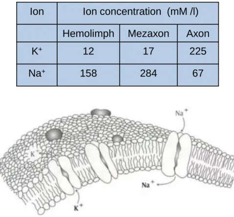
Ion Ion concentration (mM /l) Hemolimph Mezaxon Axon
K+ 12 17 225
Na+ 158 284 67
The mezaxon and the importance of the glial cells
Illustration of transmembrane proteins that function as ion channels
CONDUCTION OF ELECTRICAL RESPONSE
Phases of action potential, nerve impulse (spike) 1. Na-activation – Na
+- influx, rush into membrane
causing depolarization
2. Na-inactivation – Na
+- outflux: time-dependent closing mechanism
3. K-activation – K
+- outflux: immediately after a spike starts
4. slow after potentials (positive, than negative)
Basic neuronal functions
Features of action potential:
1. absolute refractory period (2-3 ms): during a spike a neuron cannot be stimulated
2. relative refractory period (10-15 ms): only to a very strong stimulus will elicit new response
3. maximized frequency of impulses, upper limit: 100 impulses/ms
4. size of the amplitude and length of active section of axon is characteristic of the neuron
5. speed of impulse transmission is proportional with the square root of diameter of nerve fibre
Basic neuronal functions
According to the impulse transmission a neuron can be:
- „spiking” neuron: which can conduct action potentials
electrotonus potentials are conducted on its the dendrite and action potentials are conducted on its axons
- „non-spiking” neuron: which cannot produce action potentials, conduct exclusively electrotonus potentials
(intraganglionic interneurons in the CNS; have wide importance in the initiation of rhythmic activities)
Basic neuronal functions
NEUROTRANSMISSION Types of synapses:
1. chemical synapses – through the mediation of neurotransmitters 2. electrical synapses – direct transmission of the electrical impulse
(rarely, very narrow synaptic gap 3-5 nm)
Structure of a chemical synapse:
1. On the presynaptic side:
- presynaptic neuron terminal
- presynaptic membrane voltage gated Ca2+ ion channels (rarely
intracellular bound Ca2+)
- presynaptic vesicles (50-80 nm diameter) (containing neurotransmitter or neuromodulator molecules)
Basic neuronal functions
Structure of a chemical synapse: (cont.) 2. On the postsynaptic side:
- postsynaptic membrane equipped with:
transmitter receptors: ligand gated Na+ and K+ channels or ligand gated Cl- channels
3. On the place of neurotransmission - narrow synaptic cleft (20-50 nm)
Basic neuronal functions
Basic neuronal functions
The change in membrane potential arriving at the presynapse as an action potential causes opening of voltage gated calcium channels. Calcium enters the presynaptic membrane and promotes the fusion of vesicles with the membrane (A).
Than follows the release and diffusion of neurotransmitter stored in vesicles into the synaptic gap (A).
Neurotransmission, events at the synapse
When more frequent depolarisations reach the presynaptic membrane, there is a greater fusion of vesicles and the release of more neurotransmitters (B).
Basic neuronal functions
Neurotransmitter binds to receptors on the postsynaptic membrane, and ligand gated ion channels leading to a change in membrane potential of the postsynaptic neuron (C).
Commonest insect neurotransmitters
THREE MAIN TYPES OF INTERNEURONAL SYNAPSES:
1. Excitatory synapses A) chemical synapse:
- operated by: cholinergic system
- transmitter: acetylcholin (acetylcholinesterase (breaks down) and cholin acetyltransferase (synthesizes))
- receptors can be activated by other molecules too e.g.:
nicotine or muscarine mimic the action of ACh - depolarized by an electrotonus potential
- starts: Excitatory Postsynaptic Potential: EPSP B) electrical synapse
- rare occurrence in case of insects (all synapses within the giant fibre systems of ventral nerve cord)
- requires very narrow synaptic cleft (3-5 nm)
- makes possible direct and rapid impulse transmission
Basic neuronal functions
THREE MAIN TYPES OF INTERNEURONAL SYNAPSES: (cont.) 2. Inhibitory chemical synapses
- operated by: GABA-ergic system
- transmitter: gamma-aminobutyric acid (GABA) - effects on ligand gated Cl- channels
- hiperpolarized by electrotonus potential
- starts: Inhibitory Postsynaptic Potential: IPSP
3. Nerve-muscle junctions, neuromuscular synapses - excitatory neuromuscular junctions
- operated by: glutaminergic system
- transmitter: L-glutamate (L-aspartic acid)
- in case of inhibitory neuromuscular junctions GABA is the transmitter
Basic neuronal functions
Neuromodulators
may affect either the release of a
neurotransmitters from the presynaptic
membrane or the response of the postsynaptic membrane to other released neurotransmitters.
Properties:
- affect within or near to synapses
- excreted into the mezaxon or synaptic gap - have slow and permanent effect on the
function of synapses e.g. passage of vesicles They can effect on:
- one single synapse – parasynaptic secretion - on several point of axonal surface – paracrine
secretion
Basic neuronal functions
Synthesis of biogenic amines
from tyrosine
CLASSIFICATION
According to innervated body parts:
1. somatic nervous system 2. vegetative nervous system
According to the localization:
1. central nervous system
2. peripheral nervous system
Nervous system of insects
Functional and structural units of nervous system are nerve cells (neurons)
Parts of neurons:
- cell body (soma, perikaryon) including nucleus (karion), Golgi-app.; ER..
- cytoplasmatic extensions:
sensory branches – dendrites motor branches – axons
(their joint branch = neurites) Connections among neurons
(interneuronal ~)
synapses (generally axo-dendritical)
Nervous system of insects
Neurons found in the insects nervous system
Classification of neurons:
According to their appendages:
A. monopolar or unipolar neurons (A) B. bipolar neurons (B)
C. multipolar neurons (C)
According to their function:
1. sensory neurons or afferent neurons (A)
2. motor-, motoneurons or efferent neurons (B) 3. associative neurons or interneurons (C)
4. neurosecretory neurons (D)
Nervous system of insects
Histlogical structure of CNS (Central Nervous System)
a.)cell bodies of neurons are on the verge of the tissue b.)axons and synapses are within the tissue = neuropil
Sheath and alimentary elements of CNS:
- glial cells (glia Greek for glue):
- can be: - peripheral (perineural) forming external sheath (perineurium) a blood-brain barrier
- neural lamella (amorphous, cell free layer)
Nervous system of insects
FUNCTIONAL CONCENRTATIONS, AGGREGATIONS OF NERVE CELLS:
Nerves = include only the axonal components of neurons Ganglions = include axons, perikaria and dendrites
Nervous system of insects
Configuration within a ganglion
Anatomical structure of CNS:
„ventral ganglion-chain or - cord” – like a rope-ladder
Main parts of the CNS:
• nerve-knots – ganglions (in pairs per each segment)
• longitudinal nerve-trunks - connectives
• crosswise nerve-trunks - commissures
Nervous system of insects
Original number of ganglions per tagma:
head - brain – 3 pairs - suboesophageal ggl. – 3 pairs
thorax - 3 pairs (pro-, meso- and metathoracic) abdomen - 8 pairs (usually less)
Fusion of ganglions, reduction in their number:
most frequent in case of:
- meso- and metathoracic ganglions - complex metathoracic ganglions - terminal abdominal ganglions
Nervous system of insects
Nervous system of insects
No fusions Meso- and metathoracic ggl. Complex metathoracic ggl.
STRUCTURE AND FUNCTION OF THORACIC AND ABDOMINAL GANGLIONS 1.
- soma of each neuron is peripherally arranged
- neuropils (axons + synapses) are centrally arranged
- characteristic bilateral arrangement Special location of neuropils:
- motorial elements are dorsally located
- sensorial elements are ventrally located - associative elements are medially located Motoneurons are bigger and occur in less number in neuropils.
Nervous system of insects
Two main groups of interneurons:
1. Intraganglional interneurons more frequent; serve for communication within the ganglions
2. Intersegmental (interganglional) interneurons forming nerve-tracts:
a) descending nerve-tracts b) ascending nerve-tracts
Main physiological function of thoracic and abdominal ggl-s:
1. control of rhythmical activities, movements
2. induction of reflexes
Nervous system of insects
Rhythmical activities:
• complex processes, based on harmonized operation of muscles
• coordinator: rhythm giving central units (pattern generators, neuronal oscillators, pacemakers)
• Such activities:
- stepping
- flying (mostly with asynchronously innervated muscles)
- rhythmical respiratory movements
- so called spontaneous activities (heart contractions)
Nervous system of insects
Reflexes of the thoracic and abdominal ganglions:
Types:
- according to the relation between causing effect and response to it:
1. unconditioned
reflexes(more frequent)
2. conditioned reflexes (in case of social insects)
- according to the features of muscle movements triggered:
1. kinetic reflexes 2. static reflexes
- according to the number of segments taking part 1. segmental reflexes
2. intersegmental reflexes - according to the physiological
role and function:
1. nutritional reflexes 2. respiratory reflexes 3. kinetic reflexes
4. protective reflexes 5. sexual reflexes
6. stridulation reflexes
7. reflexes relating to light emission
Nervous system of insects
STRUCTURE AND FUNCTION OF CENTRAL GANGLIONS (supra- and suboesophageal ganglions)
Anatomy of central ggl-s:
A) brain (ggl. supraoesophageum, cerebrum)
Parts:
1. protocerebrum 2. deutocerebrum 3. tritocerebrum
All three parts of brain are longitudinally fusioned but laterally only the first two (between two parts of tritocerebrum: tritocerebral commissure)
Nervous system of insects
B) Ganglion suboesophageum Connected to brain with two:
circumoesophageal connectives
Physiological role of central ggl-s:
1. local sensory, motor and reflex centres
2. integrating associative centres 3. centres for affect and control the functions of trunk ggl-s (stimulating or inhibiting)
Nervous system of insects
Structure of the protocerebrum:
Properties:
- two hemisphere like parts:
between them a ditch (pars intercerebralis)
Protocerebrum is associated with the compound eyes, ocelli, and some integumental sensory receptors.
The two large lateral
lobes containing dense groupings of neurons and neurosecretory cells within the area known as pars intercerebralis
Nervous system of insects
Structure of the protocerebrum:
(cont.)
Initial nerves:
3 (2 paired, 1 unpaired) nerves:
• nervus opticus to the ocelli of frons and vertex
• nervus connectivus to the a stomatogastric system
• nervi corporis cardiaci (to the corpori cardiaci)
Its most important regions:
• optic lobes (lobus opticus)
• mushroom bodies (corpus pedunculatum)
• central complexes
Nervous system of insects
Structure of the protocerebrum:
Nervous system of insects
Major areas of brain
Structure of optic lobes (lobus opticus) Parts:
1. lamina (lamina ganglionaris) 2. medulla (medulla externa) 3. lobula (medulla interna) in some insects it has two parts:
- lobula
- lobula plate
4. nerve-fibre intercrossings - outer chiasma
- inner chiasma
Nervous system of insects
Function of optic lobes, process and analysis of optic information:
Properties:
- inversion of pictures - retinotopic mapping
- pattern recognition with „small-field” neurons - general motion recognition horizontally and
vertically with „wide-field” neurons
Nervous system of insects
Structure of mushroom bodies (corpus pedunculatum)
State of development:
- big – social insects Hymenopteras, termites - intermediate – Coleopteras, Lepidopteras - small – Heteropteras, Dipteras
Parts: 1. cup (calyx)
2. handle (pedunculus)
3. lobes (lobi): α-, β- and (sometimes) γ-lobus Histological structure :
- nerve-fibres – mostly in the peduncles - synapses – mostly in the cups and handles
- „proper neurons „intrinsic” neurons = Kenyon-cells Physiological role:
- cognitive and associative functions relating to olfaction and vision - centres for learning and memory
Nervous system of insects
Structure and function of central bodies:
Parts:
1. protocerebral bridge 2. central body
3. lateral accessory lobe Features:
- lots of nerve-fibre intrecrossings
- rich arborisation of axons and dendrites Physiological role:
- integration of information between the two hemispheres of brain
Nervous system of insects
Structure and function of deutocerebrum Features:
- bilateral innervations of antennae – nervus olfactorius - olfactory glomeruli
- connection with sensillae of antennae
- neuromuscular junctions with muscles of antennal segments
Nervous system of insects
Protocerebrum
Olfactory glomerulus:
- ball or cap like shape (50-100 μm diameter) - rich dendrite and axon terminals
- their number is typical for an insect species
- their neurons have special interconnections (synapses) with certain neurons:
1. local interneurons
2. projective neurons (uni- and multiglomerular) 3. so called „incoming neurons”
Most important physiological role of deutocerebrum:
- processing and transmission of olfactory and partly mechanical information coming from antennae
- motorial control of antennal segments
Nervous system of insects
Structure of the deutocerebrum
Nervous system of insects
Structure and function of tritocerebrum Features:
- its two parts are connected with a commissure - its is connected to the stomatogastirc system
(ganglion frontale) with two slight nerves
- innervated regions: mostly pharynx and labrum Structure and function of ganglion suboesophageum Features:
- its three parts innervates the three parts, segments of the mouth (mandibular, maxillar and labial)
- contains a lot of nerve-fibres
- its sensory and/or motorial nerves innervate: maxillar and labial palpi, mouth parts, salivary glands, muscles
of neck region
Nervous system of insects
GENERAL EFFECTS OF CENTRAL GANGLIONS:
Brain (ganglion supraoesophageum, cerebrum) Features:
- the two hemispheres effect on own side - the two hemispheres inhibit each other
(reciprocal inhibition) Effects:
1. excite the maintenance of proper muscle tone 2. hinders locomotion activities
Ganglion suboesophageum Effects:
- excite locomotion activities on own side
(ganglion supraoesophageum inhibits directly this centre)
Nervous system of insects
Protocerebrum
VEGETATIVE NERVOUS SYSTEM:
Features:
This element of the nervous system, innervates the gut, heart, and endocrine glands and forms a network of peripheral ganglia that is associated with digestive processes. These ganglia innervate the muscles of the oral cavity, foregut, and midgut and regulate food ingestion, transport, digestion, and the excretion of wastes.
- provides innervations for visceral organs with its sensory and motorial nerve fibres
- its nerve fibres create rich arborisation in the walls of visceral organs
- contains paired and unpaired ganglions nearby the innervated regions
Nervous system of insects
VEGETATIVE NERVOUS SYSTEM: (cont.)
Parts:
1. stomatogastric system: specifically innervates the foregut and midgut
The major ganglion of is the frontal ganglion that arises from the frontal connectives issued by the tritocerebrum. The frontal ganglion innervates the foregut and controls crop emptying. It gives rise to the recurrent nerve that passes underneath the brain and expands into the hypocerebral ganglion that
innervates the corpora allata, corpora cardiaca, and the fore- and midguts
2. unpaired ventral nerve-trunk and its connections
3. caudal autonomic sympathetic system: consists of those nerves that innervate the hindgut and genitalia, usually originating in the compound terminal abdominal ganglion.
Nervous system of insects
THE STOMATOGASTRIC NERVOUS SYSTEM
Nervous system of insects
DISSECTED NERVOUS SYSTEM OF A COCKROACH
Nervous system of insects
Perception:
- Organisms constantly monitor and respond to changes in their environment (both external and internal) so as to maintain themselves under the most favourable conditions for growth and reproduction
- In contrast to mammalian skin, which has millions of generally distributed sensory structures, the surface of an insect has only a few thousand such structures, and most of these are restricted to particular regions of the body.
Stimulus:
- environmental cues are always forms of energy
- physical (mechanical, optical, thermal..) effects, chemical effects
Sensory organs of insects
Adequate or stimulus:
stimulus to which the sensory unit is the most sensitive Threshold of a stimulus:
smallest stimulus which can trigger appropriate response Receptors:
structures that can receive external and internal cues (one single sense cell or complex sensory unit or well developed
sensory organ)
According to the number of different cues perceived:
1. unimodal receptors
- are able to perceive only one type of stimuli, are specialized to one stimulus
2. bimodal or multimodal receptors
- are able to perceive two or more different stimuli
Sensory organs of insects
Sense cells (sensory neurons)
1. primary or type I. sensory neurons Features:
- always bipolar within or directly under the epidermis - short dendrite, long axon
- their synapses are in the CNS
- associated with cuticle, monitor mostly the external environment
2. secondary or type II. sensory neurons Features:
- always multipolar under the epidermis, within the muscles or walls of internal organs (e.g.: digestive system)
- their axonal synapses are located outside the CNS - monitor solely the internal environment
Sensory organs of insects
Sensory unit, elementary sense organ (sensillum, sensilla) Parts:
1. sensory neurons(s)
2. stimulus modifying and mediating elements 3. protective and steady elements
Feature:
generally they occur in high numbers, forming groups Sense organ
Features:
- bigger, anatomically well separated, complex formation - occurs in lower number and often in pairs
Sensory organs of insects
Sensory unit, elementary sense organ (sensillum, sensilla)
Sensory organs of insects
Including the sensory neurons, a tormogen cell, which creates the socket; a trichogen cell, which creates the shaft of the hair; and a thecogen cell, which produces the sheath component of the sensillum that isolates the axons from one another and provides the neuron with ions and nutrients.
Basic structure of a cuticular sensillum
Sensory organs of insects
Sensory unit, elementary sense organ (sensillum, sensilla) (cont.)
Trichoid sensilla have long hairs and 1-3 unbranched dendrites covered by a thick cuticle.
Basiconic sensilla are shorter with 1-3 branched dendrites and a thicker cuticle.
Placoid sensilla are plate-like, with 2-50 branched dendrites.
Coeloconic sensilla have a peg-like structure and are often located within a cuticular pit, innervated by 3-5 unbranched dendrites.
Campaniform sensilla refers to the bell shape appearance of the sensory structure which has only a single neuron.
Examples of external morphologies of sensillae
Basic processes of reception of cues Electrochemical effects of stimulus:
1. Initial or „on” response
- rapid depolarization or hyperpolarization - generation of a receptor potential
- electrotonus potential proportional to the strength of stimulus
- in case of permanent stimulus: decreasing amplitude (adaptation)
2. Reverse or „off” response - rapid repolarisation
Sensory organs of insects
Generation of action potential:
– stimulus have to be exceed the threshold
– amplitude characteristic to given receptor cell
Two basic forms of receptors according to adaptation:
1. Phasic receptors
– adapt rapidly to steady stimuli – react to short but strong stimuli 2. Tonic receptors
– adapt slowly to steady stimuli
– react to long lasting but less strong stimuli
Sensory organs of insects
Classification of receptions according to the place of reception:
1. reception of external stimuli (exteroception)
2. reception of internal stimuli (interoception)(reception of
internal body conditions – self perception (proprioception):
• Proprioceptors are able to respond continuously to deformations (changes in length) and stresses (tensions and compressions) in the body. They provide an organism within formation on posture and position.
• Five types of proprioceptors occur in insects: hairplates, campaniform sensilla, chordotonal organs, stretch receptors and nerve nets.
Sensory organs of insects
Main groups of receptors:
1. mechanoreceptors
2. thermo-hygro receptors 3. photoreceptors
4. chemoreceptors
MECHANORECEPCTION Features:
• well developed accessory units helping perception of stimuli
• displacement, deformation or movement of the sensillum trigger the receptor potential
Sensory organs of insects
Basic mechanoreceptors:
1. tactile hair (trichoid sensilla) 2. bell shaped sense cone
(campaniform sensilla) 3. stringed or chordotonal
sensilla
4. free nerve endings forming nerve nets
Sensory organs of insects
Tactile hair:
Features:
- reacts to deflection - pore free hair
- narrows towards its tip
Parts:
- cuticular hair
- bipolar receptor cell (with tubular dendrites) - seta forming cell (trichogen cell)
- articulation membrane forming cell (tormogen cell)
- dendrite sheath forming cell (thecogen cell) - articulates on a flexible membrane ring
- often forms hair pates
Sensory organs of insects
Campaniform sensilla Features:
- reacts to expansive and compressive forces of the cuticle Parts:
- bipolar receptor cell
- trichogen and thormogen cell - often forms plates
Sensory organs of insects
A mechanoreceptor that is activated by movement of its seta in the direction of the arrow. The deformation of the subcuticular tubular body initiates the receptor potential.
Sensory organs of insects
Stimulation of a mechanoreceptor
Sensory organs of insects
Detail of the antennal apex of an ant’s worker
Sensory organs of insects
Tip of the cercus bearing one campaniform and three coeloconical sensilla. Note the folded integument structure of the cercus.
Stringed (chordotonal) sensillum - scolopidium Features:
- basic functional and structural unit of a complex chordotonal organs
- always located under the cuticle (subcuticular location)
- special accessory cells: cap cells and scolopale cells
- special formation at the end of the
dendritical appendix = scolopale (scolopoid body) - terminal bunch can be connected to the tip of
scolopale
Sensory organs of insects
Main types of insects’ mechanoreception:
1. tactile sense 2. static sense
3. sense of vibration and hearing 4. sense of internal tensions
Forms of static sense:
- posture and position sense - sense of force of gravity - pressure sense
- sense of drift
- sense of acceleration
Sensory organs of insects
Sensory organs of insects
(A) Single chordotonal sensillum (B) Chordotonal organ
Sense of vibrations
Properties:
- vibrations can be perceived on the boundaries of mediums e.g. solid - air, air water - relatively low frequency periodic stimuli
- can be perceived only from a short distance
- generally has alarming effect or plays role in communication Insects are classified according to vibration sense:
1. sensitive species: nerve impulses are synchronous with the frequency of stimuli e.g.: Lepidoptera, Hymenoptera
- typical sense organ: subgenual organ is a complex chordotonal organ in the tibia of each leg with 10-40 sensilla to perceive substrate vibrations
2. less sensitive or indifferent species:
- nerve impulses are asynchronous with the frequency of stimuli e.g.: Heteroptera
- typical sense organs: trichoid sensilla and campaniform sensilla
Sensory organs of insects
Hearing, audition (phonoreception)
- perception of vibrations spreading in the medium (air or water) - changes in pressure effect as stimuli
- frequency is high, it can be measured generally in kHz
Sensory organs of insects
Sense of vibrations – subgenual organ
Classification of insects according to the stridulation ability and audition:
1. well hearing insects without specialized stridulation organs:
- typical hearing organ: Johnston’ organ or tympanal organ - Occurrence: Culicidae, Chironomidae, Noctuidae
2. well hearing insects with specialized stridulation organs:
- typical hearing organ : tympanal organ
- Occurrence : Acrididae, Cicadidae, Grylloidea, Tettigonioidea
Sensory organs of insects
Johnston’s organs:
- large complex chordotonal organs (there occur simple Johnston’s organs too with only a few scolopidia)
- Occurrence: within the second segment of antennal flagellum (pedicellus) of all adult and many larval insects
- consists of numerous scolopidium in radiant arrangement within two rings to perceive air-borne vibrations
Sensory organs of insects
Tympanal organs:
- the most elaborate sound reception system in insects; occur always in pairs
- typical parts: eardrum a cuticular membrane (tympanum), wall of drum cavity, drum cavity, sometimes another drum (anti-tympanum)
- drum cavity has air sack origin (there are air storage sacks within the respiratory system)
- sometimes from numerous scolopidium (~60 pc) a hearing crest (crista accustica) is formed
- receptible frequency differs according to species:
0,1-100 kHz
Sensory organs of insects
Types of tympanal organs:
1. located in the thorax (thoracal tympanal organ);
Occurrs: Noctuidae (metathorax)
2. 2. located in the abdomen (abdominal tympanal organ); Occurs: Acrididae, Cicadidae,
Geometridae
3. located in the fore legs (tibial tympanal organ);
Occurs: Gryllidae, Tettigoniidae
Sensory organs of insects
Sensory organs of insects
Cross section of Johnston’s organ at the base of the antenna.
The basal plate deflects the scolopidia when it is displaced by movements of the flagellum.
The antenna of a male mosquito
Sensory organs of insects
Surface view (A) and transverse section (B) trough the tympanal organ of a grasshoper species (Tettigoniidae)
Thermoreception
- Insects actively seek out a „preferred” temperature (see autecology)
- insects evidently detect variation in temperature, yet the function and location of receptors is poorly known (e.g.
Rhodnius spp., Cimex spp. and mosquitoes, which feed on mammalian blood, are able to orient to a heat source)
- most studied insects have antennal sensing of temperature (mosquitoes)
- transduction mechanism for thermoreceptors remains unclear