BY BERTA SCHARRER
CONTENTS
Page
I. Introduction 125 II. Endocrine Control of Postembryonic Development 126
A. Observations in Various Groups of Insects 127
B. Discussion of Results 133 III. Role of Hormones in Reproduction 138
A. Effect of Corpora Allata on Gonads 138 B. Effect of Corpora Allata on Accessory Sex Glands 140
C. Effect of Reproductive Organs on Corpora Allata 140 D. Effect of Gonads on Secondary Sex Characters 141
E. Interpretation of Experimental Data 142
IV. Hormones and Color Change 145
V. "Gene Hormones" 146 VI. Sources of Insect Hormones 150
VII. Mode of Action and Physicochemical Properties of Insect Ho'rmones 155
References 158 I. Introduction
The study of insect hormones represents a particularly active sector of the wide and relatively young field of invertebrate endocrinology (see reviews: 13,20,21,35,44,54,67,69,71,85,88,95,98,118,124,135,138,159,173, 183). In insects endocrine factors are known to play an important role in reproduction and in postembryonic development. By comparison the hormonal control of color change is of minor significance. The question as to whether sex hormones comparable to those of vertebrates are opera- tive in the insect organism is still controversial. Finally, there exist in this group of invertebrates physiologically active substances which par- ticipate in the nonautonomou« development of hereditary characters and which, because of their similarity with hormones in the commonly accepted sense of the word, have been termed "gene hormones.,,
With regard to the relationship between the endocrines of vertebrates and those of insects, only few conclusive data are available. These indi- cate that, in principle, vertebrate hormones may act on insects, and insect hormones on vertebrates. Details on this subject may be found in several monographs (71,88,173; see also 104b).
125
126 BERTA SCHARRER
The actions of insect hormones are being studied by various methods:
extirpation and implantation of endocrine organs, injection of organ extracts, denervation of endocrine glands, ligatures, blood transfusions, parabiosis, etc. Most of these methods are used in vertebrate endo- crinology and are applied to insects with only minor modifications. One of the newly developed techniques is of interest. Test organs such as skin, gonads, etc., with or without endocrine glands, are implanted into the abdominal cavity of hosts whose own physiological condition offers a "neutral" endocrine surrounding preferable to any tissue culture medium (15,170).
II. Endocrine Control of Postembryonic Development
Among the physiological processes under hormonal control in insects, the most extensively studied is postembryonic development, which, in all groups except the Ametabola, consists of a series of developmental steps leading from the larva or nymph, newly hatched from the egg, to the adult insect.
In the holometabolous insects, such as butterflies, periodic steps of growth as evidenced by molts produce larval forms (instars) of increasing sizes. The larval period is terminated by pupation, which marks the onset of adult differentiation of tissues and organs, although "internal "
metamorphosis may begin during the larval period (170). Metamor- phosis is completed at the end of the pupal stage when the adult form (imago) emerges.1
In hemimetabolous insects, for example grasshoppers, the immature forms or nymphs likewise undergo a number of molts. Each molt pro- duces a nymph which is not only larger than the preceding instar but is a step closer to the adult form. In this group of insects with "incomplete"
metamorphosis a pupal period is lacking, but during the last nymphal stage considerable morphological changes occur which at the final molt result in the fully developed imago.
Quite generally then, in normal development the larval (nymphal) period of an insect is predominantly one of growth. Little adult differ- entiation occurs before the insect has reached the appropriate stage for metamorphosis. Accordingly larval molts mark a step in growth rather than in imaginai differentiation. Within certain limits molts may occur as long as the organism remains immature; they cease to take place as soon as metamorphosis is completed. This statement holds true in spite of the fact that under certain experimental conditions it has been possi- ble to induce adult skin to molt again (115,120,182,184).
1 " Hypermetamorphosis " is not dealt with in this chapter since no experimental data are available concerning this phenomenon.
For the understanding of hormonal regulations of development it is useful to point out that there exist two types of molts: (1) larval (nymphal) molts in which an increase in size but little or no imaginai differentiation occurs; (2) molts coupled with imaginai differentiation, for instance the final molt of a hemimetabolous insect which results in the imago.
The first demonstration of a hormonal factor controlling insect development was given in 1922 by Kopec (89). He removed the cerebral ganglion (brain) from freshly molted last instar larvae of a moth, Lyman- tria. In such animals pupation does not take place unless the brain, which in this case appears to play the role of an endocrine organ, is re-implanted into the abdomen. Operations of this kind yield conclusive results only if they are performed before the so-called " critical period/' i.e., a definite period at which, within a given developmental stage, the hormone concentration in the circulation reaches an effective level (p. 152),
In the following two decades evidence accumulated supporting the conclusion that hormones are instrumental not only in pupation but also in other phases of insect development. Consequently the existence of molting hormones, pupation hormones, and metamorphosis hormones was postulated. The data available now have been obtained in repre- sentatives of various groups, i.e., the hemimetabolous Hemiptera and Orthoptera, and the holometabolous Lepidoptera, Hymenoptera, Cole- optera, Megaloptera, and Diptera. As may be seen from the following examples these various orders of insects differ somewhat from one another in their developmental physiology.
A. OBSERVATIONS IN VARIOUS GROUPS OF INSECTS
In the tropical bug Rhodnius and in other representatives of the Hemiptera (Triatoma, Cimex) the dorsal region of the protocerebrum (Fig. 8) furnishes a "molting hormone." If this part of the brain, taken from donors during the critical period, is implanted into decapitated nymphs, i.e., nymphs deprived of their own source of hormone, molting results (184). The reaction is specific and cannot be induced by implants of other parts of the central nervous system or of other organs. This localization offers strong evidence that the hormone which causes molting originates in the neurosecretory cells of the pars intercerebralis. Such cells which combine nervous and glandular characteristics have been demonstrated not only in Rhodnius (70,184) but also in a variety of other insects (p. 149).
In addition to this factor Wigglesworth (180,181) postulated the existence of a juvenile (inhibitory) hormone the source of which he
128 BERTA SCHARRER
localized on indirect evidence in the corpus allatum (Fig. 8a). The juvenile factor restrains adult differentiation for a time sufficient to permit the necessary degree of growth. In other words, it controls the rate of development. Although this view has not been accepted by all workers in the field (112,114,116,117) it is supported increasingly by experimental evidence obtained in various groups of insects (cf. Section V of the following chapter for a similar phenomenon in crustaceans).
The endocrine role of the corpora allata has been more firmly estab- lished in several species of Orthoptera (Fig. 6) in which the extirpation and implantation of this gland has been possible.
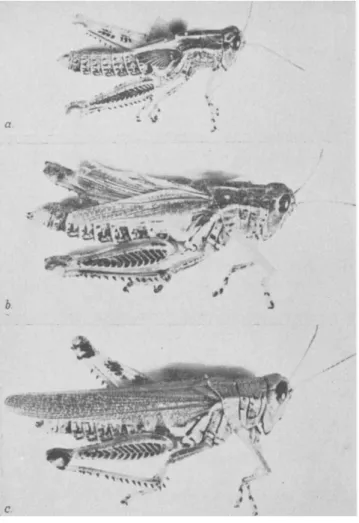
FIG. 1.—(a) Normal adult male of Leucophaea maderae. (b) Adultoid male ob- tained from allatectomized seventh instar nymph, (c) Normal male eighth instar nymph. Scale in centimeters. (From Scharrer, 140; courtesy of Charles C Thomas, Publisher, Springfield, Illinois.)
(1) After allatectomy in early nymphal instars of Dixippus (112), Bacillus (53), and Leucophaea (140; Fig. lb) adult differentiation sets in prematurely. Molts are suppressed and development is abbreviated.
(2) Implantation of corpora allata into normal last instar nymphs retards metamorphosis and is followed by supernumerary molts (110, 116,117; Fig. 2b). This effect may be obtained even with grafts from adult donors in some insects but not in others.
In the first type of experiment the animals resulting after the final molt are smaller, in the second type larger than normal adults. But difference in size is not the only characteristic that distinguishes these
experimental animals from normal adults. In the allatectomteed ani- mals, developing precociously because of lack of juvenile hormone, the developing tissues do not seem fully prepared to undergo imaginai differ- entiation. In Leucophaea they are less ready in younger nymphal stages than in older ones. Insects that resemble adults to a greater or less degree (adultoids) are the result (Fig. lb). Correspondingly, in animals which have been kept overtime in the nymphal condition by allatum implants, the organs, ready for imaginai differentiation, are subjected to the prolonged action of the juvenile hormone. These animals likewise show a mixture of nymphal and imaginai characters; they may be called nymphoids (Fig. 2b).
The role of the corpora allata in the control of development is essen- tially the same in the holometabolous Lepidoptera. Allatectomy in young larvae causes precocious development (17,18,20,22,58,92,121,123, 187). Allatum implants from pupae in diapause into hosts ready to complete adult development do not inhibit this process (187).
The endocrine significance of the brain in bringing about pupation in this group has already been referred to (p. 123). The earlier results of Kopeé (89) were later confirmed by several investigators (33,92,119,125).
The control of adult development by a substance originating in the brain has recently been demonstrated by Williams (187) in "diapausing" (dor- mant) moths. Platysamia pupae, when chilled (2-5°C.) for four to six weeks after pupation and subsequently returned to room temperature (25°C.) complete their development after an additional four to six weeks.
By contrast, in pupae left at room temperature diapause lasts at least five months. Brains from donors ready to metamorphose implanted into dormant hosts bring about adult development in the latter. Brains, up to eight in number, taken from dormant donors do not have the same effect on the host; neither do a variety of other tissues. Brainless pupae fail to complete their development, unless the extirpation has been per- formed after the critical period which is about fourteen days following the return to room temperature. Implants of "activated" (nondor- mant) brains into decerebrated pupae restore the capacity of the host to complete metamorphosis.
Of much interest is the observation that the proper effect is obtained only if the brain implants establish "intimate continuity" with tissues of the host (Williams, personal communication2; see also 8,21). The results after extirpation and implantation of brains agree with those obtained in parabiosis experiments (187).
Localization experiments showed that a specific region of the brain, i.e., the inner mass of the cerebral lobes, is responsible for the elaboration
* I wish to thank Dr. C. M. Williams for permission to use these unpublished data.
130 BERTA SCHARRER
of the active principle. This region contains two groups of neurosecre- tory cells, one medial and one lateral. Implants must contain both glandular centers to be effective; parts of brain tissue lacking one or both cell groups are incapable of terminating diapause (187b). This localiza-
FIG. 2.—(a) Normal sixth instar nymph of Melanoplus differ entialis. (b) Nymph- oid female obtained by implantation of corpora allata. (c) Normal adult female.
(From Pfeiffer, 110.)
tion of the physiological effect in the neurosecretory centers of the brain of Platysamia agrees with the results of Wigglesworth in Rhodnius (p. 123).
The brain factor has not been demonstrated in the blood. Implants
of larval brains, after undergoing a sufficient degree of development in the host and after being activated by chilling bring about imaginai development in brainless hosts.2
In addition to the brain the prothoracic glands are necessary for the completion of adult differentiation. Both sources of physiologically active substances must be implanted in order to bring about imaginai changes in a pupal abdomen isolated by means of a ligature. Unlike brain grafts, the prothoracic glands from a diapausing donor are as active as those from a nondormant pupa (187). This and other evidence (58) makes it seem quite probable that in the endocrine control of pupation as well as of imaginai differentiation the influence of the brain is super- imposed over that of the prothoracic glands.
Also in other moths (Bombyx) the endocrine significance of the pro- thoracic glands for the control of pupation and imaginai differentiation, and apparently also for molting, has been demonstrated (57,58,59; see also 8,19,22a).
Various developmental stages of Platysamia show both qualitative and quantitative differences in metabolism. The cytochrome content of diapausing pupae is considerably smaller than that of active stages (pupae shortly before emergence and larvae). These and additional data indicate a causal relationship between endocrine mechanisms and biochemical changes in the developing tissues (187b).
A hormone causing pupation and imaginai differentiation in certain Hymenoptera seems to originate in the brain (146).
In the development of Coleoptera the role of the corpus allatum was found to be the same as in other groups of insects (131).
Ligation experiments made on larvae of Sialis, a representative of the Megaloptera, demonstrate the existence of a center controlling meta- morphosis, located in the region of the third thbracic and first abdominal segments (60,61). Corresponding ligatures in pupae are without effect on the completion of metamorphosis (104). If in parabiosis experiments larvae in the beginning phase of metamorphosis are joined with younger specimens, the latter metamorphose prematurely and synchronously with their partners. The source of the hormonal substance involved is not known. Extirpation and implantation of ganglia located in the critical region have no influence on development.
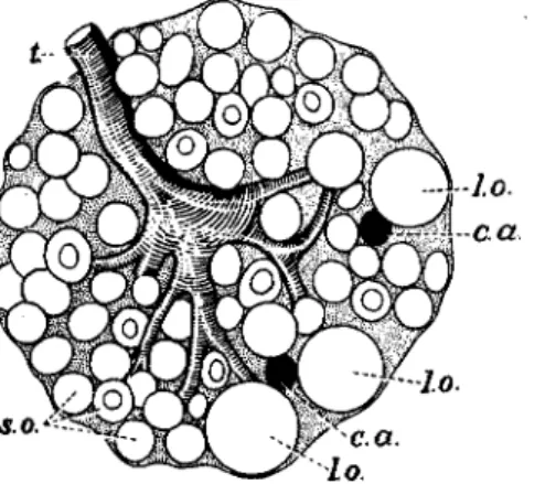
In the muscoid Diptera developmental hormones are furnished by the ring gland (Fig. 7), a composite organ containing corpora allata and cardiaca (40,128,143; see also 162a,164a). This gland controls growth and molting, pupation, and adult differentiation.
Bodenstein (16) transplanted larval heads together with ring glands
132 BERTA SCHARRER
from mature larvae into the abdomen of adult Drosophila. The result was that the transplanted heads underwent one or two molts. Molting did not occur when the heads were transplanted without the ring glands.
Puparium formation, which was found to be greatly retarded or sup- pressed in lethal larvae of the Drosophila mutant lgl and in certain hybrids, can be induced by the implantation of genetically normal ring glands (66). This action of the ring gland was confirmed in normal larvae of Drosophila (68,165) and of Calliphora (6; see also 43,55). In the latter form extirpation of the ring gland prevents puparium forma- tion (24). Pupation proper which takes place within the puparium likewise is controlled by the ring gland (103,167,167a). Implants of brains without ring glands have no effect on pupation (170) or puparium formation (68,165).
In Calliphora growth of imaginai discs is arrested after removal of the ring gland (24). Similarly the development of organ discs in Droso- phila was shown to be under the control of this gland (10,11,14,15,163, 164,167,170,172). During an earlier phase of development the ring gland mainly promotes growth of the discs, later their imaginai differ- entiation. This change in response is brought about not only by an increased hormone production as the ring gland matures, but also by the altered responsiveness of the developing tissue (tissue competence).
However, in younger discs a certain degree of differentiation also takes place (170).
In addition to the imaginai discs the differentiation of other organs of these dipterous larvae, for example the brain (165) and the gonads (p. 134), takes place under the influence of the ring gland. These changes are correlated with the dedifferentiation of larval structures such as the midgut epithelium and the fat body (167).
The hormone (GD hormone, p. 130) bringing about puparium for- mation and imaginai differentiation in muscoid Diptera originates in the larger gland cells (Fig. 7b) which are now known to be corpus cardiacum tissue (see the discussion of the homologies of the ring gland components by Poulson, 128). This conclusion is based on histological as well as experimental evidence : (a) The deficiency of lethal ring glands as com- pared with normal ones concerns the large cells (143); (b) Histological signs of secretory activity of the large cells coincide with phases of physio- logical activity of the gland (163,167,172; see also 169); (c) Implants of ring gland fragments consisting mainly of large gland cells furnish GD hormone to the host (170).
Further observations of interest are that young ring glands as well as older ones furnish the GD hormone, and that this activity apparently follows a cyclic pattern. An analysis of this cycle of hormone production
suggests that the GD hormone in Drosophila larvae also controls molting (170). An as yet unexplained observation concerns the effect of adult corpora cardiaca on Drosophila hydei larvae. Implants of these glands, in contrast to control implants of fat body or corpora allata, cause a marked delay in puparium formation (172a).
B. DISCUSSION OF RESULTS
It is evident that the information concerning the hormonal factors involved in various phases of insect development is still fragmentary.
Some of the data reported above may appear divergent. If it is assumed that each step in postembryonic development is governed by one (or several) specific hormones, i.e., molting, pupation, and metamorphosis hormones, it becomes difficult to compare the various hormonal factors in one group of insects with those in another. Obviously in a hemimetab- olous form there would be no need for a pupation hormone as in a holometabolous insect. Furthermore, even within the holometabolous group, comparable hormones such as the pupation hormones of a moth and of a fly seem to differ with regard to their source in the body.
Therefore, it may seem too early or even impossible to establish a common denominator for the data at hand. Nevertheless certain funda- mental trends are becoming increasingly apparent which justify a pre- liminary attempt at a more uniform interpretation of the hormonal control of insect development. This tentative interpretation is based on concept introduced by Wigglesworth (p. 123) and can be formulated in the following manner:
In holometabolous as well as hemimetabolous insects each develop- mental step may be viewed as governed by a balanced interaction between two developmental hormone systems on one side and the growing and differentiating tissues on the other,
It has been briefly stated before (p. 128) that the developing tissue gradually changes in its capacity to respond to endocrine stimuli. Con- sequently in a given hormonal environment the reaction is determined by the responsiveness of the tissue. For instance, in the same hormonal environment the type of response, growth or imaginai differentiation, is determined by the age of the imaginai discs (168). Furthermore, at the same stage of development various anlagen, such as salivary gland, eye, genital discs, even various regions within the same anläge may differ in their response (15).
The two types of hormone collaborating in the control of develop- ment are: (I) the "growth and differentiation hormone" (GD hormone, or hormones), (II) the juvenile hormone (inhibitory hormone, corpus allât urn hormone).
134 BERTA SCHARRER
A hormonal factor of type I (GD hormone) activates the imaginai ("imaginipetal," Vogt; 170) potencies to an extent which is regulated by the responsiveness of the developing tissue. It promotes growth and imaginai differentiation of tissues and organs. In the immature insect, growth takes place under the influence of a GD hormone in periodic steps as evidenced by the occurrence of successive molts. For this reason Wigglesworth (180,181,184) and others called this factor "molting hormone.,, When this term is used it should be kept in mind that, in addi- tion to bringing about molting, this factor also promotes imaginai differ- entiation. Therefore, the molts it causes are not "simple," i.e., larval or nymphal molts, but molts coupled with metamorphosis. It follows that the "molting hormone" alone cannot account for the occurrence of larval molts.
In order to safeguard the proper number of larval molts there exists an additional factor which stimulates the juvenile, i.e., larval or nymphal potencies of the developing tissues. This juvenile (inhibitory) hormone modifies the action of the GD hormone; the combined action of both factors causes larval molts.
By keeping the developing insect in the immature stage the juvenile hormone favors (or permits) the occurrence of molts. This fact should, however, not be interpreted as an indication that the juvenile factor as such acts as a molting hormone. According to experimental evidence the juvenile factor, when acting alone, is incapable of causing a molt. On the other hand, molts may occur in the absence of the juvenile hormone as in allatectomized animals. For this reason the use of the term "molt- ing hormone" for the juvenile hormone (13) is not recommended. Its use would also lead to confusion since this term has been previously employed by Wigglesworth and others with more justification for a different hormone.
One of the reasons why the existence of several types of hormones has been postulated where probably only one is necessary was that the GD hormone originates in organs as different as the brain of a caterpillar and the ring gland of a fruit fly larva. The following discussion is intended to show that these differences need not be considered as significant.
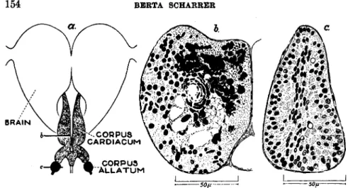
The three main sources of developmental hormones known at present are: (a) the glandular corpora allata, (b) the corpora cardiaca, consisting of nervous and glandular elements, (c) the brain, or more precisely the pars intercerebralis of the protocerebrum containing glandlike nerve cells
(Fig. 8b).
In all insect species suitable for experimental study the corpus allatum has been shown to be the source of the juvenile hormone. However, equally specific roles have not been assigned to the two remaining centers.
The physiological significance of both the corpus cardiacum and the pars intercerebralis may be better understood if they are not treated as two separate centers of glandular activity. However different they may seem at first sight, there exists an unusual morphological relationship between them, as has been demonstrated in the orthopteran Leucophaea maderae. The corpora cardiaca consisting of nervous and glandular ele- ments are innervated by fiber bundles originating in the pars intercere- bralis, a brain center which itself is characterized by the occurrence of secreting nerve cells. Furthermore, colloid masses are found along the nerves (nervi corporis cardiaci) connecting these two neuroglandular centers. It would be difficult to assume that this striking morphological feature is without physiological significance. Therefore, on the basis of this relationship, which has a counterpart in the hypothalamo-hypo- physeal system of the vertebrates (144; see also 76), it has been proposed to consider the pars intercerebralis and the corpora cardiaca as com- ponents of one neuroendocrine complex.
As to the physiological mechanism of this glandular complex there are two possibilities: either both the brain and the corpus cardiacum coop- erate in the elaboration of GD factors, or in certain animals the one, in others the other, component has become the predominant hormone source.
Considering the variability in the development of neuroglandular organs in insects one may expect to find examples of either alternative among the various groups of insects, an expectation which is borne out by data dis- cussed elsewhere (144).
Aside from the intercerebralis-cardiacum-allatum system, only the prothoracic glands of certain moths have recently been demonstrated as the source of a factor concerned with development (57,58,59,187). How- ever, this factor, lack of which prevents metamorphosis, appears to be subordinated to or otherwise linked with the GD hormone furnished by the brain.
In summary, two types of developmental hormones exist which origi- nate in two types of glands. The one type, i.e., the GD hormone (or hormones) is produced by the neuroglandular intercerebralis-cardiacum complex, the other, i.e., the juvenile or inhibitory hormone, by the glandular corpus allatum.
This "two-hormone concept" can be applied to the great majority of experimental data available at present. It has been stated (p. 123) that in hemimetabolous insects the more drastic changes leading from the immature to the mature insect occur in the last nymphal instar. But imaginai differentiation is not entirely restricted to the last stage. It takes place also in a small measure during earlier nymphal life. In an attempt to explain the hormonal mechanism it may be postulated that
136 HERTA SCHARRER
both the GD hormone and the juvenile hormone are active in all stages except the last. In the last stage none or at most only an ineffective amount of juvenile factor is released.20 Therefore, under the uninhibited stimulus of the GD hormone the final, i.e., the major, step of metamor- phosis can take place in the last stage (180,181,184).
Theoretically two possibilities exist in explanation of the small changes in younger nymphs: (a) In consecutive nymphal stages the hormone balance is shifted in favor of the GD hormone by a gradual decrease in the relative amount of juvenile hormone released into the circulation.
In this case the responsiveness of the developing tissues may be assumed to remain approximately the same in all nymphal stages, (b) The ratio of both developmental hormones remains unchanged in each nymphal stage except in the last, but the growing tissues are increasingly capable of response to the stimulus for differentiation.
These alternatives can be subjected to an experimental test by a com- parison of the effect of allatectomy on various nymphal stages in the same as well as in different insect species. In allatectomized nymphs one factor governing development, i.e.,, the juvenile hormone, has been removed. Therefore, differences in the events following these operations reflect differences in the relative effectiveness of the two remaining factors, i.e., of the GD hormone and of tissue responsiveness. Such differences are apparent, for instance, in a comparison of various stages of Leucophaea (140), a species with an average of eight nymphal instars.
Allatectomized seventh instars become adultoids at the molt following the operation which thus becomes the final molt. Allatectomized sixth or fifth instars at their next molt show only an intermediate degree of imaginai differentiation; they require one additional ("preadultoid") stage, and consequently undergo one more molt before they too become adultoids. Since both younger and older nymphs have been subjected to the influence of the GD hormone alone, without the effect of the juvenile hormone, the quantitative differences in response must have been due to differences inherent in the tissues. In Leucophaea and related insects the ratio of developmental hormones, although it may, need not change appreciably except in the last instar.
If in certain other species such as Rhodnius similar changes in tissue responsiveness exist throughout the course of nymphal development, they seem less obvious than in Leucophaea. In Rhodnius, according to
2e In a recent publication (184a) Wigglesworth suggests that the corpus allatum of last instar nymphs of Rhodnius "not only ceases to secrete the juvenile hormone, but also . . . actively removes from the blood any traces of the juvenile hormone that remain."
Wigglesworth (180), nymphal tissues prove ready for adult differentia- tion at a very early stage. "Diminutive adults'' result at the molt following the operation even when first instar nymphs are used. There- fore, in Rhodnius a gradual decrease in the activity of the juvenile hor- mone during nymphal life may account for the small changes observed in successive instars.
The conclusion that in the last stage no effective level of juvenile hormone is reached under normal conditions can be substantiated in two ways: (a) allatectomy in last instars, regardless of the species studied, has no apparent effect on the course of development (20,108,140,146,175);
(b) allatum implants in last instars cause supernumerary molts and pre- vent the completion of metamorphosis. Hence last instars also respond to the action of the juvenile factor, if it is present (20,110,123,140).
Evidence obtained in holometabolous insects likewise suggests that the two factors (GD hormone and juvenile hormone) collaborate during the phase of periodic growth (see also 76a). This larval phase is followed by a phase of adult differentiation. Pupation and metamorphosis can be explained as taking place through the action of one (or several) GD factors in the absence of an effective amount of juvenile hormone.
This conclusion is demonstrated by the fact that allatectomy and implantation of corpora allata have comparable effects in holometabolous and hemimetabolous insects. In Lepidoptera, for example, the period of growth or larval period is prematurely ended when young larvae are allatectomized, and supernumerary molts result when normal last instar larvae receive allatum implants (see above).
The "two-hormone concept'' as elaborated in the preceding analysis may or may not apply to insects other than those discussed so far. In the highly specialized muscoid Diptera, where corpus allatum and corpus cardiacum are contained in one organ, the ring gland, these sources of developmental hormones cannot be analyzed separately as readily as in other groups. The ring gland brings about molting as well as pupation and imaginai differentiation. There is good evidence that the GD hor- mone controlling these processes originates in the large (cardiacum) cells of the ring gland (p. 128), and that it is produced throughout the entire larval life in varying quantity (14,170). The precise function of the allatum cells in the development of these insects is still unknown. How- ever, there is no indication that the allatum component of the ring gland of fly larvae and pupae acts differently from the corpus allatum of other insects. The assumption that it too furnishes a juvenile hormone is based on certain histological as well as experimental evidence (25,169, 170,172a).
138 BERTA SCHARRER
III. Role of Hormones in Reproduction
In the adult insect a hormone or hormones originating in the corpora allata play an important role in reproduction. The existing relationships express themselves in two ways: (1) in effects of the corpora allata on the gonads and on the accessory sex glands, (2) in an influence of the gonads on the corpora allata. Whereas these relationships are well established, the action of sex hormones has not been demonstrated satisfactorily.
A. EFFECT OF CORPORA ALLATA ON GONADS
Ovaries. It has been shown in a variety of insect species that the normal function of the ovaries is under the control of a corpus allatum hormone. Wigglesworth (181) demonstrated in the hemipteran Rhodnius prolixus that the eggs fail to mature in the absence of the corpus allatum (see also 101a). According to Pfeiffer (108,111,174) in allatectomized females of the orthopteran Melanoplus differentialis egg development stops at the beginning of the period of yolk deposition. In another orthopteran, Leucophaea maderae, the presence of the corpora allata was shown to be necessary throughout the period of growth and yolk deposi- tion, which in this species constitutes about the first third of the total period required for the development of the eggs (141). Re-implantation of corpora allata into allatectomized females of these orthopterans restores their capacity to produce mature eggs.
A similar hormonal relationship is known to exist in the Diptera:
Drosophila (160,161,162,171; see also 16a), Calliphora, Musca (155,156;
Fig. 3), Lucilia, Sarcophaga (41), and Anopheles (47a). By means of transplantations in Drospphila larvae it was found, for example, that ovaries of D. melanogaster in D. funebris hosts develop mature eggs only if melanogaster ring glands are grafted together with the ovaries. The hormone furnished by the melanogaster ring gland seems to be qualita- tively different from that of the funebris ring gland. On the other hand, grafted ovaries of Calliphora develop in Lucilia and vice versa (156) under the influence of the ring gland of the host. In Lucilia and Sarco- phaga, denervation of the corpus allatum has the same effect on egg development as extirpation (41). In the groups of insects discussed so far, corpus allatum from male as well as female donors may furnish the hormone necessary for the maturation of the eggs (109,156,160,181).
The aquatic beetle Dytiscus (84) has an annual cycle of ovarian activity, with the laying period normally starting in March or April.
Females can be induced to lay eggs during the winter (resting period) by implantation of five or more pairs of corpora allata. The similarity of this effect to that of hypophyseal implants in winter frogs (188) is of
interest. At all times of the year allatectomy in Dytiscus prevents egg development and, as in other species studied (41,108,141,181), causes pronounced regression of the ovaries. Apparently the corpus allatum hormone makes egg development possible by suppressing the résorption of the oöcytes. The activity of the corpora allata seems to follow a cyclic pattern, hormone being released about every twelve days.
The situation is somewhat different in Dixippus (Orthoptera; 112,116), where egg maturation proceeds in allatectomized females unless the extirpation of the glands is performed during an early nymphal stage.
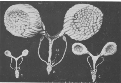
FIG. 3.—Reproductive organs of female Calliphora erythrocephala. (a) Newly emerged fly. (b) Mature female, (c) Allatectomized female, a.g., accessory sex gland; r.s., receptacula seminis. (From Thomsen, 156.)
The presence of the corpora allata is unnecessary for egg production in certain Lepidoptera (18,20,21a,187).
In some orthopterans a possible role of the fat body in egg maturation has been postulated. A similar effect has been attributed to extracts of the posterior lobe of the vertebrate pituitary (79), but this finding has not been confirmed (85).3
3 Royal jelly, the special food substance which is supposed to control sexual maturation in the honey bee, contains a gonadotropic material. Tests made with this material in rats and flies (77,158) were reported to be positive. The failure of other investigators (100) to confirm these observations may be due to differences in methods.
140 BERTA SCHARRER
Testis. No relationship between corpus allatum and adult testis has been demonstrated so far. Removal of the corpus allatum in males of various species shortly after emergence (41,111,156,181) does not disturb the course of spermatogenesis. When mated with virgin females, allatec- tomized males of the orthopterans Bacillus and Leucophaea are capable of fertilizing the eggs (53,141).
The absence of a noticeable influence of the corpus allatum on the testis in the adult was likewise observed in Drosophila by Vogt (170).
Her observation that ring gland implants increase the growth rate and accelerate spermatogenesis in larval and pupal testes is probably attrib- utable to the action of the GD hormone. As has been pointed out
(p. 128) this hormone originates in. the large gland cells (cardiacum com- ponent) of the ring gland, and implants of this part only have the same effect on the testis as do whole ring glands. A similar but less pro- nounced relationship exists with respect to the pre-imaginal ovary (see also 64).
B. EFFECT OF CORPORA ALLATA ON ACCESSORY SEX GLANDS
Regarding the action of the corpora allata on the accessory sex glands the data in the literature are divergent.
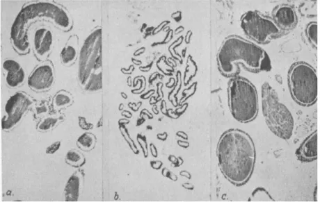
Females. In females of Melanoplus, for instance, the secretory activity of the epithelial lining of the oviduct, which corresponds to the accessory sex glands of other insects, depends on the presence of the corpora allata (108). The same situation exists with respect to the female accessory sex glands in Calliphora (156). Implantation of corpora allata into allatectomized females of Leucophaea restores the capacity of the accessory sex glands to produce normal amounts of secretory material (141; Fig. 4). In contrast to the situation in these three insect species, allatectomy has no effect on the female accessory sex glands of Lucilia and Sarcophaga (41).
Males. The male accessory sex glands of Lucilia, Sarcophaga (41), and Leucophaea (141) are not affected by allatectomy. In Rhodnius (181), however, and to some extent in Calliphora (156) the male accessory glands appear to be under the hormonal control of the corpus allatum.
C. EFFECT OF REPRODUCTIVE ORGANS ON CORPORA ALLATA
The relationship in the opposite direction, i.e., the effect of the gonads on the corpora allata has been studied by means of castration. In certain insects (Melanoplus, 109; Calliphora, 155, 156; Lucilia, 41; see also 166) ovariectomy is followed by hypertrophy of the corpus allatum. Females of Sarcophaga (41) and Leucophaea (141), as well as males of Sarcophaga, Lucilia (41), and Leucophaea (141) show no effect on the corpus allatum
attributable to the removal of the gonads or of the accessory sex glands.
According to Day (41) there is some evidence that, even in those castrates in which the histological appearance of the corpora allata shows no significant change, these glands have become "physiologically altered."
However, no such change in physiological properties could be observed in Dytiscus (84). Corpora allata from donors which had been ovari-
FIG. 4.—Accessory sex glands of female Leucophaea maderae. (a) Normal control in state of active secretion, (b) Glands of female allatectomized shortly after emer- gence, killed six weeks later. The glands resemble those of freshly emerged normal female, (c) Actively secreting glands after re-implantation of corpora allata into allatectomized female. (From Scharrer, 141.)
ectomized for several months have the same effect on the ovaries of the host as do implants from unoperated donors.
D. EFFECT OF GONADS ON SECONDARY SEX CHARACTERS
The presence of hormones determining secondary sex characters and mating behavior in a manner similar to that known in vertebrates has not been definitely proved or disproved with regard to insects. The evidence is at present more against than in favor of the occurrence of sex hormones in this group of invertebrates.
The results of experimental castration and of transplantation of gonads obtained by a number of investigators in a variety of species are on the whole negative (see reviews 54,71,88,138). Grafts of gonads of the opposite sex into castrated caterpillars do not alter the secondary sex
142 BERTA SCHARRER
characters of the adult moths. Surgical castration in larval stages, with one possible exception (129; see also 49,104a), has no influence on the development of the external secondary sex characters or on the sexual behavior of the adult.
However, the analysis of cases of "parasitic castration/' i.e., of insects whose gonads are partially or totally destroyed by parasites, is in disagreement with the experimentally obtained results. The first case of parasitic castration in an insect was described by Perez (105). In the bee, Andrena, castration by the parasite Stylops, and consequently referred to as "stylopization," was found to be accompanied by changes in the secondary sex characters. A pronounced sexual dimorphism exists with respect to the legs in that only the female possesses a pollen- collecting apparatus (pollen basket). In the infected female this modi- fication becomes reduced to such an extent that the hind legs can hardly be distinguished from those of the male. Similarly the color of the clypeus (which is a structural part of the head) changes from black, the normal color of the female, to the characteristic yellow of the male.
Corresponding changes due to stylopization take place in the male. In other insects, for instance in Chironomus (134), similar effects of parasitic castration have been described.
E. INTERPRETATION OF EXPERIMENTAL DATA
The general result of the allatectomy and gonadectomy experiments reviewed in the preceding paragraphs is that a relationship exists between corpora allata and reproductive organs in the majority of insect species studied so far. This relationship concerns primarily the female; there is only little evidence that the male sexual function depends on the endo- crine activity of the corpora allata,
There is no doubt that the action of the corpora allata on the repro- ductive organs is endocrine in character. While this general statement holds true, several problems concerning the number of existing allatum hormones and the nature of their action are yet to be solved. Thus the question is still undecided whether the corpus allatum hormone con- trolling the secretory activity of the accessory sex glands is the same as the hormone controlling egg development. The fact that nymphoids (p. 125) of Melanoplus show oviducal secretion but no yolk production (110) does not necessarily suggest that two different hormones are involved.
In ovariectomized females of Bombyx (68a), Melanoplus (108), Calliphora (156), Sarcophaga (41), and Leucophaea (141) the activity of the accessory sex glands is maintained. The influence of the corpora allata on these glands must, therefore, be direct and not by way of the
gonads, a fact which does not decide the question of the number of allatum hormones involved.
Another problem concerns the possible identity of the gonad-stimu- lating hormone with the juvenile hormone. Pfeiffer (110) discusses the possibility that both actions can be attributed to the same hormone. In support of this viewpoint are the findings that in transplantation experi- ments a hormone acting on the adult ovary is furnished by ring glands of first instar Drosophila larvae (171), and that adult corpora allata of orthopterans may provide juvenile hormone to nymphs (110,117,140; see also 184a). Finally, a factor controlling the secretory activity of the oviducts was found to be present in nymphs of Melanoplus long before their own oviducts begin to secrete (109), an observation which suggests a possible identity of this factor and the juvenile hormone.
As concerns the nature of the hormonal action of the corpora allata on the ovaries, two alternatives may be discussed:
(a) The corpus allatum produces a "gonadotropic hormone" which, similar to that in vertebrates, acts specifically on the ovary.
(b) The influence of the corpus allatum on the developing ova is merely one of the manifestations of a more general function attributable to the corpus allatum hormone.
The more specific term "gonadotropic hormone" has been used by various authors (85,156,161). At the same time others (41,115) have expressed the opinion that the various known actions of the corpora allata may be explained by the postulation of a hormone the function of which is the control of certain basic metabolic processes. This interpre- tation is strongly supported by the fact that Pfeiffer (111) recently fur- nished experimental proof of the existence of a "metabolic hormone" in Melanoplus. This hormone, originating in the corpora allata, controls changes in metabolism which are associated with egg development.
Evidence for the existence of such a hormonal activity was gained in the following way: in a series of adult females of varying age, both normal and operated (castration, allatectomy, allatectomy plus castration), the total content of fatty acid, nonfatty dry matter, and water was quanti- tatively determined.
In normal females of Melanoplus, according to Pfeiffer, the early period of adult life, i.e., several days following emergence, is characterized by a marked increase in the content of both fatty acid and nonfatty dry matter. After this period, the end of which marks the beginning of yolk production and of the secretory activity of the oviducts, the metabolic conditions change. In normal females no more increase in fatty acid occurs. The existing fat stores become reduced until a certain level is reached. At the same time the content of nonfatty dry matter and
144 BERTA SCHARRER
water continues to rise considerably. Removal of the ovaries does not alter these conditions.
In contrast, in allatectomized females with ovaries left intact or removed, the rise in fatty acid content continues at a rate comparable to that observed during the early period (see also 41). Nonfatty dry material does not increase in the manner observed in females with their corpora allata intact.
These results lead to the conclusion that under the influence of the metabolic hormone of the corpus allatum certain materials necessary for egg development are produced or mobilized, irrespective of the presence or absence of the ovaries.
Most of the known data concerning the effect of allatectomy on the course of egg development could be explained on the basis of the meta- bolic changes brought about by the corpora allata. In allatectomized Melanoplus the ova stop their development at about the time yolk deposi- tion begins (108, 111). The effect of allatectomy manifests itself at a similar period in Rhodnius (181), in Drosophila (161), and in Calliphora (156). Furthermore, egg development in Leucophaea (141) depends on the corpora allata up to the time of ovulation, i.e., throughout the period of growth and yolk deposition.
An indication, however, that allatectomy prevents egg development in some way other than by inhibiting yolk formation has recently been furnished by Joly (84). Allatectomized females of Dytiscus, when dis- sected after a suitable period of time, show complete atrophy of their ovaries: "Il s'agit donc, sinon d'une véritable castration, du moins d'un retour à l'état infantile, en quelque sorte prépubéral" (Joly, 84, p. 131).
This result may be interpreted as evidence that, perhaps at least in cer- tain species, the corpora allata furnish a specific gonadotropic hormone in addition to the metabolic hormone. More definite information is necessary, however, to establish this point.
No conclusion can be drawn at present with regard to the influence that the female gonad exerts on the corpora allata in certain cases.
Whether this effect is due to the existence of a sex hormone produced by the ovaries or is brought about in some other way remains undetermined.
It has been stated previously that the question of the occurrence of sex hormones in insects in general is still undecided. Convincing as the effects of stylopization on the secondary sex characters may seem at first sight, there is no agreement among investigators as to the interpretation of these data. The estrogenic action in vertebrates of materials extracted from insects (82,96,97,149,150) and even from certain kinds of honey (48) offers no proof that in the insect organism these substances have a com-
parable function. No effect of vertebrate estrogen (foUiculin) on insects has been observed (38,83).
It is quite possible that by an approach different from those used in the past the activity of sex hormones in insects may be established. This expectation seems justified in view of certain otherwise unexplained phenomena, such as the Correlation between flying instinct and maturity of the gonads in certain beetles (189).
IV. Hormones and Color Change
Among invertebrates, crustaceans and insects show pigmentary reactions which are under hormonal control in a manner comparable to that found in certain vertebrates. In contrast to the situation in crus- taceans (see Section III of the following chapter), color change in insects plays a minor role and is restricted to a few groups. Like other animals, insects may exhibit two types of color reactions: (a) morphological color change, a slow process consisting in the formation or destruction of pig- ments, and (b) physiological color change, brought about by pigment migration (expansion and contraction) and thus causing quick changes in appearance.
For instance, in the walking stick, Dixippus morosus, changes in the color of the background are accompanied by changes in body coloration (1,63,80,130). If Dixippus is kept on a dark background, its skin becomes dark due to pigment expansion. This prompt reaction is followed by the slow formation of additional pigment. Darkening of the body likewise occurs, irrespective of the color of the background, when the lower halves of the eyes are coated. Under normal conditions the coloration of Dixippus shows a diurnal rhythm (176).
The existence of an endocrine rather than a nervous control of this mechanism of color adaptation in insects is demonstrated by the following observations: (a) the cells responsible for the color change are not inner- vated; (b) in skin grafts the coloration changes synchronously with that of the host (81) ; (c) if in one part of the body the circulation is temporarily interfered with by means of a ligature, the isolated part assumes a pale appearance for as long as the blood supply remains inadequate. Absence of hormone in the circulation leads to pigment contraction and the cessa- tion of pigment formation.
The exact localization of the hormonal source has so far not been determined. The fact that the whole animal becomes pale following the removal of the head (80) indicates that the center of hormone formation must be in the head region. Corpora allata and corpora cardiaca may be involved, in spite of the fact that extirpation of neither of these glands
146 BERTA SCHARRER
alters pigmentary reactions (1,114). A morphological color reaction resulting in distinct color patterns is observed after denervation of the corpora allata in Dixippus. Re-implantation of these glands into alla- tectomized specimens leads to blackening of the hypodermis in the neighborhood of the implant (70,72,114,116).
Aside from Dixippus, few cases of insect color change due to hormonal action have been studied (see reviews 71,85,88,138; see also 76b).
Extracts of corpora cardiaca of several insect species have a strong chromatophorotropic effect in crustaceans (23,157; see also 72,86).
Similar but less pronounced effects have been attributed to extracts of cerebral and frontal ganglia of insects, which, however, have been tested only in crustaceans (23).
V. "Gene Hormones"
In insects certain hereditary characters are known to depend for their development on the action of diffusible substances. These substances represent the " intermediate links between the genes controlling their production and the final character" (Ephrussi, 51, p. 327). Because of certain hormone-like characteristics the gene-controlled substances have been called "gene hormones" (2,3,50,51,52,91,126). For a dis- cussion concerning the advisability of continuing the use of the term
"hormone" for the substances dealt with in this chapter, see Ephrussi (52) and Becker (5).
Some of the methods by which the existence of these diffusible sub- stances is ascertained are those used in endocrinological research. The active principle may be introduced by mouth, by blood transfusion, by injection of extracts, or by addition to organ anlagen in vitro. Another widely used method consists in the exchange of grafts between animals that contain, and others that lack, a certain gene.
With these methods it has been demonstrated that certain organs of donors, possessing a given gene, release a diffusible substance which in hosts lacking this particular gene may cause the development of a char- acter determined by this gene. Thus, for instance, the development of the genetically determined eye pigment of certain insects may be modified by the implantation of organs from a different genotype.
The first experimental demonstration of this important mechanism was given in 1933 by Caspari (28) in the mealmoth, Ephestia kuhniella.
In this species the wild race (a+a+) has a dark brown eye pigment in the development of which a gene hormone, the (a+) substance, plays a decisive role. A mutant strain (aa), lacking (a+) substance, develops red eyes. If, however, certain organs such as the testis from wild type larvae are grafted into the mutant larvae, the latter also develop normal
dark eyes instead of the expected red ones. This experiment indicates that thé nonautonomous development of eye pigment in the host must be caused by the release of (a+) substance from the grafts into the circula- tion of the host. It further indicates that the host, although deficient in its genetic constitution with respect to one gene (a) and consequently lacking (a+) substance, retains its capacity to respond to the (a+) sub- stance if furnished by a graft from a nondeficient donor.
Organs of wild type donors which in addition to the gonads may fur- nish (a+) substance to deficient (a) hosts are the eyes, the brain and ven- tral cord, the fat body, and the hypodermis (47,132).
In the experiments reported so far the effects observed are exerted by the implant on the host. The grafting procedure may be reversed.
When wild type hosts receive grafts of deficient organs, the host exerts an influence on the development of the implant. For instance, (a) testis grafts in an (a+) host assume the phenotype of the wild race, an observa- tion which leads to the conclusion that (a+) substance must be present in the circulation of the host. In a similar fashion it can be shown that, in addition to the color of the adult eye, testis, and brain, that of the larval skin, ocelli, and subesophageal ganglion also develops under the influence of the (a+) substance (31).
Another extensively studied species is Drosophila melanogaster, in which two eye color hormones were shown to exist by Ephrussi and Beadle (51): (1) the (v+) substance (for the character "vermilion"; inter- changeable with the (a+) substance of Ephestia; 127), and (2) the closely related (cn+) substance (for the character "cinnabar"). Both of these substances are released by the eyes and the malpighian tubes of Droso- phila, whereas the fat body contains only (v+) substance.
Likewise in Drosophila another gene which controls the size of the eye has been demonstrated to act through the intermediary of a diffusible substance. In larvae of the mutant Bar in which the eye size of the imago is reduced, administration of extracts of wild type Drosophila larvae (or Calliphora pupae) causes a considerable increase in the number of facets. The gene-controlled substance (B+) which thus causes the development of a phenotype resembling the wild type is not identical with (v+) substance (33b,33c).
As in Ephestia and Drosophila, so in other types of insects certain characters undergo somatic changes by means of diffusible substances.
Examples are Habrobracon, a parasitic wasp (179), and Bombyx (87).
Extracts acting similarly on the eye color development of Ephestia and Drosophila, as do the (a+), (v+), and (όη+) substances, can be prepared from a variety of insect species (51,94). However, the substances fur- nished by these insects do not necessarily have the same effect in the donor
148 BERTA SCHARRER
as they do in the host. In Ptychopoda seriatüy for instance, a mutant (dec) exists whose eye color is light yellow instead of the blackish brown of the wild type (dec+). Implantation of (dec+) gonads into (dec) larvae has no influence on eye pigmentation; yet both (dec+) and (dec) grafts furnish (a+) substance when tested in (a) Ephestia (148). Evidently the donor itself cannot utilize the (a+) substance. This result in Ptychopoda suggests that gene-controlled reactions other than those resulting in
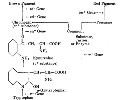
(a+, v+, cn+) substance may be involved in the process of eye pigment formation. Actually, in Drosophila the intervention of two genes, (cd+,
Brown Pigment
«— st+ Gene Î - cd+ Gene Chromogen <
(cn+ substance)
«— cn+ Gene
O
1
Red Pigment bw+ Gene ■>
; -> Precursor
A 4 - C H , - <
V x
CH—COOH NH2
Substrate, Carrier, or Enzvme
î
| <— w+ Gene NH2 Kynurenine
| (v+ substance) '
1—CH2—CH—COOH NH2
Î α-Oxy tryptophan I «— v+ Gene
Tryptophan
FIG. 5.—Scheme of development of eye pigments in Drosophila. Vertical arrows indicate steps in the reaction chain; horizontal arrows indicate the places where normal (wild type) genes are necessary for the next step of the reaction. (From Beadle, 2.)
"cardinal") and (st+, "scarlet"), in addition to (v+) gene and (cn+) gene, is necessary for the formation of the brown pigment (Fig. 5).
In recent years a series of investigations reported in rapid succession led to the determinination of the chemical nature of the eye color hor- mones (see 51,52). First an analysis of the chemical properties of puri- fied extracts suggested that the eye color hormones resemble amino acids.
Feeding experiments then established tryptophan as a most likely precursor of (v+) substance. Tatum and Beadle (153) succeeded in crystallizing a material having the physiological effects of (v+) substance, which they had obtained from bacterial synthesis (151). The (v+, a+)
substance could finally be identified as kynurenine, a derivative of tryptophaii, by these means: (1) Butenandt, Weidel, and Becker (26) showed that L-kynurenine has the same physiological and chemical prop- erties as (a+, v+) substance, while D-kynurenine as well as kynurenic acid cannot replace either the (v+) or the (cn+) substances; (2) the active principle synthesized from L-tryptophan by certain bacteria was identi- fied as a sucrose ester of L-kynurenine, the L-kynurenine being the active portion of the molecule (154); (3) kynurenine was demonstrated to occur in Drosophila pupae and Bombyx eggs (87).
In the insect organism kynurenine is apparently formed by way of 2-hydroxytryptophan (α-oxytryptophan, prokynurenine) from trypto- phan. This chain of reactions appears to take place by means of an enzymic system which is activated by the (v+, a+) gene (27,87). Simi- larly the next step, from kynurenine to (cn+) substance, depends on the action of the (cn+) gene. In vitro experiments show that the pigmentation of explanted Drosophila eyes in a medium containing kynurenine may be inhibited by the addition of KCN (37). In the mutant strains the enzymic oxidation of tryptophan may be inhibited, an assumption which is supported by the finding that (aa) Ephestia contains signif- icantly more tryptophan than (a+a*1) Ephestia (30,32a,32b). An alternative would be that in (aa) Ephestia less tryptophan is available due to the synthesis of qualitatively different proteins in this strain (32).
The most recently discussed questions concern the nature of the (cn+) substance, and the mechanism by which gene-controlled substances influence the development of eye pigments. There is good evidence to support the assumption that the eye color hormones are chemical pre- cursors of certain eye pigments (32b). A quantitative study in Ephestia (90) showed that the amount of eye pigment formed is directly propor- tional to the amount of kynurenine administered. The hypothesis that the (cn+) substance which is derived from kynurenine represents the chromogen of the brown eye pigment of Drosophila (an "ommochromeM; 5) is based on the finding (87) that Drosophila strains containing (cn+) substance yield a positive Ehrlich diazo reaction. The conclusion that it is the (cn+) substance itself which brings about the positive reaction is suggestive, although it has not been definitely proved.
Accordingly, the development of one of the two independent eye pig- ments of Drosophila, the brown pigment, may be visualized to take place as indicated in Fig. 5.
The mechanism by which the red eye pigment of Drosophila develops is as yet little understood; it is known, however, that its development does not depend upon the presence of diffusible tryptophan derivatives.
There exists a common step in the development of the brown and red
150 BERTA SCHARRER
pigments, but the reaction chains leading to the formation of these pig- ments are different.
VI. Sources of Insect Hormones
The organs in the insect body which are either known or assumed to be sources of hormones are summarized in Table I. Among these the corpus allatum is perhaps the most versatile gland of internal secretion in this group of invertebrates. Its action in developmental and repro- ductive processes as well as color adaptation has been discussed (see Sections I, II, III). Aside from these functions a certain influence of the corpora allata on tissue growth and maintenance has been demonstrated:
(a) allatum implants in adult Dixippus restore the capacity to regenerate lost extremities (115); (b) degenerative processes or uncontrolled tumor- like growth may take place in Dixippus nymphs after allatectomy in certain parts of the body (musculature, malpighian vessels, fat body, nerv- ous system; 114);4 (c) after removal or denervation of corpora allata in newly emerged adult flies the imaginai fat body and the oenocytes show signs of regression while the larval fat body fails to disappear completely (41). Furthermore, there exists in Melanoplus a possible correlation between corpus allatum hormone and blood color (110). It is possible that all these actions are correlated with the regulative effect which the corpora allata appear to have on metabolic processes.
Implantation of corpora allata into normal last instar nymphs causes regression of the corpora allata of the host (116).
The number of existing allatum hormones is not known (p. 139).
Certain investigators maintain that one hormone may account for all the various effects attributed to the corpus allatum.
The corpus allatum (Fig. 6) is a glandular organ whose morphological relationships have been studied extensively (33a,46,73,75,75a, 102,103a).
It is situated in the head or anterior thorax and is paired in most insects but unpaired in some, such as Rhodnius (Fig. 8). Histological signs of secretory activity (cytoplasmic inclusions, acidophilia, vacuoles, lobated nuclei) are more pronounced in some insect species than in others (42,62, 112,147,169). The increase in size of the corpus allatum during the adult stage (47a,78,156) and the sometimes pronounced sexual dimorphism (75,113,147) seem to be manifestations of differences in physiological activity.
4 It may be remarked that in this case tumor-like growths appear actually to be caused by an endocrine disturbance, inasmuch as their occurrence can be prevented by corpus allatum implants. Tumors in various organs were also observed in a different species (Leucophaea maderae) after allatectomy, but their origin has definitely been traced to the incidental cutting of the recurrent nerve (139).