Hormones in Crustaceans
B Y F R A N K A . B R O W N , JR.
CONTENTS
Page
I. Introduction 159 II. H o r m o n e s a n d Sex Characteristics 160
A. General 160 B. Male Sex Characteristics 161
C. Female Sex Characteristics 163 D . General Conclusions 163 III. Hormones and Color Changes 164
A. Geneial 164 B. T h e Chromatophores and Their Normal Activity 164
C. Hormonal Control of Chromatophores 166 1. General Historical Background 166
2. T h e Sinus Gland 170 a. Structure and Innervation 170
b. Chromatophorotropic Activity 171 c. T h e Number of Principles and Their Activities 173
3. Chromatophorotropic Hormones from Central Nervous S y s t e m . 175
4. Properties of the Chromatophorotropic Hormones 180 5. Identities and Phylogenetic Distribution of the Hormones 181
6. Control of Secretion of the Hormones 181
D . General Summary 182 IV. Hormones and Retinal Pigment M o v e m e n t s 182
A. Retinal Pigments and Their Normal Activities 182
B. T h e Role of Hormones 184 C. General Summary 187 V. Hormones and Molting and Growth 187
A. T h e Molting Process 187 B. T h e R o l e of Hormones 188 C. General Summary 191 VI. Hormones and Other Activities 192
A. Viability 192 B. Heart Rate 193 C. Blood Sugar 194 D . L o c o m o t o r Activities 194
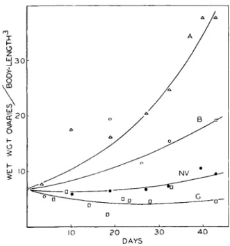
E. Ovarian Development 194
References 195 I. Introduction
A number of hormones, produced at specific points within the body and transported in the blood, have now been satisfactorily shown to be
159
concerned in coordination and integration in crustaceans. There is, furthermore, strong suggestion that still other processes are normally influenced by hormones, though reasonable proof of these latter is still lacking. It appears that the same general types of functions are con- trolled or influenced by hormones in the crustaceans as in the vertebrates.
Included in a list of such functions are color change, molting and growth, certain aspects of general metabolism, and differentiation and main- tenance of sex characteristics. The sequence in this list is also roughly the order of decreasing extent of our knowledge regarding the details of the total hormonal mechanism which is involved. In no instance has a hormone been obtained in a pure state. Experimental work in no case has proceeded beyond studies of the results of extirpation of tissues or organs containing the source of hormones, implantations of these tissues or organs, blood transfusions, or injections of either crude extracts of the glandular tissue or of partially fractionated extracts still probably con- taining a wide variety of substances. Despite this, there has accumu- lated a fairly considerable body of information as to the roles that certain endocrine tissues and their hormones play in the economy of the indi- vidual. In the absence of chemical isolation or purification of the active principles, however, it is frequently very difficult to delineate the exact role of each hormone by itself. Hence it has not been possible, in general, to demonstrate the identities of similarly acting hormones from different species and to apply names to these principles with any real degree of assurance.
Compared with the state of our knowledge of vertebrate hormonal mechanisms, our knowledge of those of the crustacean is in a most ele- mentary and fragmentary state.
A number of reviews have been written on the general subject of invertebrate hormonal mechanisms (62,65,91,93,97,127,154). Other and more recent reviews have been restricted to crustaceans (27,30,81).
II. Hormones and Sex Characteristics
A . GENERAL
The malacostracan crustaceans, in general, are dioecious and show a distinct sexual dimorphism, with the two sexes readily distinguishable on the basis of a number of characteristics. The first suggestion of a hor- monal activity within crustaceans came from observations on the develop- ment and maintenance of these secondary sexual characteristics, and was first called forth as an hypothesis to explain the results of parasitic cas- tration (50) of male decapod crustaceans.1 The earlier observations are
1 Cf. Section III, D of the preceding chapter (p. 138) for the effects of parsitic cast- ration in insects.
ably summed up in the excellent paper of Tucker (140). Parasitic castration by rhizocephalans such as Sacculina, Peltogaster, Triangulus, and Lernaeodiscus has been described frequently. These organisms, after a brief existence as free-swimming larvae, become attached to the body of their host and eventually become little more than sacs containing reproductive organs and with nutritive roots growing deeply through the host, destroying tissues and organs, and robbing host nutrients. Other common parasites of crustaceans involved in castration are epicarid isopods such as Gyge and Bopyrus. These latter parasites enter the branchial chamber of the host as free-swimming larvae, become attached, metamorphose, and feed upon the body fluid. An infestation by such parasites leads to more or less suppression, degeneration, or occasionally even destruction, of the gonads, the extent of the effect varying greatly with the host species, parasite species, and the specific case.
B. M A L E S E X CHARACTERISTICS
Parasitized males commonly show incomplete differentiation of such typical secondary sexual characteristics as the specialized copulatory pleopods, the narrower abdomen, and the larger male-type chelipeds.
These portions of the body tend to assume forms resembling more closely the homologous parts in the female.
This modification of the sex characteristics has been explained in various ways by different investigators. Smith (132,133,134) noted that females showed a greater rate of fat production than males and that the parasitized males resembled females in this regard. Smith formulated the hypothesis that the parasite in the male imposed the same metabolic demands on the host as the normally active ovary of the female. Both utilized large amounts of fats. He believed that sexual-formative stuffs, related in some manner to the fat metabolism, were involved in influenc- ing ovarian or testes activity and, in parallel, the secondary sexual char- acteristics. This hypothesis has been supported by Robson (124), Tucker (140), and Hughes (71).
Another hypothesis was that set forth by Biedl (17), who suggested that the parasite was female and liberated a feminizing hormone into the host's blood. Others have criticized this view on the ground that the parasite is not a female but a hermaphrodite.
The first investigator to suggest action of a male hormone was Courrier (43) working on parasitized male Carcinus. He could find, however, no correlation between the degree of suppression of the gonad and the extent of influence on the secondary sex characteristics. Therefore, he con- cluded that the male hormone must be formed in some tissue other than the gonads, and that this source must be suppressed or destroyed by the
parasite. Okada and Miyashita (107), working on the crab, Eriocheir, confirmed Courrier in finding no significant correlation between the degree of suppression of the gonads and of the secondary sex characteristics.
Lipschutz (100), on the other hand, assumed a normal liberation by the testis of a male hormone. Its absence or reduction following parasitiza- tion was considered to result in a change of the host toward a neutral form. This view was also upheld by van Oordt (143,144) and was given strong support by the work of Brinkman (20), who found in an extensive study of parasitization of the male crab, Munida, with three species of rhizocephalans that there was (1) a good correlation between the degree of suppression or degeneration of the testis and the extent of modification in the female direction, and (2) no similar correlation between the size of the parasite (and hence the nutritional drain on the host) and the change.
Brinkman believed, however, that both a male hormone and malnutrition were involved in the explanation of the feminization.
Another interpretation of the results of parasitic castration in male crustaceans was that of Goldschmidt (51), who considered the parasitized form to be an intersex as a result of an influence of the parasite upon the normal expression of the sex genes. A somewhat similar point of view was adopted by Callan (38), who found no perceptible influence of castra- tion upon the secondary sex characteristics of the male shrimp, Leander.
The latter investigator believed that the different species of crustaceans varied widely in the stability of their sex-determining mechanism, with parasitic infestation being able to tip the balance much more readily in some than in others. On the basis of this hypothesis Leander appeared to show a rather stable condition. Less stable conditions were found by Potts (119), who reported that parasitized male Eupagurus showed pro- duction of ova in the testis. Comparable observations were made by Smith (132) for Inachis, and Tucker (140) for Upogebia. Evidence for a normal tendency toward hermaphroditism in higher crustaceans has been reported by Rünnstrom (125) and Turner (141).
Certain seasonal variations in secondary sexual characteristics in the male crayfish also suggest an action of hormones. The copulatory appendages typically show a seasonal variation in form, assuming a sexually functional form at the summer molt and a nonfunctional form at the spring molt. Scudamore (130) has pointed out that the time of metamorphosis to the functional condition is a time of high testis activity, according to Fasten (47), and that there is a minimum of testis activity at the time of the spring molt. Scudamore also found that molts experi- mentally induced by removal of the eyestalks during the winter months invariably yielded the nonfunctional type, and correspondingly this also was a time of low testis activity.
C . FEMALE S E X CHARACTERISTICS
Female crustaceans in general do not appear to show an extensive modification of their secondary sexual characteristics upon parasitic castration as do males. Potts (119) working on Eupagurus, and Miya- shita (105) on Eriocheir have described a tendency of parasitized females to retain juvenile characteristics. Potts also described a slight change of form of pleopod from the typically biramous type of the female toward the uniramous one of the male.
The female does, however, commonly exhibit certain seasonal modi- fications of body form associated with breeding activity. These changes may involve brood pouches, incubatory chambers, and related structures.
Le Roux (98,99) has observed that parasitic castration of Gammarus by a worm, Polymorphus, usually results in failure of development of charac- teristic marginal setae of the oöstegites. These setae were similarly inhibited when ovarian activity was suppressed by irradiation, but did eventually develop, along with a restored ovarian activity, following cessation of irradiation treatments. Haemmerli-Boveri (52) working on Asellus, and Mori (106) working on Daphnia reported that irradiation resulted in suppression both of ovaries and of brood pouch formation.
Callan (38) found with female Leander that parasitic castration and x-ray castration were both associated with failure of the typical incuba- tory chamber to differentiate during the breeding season. The typical pattern of white-reflecting chromâtophores of the female was also absent (87). Callan leaned toward an explanation of his results in terms of the activity of a hormone arising in the ovary, but realized that other inter- pretations of the results were not ruled out.
Recently McVay (101) has reported finding a seasonal fluctuation in abundance in female crayfish of a chromatophorotropic factor from the brain concentrating white pigment. Males and females showed similar quantities during the nonbreeding period, while females possessed sub- stantially less during breeding activity.
D . GENERAL CONCLUSIONS
We may sum up by saying that many observations suggest strongly that hormones are operative in the development and maintenance of male and female sexual characteristics. Such a suggestion arises chiefly from the often-demonstrated fact that suppression or degeneration of the gonads following parasitic castration or irradiation is commonly asso- ciated with more or less modification of the sex characteristics in the direction of either a neutral form or of the opposite sex. The extent and character of the modifications vary with the host and the parasite species and with the individual case. Crucial experiments have not yet been
performed to enable us to conclude definitely that specific sex hormones are actually operating, and, if so, in what tissues they arise. Resolution of this problem must await results of conventional endocrinological experi- ments involving studies of the effects of tissue extirpations and replace- ments, and the isolation and physiological study of the active principles.
III. Hormones and Color Changes1
A. GENERAL
The first decisive demonstration of hormonal activity in the crusta- ceans came from a study of the controlling mechanism of physiological color changes, and it is on this subject that the largest amount of research on crustacean hormones has been done. Just as a similar approach to vertebrate hormonal mechanisms would have probably soon led to two very important hormone sources, the adrenals and the pituitary, so it happened that this attack on the crustacean led to two rich sources of hormones. One of these, the sinus gland, usually within the eyestalks, has already been shown to possess a number of important functions other than control of color change. Other sources, within the central nervous organs, also appear to give some indication of possessing functions within the organism of more fundamental importance than control of color changes.
B . T H E CHROMATOPHORES AND T H E I R NORMAL ACTIVITY
Color changes in the crustaceans are brought about by the activities of chromatophores. The earlier literature on this subject has been extensively reviewed (6,48,112). The chromatophores comprise numer- ous small syncytial bodies in the hypodermis, or directly beneath it, and over certain of the internal organs. According to the opinion now most generally held, these bodies have diffusely branched, radiating processes of a permanent character. Within a single animal the chromatophores over the body may contain pigments of one or, more commonly, several different colors. Crustacean pigments include a black or sepia (particu- late melanin), reds and yellows (Carotinoids), blue (a carotinoid-protein complex), and reflecting white (particulate guanine.) Each pigment within a chromâtophore possesses the capacity of (1) moving centripetally to form a small mass near the chromatophore center (pigment concentra- tion), or (2) dispersing centrifugally until the whole of the chromato- phore, even to the tips of its branches, is filled with the pigment (pigment dispersion), or (3) maintaining any intermediate degree of dispersion or concentration. The degree to which any given pigment contributes to the gross coloration of the animal is a function of the degree of dispersion
1 Corresponding phenomena in the vertebrates will be treated in Volume 2.
of that pigment within the chromatophore. Only the blue pigment commonly appears outside of chromatophores where it often seems to pervade the general body tissues.
The chromatophores may be classed as monochromatic, dichromatic, or polychromatic depending upon the number of types of pigment found within each. When more than one pigment is present within a single chromatophore, these pigments usually remain separated from one another and usually each possesses its own chromatophore branches into which it disperses.
The chromatophore system of many crustaceans possessing trans- parent or translucent cuticles constitutes a very effective mechanism for enabling the individual to mimic rather closely the shade, and often even the tint, of the background upon which it comes to lie. The common shrimp, Palaemonetes, possesses within its chromatophore system red, yellow, blue, and white pigments. By the appropriate differential dis- persion or concentration of these four pigments in sympathetic response to backgrounds, the animal is able to assume any spectral color, and may even closely approximate black on the one hand, or almost complete transparency on the other. The shrimp, Hippolyte, appears to possess, in addition to these abilities, power to assume adaptive color patterns as well (49,104). The sand shrimp, Crago, which contains black, brown, red, yellow, and white pigments, lacks the ability to assume bluish or greenish tints though displaying otherwise considerable powers of modify- ing its shade, tint, and general pattern of coloration to conform to its background (89). Such remarkable powers of chromatic adjustment are obviously possible only in terms of relatively independent control, on the part of the animal, of each of its various pigments.
The chromatophore system also typically responds to changes in light intensity, usually assuming one state characteristic for the species during darkness at night, and another in light during the daylight hours. In some species this appears to be exclusively determined by the light intensity change while in others the response is strongly conditioned or even determined by an inherent diurnal rhythm within the animal. In the latter instances the typical diurnal changes may proceed even under conditions of a constant state of illumination and background. A species showing the former type of response seems to be Palaemonetes, where the animal on a black background pales in darkness and darkens in light without relation to the diurnal cycle. The fiddler crab, Uca, on the other hand, normally pales at night and darkens by day despite the maintainance of constant illumination and background. The latter species shows relatively little adaptive response to color of background in light.
C. HORMONAL CONTROL OF CHROMATOPHORES
1. General Historical Background
It was assumed for many years that the chromatophores of crusta- ceans were directly innervated organs, though it gradually came to be realized that no one had demonstrated histologically any nerve endings at these organs, nor could workers show that the types of nerve transec- tion which they performed interfered significantly with color changes.
Koller (88,89) working on the shrimp Crago vulgaris, was the first investi- gator to provide positive evidence that controlling agents for crustacean color changes are carried in the blood. Koller's experiments involved transfusion of blood from one animal to another. He noted that when blood from a black animal was transfused to a white one, the latter darkened even though kept upon a white background. Blood from a white donor had no such effect, though neither did it lighten black recipients maintained upon a black background. Blood from a yellow- adapted animal induced distinct yellowing of a white animal. These blood transfusions brought about the color changes at approximately the same rate as normally followed the corresponding background changes.
Perkins (116) found no evidence whatsoever that the chromatophores of the shrimp, Palaemonetes vulgaris, were under the control of nerves.
He could discover no direct nerve innervation, nor would extensive nerve transection experiments interfere with the color changes in this form.
When, on the other hand, blood flow in the dorsal abdominal artery supplying the posterior portion of the body was stopped, color change posterior to the point of stoppage immediately ceased. Later when blood flow resumed, the posterior portion of the body quickly assumed the color of the remainder of the body. Perkins interpreted these results as due to blood-borne factors inducing pigment concentration and dispersion.
In attempting to determine the origins of these hormones, he extracted separately, in sea water, numerous organs and tissues of the body and observed the action of injections of these extracts into black- and white- adapted individuals. Of the numerous extracts tested, only one—
extracts of the eyestalks—resulted in lightening of dark specimens, and none produced darkening of light individuals. Perkins also found that extracts of eyestalks from white-adapted donors were much more effec- tive than those from black-adapted ones. Animals from which the eye- stalks were removed darkened and remained so permanently. On the basis of these experiments Perkins concluded that the eyestalks contained the source of a hormonal substance which lightened the body through con- centration of the red and yellow pigments.
These results were completely confirmed for Crago, Leander, and
Processa by Koller (90), who also demonstrated that the eyestalk hormone was not species or even genus specific. Koller sought further for the source of the blood-borne principle which resulted in the darkening of white-adapted Crago which received a blood transfusion from a black one.
He found that injection of extracts of the rostral region of black-adapted Crago or Leander caused darkening of white-adapted Crago. White- adapted animals darkened after feeding on the rostral region. Cautery destruction of this region deprived animals permanently of the ability to darken again. Koller concluded that an endocrine gland was located in the rostral region and that it produced a principle influencing the dark pigments of the body antagonistically to one from the eyestalk.
Following these initial demonstrations of hormonal control of crusta- cean color changes, many crustaceans were examined to determine how generally the hormonal activity was present within the group. The eye- stalks, or occasionally the heads instead, of some seventy to eighty species of crustaceans were shown to yield sea water extracts with strong chroma- tophorotropic activity upon the chromatophores of injected animals.
This activity was generally similar to that which had been found in the eyestalks of Palaemonetes and Crago. The chromatophorotropic mate- rial appears so commonly present among the higher crustaceans that the three isopods, Oniscus, Porcellio, and Mesidothea, reported not to have it (137,138) should certainly be thoroughly re-examined.
Attempts to repeat Koller's observations on the rostral-region gland have met with almost uniformly negative results. Beauvallet and Veil (12) working on Leander reported confirmation of Koller's rostral gland, but Panouse (109) and Carstam (41) also using Leander failed to confirm it. Attempts to discover a rostral gland for Palaemonetes by Perkins and Snook (117) and Brown (22), for Cambarus by Hanström ,(60), and even to confirm the presence of one in Crago itself by Kropp and Perkins (95), Kleinholz (80), Panouse (109), and Carstam (41) all led to negative results. It appears therefore that no endocrine gland with the function ascribed to it by Koller lies in the rostral region. There is now reason to suspect that a hormone originating in anterior central nervous organs was responsible for the positive results which were obtained occasionally.
This latter possibility will be considered later in Section 3, page 175.
Numerous observations have been made of the effects of extirpation of the eyestalks upon the state of the chromatophores in a number of species of crustaceans. It is unfortunate for the interpretation of the results that the operation removes not only the gland in question (see Section 2 below), but also important central nervous organs and the principal photoreceptors known to be essential to the normal reflex color adaptations of the animals. The observations at hand suggest that
crustaceans fall into three groups (Fig. 1) with respect to the character of their response to eyestalk removal. One group, exemplified by the shrimp, Palaemonetes, includes the majority of Isopoda, Mysidacea, Natantia, and Astacura which have been investigated. The dominant dark pigments of these animals disperse widely, yielding a permanently darkened condition of the body. Injection of eyestalk extract into these eyestalkless animals induces rapid lightening.
EG. PALAEMONETES GROUP Ή
ε.α CAAGO GROUP ITT
ε.6. UCA
ετεετΑΜα HEUOVEO
INITIAL FINAL αεςροΝεε ςτΑαε
INJECTεO WITH SINUS-GLAND
EXTRACT
BLACK WHITE
F I G . 1.—Diagrammatic representation of the results of removal from crustaceans of the eyestalks with their included sinus glands upon the coloration and dominant chromatophore types (top r o w ) . All crustaceans so far investigated fall into one or another of the three groups. The b o t t o m row shows the influence of injection of eye- stalk or of sinus gland extracts from other animals of the same group into the eyestalk- less specimens. Dotted arrows indicate an alcohol-soluble fraction only; dashed arrows indicate only an alcohol-insoluble fraction. Reciprocal injection experiments among the three groups show that crustaceans of group I I I possess no telson- and uropod-lightening activity for Crago of group I I , but otherwise there are no qualitative differences.
A second type of response is found in Crago. Eyestalkless Crago show an intermediate and mottled coloration (24). Some of the dark chromatophores have their pigment broadly dispersed, others are in an intermediate condition, while still others have theirs fully concentrated.
These animals respond to eyestalk extract injection by uniform blanching.
A third type of response, exemplified by the crab, Uca, is exhibited by all the Brachyura (true crabs) which have been investigated. Eye- stalk removal in these yields a permanently pale condition of the body due to maximum concentration of the dominant dark pigment (1,39).
Injection of eyestalk extract results in rapid darkening of the body, the reverse response to that seen in the first two types.
The state of the pigments following eyestalk removal seems to differ in various crustaceans, with each species possessing its own character- istics. In view of this situation and the fact that reciprocal injection experiments seemed to suggest that regardless of the species contributing the eyestalks, their extracts would call forth the same reaction as an extract of stalks from the same species, Abramowitz (4) proposed the hypothesis that all the crustacean pigmentary behavior could be explained through the action of one hormone, eyestalk hormone—ESH of Abramo- witz and Abramowitz (8). The differences in response among species were believed explainable in terms of differences in the thresholds and in the characters of response of the various chromatophores to the single hormone. This concept became known as the "unitary hormone hypoth- esis " and has been supported by a number of investigators.
In contrast with the unitary hormone hypothesis was the "rhultiple"
one. According to this concept all the observed pigmentary responses could not be explained in terms of a single chromatophorotropic prin- ciple. This view was implied in the work of Perkins (116) by his factors for concentration and dispersion, and definitely supported by Koller (89,90,92) with his work on the eyestalk hormone, the rostral-region hormones, and his yellow factor from elsewhere in the body. Also the work of Koller (89) on Crago, Brown (22) on Palaemonetes, Abramowitz (1) and Hitchcock (69) on the crab, Portunus, showed that in adaptation of these animals to colored backgrounds, various combinations of pig- ments displayed ability to distribute themselves within the chromato- phores more or less independently of one another. Such relatively independent activity of the pigments had been known for many years to be true for the shrimp, Hippolyte (Keeble and Gamble, 74, and Min- kiewicz, 104). Brown (22) proved by nerve transection experiments that the independent activity of the four pigments of Palaemonetes was wholly the result of hormonal action and suggested that at least four chromato- phorotropic hormones were present to account for the observed phe- nomena. Parker (114) pointed out that three principles would account for the behavior in this species. Smith (135) by very ingenious experi- mentation has presented evidence for separate body-lightening (W-factor) and body-darkening (B-factor) principles in an isopod. Carstam (41), working on Leander, demonstrated separate controlling factors for the red and yellow pigments in this shrimp.
In addition to the preceding work, several experiments indicated that chromatophorotropically active substances could also be extracted from central nervous organs of crustaceans. Brown (21) discovered that
extracts of these organs of Palaemonetes would induce paling of dark, eyestalkless animals though no other organ of the body except the eye- stalks would do likewise. This observation was confirmed for two species of Penaeus by Hosoi (70) and Hanström (60). Knowles (86) found that extracts of central nervous organs concentrated white pigment in Leander.
Brown (22) noted that the dark and white pigments of eyestalkless Palaemonetes could be made to concentrate within the chromatophores in response to electrical or heat stimulation of the cut ends of the optic nerves. This last observation found a reasonable interpretation in the activity of hormonal material originating in the central nervous organs.
2. The Sinus Gland
a. Structure and Innervation. Hanström in 1933 (54) described for the first time in the crustacean eyestalk a gland which he first called the blood gland but later (60) named the sinus gland. Since that time the gland has been found in all the higher crustaceans in which it has been
A B C
F I G . 2.—The sinus glands in the eyestalks of A , Palaemonetes, B, Crago, and C, Uca, as seen from the dorsal view. In species such as Crago and Palaemonetes possessing transparent cuticles the gland is clearly visible in the intact living specimen in which it appears as a bluish-white opaque organ against the more transparent grayish-white underlying nervous tissue. (Modified from Brown, 26.)
sought (26,31,41,60,131,138). In the vast majority of the stalk-eyed crustaceans it lies in the eyestalk (Fig. 2). In some stalk-eyed species (e.g., Upogebia and Emerita) and in species without eyestalks the gland lies close to the supraesophageal ganglion in the head. In the decapod crustaceans upon which most experimental work has been done the gland occupies a dorsal or dorsolateral position in the eyestalk, most commonly lying opposite a point between the medulla externa and interna. Less commonly it lies opposite the medulla interna, while in a few species it has an attenuated form and occupies a position opposite the medulla interna and medulla terminalis. In shrimp with highly transparent cuticles, such as Palaemonetes and Crago, the giand is clearly visible in the intact living animal held in bright incident illumination. The gland possesses a more bluish-white coloration than the remainder of the stalk tissue, probably due to the large amount of intracellular inclusions of the gland
cells. In species with thick opaque cuticles the gland may be seen in fresh tissue by dissecting away the dorsal exoskeleton and hypodermis of the stalk. The gland occupies less than the volume of the eyestalk in the crayfish, Cambarus (31).
Hanström (60) believes that the gland originates phylogenetically as a thickening of the neurilemma over the nervous elements of the eyestalks, with its simplest and most primitive form found in certain mysids, euphausids, isopods, and amphipods. In the Natantia with few excep- tions, the gland occupies a portion of the neurilemma at a point where a blood sinus within the central nervous system opens into the large super- ficial sinus of the stalk and thus the gland possesses a beaker-shaped form.
In the Astacura the inner blood sinus has become complexly branched, and, with this, the sinus gland which occupies its walls. In most of the Brachyura examined, the gland has the form of a hollow sphere. Here the primitive gland is believed to have separated from the neurilemma, become invaginated, and liberate its products into the lumen which is connected with both the inner and the outer blood sinuses. In those decapods in which the gland is in the head instead of the eyestalks, as in certain anomurans, the gland appears secondarily simplified to form a simple plate of glandular tissue in contact with only an outer blood sinus into which the contents appear to be discharged directly.
The cells comprising the sinus glands appear richly charged with acidophilic inclusions, staining with eosin, acid fuchsin, and light-green (Hanström, 60). Also described are basophilic inclusions with the rela- tive abundance of the two types of granules varying with the different stages in the molting cycle in the crayfish, Cambarus (120). Hanström (60) also described for the cells fine secretory canals for the conductance of gland products to the sinus.
The gland is richly innervated. It is supplied on its inner surface by a large nerve arising in the medulla terminalis (60), and at least in Cambarus some fibers of this nerve appear to arise in the supraesopha- geal ganglion (153). In this latter species Welsh (153) has also described a branch of the oculomotor nerve passing to the region of the gland.
Thus, the gland appears to have a triple innervation.
b. Chromatophorotropic Activity. Shortly following upon the demon- stration that the eyestalks of crustaceans produced hormonal material active upon chromatophores came attempts to localize the source within the stalk. Koller (92) divided the stalks of Crago transversely into two portions, the sensory portion and the remainder. Since the sensory portion showed slight activity, even though by far the greater part of the activity lay in the remainder of the stalk, Koller concluded that the blood gland at the base of the retina was the source. Destruction of this sup-
posed source by cautery produced lasting body darkening, therefore adding apparent confirmation to his conclusion.
Hanström (60) believed that Koller's blood gland could not be the actual source of the active material since it was not present in the chroma- tophorotropically active eyestalks of some species such as Palaemonetes.
Furthermore, the gland was not innervated as would perhaps be expected.
Nor did Hanström believe that the source of the hormone in question was the X-organ of the crustacean eyestalk (18,41,53,54,55,56,57,58,60, 66,138) even though this organ appeared to possess the histological char- acteristics of an endocrine gland. The X-organ, also, was not found in a few species bearing active eyestalks (e.g., Astacus, Uca). On the other hand, Hanström's sinus gland was found in all the numerous malacostracan crustaceans in which it was sought; its cells showed every indication of active secretory activity, and it was well innervated as it seemed reason- able to expect in view of the reflex nature of crustacean color changes.
Hanström (59,60) carried out an extensive survey of a wide variety of species of crustaceans in which the eyestalks, or heads of those species in which the eystalks were inactive, were sectioned in various ways and the portions extracted and injected into species with active, readily observable chromatophores. His assay animals consisted in different experiments of Palaemonetes, Uca, or Penaeus. In these experiments Hanström utilized in a very ingenious manner the species differences in the position of the sinus gland with respect to other organs in the eyestalk or head. He showed quite conclusively that every active portion always contained the sinus gland in whole or in part and that no inactive portion ever did. Furthermore, he managed by judicious selection of species to get one by one every conspicuous organ of the typical crustacean eyestalk into a portion without the sinus gland and found extracts of each one in turn to be inactive. The sinus gland therefore appeared to be the exclusive source of hormonal material blanching the bodies of dark shrimp on the one hand, and darkening the bodies of pale fiddler crabs on the other.
These conclusions were fully confirmed by Brown (26), who removed the sinus glands by themselves from a number of crustaceans: Callinectes
(blue crab), Carcinides (green crab), Crago, Libinia (spider crab), Pagurus (hermit crab), Palaemonetes, and Uca. The activities of extracts of the glands by themselves were compared with extracts of the remainder of the eyestalks in their action in concentrating the red pig- ment of Palaemonetes on the one hand and dispersing the black pigment of Uca on the other. It was found that approximately 80% of the activity of the whole eyestalks was present in the sinus glands which occupied less than 1% of the total volume of the stalks. In dissecting
out the sinus glands from the stalks a bluish-white cloud of colloidal material could usually be seen to pass out of the gland into the surround- ing tissues. Such an escape of substance could reasonably be expected to account for the remaining 20% of the activity seen in the residual stalk tissue. It was found, furthermore, that the activity of the glands by themselves was the same as that of the remaining stalk tissue in relative effectiveness upon the two types of chromatophores, further suggesting the gland as being the sole source in the stalk of hormonal material influencing these two chromatophore types. Still further confirmation was also found in the action of implants of sinus glands in the ventral abdominal sinus of Palaemonetes. A single implant maintained the red pigment in eyestalkless specimens more or less concentrated for as long as five days, i.e., many times as long as that ever found following injection of highly concentrated extracts of eyestalks.
The only attempt that has been made to remove the sinus glands alone from the eyestalks for the study of chromatophoric responses (33) involved the bilateral removal of the glands by microaspiration from several specimens of Palaemonetes. Such sinus-glandless animals became dark and showed no ability to concentrate their dark pigments in response to white backgrounds. Proof that the glands were com- pletely removed was afforded by injecting into test animals extracts of the stalks of sacrificed animals.
The sinus gland has also been shown to affect other types of pigments than the two mentioned above. Each of the eight physiologically differ- ent pigments of Crago shows its own response to injection of sinus gland extract (32,37). The pigments are induced to concentrate, or disperse, to different relative extents. The white pigment of Cambarus has been shown to disperse, and the red to concentrate, under the influence of sinus gland extracts (34).
From the preceding account it is seen that much evidence exists that the sinus glands are chromatophorotropically active, and there is no evidence that any other eyestalk organ is active in this regard.
c. The Number of Principles and Their Activities. The eyestalks, or the sinus glands by themselves, yield hormonal material which upon injection produce within the shrimp at least grossly the same response (body lightening) as that normally called forth by a white background in light, and within true crabs the typical dark coloration seen in the day- time phase of their diurnal cycle (4). The removal of the eyestalks from the shrimp brings about a state of the coloration which tends in the gen- eral direction of that seen in normal response to an illuminated black background, and in the crab to a condition simulating the nighttime phase of its diurnal cycle. However, the coloration of eyestalkless
shrimp is distinguishable from black-adapted ones. Palaemonetes always remains more reddish-brown, and it will be recalled that Crago simply reaches finally an intermediate mottled coloration. Supporters of the "unitary hormone hypothesis" discussed earlier assumed that a single chromatophorotropic hormone (ESH) was produced by the sinus gland. However, in view of the overwhelming weight of the evidence indicating that two to several chromatophorotropic hormones must be present within the animals, there seemed to be a reasonable possibility that the sinus gland itself was responsible for more than one of them, especially since all of the numerous pigmentary types investigated among crustaceans were shown to be affected by extracts of this gland.
Brown and Scudamore (36) sought to determine whether or not a single hormone from the sinus gland is responsible for all of the observed reactions of the chromatophores to extracts of the gland. They made a comparative survey of the effects of eyestalk and sinus gland extracts from Crago, Carcinides, Libinia, Uca, Pagurus, Callinectes, and Palae- monetes simultaneously upon Uca-black, and Palaemonetes-red, pig- ments. The ratio, (effect upon Uca-black)/(effect upon Palaemonetes- red), differed in a repeatable manner with the species source of the gland.
The order of decreasing size of the ratio was the order of species listed above. This order bore no relationship either to the relative weights of the animals or to the apparent relative concentrations of ESH as deter- mined by Abramowitz (3) upon Uca, which were respectively: in grams, 1, 60, 50, 2, 11, 100, 1; and in Uca units, 0.25, 1.25, 4.0, 1.0, 1.25—, 0.36.
A hypothesis that the differences observed for the ratios were the result of differing concentrations of two factors, a Uca-black-dispersing hormone and a Palaemonetes-red-concentrating hormone, was fully borne out by the partial separation of the extract into two fractions, one relatively soluble in 100% ethyl alcohol and the other relatively insoluble in this solvent. The alcohol-insoluble fraction showed a significantly higher Uca/Palaemonetes ratio and the alcohol-soluble fraction showed a dis- tinctly lower one than sea water extracts of whole glands. Furthermore, a ratio closely approximating that shown by whole-gland extract was re-obtained by mixing the two fractions. These results seem most reasonably explained in terms of possession by the glands of two different hormones, with the glands of the seven species examined varying in the relative amounts of the two present.
Evidence from studies of the comparative influence of sinus gland extracts upon a third chromatophore type, the melanophores of the telson and uropods of Crago, by Brown and Ederstrom (32) point to the presence of still a third principle. Sinus gland extracts from Crago and Palae- monetes, but not of Uca and Carcinides, will produce a very powerful and
rapid concentration of this pigment. The effectiveness of the extracts in bringing about this action appears to bear no relationship to the ability of the extracts to influence either Palaemonetes-red pigment, on the one hand, or Uca-black, on the other. Therefore it appears that sinus glands of Crago and Palaemonetes contain a hormonal substance not present in significant amounts in the glands of the crabs. Brown and Wulff (37) showed that this hormone remains in the alcohol-insoluble fraction of those sinus glands which possess it (Fig. 1, page 168).
For the purposes of convenience of reference and also in order to focus attention upon three differently acting chromatophorotrophic principles of the sinus glands of crustaceans it is proposed at this time that they be named: (1) Palaemonetes-lightening hormone or PLH, (2) Uca-darkening hormone, or UDH, and (3) Crago-telson-lightening hormone or CTLH.
These terms are not applied with any intended implication that these three together necessarily constitute the total of the sinus gland hormones which influence chromatophores, nor that it is expected that it will be found eventually that any single hormone influences exclusively a single chromatophore type. On the contrary, it seems reasonable to expect that other principles will be found in the glands, with single ones influencing more than one type of pigment cell.
3. Chromatophorotropic Hormones from Central Nervous system Crustaceans from which the eyestalks with their included sinus glands have been removed normally reach a condition of the chromatophores characteristic for each species and which they maintain within rather narrow limits for an indefinite period, if the animals are not disturbed.
These eyestalkless animals are, nevertheless, capable of showing changes in the state of their chromatophores upon appropriate stimulation of the stubs of the optic nerves. These changes in those cases which have been analyzed for the means of chromatophore influence indicate that the changes are due to activity of blood-borne agents.
Uca, after eyestalk removal, continue to show a diurnally rhythmic activity of the dark pigment though in greatly reduced degree (5,36).
Eyestalkless Hippolyte continue to respond to darkness and light by concentration and dispersion respectively of their dark pigment (49,85).
Undisturbed eyestalkless Crago are occasionally seen to exhibit a transi- tory blackening of their telson and uropods (apparently associated with molting activity) or even of their whole body. Observations such as these suggest strongly that there is a normal source of chromatophoro- tropic hormonal material in a tissue outside of the eyestalks, and that this source could produce coloration changes either in the same direction as those induced by sinus glands (Uca, Palaemonetes) or oppositely (Crago).
In this connection it has been found recently (unpublished) that extracts of the central nervous system of Uca exhibit great effectiveness in darken- ing eyestalkless animals of this species. The influence of injections of extracts of central nervous systems upon the color changes of eyestalkless crustaceans is diagrammatically represented in Fig. 3.
The observations of Koller (90,92) and Brown (24) on Crago sup- ported very strongly the hypothesis that a hormone antagonistic to an eyestalk-originating one lay in a region of the body other than the eye-
F I G . 3.—Diagrammatic representation of the influence of extracts of the central nervous organs of crustaceans of each of the three differently responding groups upon the coloration and major chromatophore types of eyestalkless specimens (top row) of the same group. Dotted arrows indicate action of an alcohol-soluble fraction ; dashed arrows indicate action of the alcohol-insoluble fraction. Reciprocal injection experi- ments among the three groups show group I I I to lack the b o d y - and the telson- and uropod-darkening activity for specimens of group II, but otherwise no qualitative differences seem to exist.
stalks in this species. It has been pointed out that Koller believed the source lay in the dorsal rostral region, in the blood cell gland located there (44,94). Stimulation of the stubs of the optic nerves in eyestalkless individuals always resulted in the blackening of the telson and uropods.
The rest of the body responded more variably, sometimes lightening, at other times darkening, or showed intermediate response involving an initial lightening followed by darkening (26). These responses con- tinued after nerve transection, indicating their dependence upon blood- borne agents. Brown and Ederstrom (32) surveyed the tissues of Crago, injecting extracts of each into eyestalkless specimens of the same species.
The midregion of the circumesophageal connectives, including the con- nective ganglia, was found to be most effective in blackening the telson and uropods in these animals, with positive responses resulting in more than 90% of the experiments. Activity of the nervous system dropped off sharply along the connective anteriorly and posteriorly from this region, dropping off much more abruptly in black-adapted than in white- adapted specimens. No other tissue of the body gave a similar darkening response. These workers postulated that a telson- and uropod-darkeniug principle was produced in the connective ganglia or in the connective just posterior to it. A normal function of such a hormone is indicated by the fact that it is rather common to collect in the field specimens of Crago with a coloration (black "tail" and light trunk) indistinguishable from that of eyestalkless specimens injected with connective extract.
These observations were extended by Brown and Wulff (37), who found that extracts of the connectives affected each of the eight differ- ently responding chromatophore types within the species. The action appeared to be supplementary to the action of the sinus glands with regard to some pigments (black, brown, and red) of the body and to antagonize it with respect to others (black and red pigments of the telson and uropods and all the white pigment). The darkening action on the telson and uropods was found to reside only in an ethyl-alcohol-insoluble fraction of the connectives, while the rest of the activity was readily alcohol soluble. Therefore two active principles appeared present in the connectives. These were, in general terms: (1) a telson- and uropod- darkening principle, and (2) a body-lightening principle. In the experi- ments it was not possible to obtain a telson- and uropod-darkening fraction without body-lightening activity present. The two general types of activity were also spatially separated within the central nervous organs.
Only the connectives possessed the former, but all the major parts of the system contained the latter. It was suggested that if the telson- and uropod-darkening principle of the connectives possessed a general body- darkening action when present without the body-lightening principle, an explanation would be at hand for the earlier observations of Koller and of Brown. Supporting, but not proving, such an hypothesis were the observations that mild stimulation of the eyestubs of eyestalkless Crago resulted in blackening of both body and "tail," whereas stronger stimula- tion called forth blackening of the "tail" and simultaneous lightening of the body. In terms of this concept, weak stimulation could be considered to result in a liberation of only one of the two principles, while strong stimulation would cause indiscriminate liberation of both.
Further experiments aimed at localization of the source of the telson- and uropod-darkening hormone of Crago were carried out by Brown (26),
who found the activity to reside almost exclusively in the tritocerebral commissure (Fig. 4) lying posterior to the esophagus, and passing between the two circumesophageal connectives, together with the immediately adjacent medial aspect of the connective lying between the origin of the commissure and the connective ganglion. The tritocerebral commissure by itself showed by far the greater part of the total activity indicating that in it, or on it, was the actual cellular source of the hor- mone. Therefore practically all the activity in this regard has been
localized to a relatively minute portion of the nerv- ous system. It is very suggestive that this portion of the nervous system is closely associated with the stomatogastric, or sympathetic, system in these forms, as is the case with the corpora cardiaca of cockroaches, which Brown and Meglitsch (34) have shown to possess powerful chromatophorotropic activity upon crustacean c h r o m a t o p h o r e s . Extracts of tritocerebral commissures and the adjacent region of the connectives of Crago invar- iably darken the telson and uropods, but show varying degrees of body lightening followed by body darkening which may readily be interpreted in terms of the hypothesis of differing relative amounts of two hormones, (1) a body- but not a "tail''-lighten- ing one and (2) a body- and " tail "-darkening one.
This hypothesis was recently verified thoroughly by Brown and Klotz (33a), who were able to sep- arate quite completely the activity in extracts of the tritocerebral commissure into two fractions, through utilization of their solubility differences in alcohol and water. The alcohol-soluble fraction, as earlier predicted, blanched the bodies of eye-stalkless Crago, while the alcohol-insoluble fraction blackened both body and "tail" (Fig. 5).
A survey of other crustaceans for the presence of the Crago-" tail "- darkening principle by Brown and Saigh (35) found no other crustacean with the commissures alone active. Other crustacean groups showed : (1) other regions within the nervous system active with maximum activity in the posterior thoracic cord (the anomurans, Pagurus, Emerita, and Upogebia), or (2) nearly uniform activity for all the central nervous organs (the astacurans, Homarus and Cambarus and the natantian Palaemonetes), or (3) no activity in any part of the system (the bra- chyurans, Carcinides, Libinia, Uca, etc.). All these other crustacean nervous systems examined, with the exception of those of the astacurans,
- - CIRCUMESOPHAGEAL CONNECTIVE
TRITOCEREBRAL COMMISSURE
- - THORACIC CORO
FIG. 4.—A diagram showing the relation- ship of the tritocere- bral commissure to the other parts of the anterior central nervous system ο f Crago.
also showed strong Crago-body-lightening activity, this lightening activ- ity showing in general complemental distribution to the darkening activity. Homarus and Cambarus showed relatively slight lightening- activity, with certain parts of the central nervous system strongly black- ening both the "tail" and body proper in eyestalkless Crago. All these observations lend further support to the hypothesis previously set forth that the Crago-"tail"-darkening principle, in the absence of an antag- onistic body-lightening one, is an effective body darkener.
F I G . 5.—A photograph showing the influence of chromatophorotropins originating in the tritocerebral commissure of the central nervous system of Crago upon eyestalk- less Crago. T h e three animals were initially matched in coloration. Fifteen minutes before the photograph was taken Β was injected with sea water and served as a con- trol. A and C each received injections of the equivalent of one quarter tritocerebral commissure either as a sea water extract ( A ) or as an alcohol-insoluble fraction ( C ) .
Evidence for the presence of two chromatophorotropic hormones within the central nervous system of Cambarus has also been obtained by McVay (101), who studied the relative effects of extracts of nervous organs upon isolated red and white chromatophores of Cambarus. It is quite possible that this investigator was dealing with the same two prin- ciples previously studied.
These experiments give strong evidence favoring the existence of two hormones originating from certain loci within the central nervous system.
It is proposed that these be called, on the basis of the responses through
which they have been differentiated: (1) Crago-darkening hormone or CDH, and (2) Crago-body-lightening hormone or CBLH.
4. Properties of the Chromatophorotropic Hormones
We still know very little of the chemical nature of the color change hormones of the crustaceans. It is very evident that all the hormones are readily soluble in water and are all insoluble in such fat solvents as ether, benzene, and chloroform (2,3,5,7,40,101). Practically every investigator in the field, beginning with Koller (92), has dealt with boiled extracts, indicating that all are stable in neutral solutions during short periods of boiling. In fact there are some reports of potentiation of eye- stalk extract upon boiling. This has been found for Palaemonetes (116) and for Cambarus (60) although denied for Callinectes and Carcinides (36,101). Chromatophorotropic activity has been found to persist in dried eyestalks for long periods, even up to several months, by Perkins and Snook (117) and Hanström (60).
The solubilities of the color change hormones in alcohol show greater differences. Some are more soluble in ethyl alcohol than others. Carl- son (40) and Abramowitz (3,5) found some (up to 60%) of the activity of eyestalks in darkening eyestalkless Uca to be soluble, although Brown and Scudamore (36) found this fraction (UDH) much less soluble in this solvent than a fraction (PLH) with greater influence upon red pigment of Palaemonetes. Similarly, a third hormone of the sinus gland (CTLH) is relatively insoluble in alcohol. The hormones of the central nervous organs similarly show differences in their solubility in ethyl alcohol; one (CDH) is relatively insoluble in this, while the other (CBLH) is quite soluble.
Carlson (40) originally showed that the Uca-darkening activity of sinus glands was resistant to brief boiling in dilute HCl and NaOH but Abramowitz (3) found that longer boiling in NaOH, but not HCl, resulted in its total inactivation. Brown and Suter (unpublished) dealing with a factor influencing Cambarus red pigment found that in boiling in 0.1 Ν NaOH there was potentiation during the first 45 minutes, then rapid destruction which was complete in one and a half hours.
The most successful attempt at purifying one of the hormones was that of Abramowitz (7) using black pigment of Uca for assay. With adsorption techniques he was able to increase the concentration of the hormone nearly two hundred times. The purified substance showed reactions characteristic of amino bases.
Carlson (40) found that active material from eyestalk extract would readily diffuse through cellophane, thus indicating a relatively low molecular weight.
5. Identities and Phylogenetic Distribution of the Hormones
Of the three active principles apparently present in crustacean sinus glands, two, namely, UDH and PLH, appear to be present in all of the species examined. CTLH, on the other hand, is abundantly present in the sinus glands of the Natantia examined and absent, or nearly so, from all brachyurans.
One of the two principles occurring in the central nervous systems (CDH) is found in one or another part of all nervous systems except those of the brachyurans, while the other one (CBLH) seems to be present in all, though relatively least abundant in the Astacura.
There is as yet no clear indication that any of the color change hor- mones are identical with those found in insect heads or corpora cardiaca (34,62,64), in Limulus central nervous system, or in vertebrates, although there is a certain degree of similarity in some instances. Abramowitz (5) examined the action of eyestalk extract upon vertebrate chromatophores and the action of intermedin on crustacean ones. He found similar but not identical action. Intermedin and the eyestalk hormone influencing Uca pigment (UDH) were also shown to have many physicochemical properties in common. It was not possible, however, to balance com- parable doses of eyestalk extract and intermedin. Furthermore, inter- medin dispersed Crago black pigment (19) while eyestalk extract concentrated it.
6. Control of Secretion of the Hormones
The secretion of hormones by both the sinus glands and the sources within the central nervous organs appears controlled jointly by two factors: (1) an inherent diurnally rhythmic mechanism, and (2) the reflex responses of the animals to stimulation of the compound eyes. The relative importance of the two appears to vary with the species and the chromatophore type. At one extreme lies Uca in which stimulation of the compound eyes by changes in light intensity or background induces relatively minor changes in the chromatophore state, while a striking, diurnally rhythmical change continues regardless of eye stimulation.
At the other extreme lie such crustaceans as Palaemonetes in which the state of the chromatophores is almost entirely dependent upon the light intensity and the background, with the responses mediated through the eyes.
We know almost nothing of the mechanism of the 24-hour rhythm and very little more about the relationship between the compound eyes and the state of coloration. The darkening and lightening responses are dependent upon the relative degrees of stimulation of dorsal and ventral
portions of the retina (4,60,63,75,135); however, it is still too soon to do more than just speculate upon just what hormones, and in what propor- tions, are responsible for any given state of the chromatophore system.
D . GENERAL SUMMARY
The chromatophore system of crustaceans is controlled almost exclusively by hormonal substances arising within the sinus glands of the eyestalks or head, and within central nervous organs. The active locus or loci within the central nervous organs varies with the species but is relatively constant within each of the major groups of decapods. The sinus glands appear to possess three principles which have been named on the basis of the principal activity by which each was differentiated from the others, as: (1) PLH (Palaemonetes-lightening hormone), (2) UDH (Uca-darkening hormone), and (3) CTLH (Crago-" tail "-lightening hormone). The first two are found within all sinus glands tested, while the third is absent, or practically so, from all the true crabs (Brachyura).
The central nervous organs contain at least two active principles: (1) CDH (Crago-darkening hormone), produced in the tritocerebral com- missures of Crago and in other portions of the nervous system of other crustaceans (except the true crabs, from which it is entirely absent), and (2) CBLH (Crago-body-lightening hormone), of general distribution through all the decapods examined, being least abundant in the asta- curans, lobster, and crayfish.
These hormones have not yet been identified with any noncrustacean hormones, though one, UDH, resembles intermedin in many respects.
The hormones are all water soluble, some are relatively soluble in ethyl alcohol, and none are soluble in the common fat solvents. The control of hormone liberation is in part internal through a diurnally rhythmic mechanism, and partly reflex involving stimulation of the compound eyes, with the relative importance of the two varying with the species.
IV. Hormones and Retinal Pigment Movements A . RETINAL PIGMENTS AND T H E I R NORMAL ACTIVITIES The principal photoreceptors of the higher crustaceans are the com- pound eyes, each of which is composed of a relatively large number of units, the ommatidia. The determination of the manner in which these eyes function and the physical adaptation of the eyes to changes in light intensity are both affected by the movements of pigments within certain cellular elements within the eyes. This subject has been reviewed by Parker (113). The pigments participating in these functions in crusta- ceans fall into three groups: (1) the distal retinal pigment, (2) the proxi- mal retinal pigment, and (3) the reflecting pigment (Fig. 6).
The distal retinal pigment is the black pigment, melanin. This pig- ment occupies two cells which surround the distal portion of each rodlike ommatidium to form a light-absorbing, sleevelike casing. In bright light the pigmented sleeve elongates and encases the whole length of the dioptric portion of the ommatidium, effectively providing that all light
F I G . 6.—Ommatidia from the eyes of Palaemonetes vulgaris in light ( L ) , dark ( D ) , and in dark following injection of extract of eyestalks from light-adapted specimens (E).
C, cornea; D P , distal pigment; P P , proximal pigment; B M , basement membrane; R P , reflecting pigment; R H , rhabdome. (From Kleinholz, 77.)
which passes through an eye facet of a given ommatidium remains within that particular one. Thus, in bright light the eye functions as a mosaic type with only the light entering an ommatidium finally stimulating the sensory elements of that ommatidium. In darkness or in very low light intensity the pigmented sleeve is reduced in length and surrounds only the distal region of the dioptric apparatus of the eye. This condition allows light to pass abundantly from the refractive apparatus of one ommatid-
ium to other neighboring ommatidia. In this condition the refractive bodies of several adjacent ommatidia may cooperate to bring more light to bear upon the sensory portion of a single one. The small amount of light may thus be used more efficiently. In these roles the distal retinal pigment cells are supported by the activity of the proximal retinal pig- ment cells which also contain melanin.
The proximal retinal pigment migrates within the retinula cells. In bright light the pigment spreads throughout the retinula ceils to form an elongated collar surrounding the central receptive rhabdomes, effectively preventing the passage of light from one rhabdome to neighboring ones.
In maximally light-adapted eyes, the distal retinal pigment together with the proximal may form almost a continuous sheath of pigment extending the whole length of the ommatidium. In darkness the proxi- mal pigment migrates proximally even to a point beneath the basement membrane.
The third pigment is the white-reflecting pigment, guanine, which comprises the tapetum of the eye. This granular pigment in bright light typically migrates proximally to a position beneath the basement membrane, while in very low light intensity or in darkness it moves distally to surround the rhabdomes, where it is believed to function to increase the stimulative efficiency of the weak light entering the eye by reflecting any light which strikes it back over the receptive elements.
The three pigments typically respond to light intensity changes as have just been described, but the responses are often complicated by the possession by the animal of a diurnally rhythmic activity of the retinal pigments with one or more of the three pigments exhibiting, independ- ently of light intensity changes, movements to the dark- and light- adapted conditions during nighttime and daytime, respectively (15,78,79,145,147,148).
B . T H E R O L E OF HORMONES
Bennitt (13,14) was the first investigator to suggest that hormones might be involved in the control of the movements of retinal pigments.
Bennitt's experiments consisted of stimulating one eye of crustaceans of several species and observing the effect of this stimulation upon the contralateral eye maintained in darkness. He noted that the shielded eye also tended to assume the light-adapted condition. This removed the possibility of the retinal responses being exclusively that of independ- ent effectors but did not permit any decision as to whether the control was through nervous innervation or through blood-borne hormones.
Bennitt favored the hormonal alternative in view of the apparent absence of any histological evidence of innervation of the active, distal-retinal-
pigment cells. An endocrine interpretation was supported by the observations of Welsh (146) that dark-adapted Palaemonetes subjected to light for twenty minutes would rapidly commence retinal light adapta- tion through appropriate migrations of their pigments. This change for the distal retinal pigment continued for many minutes after the animals were returned to darkness. This fact appeared to find its most reason- able explanation in terms of the continued activity of a light-adapting hormone which persisted in the blood for some time after the stimulus inducing its discharge had ceased.
The first direct evidence in support of a hormonal hypothesis of con- trol of crustacean retinal pigments was provided by Kleinholz (76,77), who noted that when aqueous extracts of eyestalks of light-adapted Palaemonetes were injected into dark-adapted animals kept in darkness, the latter became light-adapted with respect to their distal and reflecting retinal pigments (Fig. 6). The proximal pigment showed no response.
With doses containing the equivalent of one to three eyestalks, the rate of the light adaptation was very similar to that normally induced by light.
Support for the assumption that the eyestalks contained the source of a hormone normally involved in this role came in the observation that eye- stalks of dark-adapted specimens were significantly less effective. Mus- cle extracts, or physiological salt solutions by themselves, had no effect.
Injection of fully light-adapted Palaemonetes with eyestalk extract pro- duced no changes. The eyestalks of a number of other species of crusta- ceans (Cancer, Libinia, Uca, Callinectes, and Carcinides), all brachyurans, were extracted and these extracts assayed upon dark-adapted Palae- monetes in darkness. All the extracts except those of Callinectes showed strong light-adapting activity on distal retinal pigment; Callinectes extracts gave only weak responses.
The activity of eyestalk extract upon retinal pigment migration was confirmed by Welsh (151) working upon Cambarus. Welsh found that boiled extracts were fully effective, and that the response obtained upon injection of Cambarus eyestalk extract into Cambarus varied with the dosage. With doses containing about one quarter of an eyestalk, only the distal retinal pigment responded, but with doses equivalent to about two eyestalks both the distal and proximal pigments responded. It will be recalled that Kleinholz had found no response of the latter pigment of Palaemonetes to the injections of Palaemonetes eyestalk extract. On the basis of his experiments Welsh believed that both of the pigment cell types were under control of a single hormone produced in the eyestalks with the two pigments differing in their threshold of response. Attempts to locate the specific source in the eyestalk of the principle involved led Welsh (153), still working on Cambarus, to find that the sinus gland was