LIONEL I. REBHUN
Department of Biology, Princeton University, Princeton, New Jersey, and the Marine Biological Laboratory, Woods Hole, Massachusetts^
Introduction
In this paper we shall deal with a kind of intracellular particle movement that does not follow the statistics associated with Brownian motion. In the latter type of movement, the total path length traversed by a particle in a Newtonian fluid is proportional to the square root of the time considered for all sufficiently long time intervals (Mysels, 1959). In the movement considered here, particles may show little or no motion for intervals of minutes and then suddenly undergo transla- tory motions involving distances of 20-30 μ in a period of a few seconds, only to become quiescent again. Not all types of particles participate in this movement in a given cell type and, indeed, particles may at one time undergo Brownian movement and suddenly undergo a process con- verting this to sudden, discontinuous motion, i.e., saltatory motion.
There is a wide range of particle types in different cells which may at times in the cell cycle participate in saltatory movement.
In addition to the startling characteristics of saltatory movement itself, cells undergoing mitosis often show a remarkable ability to orga- nize the saltatory movements in such a way that the particles become differentially located around the centrosomes. That is, particles will move into the centers by a kind of in and out movement with the path traversed toward the center, of greater length than that away, the net result being an aggregation of particles surrounding the astral centers.
Such aggregations may persist on the nuclear surface through interphase to the following mitosis in some instances (Spisula) and in other cases the aggregations break up (Pectinaria), re-forming again at the succeed- ing mitosis.
The evidence presented later leads us to the conclusion that the movements are not autonomous but are induced by the interaction of the particles with some other system in the cytoplasm. We have thus
1 This work has been supported at various times by the NSF (G 13422), the NI Η
<GM-07426), and the American Cancer Society (P 51 and Ρ 267).
503
504 LIONEL I. REBHUN
been led to search for some widely distributed system in the cell, present in interphase in essentially unoriented form (since the particle move- ments are unoriented in interphase) but which undergoes an orientation radial to the asters at mitosis, and which may act as the movement- inducing system. We have in a previous paper tentatively concluded that the endoplasmic reticulum (ER) or some elements associated with it may represent such a system (Rebhun, 1963). Evidence presented later also suggests that mechanisms of saltatory particle movement have many characteristics in common with mechanisms of spindle action (see also, Rebhun, 1963; Ostergren, et al., 1960). If this is so, then we are forced to consider the possibility that fibrils such as have been seen both in spindles and asters (Kane, 1962; Harris and Mazia, 1962) may be in- volved in these movements although no evidence for the extensive, un- oriented deployment of these elements in the cytoplasm during inter- phase has been described. These matters will be discussed at length later. Much of this material has been discussed in a recent paper (Reb- hun, 1963) and so a complete description of experimental procedures and of electron micrographs will not be given, but the reader will be referred to that paper for details.
Observations and Literature Review
The personal observations reported here have been made on particle movements in cells of four different organisms with primary emphasis on metachromatic granules in eggs of the surf clam, Spisula solidissima.
These eggs were treated with methylene blue or with toluidine blue in dilute solution in sea water and then observed. The cytoplasm can be seen to contain many particles, which, in the case of toluidine blue, are stained metachromatically. In the unfertilized egg the particles are uni- formly distributed throughout the cytoplasm. On observing them in detail, either directly through the microscope or by means of time-lapse movies taken at rates of 1 frame/sec or 1 frame/2 sec, it can be seen that the particles undergo saltatory movements which have the following detailed characteristics:
1. T h e motion of observed particles shows a discontinuous velocity distribution in the sense that a given particle may show little or no observable movement for minutes and then may suddenly move for distances up to 30 μ at velocities up to 5 μ/sec, this motion ceasing as suddenly as it started.
2. The movement of a given particle is not influenced by its proxi- mity to other particles. Thus, of two neighboring particles separated by distances as little as 1 μ, one particle may undergo saltatory movement and the second remain stationary. However, occasionally, groups of 2
to 5 particles move in one extended continuous movement with no observable relative motion between them, i.e., they all move together.
This is especially true of the aggregates of particles often occurring at mitosis (Rebhun, 1959).
3. Only the metachromatic granules show this behavior in the unfertilized egg, as far as can be observed. T h e various refractile bodies, presumably yolk, lipid, and mitochondria, do not show this movement and are not influenced in their movement by metachromatic granules which are moving very close to them.
4. With methylene blue, the particles may grow considerably larger if eggs are left in stain for long rather than for short periods, and some of them may attain diameters of 2-3 μ. Observation of these particles compared to particles in eggs stained for short periods (such particles being about 0.5 μ in diameter) reveal no difference in saltatory behavior, including distances moved and maximum velocities attained. Further, the larger particles are actually denser than the smaller ones, moving to a more centrifugal position in the egg on stratification in a centrifuge.
5. For most of the movement, especially if the distance traversed is large, the velocity appears to be uniform so that the force applied to the particle must be applied continuously during the motion in over- coming viscous drag. That is, the motion does not appear to be one in which an impulse is applied during a short interval with the particle then gradually coming to a halt.
6. Where verifiable, i.e., with particles large enough for clear obser- vation, no change in form or size of particles can be observed during motion.
The previously mentioned characteristics of the motion are identical to those of yolk granule movements in the unfertilized egg of the annelid, Pectinaria [the older name, Cistenides, was used previously (Rebhun, 1963)], except that many fewer granules undergo these movements in this egg at any one time. Again, in melanin granules in the migrating melanocytes of the 3-4 day embryo of the minnow, Fundulus, the same set of characteristics describe the melanin particle movements. In addi- tion, countermoving streams of particles may often be observed in these cells, with an occasional particle reversing its direction in the middle of the stream and moving opposite to that of the rest of the particles. In the final case with which we have had direct experience, that of echino- chrome granules in the unfertilized eggs of the sea urchin, Arbacia, the same set of characteristics of the movement may be seen, as has been partially reported by Parpart (1953) and more fully in this Symposium.
Considerably more detail concerning these processes may be obtained in previous papers (Rebhun, 1959, 1960, 1963).
506 LIONEL I. REBHUN
Observations of saltatory particle movements, though not usually thoroughly documented, are numerous in the literature and exist for a wide variety of cell types. A cell for which the observations are detailed is the newt heart fibroblast, as studied by Taylor (1957). In this case the fibroblasts were observed in tissue culture. Taylor was interested in studying the viscosity of the cytoplasm at different mitotic stages by using statistical analysis of the movements of particles showing Brown-
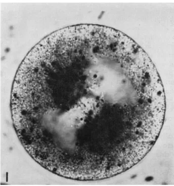
FIG. 1. Fertilized egg of Spisula at about 2-3 min subsequent to germinal vesicle breakdown. A tight aggregation of methylene blue particles can be seen in the asters of the forming first polar body spindle.
ian motion. T h e particles studied were the so-called lipid granules.
These granules, however, not only showed Brownian movement, but on occasion would engage in long saltatory movement. Indeed, as may be verified in the film by Bloom and Zirkle (undated), the particles show the same behavior characteristics (1 to 6) as described for the cells previously. In this case, however, when not engaged in saltatory move- ment, the particles can be seen to be engaged in Brownian movement, whereas in the case of egg particles, this is not always easy to see, al- though it probably occurs.
In protozoa, movements similar to those described previously have
been reported by Andrews (1955; see also Seif ritz, 1952), and have been seen by others (Allen, personal communication). Most important is the observation of Andrews that ingested carmine or ink particles undergo such movements. An important case of saltatory particle movements in plant material is that which occurs in extruded cytoplasm of Chara and Nitella, reported by Jarosch (1956) and Kamiya (1959). A final probable case of such movement occurs in HeLa cells (Rose, 1957). In
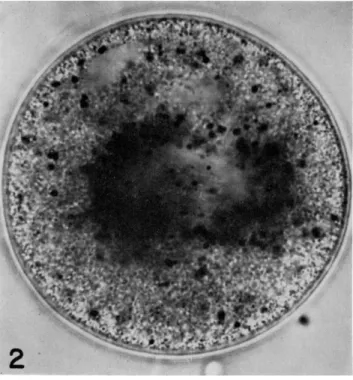
FIG. 2. Completed first polar body spindle in the center of the egg of Spisula at about 20 min after fertilization. T h e degree of astral aggregation of methylene blue particles seen in Figs. 1 and 2 is higher than in most eggs at this stage but is not rare.
this case Rose was describing microkinetospheres and their ability to move against the general stream of material entering the cell by pinocy- tosis. His description of their "peculiar" movements is suggestive of the movements we have been describing. Indeed, our own informal observa-
tion of many films of cells in tissue culture, lead us to conclude that almost all such cells possess a class of particles whose movements appear to be saltatory in the sense of characteristics 1 to 6 described previously.
If the vitally stained eggs of Spisula are fertilized, further important behavior characteristics appear. T h e particles begin to show an orienta- tion in their movement which was previously absent, Figs. 1 and 2. At
508 L I O N E L L R E B H U N
first, this orientation is variable from cell to cell, but generally, by the polar body stages (Spisula eggs only undergo maturation after fertiliza- tion) most of the particles have moved into the asters; specifically, at this stage they surround the central aster, few, if any, actually moving out with the polar bodies. By the time the first cleavage spindle has formed in the center of the egg most of the particles are in the astral regions surrounding both centrosomes but not penetrating into them,
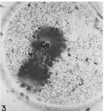
FIG. 3. First cleavage spindle in Spisula, rotated about 80° from its normal orien- tation by pressure on the cover slip. Note the strings of particles on the spindle.
Note also the clear centrosomal region (Ce). T h e degree of aggregation of methylene blue particles seen is universal in normal eggs at this stage.
Fig. 3. At the conclusion of telophase, and after formation of the blasto- mere nuclei, the particles remain aggregated on the peripheral pole of the nucleus (that is, the pole between the nucleus and the egg surface).
During prophase of the next division the mass of particles separates into two masses, each one surrounding the aster of the newly formed spindle, Fig. 4. This cycle is repeated until at least the fifth cleavage.
This description concerns the average location of the particles at various stages but does not describe the motions of the particles during their transport into the asters. During this period the number of par-
tides moving at any one time increases considerably, especially in Pec- tinaria, and the movements gradually become oriented relative to the centrosomes, although always maintaining a saltatory character. That is, individual particles still show the characteristics 1 to 6 (described previously) but now many more are involved in the movement, and the movements are directed. However, though the movement of particles is oriented radial to the astral centers, some particles may move away
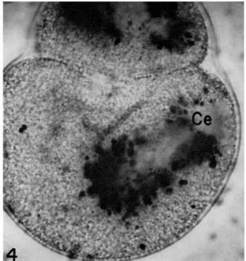
FIG. 4. Beginning of second cleavage in Spisula egg. Note clear peripheral centro- some (Ce).
from the center while other particles are moving toward it; that is, contiguous particles may move in opposite, but radial, directions in the aster. However, the average distance traversed into the centers is greater than that out of them and the net result is the accumulation of the particles around the centrosomes. This is, however, a dynamic accumula- tion in the sense that particles still show bidirectional movement of small amplitude even after the accumulation has resulted in a tight ingathering of particles. T h e result is the same for Pectinaria as for Spisula, except that in Pectinaria the particles disperse after the re- formation of the blastomere nuclei in telophase, whereas in Spisula, as
510 LIONEL I. REBHUN
indicated previously, the particles remain aggregated on the peripheral pole of the blastomere nucleus.
These observations may be summarized in the following way:
7. At times in the cell cycle when a mitotic apparatus is present, the number of particles moving with saltatory movements at any one time increases and the movements possess the characteristics 1 to 6, previously listed.
8. The particles statistically orient their movements radial to the centrosomes and aggregate tightly about them by a process in which the independent movements of particles into the asters have greater path lengths than those away from the centers. More detail is contained in previous reports (Rebhun, 1959, 1960, 1963).
This ingathering of particles into the aster has been seen by several other investigators, the first report, to our knowledge, having been that of Fischel (1899), who dealt, in part, with the astral distribution of neutral red particles in sea urchin eggs. Iida (1942) also reported the astral location of neutral red particles in sea urchin eggs and referred the movement of the particles to the growth characteristics of the aster.
Kojima (1959a,b) in two excellent papers studied the neutral red par- ticles in three species of sea urchins and an echiuroid, from the point of view of astral orientation and the possible involvement of the par- ticles in furrowing activity of the egg, and in ciliary development in later stages of embryogenesis. A considerable amount of work on par- ticles in marine invertebrate eggs stainable with basic metachromatic dyes and showing the movements 1 to 8 described previously has been done by the Belgian group at Brussels (Pasteels, 1955, 1958; Pasteels and Mulnard, 1957; Mulnard, 1958; Dalcq, 1957; Dalcq et al, 1956). This group has stressed the relationships of several types of particles in eggs that show metachromatic reactions to toluidine blue and which show relations to and movements toward the mitotic apparatus similar to those already described. A natural pigment granule present in the echiuroid, Urechis, has been described by Taylor (1931) as aggregating in the cleavage asters, by a process of "sliding down the astral rays."
The description, though not complete, strongly suggests the process we are discussing here. Such particles were identified as containing heme by Horowitz and Baumberger (1941). Finally and most important, Chambers (1917) reported that 2-4 μ oil droplets in the egg could also be transported toward the centrosome after being pushed into the aster with a microneedle, from which he inferred the presence of centripetal currents in the aster.
It is thus clear that the phenomenon of differential distribution of particles into the asters in cleavage of marine invertebrate eggs is quite
widespread and that several different particle types may be involed in these movements in different eggs. In a given egg in mitosis, however, it is possible for more than one type of particle to participate in saltatory movements into the asters. For example, in Spisula, at later stages some (not all) cortical granules may move into the astral centers by indepen- dent saltatory movements although refractile granules (presumably lipid, yolk, and mitochondria) definitely do not. Again, in Arbacia (sea urchin), some of the echinochrome granules which do not move into the cortex during the immediate postfertilization stage, generally become located in the aster at the time of cleavage along with the vitally stained granules (the same astral location of neutral red particles exists in Arbacia as Kojima finds in other sea urchins). However, other particles in the cell do not show this differential distribution at these times, although yolk granules in unfertilized Arbacia eggs may undergo saltatory movement of smaller amplitude than that of echinochrome granules (Parpart, personal communication).
The best-studied case of the influence of the mitotic apparatus on the saltatory movement of particles in cells other than egg cells is that of the lipid granules in newt heart fibroblasts. In this case, as in those already discussed, both properties, 7 and 8 (listed previously), can be verified for many of the cells in mitosis, although in some of the cells many particles remain in the cytoplasm still undergoing unoriented saltatory movement during mitosis. This behavior may be compared to particles in other fibroblasts which undergo the extensive ingathering of particles shown by eggs during cleavage. However, some differential localization of particles, in many cases extensive, does take place in all these cells, and by processes whose description is identical to that of the egg granules described previously (see Taylor, 1957; Bloom et al, 1955; Bloom and Zirkle, undated film).
We have seen no other reports of the differential movement of par- ticles into the asters in living cells but wish to point out the existence of some phenomena which may be related to our discussion, namely, those of dictyokinesis and of chondrokinesis (see extensive review in Wilson, 1925). In the former case, particles identified as Golgi bodies (dictyosomes) are shown to assume an astral location in mitosis similar to that assumed by the particles discussed previously. In chondrokinesis, the mitochondria are described as undergoing similar differential dis- tributions in some cells. The identification of the particles involved as mitochondria is not always certain, and it is not clear that the differen- tial distribution attained by the particles is due to saltatory movement.
If so, however, this latter process might be an example of mitochondria in some cells undergoing saltatory movements. Other then these pos-
512 LIONEL I. REBHUN
sible cases, no other examples of mitochondrial aggregation by saltatory movements are known to us. [The aggregation of the long mitochondria of insect spermatocytes about and parallel to the spindle (e.g., Michel, 1943; Barer and Joseph, 1957) during mitosis it not accomplished by saltatory movement.]
Another possible case is that of the aggregation of acid phosphatase particles about the spindle ends (and to some degree also on the spindle equator) of cells in mitosis in regenerating rat liver. This phenomenon has been studied in detail by Dougherty in our laboratory (it appears to have been first reported by Holt, 1957), and Dougherty has shown with the electron microscope that two kinds of particles show this differential localization—dense bodies and microbodies (Dougherty, 1963). Which of these bodies correspond to the acid phosphatase particles seen in the light microscope is not clear. These particles are located at the bile canaliculi during interphase, however, and there is therefore a problem as to the transport mechanism from canaliculi to spindle pole. Again, this looks like a case in which saltatory movement may be involved, but one about which nothing definite can be said.
Discussion
We have reviewed actual and possible cases in which saltatory par- ticle movement occurs and in which this movement may be oriented by processes in the cell so that a nonuniform location of the particles occurs.
In the cases described here, the process, mitosis, results in the transport of the particles into the asters surrounding the centrosome. In the case described by Parpart in this Symposium, the processes set off by fertiliza- tion in Arbacia result in the movement of the echinochrome particles into the cortex. In all the cases described, where actual particle move- ments have been studied, almost the same verbal description characteriz- ing the movement can be given and the major characteristics have been summarized in propositions 1 to 8. The problem which now confronts us is that of attempting to see if some reasonable hypothesis may be found which might help orient us in our understanding of this complex set of events. Several hypotheses concerning the genesis of the move- ment immediately present themselves: (a) that the movements are auton- omous in the sense that some process within the particles themselves initiates the movement, (b) that the movements are induced in the par- ticles by the transfer of momentum to the particles by some external force generating system, and (c) that a combination of external and internal forces are involved. None of these ideas can be completely eliminated on the basis of the data which are on hand. The most potent argument in support of movement arising from internal processes comes
from the observation that the particles do, indeed, seem to move inde- pendently, that is, that the first part of proposition 2, listed previously, holds. It can be seen that the major stumbling block to any theory of externally induced movement is precisely this fact that adjacent, and in fact, contiguous particles are influenced in entirely different ways.
Nevertheless, it is difficult to imagine what kind of processes could be involved in particles as different as lipid droplets, melanin granules, echinochrome granules, heme granules, and microkinetospheres, which would result in the same detailed type of movement, since most of the granules mentioned have considerable claim to being considered as metabolically inert. Most cogently, oil droplets in eggs and carmine and ink particles in protozoa undergo similar movements. Also, hypotheses such as those involving propulsion of a particle by some form of jet process originating, for example, by local influx and efflux of ions, water or other molecules, due to some permeability change in a surface membrane, should certainly predict form changes on the part of the particles, especially in processes involving movements for distances as great as 30 μ and such changes are not observed. Again, one would ex- pect considerable differences in velocities and distances attained between large and small particles, which is not observed. Finally, it is not rare to find groups of 2 to 5 particles moving with the same velocity and for the same distance but not changing the relative distances between them, which may be of the order of a micron. It is very difficult to see what kind of process would correlate the movements of such a group of par- ticles with such precision if the movement arose from some internal proc- ess. We feel that these considerations effectively, if not absolutely, rule out internal processes as being totally responsible for the movement.
A modification of this might suggest that the movement is due to the combination of some process in the cell as a whole with some proc- ess in the particle. For example, it might be suggested that some force field, such as an electric field set up by metabolic processes centered on some element in the cell; for example, the centriole might interact with charges that develop on the particles, which would result in movement in one or another direction depending on charge sign. Similarly, rever-
sal of direction might be correlated with change of sign on the particle.
In addition to the completely ad hoc nature of such a hypothesis the same arguments as used before against theories suggesting processes originating within the particle as causing movement may be used here.
In addition, the unoriented movements during interphase would be most difficult to explain on this basis. We again conclude the unlikely nature of this specific possibility and others like it.
We are thus left with the middle alternative, namely, that the particle
514 LIONEL I. REBHUN
moves because of some process which originates external to the particle itself. Again we may distinguish hypotheses that postulate the inter- action of the particle with some general process occurring in the cell and those that involve a direct interaction of the particle with some physi- cally existing structure in the cell which transmits momentum to it. An example of the first kind might be the hydrodynamic forces postulated by Bjerknes as causing chromosome movement (see Schräder, 1953). He showed that oscillating or pulsating spheres in a liquid medium can set up standing waves, and depending on the phases of the spheres (that is, whether they pulsate or oscillate in or out of phase), particles of a density greater than the medium move in one direction relative to the spheres whereas particles less dense than the medium move in the oppo- site direction. Aside from the fact that the particles, in actuality, do not all move at once, which would seem to be required by this hypothesis, it is difficult to imagine particles changing density as rapidly as would be required by the suddenness of the onset and termination of the move- ment. Also, other than Pfeiffer's (1956) report of pulsating centrosomes, we know of no other reports of the existence of oscillating spheres in the cell.
We are finally brought to what we consider to be the most likely hypothesis for mechanisms of saltatory movement, namely, the hypothesis that some system in the cell can develop forces which it can transmit to the particles, inducing them to move. It is clear that we have by no means completely eliminated (or enumerated) all other hypotheses, but we feel that arguments similar to those used previously, plus the positive attributes of our suggestions, discussed later, make the suggestion of the external-force transmitting system the hypothesis most likely to succeed.
Before inquiring into the requirements which such a system must satisfy, we shall describe two further sets of observations which add to the criteria we shall list.
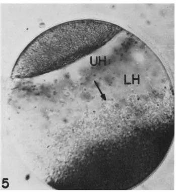
If fertilized eggs of Spisula are centrifuged, the contents of the egg stratify to definite layers; from centrifugal to centripetal poles the layers are cortical granules, yolk, mitochondria, lower hyaline zone (contain- ing primarily ribosomal material), upper hyaline zone (containing mainly elements of the endoplasmic reticulum, Golgi bodies, and multivesicular bodies) and, most centripetal, the lipid layer. Spindles and nuclei stratify into the boundary between the upper hyaline and the lower hyaline layers (see Figs. 5 to 9). If eggs which have been lightly stained with vital dyes are centrifuged so that they are stratified, the stained particles also gather primarily at the boundary between the upper and lower hyaline regions, Fig. 5. However, if eggs are heavily stained so that the stained particles become large (see point 4, p. 505), they stratify into the
yolk region. Observation of these particles reveals differences in their salta- tory movements. Thus, the particles stratifying to the hyaline zones can still be observed to undergo saltatory movement, generally parallel to the boundary between upper and lower hyaline zone. These particles will tend to gather tightly about the re-forming asters when redistribu- tion gets underway, if the cell has been centrifuged in a stage containing a spindle. On the other hand, the particles going to the yolk layers do not undergo saltatory movements. However, eggs generally recover from the effects of centrifugation, and after variable periods from ^ to 1 hr, may cleave again. In this latter case the heavy particles will again par- ticipate in saltatory movements and will gather about the aster centers.
Thus, particles stratified to the region bordering the upper hyaline zone continue to exhibit saltatory movement and may participate in redistribution into the asters as these re-form in the hyaline zone. Par- ticles stratified into the yolk layer lose their ability to undergo saltatory movement, but subsequently, and much later, regain that ability as the contents of the egg redistribute and cleavage begins to occur (see Rebhun 1959, 1960, 1963 for further details on some of these processes).
The relationship of these observations to those on echinochrome granules as reported by Parpart (Parpart, 1953 and this Symposium) for unfertilized eggs is clear. I n fact, their significance was not realized by us until we had considered them in relation to the work of Parpart.
The second set of phenomena, which we feel are related to saltatory movements, occur in certain phenomena to be seen on the mitotic spindle. In 1960, Östergren et al. put forward a rather unique hypothesis for the mechanism of spindle action, which postulated as its main ele- ment, the existence of a "pumping" activity on the part of the spindle, which was controlled by and which interacted with the chromosomal fibers resulting in the relative motion of spindle and chromosomal fiber (with attached chromosome) during anaphase separation. Stripped of the language used by these authors, the hypothesis suggests that spindles work because they can develop forces that can move particles relative to themselves without the spindle itself necessarily changing in form or shape during that movement; i.e., spindles are able to develop sliding or shearing forces at their surfaces. Östergren et al. (1960) showed that many particles other than chromosomes with chromosomal fibers may be moved to the poles by the activity of the spindle; for example, acen- tric chromosomal fragments, pieces of nucleoli and other cytoplasmic particles accidentally caught on the spindle are so moved. They also suggested that the particle aggregations seen in newt heart fibroblasts could have their origin in the same type of force generated in the asters as is generated in the spindle. The major suggestion which we would
516 LIONEL I. REBHUN
like to glean from this work is that the spindle may work by generating a shearing or sliding force at its surface and that mechanisms for saltatory particle movement may be related to mechanisms for spindle activity.
Our own observational data had also suggested the possible relation of spindle activity to saltatory activity prior to the appearance of the work by Östergren et al. (1960) since we had often seen metachromatic particles moving on the surface of spindles during cleavage in Spisula.
FIG. 5. A Spisula egg stratified just prior to first cleavage. Note the toluidine blue particles at the junction of the upper hyaline zone (UH) and lower hyaline zone (LH). T h e arrow points in the centrifugal direction.
Specifically, particles may often be seen on the spindle surface engaging in what appear to be directed pole-to-pole movements (see Fig. 3).
These have many of the characteristics of saltatory movement in that the movements are discontinuous in time, involve continuous motions of the order of 30 μ (the spindle length in first cleavage spindles in Spisula eggs), do not differ for large or small particles, and may involve particles moving simultaneously in opposite directions. These charac- teristics are so similar to the ones described previously for astral par- ticles as to have suggested to us that spindle mechanisms and saltatory particle mechanisms might, indeed, be identical (Rebhun, 1963).
A system in the cell involved in saltatory movement should, therefore, satisfy the following criteria:
(7) It must satisfy the orientation requirements set by the behavior of the particles both in interphase and during mitosis. That is, it should be a system unoriented in interphase, but with parts long enough to be physically extant over distances up to 30 μ and it should be ori- ented radially to the astral centers during mitosis.
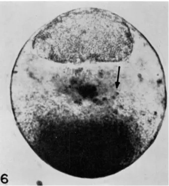
FIG. 6 . A Spisula egg 1 0 - 1 5 min after stratification (prior to first cleavage). Note the aggregation of toluidine blue particles about a center in the lower part of the lower hyaline zone.
(2) It should be the only system in the cell exhibiting these prop- erties.
(3) There should be some evidence that this system might be ca- pable of performing the tasks required of it.
(4) With the orientation properties of the system in centrifuged eggs one should be capable of explaining the phenomena seen there, especially, the reversible cessation of particle movements in the case of echinochrome granules in Arbacia and in the case of heavy (but not light) vitally stained granules in Spisula.
LIONEL I. REBHUN
FIGS. 7 to 9. Stratified Spisula eggs 10-15 min after stratification. Note the radial chains of cis- ternae (Ci) coming from the upper hyaline zone. In Fig. 7 can be seen the lipid (L), upper and lower hyaline zones, mitochondrial layer (M), and yolk layer (Y). The cortical granules (C) have not formed a distinct layer but are concentrated to some extent in the lower (centrifugal) hemisphere. In Figs. 7 and 9, V represents the vitelline membrane. (For more details, see Rebhun 1960, 1963.)
520 LIONEL I. REBHUN
(5) The system should also be present in the spindle, or on its surface. This criterion is more speculative than the previous four but we feel that the observations that generate it are sound.
With these criteria in mind, we shall now examine results from ultrastructural studies of cells to see if any system present in the cell does satisfy the previously mentioned requirements, and, especially, whether criteria (3) and (5) can be verified.
Ultrastructural Studies of Cells and General Discussion More complete description of our work on the ultrastructure of egg cells with respect to this problem has already been published (Rebhun, 1960, 1962, 1963). In these papers it is shown in some detail that the endoplasmic reticulum does show the orientation properties required in criterion (7) listed previously. Thus, elongated elements of this sys- tem are present in the unfertilized eggs of both Spisula and Pectinaria, and, in both, this system shows an orientation radial to the centers when the spindle begins to form. In addition, in centrifugally stratified eggs, the endoplasmic reticulum of Spisula eggs occupies the upper hyaline zone and generally has its elongated elements (cisternae) lying parallel to the boundary of the upper and lower hyaline zones. As eggs begin to redistribute after centrifugation the elements of the endoplasmic re- ticulum orient radially to the centers of the asters (see Figs. 7-9). It is clear then that the E R forms one component of the visible aster and does show distribution characteristics required of it if it is to be the system involved in saltatory movements. Further, the yolk zone is en- tirely free of elements of the E R after centrifugation. Finally, in the case of Arbacia eggs, our electron micrographs show that the E R strati- fies to the hyaline zone and is completely absent from the zone occupied by the echinochrome granules. This agrees with the distribution of the E R as seen in other sea urchins by Pasteels et al. (1958). Unfortunately, the studies of E R have not yet been extended to redistributing eggs after centrifugation. However, from the observations we have obtained the E R appears to be a system satisfying criteria (7) and (4).
Other work indicates a similar distribution of E R relative to asters in egg material. For example, Harris (1961, 1962) and Harris and Mazia (1962) report a radial orientation of E R in asters of eggs of the urchin, Strongylocentrotus, similar to that seen here. In division figures in onion root tips, Porter and Machado (1960) also report orientations of E R ra- dial to the asters. A similar, though complex orientation of a smooth E R appears to occur in spermatocyte divisions (Ito, 1960) in Drosophilia.
As mentioned, the criteria (7) and (4) described in the preceding sec-
tion are satisfied by the E R . However, we have obtained no evidence lor the existence of E R in or on the spindle as a constant component in the egg material we have studied, although on occasion small pieces of this system are present (this is in contrast to the presence of "spindle lamellae" in root tip cells (Porter and Machado, 1960) and in carcinoma cells (Buck, 1961). Thus, if we are to retain criterion (5), we find dif- ficulties in the absence of E R from egg cell spindles. Further, criterion (2) may not hold; that is, there has now been a second system described in cells which has many of the orientation characteristics that we require.
This is the system of spindle filaments which have been described so well in spindles of amebae by Roth and Daniels (1962) and Roth (this Symposium) and in spindles and asters of sea urchin eggs by Kane (1962) (in isolated mitotic apparatuses), by Harris (1961, 1962), and by Harris and Mazia (1962). These filaments, or "tubules," are about 150 A in diameter and occur both in chromosomal fibers and in pole-to-pole fibers in the spindle. More important for our consideration, however, is the fact that they extend into the asters of the mitotic apparatus, and, ac- cording to Harris and Mazia (1962) may extend out as far as the egg cortex. They have the proper orientation characteristics needed to ac- count for saltatory movement and are present in force in the spindle.
However, there is no evidence that they exist as an extensively deployed component in interphase cells, although they appear prior to nuclear membrane breakdown (Harris and Mazia, 1962). It may be that a search for them in interphase cells would reveal them, although their presumed lack of orientation, if they are present, would create great technical difficulties in identifying them.
We are thus left with the disturbing situation in which the only ultrastructural elements, visible with present electron-microscopical tech- niques, which might be responsible for saltatory movements do not simultaneously satisfy all the criteria we have set up for the system. It is, of course, always possible that a further system as yet not visualized in the cell may be the responsible element. However, a possible solu- tion may be contained in observations such as those of Jarosch and Kamiya described next.
In a series of papers (Jarosch, 1956, 1957, 1958, 1960) a fibrillar cyto- plasmic system was described which consisted of moving filaments in the cytoplasm. These filaments appear to be capable of exerting a force on the medium surrounding them, which results in their forward pro- pulsion. Such systems have been also described by Kuroda and Jarosch in this Symposium. Jarosch and later Kamiya (1959) postulated deep involvement of these filaments in certain movements in the cytoplasm
522 LIONEL I. REBHUN
of the exuded cytoplasmic droplets they were concerned with. Specifi- cally, they attempted to account for saltatory movement as a movement of particles along the filaments but in a direction opposite to that in which the filaments are moving. Further, they observed that the fila- ments appeared to attach to and detach from the nuclear surfaces and that the filaments with their associated countermoving streams appeared to be correlated with the rotations of the nuclei. Similar phenomena were observed for moving chloroplasts. Thus, they considered that the nuclear rotations and chloroplast movements were due to the concerted efforts of filaments attached to the nuclear or chloroplast surface exert- ing their influence in a fixed direction. The observations on which these ideas are based appear to be well founded and suggest, therefore, that the nuclear surface may form reversible associations with a fila- mentous component of the cytoplasm which may be able to exert shear- ing forces on the medium in which it is immersed. Since, however, most recent work indicates that the nuclear surface is derived from, is con- tinuous with, and can contribute to the endoplasmic reticulum, and thus is part of that system (see Porter, 1961), it is not too far-fetched to sug- gest the possibility that an ER-filament association may be possible, and that force transmission to the particles from the E R may, indeed, be due to this associated filament system. Thus, if saltatory particle movements in these cytoplasmic droplets are due to interaction with a filament ca- pable of exerting a shearing force on the medium, then this may also be the mechanism involved in other cells, if it is justifiable to assume all saltatory motion to have a common mechanism.
We have thus arrived at a hypothesis for saltatory particle move- ment which has been obtained by consideration of the most diverse phenomena, namely, that these movements are due to sliding or shear- ing forces exerted on particles in cells by the activity of filaments which may occur in the spindle, in the cytoplasm, or are associated with the endoplasmic reticulum. Many auxiliary hypotheses are used in this work. T h e highly hypothetical nature of this discussion is made clearer, if we reiterate all the hypotheses used with an estimate of their worth.
A similar and extended discussion has been previously published (Reb- hun, 1963).
a. Because the details of saltatory movement are so similar in dif- ferent cells, we have assumed that the mechanism of saltatory movement is likely to be the same. We feel that this hypothesis is quite strong.
b. An analysis of the details of the movement characteristics of the particles leads us to consider that the most reasonable hypothesis for the mechanism of saltatory movement is that of the transfer of momentum
to the particle by some outside agency, with the particle itself being quite passive in the process. This hypothesis we feel to be very likely, but not as strong a hypothesis as (a).
c. A consideration of some of the phenomena associated with spin- dle action, especially as it relates to saltatory movement, leads us to con- sider the possibility that saltatory movement and spindle action are mechanistically similar. This is clearly a hypothesis on a lower level of support than (a) and (b). This leads to the next hypothesis.
d. Some structure present in the spindle is also present in the cyto- plasmic system responsible for saltatory movement. Since the only vis- ible structure consistently present in the spindle is the 150 A filament (or tubule) and since this is present in asters, we have suggested that this filament or one related to it may be responsible for saltatory move- ment. An even more tenuous suggestion is the final one necessary to tie in the observation on the E R in this process.
e. T h e E R is involved in saltatory movement because of its asso- ciation with the true force-producing structure, the filaments. Evidence for such an association comes from observations of filaments associated with rotating nuclei in isolated cytoplasmic droplets of Chara and Ni- tella.
The essence of the last three hypotheses is the suggestion that there is available to cells a structure, filamentous in nature, which can exert a force on its surroundings in such a way as to propel itself if it is free to move, or, if not, acting in concert with other similar structures, to propel the surrounding medium or objects in it. Such ideas have orig- inated with Jarosch and Kamiya in the papers cited previously, and though the universality of these ideas is by no means clear, we feel that they deserve consideration in the present context. Finally, it is not sug- gested that the filaments in different cells are made of the same mate- rials or are even necessarily organized in the same way on a molecular level. We merely require that they act alike, namely, exert forces ca- pable of moving themselves or external objects parallel to their axes, possibly by processes such as those described by Jarosch in this Sym- posium.
It remains to be seen whether the concepts introduced will prove adequate to interpret these phenomena, or whether concepts drawn from a less structural and morphological sphere will ultimately have to be invoked in the explanation of the mechanisms of saltatory particle movements.
Some discussion on saltatory motion can be found in the Free Discussion of Part IV.
524 LIONEL I. REBHUN
REFERENCES Andrews, Ε. A. (1955). Biol. Bull. 1 1 4 , 113-117.
Barer, R., and Joseph, S. (1957). In "Mitochondria and other Cytoplasmic Inclusions,"
Society for Experimental Biology Symposium, Vol. X . Cambridge Univ. Press, Cambridge, England.
Bloom, W., and Zirkle, R. (undated). "Mitosis of Newt Cells in Tissue Culture," film- print No. 64. Univ. of Chicago Bookstore, Chicago, Illinois.
Bloom, W., Zirkle, R., and Uretz, R. (1955). Ann. Ν. Y. Acad. Sei. 5 9 , 503-513.
Buck, R. C. (1961). ./. Biophys. Biochem. Cytol. 1 1 , 227-236.
Chambers, R. (1917). / . Exptl. Zool. 2 3 , 483-505.
Dalcq, A. (1957). Bull. Soc. Zool. France 8 2 , 296-316.
Dalcq, Α., Pasteeis, J . J . , and Mulnard, J . (1956). Bull. Acad. Roy. Belg. Classe Sei., Ser. 5, 42, 771-777.
Dougherty, W. J . (1963). Ph.D. Dissertation, Dept. Biol., Princeton Univ., Princeton, New Jersey.
Fischel, A. (1899). Anat. Heften 3 7 , 463-504.
Harris, P. (1961). / . Biophys. Biochem. Cytol. 1 1 , 419-432.
Harris, P. (1962). / . Cell Biol. 1 4 , 475-488.
Harris, P., and Mazia, D. (1962). In "The Interpretation of Ultrastructure" (R. J . C.
Harris, ed.), Vol. I. Academic Press, New York.
Holt, S. J . (1957). 9th Intern. Congr. Cell Biol., St. Andrews, Scotland.
Horowitz, Ν. H., and Baumberger, J . P. (1941). / . Biol. Chem. 1 4 1 , 407-415.
Iida, T. T. (1942). Dohutsugaku Zasshi 5 4 , 364-366.
Ito, S. (1960). / . Biophys. Biochem. Cytol. 7, 433-442.
Jarosch, R. (1956). Phyton (Buenos Aires) 6 , 87-107.
Jarosch, R. (1957). Biochim. Biophys. Acta 2 5 , 204-205.
Jarosch, R. (1958). Protoplasma 5 0 , 93-108.
Jarosch, R. (1960). Phyton (Buenos Aires) 1 5 , 43-66.
Kamiya, N. (1959). Protoplasmatalogia 8 , 13a.
Kane, R. L. (1962). / . Cell Biol. 1 5 , 279-287.
Kojima, M. K. (1959a). Embryologia (Nagoya) 4 , 191-209.
Kojima, M. K. (1959b). Embryologia (Nagoya) 4, 211-218.
Michel, K. (1943). Film No. C443, Institut für den Wissenschaftlichen Film, Göttingen.
Mulnard, J . (1958). Arch. Biol. (Liège) 6 9 , 645-685.
Mysels, K. J . (1959). "Introduction to Colloid Chemistry." Interscience, New York.
Östergren, G., Molé-Bajer, J . , and Bajer, J . (1960). Ann. N. Y. Acad. Sei. 9 0 , 391-408.
Parpart, A. K. (1953). Biol. Bull. 1 0 5 , 368.
Pasteels, J . J . (1955). Bull. Acad. Roy. Belg. Classe Sei., Ser. 5, 4 1 , 761-768.
Pasteels, J . J . (1958). Arch. Biol. (Liège) 6 9 , 591-619.
Pasteels, J . J . , and Mulnard, J . (1957). Arch. Biol. (Liège) 6 8 , 115-163.
Pasteels, J . J . , Castiaux, P., and Vandermeersche, G. (1958). Arch. Biol. (Liège) 6 9 , 627-643.
Pfeiffer, H. H. (1956). Protoplasma 4 6 , 585-596.
Porter, K. R. (1961). In "The Cell" (J. Brächet and Α. E. Mirsky, eds.), Vol. II.
Academic Press, New York.
Porter, K. R., and Machado, R. D. (1960). / . Biophys. Biochem. Cytol. 7, 167-180.
Rebhun, L. I. (1959). Biol. Bull. 1 1 7 , 518-545.
Rebhun, L. I. (1960). Ann. N. Y. Acad. Sei. 9 0 , 357-380.
Rebhun, L. I. (1961). / . Ultrastruct. Res. 5 , 208-225.
Rebhun, L. I. (1963). In "The Cell in Mitosis" (L. Levine, ed.). Academic Press, New York.
Rose, G. G. (1957). / . Biophys. Biochem. Cytol. 3, 697-704.
Roth, L. E., and Daniels, E. W. (1962). / . Biophys. Biochem. Cytol. 12, 57-78.
Schräder, F. (1953). "Mitosis," 2nd ed. Columbia Univ. Press, New York.
Seifritz, W. (1952). In ''Deformation and Flow in Biological Systems" (A. Frey-Wyssling, ed.). North Holland, Amsterdam.
Taylor, C. (1931). Physiol. Zool. 4, 423-460.
Taylor, E. W. (1957). Ph.D. Dissertation, Committee on Biophysics, Univ. of Chicago, Chicago, Illinois.
Wilson, Ε. B. (1925). "The Cell in Development and Heredity," 3rd ed. Macmillan, New York.