https://doi.org/10.1007/s42977-020-00047-z ORIGINAL PAPER
Electrophysiological and behavioral properties
of 4‑aminopyridine‑induced epileptic activity in mice
F. Z. Fedor1,2 · C. Paraczky3 · L. Ravasz2 · K. Tóth4 · Z. Borhegyi7 · Z. Somogyvári8 · G. Juhász7 · Z. Fekete5,6
Received: 3 June 2020 / Accepted: 14 September 2020
© Akadémiai Kiadó Zrt. 2020
Abstract
4-aminopyridine (4-AP) is a widely used drug that induces seizure activity in rodents, especially in rats, although there is no consensus in the literature on the dose to be used in mice. The aim of the present study was to investigate the effect of the intraperitoneal administration of 4-AP in two doses (4 and 10 mg/kg) in vivo. EEG, movement, and video recordings were made simultaneously in male B6 mice to specify the details of the seizures and to determine whether there is a suitable non- lethal dose for seizure induction and for further molecular studies. Seizure behavior in mice differs from that seen in rats, with no characteristic stages of epileptic seizures, but with spiking and seizure activity. Seizure activity, although produced at both doses without being lethal, induced different changes of the EEG pattern. Smaller dose induced a lower amplitude seizure activity, decreased spiking activity and later onset of seizures, while higher dose induced a much more intense brain seizure activity and severe trembling. It is concluded that the intraperitoneal administration of 4-AP at a dose of 10 mg/kg induces explicit seizure activity in mice which is repeatable and can be suitable for further molecular research.
Keywords 4-Aminopyridine (4-AP) · Epilepsy model · EEG · B6 mice · In vivo studies · Behavior
Introduction
Epilepsy is a mostly convulsive brain disorder affecting mil- lions worldwide (WHO 2019), although most commonly, it has no identifiable origin (idiopathic epilepsy). There are several other cases, when the reason of the disease is revealed (symptomatic epilepsy) for example severe head injury, ischemic conditions in the brain (either because of pre- or perinatal injury or stroke), brain infection, and genetic syndromes (Leonardi and Ustun 2002). Treatments applied recently use controlled inhibition of seizures that may result in endogenously performed self-reparatory pro- cesses recovering the subjects and may allow to leave antie- pileptic treatment later (Schachter 2018).
Actually, most of the studies use mouse models of neuro- degenerative diseases (Trancikova et al. 2011), because their genetic constitution is well known and easily manipulated while they have a rapid reproduction period with numerous offsprings (White 2016). To observe spontaneous epilep- tic activity, countless genetic models have been developed, such as El mouse (King and LaMotte 1989), apathetic mouse (Chung et al. 2009), epf mutants (Hare and Hare 1979), and tottering mutants (Tsuji and Meier 1971). These are engi- neered epilepsy model animals having one or more genetic
* F. Z. Fedor
fedor.flora@gmail.com
1 Doctoral School of Chemical Engineering and Material Sciences, University of Pannonia, Veszprém 8200, Hungary
2 ELTE NAP Neuroimmunology Research Group, Department of Biochemistry, Institute of Biology, Eötvös Loránd University, Budapest 1117, Hungary
3 Institute of Experimental Medicine, Budapest 1083, Hungary
4 Laboratory of Molecular Neuroendocrinology, Institute of Experimental Medicine, 1083 Budapest, Hungary
5 Research Group for Implantable Microsystems, Faculty of Information Technology and Bionics, Pázmány Péter Catholic University, Budapest 1083, Hungary
6 Centre for Energy Research, Budapest 1121, Hungary
7 Department of Biochemistry, Eötvös Loránd University, Budapest 1117, Hungary
8 Theoretical Neuroscience and Complex Systems Research Group, Department of Computational Sciences, Wigner Research Centre for Physics, Budapest 1121, Hungary
aberrations that result in epileptic activity (Seyfried and Gla- ser 1985). Another widely used method to induce epilepsy- like seizures in varied animal models is chemical induction.
Chemical induction can be performed with several agents such as pilocarpine (Cavalheiro 1995), pentylenetetrazole (PTZ) (Klioueva et al. 2001), bicuculline (Blennow et al.
1978), kainic acid (KA) (Ben-Ari and Lagowska 1978), and 4-aminopyridine (4-AP) (Szente and Pongrácz 1979).
4-AP is a well-known voltage-gated K+ channel blocker that results in prolonged action potential, stimulates neu- rotransmitter release and facilitates inward Ca+ currents, and therefore causes neuronal hyperactivity (Thesleff 1980;
Tapia 1982; Tapia et al. 1985; Rogawski and Barker 1983;
Rudy 1988; Szente and Baranyi 1987). By reason of its simple and well-described mechanism of action, 4-AP is proved to have a useful role in the fundamental research of seizure development (Pasantes-Morales et al. 1987). It has strong convulsive effect both in humans (Spyker et al.
1980) and in animals (Schafer et al. 1973; Gandolfo et al.
1989; Fragoso-Veloz and Tapia 1992; Morales-Villagrán et al. 1996); hence, it is commonly used in epilepsy research both in vivo and in vitro in rodents (Schafer et al. 1973;
Spyker et al. 1980; Gandolfo et al. 1989; Voskuyl and Albus 1985; Avoli 1990; Gean 1990). It is also known that 4-AP elevates blood pressure and produces muscle contractions and its intraperitoneal administration induces discharges in the EEG such as isolated spikes, poly-spikes, and spike-wave complexes (Fragoso-Veloz et al. 1990; Pasantes-Morales and Arzate 1981).
The sequence of behavioral changes associated with 4-AP in mammals was first described as the following: “hyper- excitability, salivation, tremors, muscular incoordination, clonic and tonic convulsions, cardiac or respiratory arrest, and death” (Schafer et al. 1973). Later Yamaguchi concluded that appearing behavioral signs are in a typical order; how- ever, there are exceptions when behavioral changes do not occur or not in the described order which is: hyperreactivity to touch or loud noise, vocalization, unsteady gait/immobil- ity, blinking/eye closing, hyperpnea, straub tail 1–10 min after injection, trembling, intermittent forelimb/hindlimb clonus 3–15 min after injection, explosive running, hindlimb extension, opistotonus, continuous clonic limb movements, and death 6–20 min after injection (Yamaguchi and Rogaw- ski 1992). There is no uniform scoring for rating behavioral features after administration like the Racine Scale for rats, although a revision of the Racine Scale to accommodate murine epilepsy-like seizures has recently been proposed in case of the pentylenetetrazole (PTZ) epilepsy model (Van Erum et al. 2019).
Investigations of the in vivo effects of different doses began as early as in the 1960s, when Humphreys adminis- tered 4-AP at three different doses (16 mg/kg, 12.8 mg/kg, and 10.3 mg/kg) to male CDBA mice. He found that, at the
highest dose, the death rate was 5/6 in the next 24 h, whereas at the two lower doses all subjects survived (Humphreys et al. 1962). Later, Schafer determined LD50 at 14.7 mg/
kg by intraperitoneal (i.p.) administration with a confidence limit of 13.9–15.5 mg/kg (Schafer et al. 1973). In male Swiss mice, ED50 for subcutaneous administration was found to be 10.9 mg/kg. At 10 mg/kg, only two out of eight animals exhibited hindlimb extension and lethality. Death occurred in these two with a mean latency of 37 min (Yamaguchi and Rogawski 1992). At 5 mg/kg i.p. dose, 90% of the mice developed clonic seizures in 5–7 min after injection, while 92% developed tonic seizures in 17–24 min after injection, and 80% produced post-convulsive mortality (Pasantes- Morales and Arzate 1981). However, all dose dependence studies on mice aimed to establish the lethal dose but not the dose dependence of EEG and behavioral symptoms.
To improve the applicability of the 4-AP model of epi- lepsy, this experiment is designed to show the effect of i.p.
administered 4-AP in 4 mg/kg and 10 mg/kg doses on the EEG and behavior of B6 mice. The two doses were estab- lished based on the previous researches described in the relevant literature. Dose dependency, development of the seizures, and the correlation of behavior and brain activity are presented.
Materials and methods
Animal experiment conditionA total of 18 in-house bred 3-month-old male B6N mice served as experimental subjects and as their own controls in this study. For 4 days prior to the surgeries, animals were housed in groups of 3–4 per cage (diurnal cycle, lights on from 9:00 to 18:00), with ad libitum access to food pellets and water in order to habituate them to the environment and to minimize stress caused by the transportation. After three days, the surgical procedures were performed.
Surgery
In all cases, our experiments were conducted in compliance with EU directive in force (2010/63/EU) and in accordance with the applicable government decree (40/2013. (II. 14.)), making sure that the animals suffer as little as possible. Dur- ing the surgeries, animals were kept in inhalational anesthe- sia (Isoflurane, 0.5–1%, Forane, Abbott). First, the surgical area was depilated, and then, the head of the animal was fixed in a stereotaxic frame while it was already in anesthe- sia. The surgical area was exposed and cleaned to provide a suitable surface for electrode implantation. Small crani- otomies were drilled through the skull, and then, stainless steel screw electrodes were placed on the top of the dura
and fixed to the skull using dental cement. The arrangement of the three electrodes: two inserted anterior and posterior to the left parietal bone (one rostral and one caudal both 3 mm-s from the mid-sagittal line) functioned as data chan- nels from the somatosensory cortical area, and one used as a ground and reference for the measurements was inserted to the right side of the occipital bone just above the cerebellum.
One additional stabilizing stainless steel screw was placed opposite to the electrodes also above the somatosensory cortical area. After the arrangement of the electrodes and the screw, the electrodes were soldered to a connector and dental acrylic cement (GC America Inc., USA) was used to cover the construction. The operations lasted for 45 min maximum and were followed by a one-week-long recovery period, while the animals were housed separately to avoid infections.
Induction of seizure activity
Prior and after (1 day) the induction, one and a half hours of control recordings were made. Seizures were induced by intraperitoneal injections of 4-aminopyridine dissolved in 0.85% NaCl solution (4 mg/kg or 10 mg/kg dose, Merck, Germany) and followed by the recording of the EEG for at least one and a half hours.
Electroencephalographic recording
During the experiments, animals were housed 12–12 h of light–dark cycle using light intensity of 500 lx. Electro- physiological measurements were taken in a Faraday cage in darkness (under 0.17 msd), using a 32-channel Amplipex KJE-1001 (Amplipex Ltd., Hungary) amplifier. The behav- ior of the animals was recorded with a camera, and their movements were monitored with a 3D accelerometer (ACC- 2x, Supertech, Hungary). Spike2 (Cambridge Electronics Design Limited, UK) program was used for the measure- ments at a 2 kHz sampling rate for the EEG and 500 Hz sam- pling rate for the accelerometer channels. EEG was recorded in five channels, two for the implanted cortical screw elec- trodes and three for the accelerometer channels (x, y, z direc- tions). The reference and ground for the measurements was a third screw electrode occipitally implanted. During the recordings, animals were freely moving.
Data processing
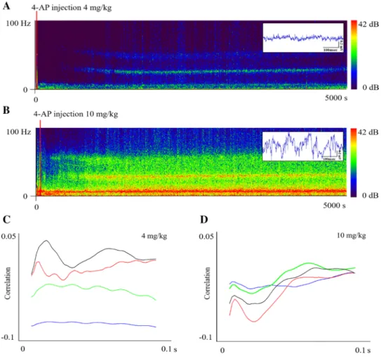
Data analysis was performed with Spike2 (Cambridge Elec- tronics Design Limited, UK) and Origin (OriginLab Cor- poration, USA) programs. EEG recordings were analyzed offline. Sonography (Figs. 1, 3) was made from caudal screw electrode data and the X-axis off the accelerometer using 8192 block size and a 14 dB range for the EEG and 24 dB
range for the accelerometer. Power spectrum density estima- tions (Fig. 3) was calculated from 0.5 h of EEG and accel- erometer data using a 65,536 sized fast Fourier transfor- mation represented applying a Hanning window correction.
In the case of Fig. 1c, the sections used for analysis were half an hour long data from 0 to 1800s control and 2000 to 3800 s seizure activity. The points between the lower edge and 2 mV were calculated, and then, average and standard deviation were estimated from 9 lower- and 9 higher-dose subjects. Figure 1d shows seizure peaks estimated using 8 higher- and 8 lower-dose subjects after filtering data (EEG:
20–40 Hz, ACC: 25–55 Hz) using RMS amplitude (100 s constant). In this case, peak means the time between the 4-AP administration and the maximum of amplitude in Fig. 1e, f, and correlation of movement and brain activity were calculated in four stages (control, beginning of sei- zure, seizure, and end of seizure) of two doses from 1000 s long data stream. Half an hour long control and seizure data (Fig. 3c) were analyzed at 0.01 bin size, while the number of bins was set to 300. The lower limit for the analysis was estimated individually according to their own control (one day before) limit, which was considered as 100%. Start of spiking activity enrichment was calculated from raw data analysis and the comparison of the high-frequency activity of both EEG and accelerometer channels. The spike number count analysis was performed with the amplitude histogram script of Spike2 program. The amplitude threshold from which the program counts spikes was estimated based on the analysis of control activity. In each subject, the threshold for control and previously injected data was the same, and then, seizure spike number was normalized with the control data. Peak of seizure activity was determined according to the highest frequency of spiking activity. Start of enrich- ment, peak, and spike number characterizes a seizure and allows a comparison between doses. Statistical analysis was performed using data from 9 low (4 mg/kg of 4-AP)- and 9 high (10 mg/kg of 4-AP)-dose injected subjects.
Results
The applicability and dose-dependency of epileptic sei- zure activity evoked by intraperitoneally injected 4-AP in mice were examined. It was observed that the onset of after administration EEG spike enrichment is different for the two doses, starting on average 954.1 ± 250.7 s after the administration of 4 mg/kg and 580.3 ± 171.5 s in the case of the administration of the higher dose. The locomo- tor activity follows the brain activity in both cases (4 mg:
318.2 ± 389.6 s; 10 mg: 468.8 ± 190.4 s, on average).
Compared to the control recordings, there is a remark- able difference after administration at both doses, but the maximum amplitude at the higher dose is the double of the
lower dose respectively (Fig. 1a, b). The average number of spikes during the seizure increased to 325.6% (4.9 ± 4.4 to 20.8 ± 27.2) at the 4 mg/kg dose, compared to 339.1%
(5.8 ± 5.6 to 25.3 ± 17) at the 10 mg/kg dose, although as standard deviation shows data were variable (Fig. 4a). After data normalization with control spike number, the increase in spiking activity was 222% bigger in case of the higher dose. Spike shape sorting was also made but revealed no significant results, since spike shapes were heterogeneous at both doses. The peak of movement was determined based on the accelerometer, while the peak of the seizure in terms of brain activity was determined based on brain activity measurements.
Regarding the seizure peak of brain activity calcu- lated from the highest frequency of spiking activity, EEG reached its seizure activity peak 358.3 s earlier in the case of the higher dose (4 mg peak: 2825 ± 512.3 s, 10 mg peak:
2466.7 ± 367.4 s); meanwhile, motor activity calculated from the highest frequency spiking activity of the accel- erometer reached its peak 611.1 s earlier after the admin- istration of the lower dose (4 mg peak: 2966.7 ± 758.3, 10 mg peak: 3577.8 ± 917.6). The peak of motor activity at the 4 mg/kg dose is 141.7 s delayed comparing to cerebral activity at the same dose, whereas at the 10 mg/kg dose,
the peak of the locomotor activity is preceded the seizure activity by 1111.1 s (Fig. 4b).
The correlation of the Y-axis direction displacement and the cortical seizure frequency was investigated to study the motor activity-EEG relationship. In the case of the 4 mg/kg dose, the correlation pattern was variable from seizure onset to end, while at 10 mg/kg, the positive correlation was steady and maintained steady during the progression of the seizures (Fig. 1c, d). As the mortality is concerned, it was revealed that the 10 mg/kg dose did not cause death in the studied subjects, but evoked severe changes both in brain activity and in behavior.
To determine whether the 10 mg/kg dose is suitable for epilepsy research, the changes of the different stages of brain activity and behavior were studied. Seizures reach their peak at an average of 2466.7 ± 367.4 s after 4-AP administration. In one and a half hours, the EEG pointed toward normalization (approaching control). Nonetheless, spiking activity was generated occasionally in the EEG even one and a half hours after but in decreasing numbers.
The control recording on a day after 4-AP administration was the same in amplitude and frequency as the pre- administration control without spiking activity (Fig. 2).
Fig. 1 Seizure EEG in two doses represented in two sonograms with raw data on the satellite (a, b). c and d The cor- relation of axis-Y accelerometer data with cortical EEG in two doses and four states (con- trol—blue, beginning—green, seizure—red, and end—black)
Another aim was to characterize the behavior using video recordings, yet the application of the Racine scale on mice proved to be ineffective. Instead, accelerometer data were used to examine the behavior. It was observed that the onset of seizure activity and motor activity did not coincide with the EEG. Brain seizure activity appeared at 468.8 ± 190.4 s earlier on average than the motor activity. In addition to the appearance of EEG spiking activity, clonus appeared as shown by the accelerometer (Fig. 3a). Motor activity started to attenu- ate from 5061.2 ± 460.8 s after 4-AP application on average
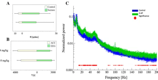
(Fig. 3a). The power spectra of accelerometer recording and EEG showed a 7 Hz peak before 4-AP administration, which is increased by 4-AP, and a novel 29 Hz peak appeared in the EEG power compared to the control. Additionally, in the accelerometer recordings, 4-AP produced a new peak at 16 Hz and peaks above 50 Hz, but the 40 Hz peak that was present in control decreased (Fig. 3b). It is examined how the number of spikes changed as a result of 4-AP administration.
EEG and accelerometer also showed remarkable increase in spike number, but the spiking activity on the EEG had higher
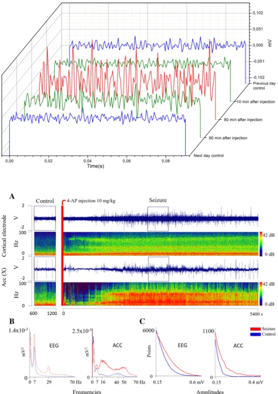
Fig. 2. 0.1 s EEG traces are showing the effect of i.p.
injected 10 mg/kg 4-AP in the mouse. Traces from control EEG recordings one day before and after 4-AP injection are colored with blue. EEG sample is represented shortly (10 min) after 4-AP injection and at the end (90 min) of recording (green). A trace from a devel- oped seizure (80 min) is shown with red
Fig. 3 In a control and seizure activity data from cortical electrode and accelerometer (X-axis) are compared, both presented in sonogram also.
Power spectrum from EEG and accelerometer is shown in b (blue—control, red—seizure activity). Spiking activity is rated by counting spikes in different amplitudes (c), colors correspond to b
amplitude, whereas accelerometer shows lower amplitude, high-frequency spikes than in the control (Fig. 3c). The aver- age number of spikes during the seizure increased to 325.6%
(4.9 ± 4.4 to 20.8 ± 27.2) at the 4 mg/kg dose, compared to 339.1% (5.8 ± 5.6 to 25.3 ± 17) at the 10 mg/kg dose, although as standard deviation shows data were variable (Fig. 4a). After data normalization with control spike number, the increase in spiking activity was 222% bigger in case of the higher dose.
Spike shape sorting was also made but revealed no signifi- cant results, since spike shapes were heterogeneous at both doses. The peak of movement was determined based on the accelerometer, while the peak of the seizure in terms of brain activity was determined based on brain activity measurements.
Regarding the seizure peak of brain activity calculated from the highest frequency of spiking activity, EEG reached its seizure activity peak 358.3 s earlier in the case of the higher dose (4 mg peak: 2825 ± 512.3 s, 10 mg peak: 2466.7 ± 367.4 s); meanwhile, motor activity calculated from the highest fre- quency spiking activity of the accelerometer reached its peak 611.1 s earlier after the administration of the lower dose (4 mg peak: 2966.7 ± 758.3, 10 mg peak: 3577.8 ± 917.6). The peak of motor activity at the 4 mg/kg dose is 141.7 s delayed comparing to cerebral activity at the same dose, whereas at the 10 mg/kg dose, the peak of the locomotor activity is preceded the seizure activity by 1111.1 s (Fig. 4b).
Discussion
In this study, the electrophysiological and behavioral prop- erties of the already described 4-AP mouse epilepsy model were examined. Since there was a lack of data about the dose dependency of EEG seizures and locomotor activity especially in case of B6N mice, correlation which allows the more sophisticated use of mouse 4-AP epilepsy model is described. The convulsive effect of 4-AP in different species was demonstrated in the early 1970s. Since then, it is known that 4-AP has a strong effect in mammals, they show the following symptoms: hyperexcitability, salivation, tremors, muscular incoordination, clonic and tonic convulsions, cardiac or respiratory arrest, and death (Schafer et al. 1973). Our findings support that 4-AP has a reliable electrographic and behavioral convulsive effect in mice. Although there are several chemical models of epi- lepsy in rodents, there seem to be a need for a well-inves- tigated model in mice for the benefit of further molecular research, as molecular research uses mice as model animal owing to the fact that they have short reproduction time and several genetic alterations have already been formed in them. The main finding of our study is that there is a remarkable difference between the effects of low and high
Fig. 4 Spike number average and standard deviation is compared in two doses both during control (white) and seizure activity (green) on a in two doses. Average peak of behavioral activity (accelerometer data—white) and average EEG seizure activity data (green) correla- tion is shown on b in two doses. Normalized power spectra compari- son is presented on c where different degrees of ketamine effect are
compared. Red stars indicate the sections where there is a significant difference between the two sections at p = 0.05 significance level with Bonferroni correction (2000 frequency value, 10 state comparison).
Averages are calculated from 30, 10 s long data for each state and their Standard Error of Mean (SEM) are shown
doses of 4-AP regarding correlation and frequency of EEG seizures and motor activity. Lower dose is characterized by a subsequent increase in lower amplitude range, number of spikes, later onset of seizures, and more variable seizures as shown by standard deviations. In this case, the develop- ment of behavior also shifted relatively to the enrichment of EEG spiking activity, and the pattern of correlation between movement and EEG was variable. Higher dose is characterized by intense EEG spiking activity and motor action positively correlating with the EEG constantly.
Behavior in mice definitely differs from that seen in rats, with no characteristic stages of epileptic seizures, instead mice perform a continuous increasing and then decreas- ing spiking activity in the cortex presented in Fig. 3. The increase of activity in the beta-, theta-, and gamma-bands indicates that the animal performed an exploration-like activity, constantly monitoring its environment. The sei- zure activity is rapidly spreading to the entire brain. At 10 mg/kg dose, the behavior follows the brain activity with a constant delay, the amplitude of spikes approximately doubles compared to that in the lower dose, and the seizure reaches its peak earlier but lasts for at least one and a half hours (Fig. 4). It is concluded that the 10 mg/kg dose set by Yamaguchi & Rogawski in Swiss mice is effective for B6N mice and this study also supports Van Erum et al.’s suggestion of revision of the Racine-scale in case of mice (Yamaguchi and Rogawski 1992; Van Erum et al. 2019).
Based on the results, we can suggest 4 mg/kg dose to study the seizure onset stage of epilepsy in mouse models. Nev- ertheless, 10 mg/kg dose is reliable to be used for severe epileptic activity in spite of the fact that mouse has no status epilepticus as rats. It is also revealed that the cor- relation of motor activity and seizures can be achieved at 10 mg/kg dose but not lower. Adjusting the dose of 4-AP, the mouse model can be tuned from the early stage to late stage of epileptic activity and can be applied for molecular studies with higher confidence in the future. However, it is emphasized that the mouse model of epilepsy is very dif- ferent from the human disease because of the continuous EEG spike genesis instead of bursts of seizures.
Conclusion for future biology
For the better understanding of human diseases, animal models are needed in most cases. These models must be designed for the further benefit of human research as well as the benefit of animal welfare. It is important to keep in mind the applicability, the practical implementation, and the repeatability of the experiments based on these models. In addition to developing new models, the revision of already standard methods is required from time to time. This article represents a supplement for the already described and used
4-aminopyridine rodent epilepsy model. According to the authors, as most of the studies use mouse models of neuro- degenerative diseases because of their well-known genetic constitution, easy manipulation and rapid reproduction period with numerous offspring, a strain-specific description is required. The findings of the present study are confirming the applicability and the dose of the 4-aminopyridine epi- lepsy in B6N mice; however, the authors wish to draw atten- tion to the fact that there is a remarkable difference between rat and mouse characteristics of seizures and there may be differences between mouse strains and between doses.
Acknowledgements The supportive work of the colleagues at ELTE Research Group of Proteomics and Institute of Experimental Medicine is highly appreciated.
Funding This work was supported by the National Research, Devel- opment, and Innovation Office of Hungary Grants (2017-1.2.1-NKP- 2017-00002 to F.Z.F., and Z.F.) the National Research, Development and Innovation Office (grants: NKFIH K 120143, K 113147) and FIEK_16-1-2016-0005 Grant to L.R., Zs. B. and G. J. The Bolyai János Scholarship of the Hungarian Academy of Sciences and Scholarship of the New National Excellence Foundation (UNKP-19-4-PPKE-9) granted for Z.F. is also acknowledged.
Data availability The raw generated data are available from the cor- responding author (FZF) on request accessibility.
Compliance with ethical standards
Conflict of interest The authors declare that they have no conflict of interest.
Ethical statement In all cases, our experiments were conducted in compliance with EU directive in force (2010/63/EU) and in accordance with the applicable government decree (40/2013. (II. 14.)), making sure that the animals suffer as little as possible.
References
Avoli M (1990) Epileptiform discharges and a synchronous GABAergic potential induced by 4-aminopyridine in the rat immature hip- pocampus. Neurosci Lett 117(1–2):93–98
Ben-Ari Y, Lagowska J (1978) Epileptogenic action of intra-amyg- daloid injection of kainic acid Comptes rendus hebdoma- daires des seances de l’Academie des sciences. Ser D Sci Nat 287(8):813–816
Blennow G, Brierley JB, Meldrum BS, Siesjö BK (1978) Epileptic brain damage: the role of systemic factors that modify cerebral energy metabolism. Brain J Neurol 101(4):687–700
Cavalheiro EA (1995) The pilocarpine model of epilepsy. Italian J Neurol Sci 16(1–2):33–37
Chung WK, Shin M, Jaramillo TC, Leibel RL, LeDuc CA, Fischer SG et al (2009) Absence epilepsy in apathetic, a spontaneous mutant mouse lacking the h channel subunit, HCN2. Neurobiol Dis 33(3):499–508
Fragoso-Veloz J, Tapia R (1992) NMDA receptor antagonists protect against seizures and wet-dog shakes induced by 4-aminopyridine.
Eur J Pharmacol 221(2–3):275–280
Fragoso-Veloz J, Massieu L, Alvarado R, Tapia R (1990) Seizures and wet-dog shakes induced by 4-aminopyridine, and their potentia- tion by nifedipine. Eur J Pharmacol 178(3):275–284
Gandolfo G, Gottesmann C, Bidard JN, Lazdunski M (1989) Ca2’
channel blockers prevent seizures induced by a class of K+ chan- nel inhibitors. Eur J Phamzacol 160:173–177
Gean PW (1990) The epileptiform activity induced by 4-aminopyridine in rat amygdala slices: antagonism by non-N-methyl-d-aspartate receptor anatagonists. Brain Res 530(2):251–256
Hare E, Hare AS (1979) Epileptiform mice, a new neurological mutant.
J Hered 70(6):417–420
Humphreys SR, Venditti JM, Ciotti CJ, Kline I, Goldin A, Kaplan NO (1962) Toxicity and antileukemic effectiveness of pyridine derivatives and 1, 3, 4-thiadiazole derivatives in mice relationship to nicotinamide antagonism. Cancer Res 22(5 part 2):483–550 King JT Jr, LaMotte CC (1989) El mouse as a model of focal epilepsy:
a review. Epilepsia 30(3):257–265
Klioueva IA, van Luijtelaar ELJM, Chepurnova NE, Chepurnov SA (2001) PTZ-induced seizures in rats: effects of age and strain.
Physiol Behav 72(3):421–426
Leonardi M, Ustun TB (2002) The global burden of epilepsy. Epilepsia 43:21–25
Morales-Villagrán A, Urefia-Guerrero ME, Tapia R (1996) Protec- tion by NMDA receptor antagonists against seizures induced by intracerebral administration of 4-aminopyridine. Eur J Pharmacol 305:87–93
Pasantes-Morales H, Arzate ME (1981) Effect of taurine on seizures induced by 4-aminopyridine. J Neurosci Res 6(4):465–474 Pasantes-Morales H, Arzate ME, Quesada O, Huxtable RJ (1987)
Higher susceptibility of taurine-deficient rats to seizures induced by 4-aminopyridine. Neuropharmacology 26(12):1721–1725 Rogawski MA, Barker JL (1983) Effects of 4-aminopyridine on cal-
cium action potentials and calcium current under voltage clamp in spinal neurons. Brain Res 280(1):180–185
Rudy B (1988) Diversity and ubiquity of K channels. Neuroscience 25(3):729–749
Schachter SC (2018) Determining when to stop antiepileptic drug treat- ment. Curr Opin Neurol 31(2):211–215
Schafer EW Jr, Brunton RB, Cunningham DJ (1973) A summary of the acute toxicity of 4-aminopyridine to birds and mammals. Toxicol Appl Pharmacol 26(4):532–538
Seyfried TN, Glaser GH (1985) A review of mouse mutants as genetic models of epilepsy. Epilepsia 26(2):143–150
Spyker DA, Lynch C, Shabanowitz J, Sinn JA (1980) Poison- ing with 4-aminopyridine: report of three cases. Clin Toxicol 16(4):487–497
Szente M, Baranyi A (1987) Mechanism of aminopyridine- induced ictal seizure activity in the cat neocortex. Brain Res 413(2):368–373
Szente M, Pongrácz F (1979) Aminopyridine-induced seizure activity.
Electroencephalogr Clin Neurophysiol 46(5):605–608
Tapia R (1982) Effect of 4-aminopyridine on transmitter release in synaptosomes. Brain Res 250(2):291–299
Tapia R, Sitges M, Morales E (1985) Mechanism of the calcium- dependent stimulation of transmitter release by 4-aminopyridine in synaptosomes. Brain Res 361(1–2):373–382
Thesleff S (1980) Aminopyridines and synaptic transmission. Neuro- science 5(8):1413–1419
Trancikova A, Ramonet D, Moore DJ (2011) Genetic mouse models of neurodegenerative diseases. In: Progress in molecular biology and translational science, vol 100. Academic Press, pp 419–482 Tsuji S, Meier H (1971) Evidence for allelism of leaner and tottering
in the mouse. Genetics Res 17(1):83–88
Van Erum J, Van Dam D, De Deyn PP (2019) PTZ-induced seizures in mice require a revised Racine scale. Epilepsy Behav 95:51–55 Voskuyl RA, Albus H (1985) Spontaneous epileptiform discharges
in hippocampal slices induced by 4-aminopyridine. Brain Res 342(1):54–66
White BH (2016) What genetic model organisms offer the study of behavior and neural circuits. J Neurogenet 30(2):54–61
World Health Organization (2019) Epilepsy. https ://www.who.int/
news-room/fact-sheet s/detai l/epile psy.
Yamaguchi SI, Rogawski MA (1992) Effects of anticonvulsant drugs on 4-aminopyridine-induced seizures in mice. Epilepsy Res 11(1):9–16.7