BY KENNETH V. THIMANN
CONTENTS
I. Historical Development and Definitions 2
II. Assay Methods 4 A. Avena test 4 B. Other Curvature Tests Using Agar Blocks 7
C. Straight Growth Measurements 8 D. Curvature of Slit Organs 9
E. Epinastic Curvature of Petioles 10
F. Other Methods 11 III. Chemistry of Auxins 11
A. "Auxin o and 6." 11 B. Indole-3-acetic Acid 12 C. Synthetic Substances Not Known to Occur Naturally 13
D. Nature of Auxin Precursors 17
IV. Transport of Auxin 22 A. Polar Transport and Its Mechanism 22
B. Upward Transport 26 V. Role of Auxin in Tropisms. 26
A. Geotropism 27 B. Phototropism 28 C. Other Tropisms 30 VI. Root Formation 31
A. Auxin as a Root-Forming Hormone 31
B. Substances Active 32 C. Interactions between Factors 32
D. Anatomical Studies 33 E. Methods of Treatment 34 VII. Phenomena of Inhibition and Toxicity 34
A. Bud Inhibition 35 1. The Facts 35 2. Mechanisms 35 3. General Significance 38
B. Root Inhibition 39 C. Inhibition of Abscission 42 D. General Toxicity 43 VIII. Other Actions of Auxin 44
A. Cell Division 44 1. Tissue Cultures 44 2. Cambium 45
1
3. Other Tissues 47 4. Pathological Changes 47 B. Formation of Fruits. 48 C. Role of Auxin in Seed Germination 50
IX. Mechanism of Action 51 A. Effects on Cell Wall 51 B. Mobilization of Special Hormones 53
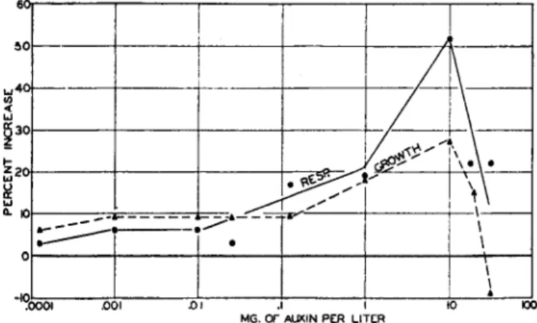
C. Relation between Respiration and Growth 54 D. Relation between Growth and Protoplasmic Streaming 56
E. Growth and Uptake of Water 59
F. Conclusions 60 References 61 Addendum 70 Supplementary References 70
I. Historical Development and Definitions1
The concept of hormones in plants developed from the study of tropisms or curvatures. Growing shoots typically curve toward a source of light (positive phototropism), and away from the earth (negative geotropism), while roots curve toward the earth (positive geotropism), and in some cases away from light (negative phototropism). Curva- tures may also occur away from or toward wounds (traumatotropism), electrodes (electrotropism), water (hydrotropism), etc. All these curva- tures depend fundamentally on a difference in growth rate between the two sides of a growing organ—the convex side grows faster than the concave.
The careful studies of Charles and Francis Darwin on the geotropism and phototropism of seedlings (72) made it clear that the perception of light and gravity is centered in the tip of the growing organ; thus photo- tropism of the coleoptile of Phalaris (a grass) was prevented completely by covering the extreme tip with a black paper cap. Nevertheless, the Darwins observed that the curvature in such tropisms is not restricted to the tip but spreads downward to the basal regions. They concluded that some "influence" is "transmitted" from the tip to the basal regions.
Thirty years later Boysen-Jen^en (41,42) showed that this influence can cross a discontinuity. He cut off the tips of Avena (oat) coleoptiles and stuck them on again in situ with gelatin. On now illuminating the tips, curvature appeared first in the tip and then also in the base. Evidently the influence which is transmitted must be of a "material nature/' This experiment was repeated with numerous variations, refinements, and controls by Paâl (239). More important, however, was the following experiment (done with Coix coleoptiles): the tip was cut off and then replaced, not symmetrically, but a little to one side. Without any illumination the plant now curved so that the side in contact with the tip
1 For a fuller treatment see Went and Thimann (360) Chapter 2, and also Boysen·
Jensen (46) Chapter 1.
was convex. This side, therefore, grew more than the other, and Paâl deduced that in the tip "a substance (or a mixture) is formed and inter- nally secreted." This substance diffuses into the lower regions and controls growth there. In normal growth, this substance would be symmetrically distributed, but curvature would be due to asymmetric distribution, caused in some way by the light (or gravity).
This conclusion led to experiments on normal, not curved, growth.
Söding (287,288) showed that indeed the tip controls straight growth of the part below it. Decapitation slows the growth greatly, though after some hours there is an acceleration due to " regeneration of the physio- logical tip" in the apical part of the remaining shoot. This regeneration was subsequently shown (78,288) to be due to production of the growth- promoting substance by the most apical remaining tissue.
Extracts of various tissues mixed with agar and applied to one side of decapitated coleoptiles (294) gave no evidence of containing a growth substance, though the technique of these experiments was a valuable advance. Certain enzyme extracts applied in agar did produce curva- tures, however (Seubert, 269).
Finally, Went (347,348) placed cut-off coleoptile tips on agar and applied this agar to decaptitated coleoptiles. This caused curvature, the side in contact with the agar being convex. Evidently the growth substance, although it could not be extracted by crushing the tissue, would "diffuse" from the intact tip into agar. The curvature was shown to be proportional, within limits, to the amount of growth substance in the agar, i.e., to the number of the tips placed on each block and the length of time they had been in contact. This procedure has formed the basis for the assay method described below, by means of which three naturally occurring substances of similar growth-promoting action have been isolated and many aspects of growth physiology have been studied.
The growth hormones have been named "auxins" and this name has since been applied to the whole group of synthetic substances of similar activity.
The remaining historical development will be treated in the appro- priate sections.
Definitions
Considerable confusion in the use of the terms: growth substance, growth hormone, growth regulator, Wuchsstoff, phytohormone, forma- tive substance, and auxin has arisen. The following definitions, which are practical rather than rigid, are put forward to simplify the situation.2
* Similar, but not identical, definitions have been proposed by van Overbeek (234a)
Auxin. An organic substance which promotes growth (i.e., irreversible increase in volume) along the longitudinal axis, when applied in low concen- trations to shoots of plants freed as far as practical from their own inherent growth-promoting substance. Auxins may, and generally do, have other properties, but this one is critical. The definition excludes nutrient salts, and in order to exclude sugar, which unquestionably promotes longitudi- nal growth, the term "low concentrations" may conveniently be inter- preted as "below ilf/1000." Most auxins produce clear-cut growth effects at 10~e M or even considerably below.8
Phytohormone. An organic substance produced naturally in higher plants, controlling growth or other physiological functions at a site remote from its place of production, and active in minute amounts. This definition includes those auxins which are of natural occurrence, certain of the vitamins, and other hormones such as those stimulating wound growth, or the postulated hormones of flowering, etc.
II. Assay Methods
Like vitamin assays, auxin assays can only be reliably carried out with auxin-deficient test objects. The most convenient of these is the dark- grown oat coleoptile from which the tip has been removed.
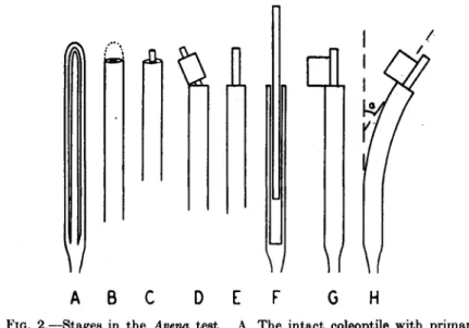
A. Avena TEST
As developed by Went and modified by numerous workers this is carried out as follows:
(1) Seeds of a pure line (the variety "Victory" or "Segerhavre" is the most commonly used) are husked, soaked for two hours in water, and laid out on wet filter paper with the embryo upward for 24 hours at 25°C. in weak red light.
(#) When the roots are about 2 mm. long they are planted in glass holders (see Fig. 1) with the root dipping into water contained in a zinc or glass trough. The holders are held in brass clips in rows of twelve.
They can be adjusted in two planes so that the shoots can be made strictly vertical. Some laboratories prefer to grow the plants in sand or soil, either in individual vials or in long narrow boxes.
(8) The seedlings are allowed to grow for about 48 hours at 25° in a dark room. The humidity must be controlled at 85-90% (relative) both to avoid drying and shrinkage of the agar blocks, and because plants grown in lower humidities are lefes sensitive (Gorter and Funke, 104), while at higher humidities guttation may occur. Small cabinets have been designed to take the place of a controlled dark room (Avery et al.,
18) but the latter is more convenient.
a Malic and other organic acids promote growth of the coleoptile at ΛΓ/1000 and below (335a) but only in presence of auxin.
(4) Straight seedlings of the same height are selected and the tips of the coleoptiles, to a length of about 1 mm., removed with sharp scissors (stage B in Fig. 2). This and all subsequent operations are carried out
rack trough
FIG. 1.—Avena seedling in glass holder with roots in water. Arrows show the directions in which adjustments can be made. (From Went and Thimann, 360.)
in orange or red light free from wavelengths shorter than 5900 A. Shorter wavelengths, except at extremely low intensities, produce phototropic curvature.
/iï\ A fci dl
ÏÏ
A B
H
FIG. 2.—Stages in the Avena test. A. The intact coleoptile with primary leaf within. B. First decapitation. C. Three hours later. D, E. Second decapitation.
F. Primary leaf pulled loose. G. Agar black in place. H. Curvature; the angle measured is a. (From Went and Thimann, 360.)
(5) Blocks of agar containing the substance to be tested are made by melting 3% agar and mixing with an equal volume of the test solution.
(Formerly blocks of pure washed agar were soaked in the solution but
this gives unreliable results.) For experiments of the diffusion type, the plant parts are placed directly on 1.5% agar. The blocks are cut up into small blocks of standard size, commonly 10 mm.3 The size is, however, not critical, since the curvature is essentially proportional rather to the concentration than the amount of the auxin contained in the block (Thimann and Bonner, 318); with 10 mm.3 blocks, 15% of the amount present enters the plant.
(6) Three hours after decapitation, when growth has slowed down and regeneration of the physiological tip begun (see pp. 12, 32) a further 4 mm. is cut off (stage D, Fig. 2). This is preferably done with special scissors with adjustable closure (see Went and Thimann, 360, Fig. 12).
The protruding primary leaf is pulled until it breaks off at the base, but left inside the coleoptile as a support (stage F, Fig. 2).
(7) The agar blocks are placed on one side of the decapitated coleop- tile, resting against the leaf (stage G, Fig. 2). From six to twelve or more plants are used for each determination. After a standard time (90 or 110 min.) shadowgraphs of the resulting curvatures (stage H, Fig. 2) are taken. This time is set by the "regeneration of the physiological tip," which causes formation of auxin on both sides and consequent regression of the curvature with increased growth rate.
(8) The curvatures are measured in degrees with a simple goniometer, and from the averages the concentration of auxin in standard units is determined. The plants for each test are calibrated by using blocks containing 0.025 mg. indoleacetic'acid per liter of agar, which gives a curvature within the range of proportionality, and a concentration five times higher, which gives the maximum curvature obtainable. The relation between concentration of auxin and curvature depends on the agar concentrations and the method of preparation of the plants. For times, age, and conditions similar to those given above, this relation is shown in Fig. 3. With higher agar concentrations the proportionality curve does not pass through the origin; with lower concentrations the curve is convex to the abscissa (326). The curvature may also be expressed in terms of d, the difference in growth between the two sides.
This is done by measuring r, the radius of curvature, and Z, the length of the curved zone, by means of a series of circular arcs.drawn on paper.
Then:
, tl a = —
r
where t is the thickness of the coleoptile, usually about 1.5 mm. This method was introduced by Purdy (244) and is used mainly by Boysen- Jensen and co-workers, who also grow their plants in sand or soil rather
than in glass holders. The relationship between d (in mm.) and curva- ture (in degrees) is approximately linear, a d value of 1 being about 47.8°.
Although the dark-room conditions are essentially constant, the sensitivity of the test varies with the time of day, being highest in the early morning (167,360). In spite of several attempts (167), no explana- tion has been found for this. In carrying out the test in diffuse light,
0.5 I 1.5 2 mg x I0"6 3 Amount of Auxin in the Block
FIG. 3.—Curvature (dotted) and straight growth (solid line) of Avena coleoptiles as a function of the amount of auxin applied. (After Thimann and Bonner, 319.) Söding and Funke (293b) found the sensitivity lower in warm weather than in cold, although this is not a direct effect of temperature.
B. OTHER CURVATURE TESTS USING AGAR BLOCKS
The characteristic feature of the Avena test is the use of an agar block of small volume. This makes possible the determination of very small quantities of auxin. In the standard test above, 0.025 mg. indoleacetic acid per liter of agar gives a curvature of about 10°, measurable to about 10%. The amount of indoleacetic acid in each block of volume 10 mm.3 is thus 2.5 X 10~7 mg. or 0.0025 7.
The "deseeded11 test (Skoog, 271) uses the oat coleoptile as above, but the seeds, i.e., the endosperms, are removed, without damaging the embryo, at twelve to eighteen hours before the test. The plants are held in the holders by cotton wool. Since the endosperm of the seed furnishes the precursor which is converted to auxin in the regenerated physiological tip (see pp. 22-24), these seedlings do not show regen- eration. Hence, the curvatures continue to increase up to six hours after application, and consequently the test, if curvatures are recorded at six hours, is three to five times as sensitive as the ordinary Avena test.
The Cephalaria test (Söding, 291,293) is carried out in diffuse daylight with decapitated hypocotyls of Cephalaria. Because this seedling has a
solid structure, unlike the hollow coleoptile of the grasses, it is less easy to apply the agar to one side. Accordingly the hypocotyl is cut through obliquely and the block placed at the lower end of the cut. The sensi- tivity of the test has an unaccountably large variation with the season : in June and July it is 400 times as sensitive as the Avena test, but in winter it is only about half as sensitive, according to Söding (293). It has been little used.
A curvature test with Raphanus hypocotyls was worked out by van Overbeek (226). The two cotyledons were removed, and in their place agar blocks were applied to the petioles—plain agar to one and the test block to the other. The curvatures were photographed after two hours.
C. STRAIGHT GROWTH MEASUREMENTS
Since the action of auxins in nature is to control straight growth, it is in principle desirable that assays should be checked by straight growth measurements, if not actually founded upon them.
Straight growth of rapidly growing organs is readily measured over short periods with a travelling microscope. In this way Söding (288) demonstrated uregeneration," i.e., renewed auxin formation in the coleop- tile stump some hours after decapitation. At Utrecht an automatically recording growth-measuring device, or "auxanometer," has been used in some critical studies (e.g., that of Dolk, 78). Measurements of enlarged photographs taken at intervals during growth were used by Thimann and Bonner (319) and showed, inter alia, that straight growth, like curva- ture, increases with the applied auxin concentration up to a clearly- defined maximum (see Fig. 3). Straight growth of decapitated coleoptiles has been used for comparing the activity of different auxins (260).
Decapitated Lupinus seedlings almost stop growth when exposed to light, and if auxin is applied to them, the resultant elongation, for whole hypocotyls, is linearly proportional to the logarithm of the concentration (108), while short sections show a direct linear relationship very much like that of Fig. 3 (Dijkman, 76).
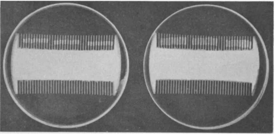
Straight growth of isolated coleoptile sections is conveniently meas- ured by placing the sections on fine glass rods (Bonner, 30; Thimann, 310) or better still on the teeth of fine combs (Schneider, 262) and floating these on the test solution (see Fig. 4). Sections of coleoptiles growing vertically on agar have been used by Monselise (209) and this method can be used with the auxin in agar blocks like the tests in B and C above.
Decapitated, isolated coleoptile sections, growing vertically, have also been used by Funke (88a) for assay of the growth inhibitor of maize seeds.
When the sections are used in solution the pH must be brought to 6.0, because acid pH increases the growth by increasing the fraction of
the auxin in undissociated (as opposed to salt) form (31,259a). The sections should not be submerged in the solution, but should break the surface (335a).
FIG. 4.—Sections 3 mm. long cut from coleoptiles, mounted on combs and immersed in solutions in petri dishes, photographed after 90 hours. Left: sections in water;
elongation about 10%. Right: Sections in auxin, sucrose, and KC1; elongation about 100% with some growth in thickness. (From Schneider, 262.)
D. CURVATURE OF SLIT ORGANS
It was found by Went (351) that the internodes of pea stems, if slit lengthwise and immersed in auxin solutions, curve inward (toward one another), the curvature being more nearly proportional to the logarithm of the auxin concentration than to the concentration itself. Jost and Reiss (150) used slit dandelion flowerstalks; and Thimann and Schneider (328) found slit coleoptiles of oats or corn very sensitive. Helianthus hypocotyls have been used by several workers, especially Diehl et al. (75), but with the auxin applied in lanolin paste. With all such objects, in water alone the halves curve outward, due to tissue tension. In very dilute auxin solutions there is often a slightly increased outward curva- ture, more marked with some auxins than with others (327). Acid pH has the same effect, probably due to liberation of auxin at the cut surface (31). The inward curvature is, like thé curvature of Avena coleoptiles in the test under A above, due to a difference in growth between the two sides of the organ, the outer side growing more than the inner (van Overbeek and Went, 238), but in this case the auxin is applied sym- metrically and the differential response is inherent in the plant tissue.
Van Overbeek and Went concluded that the curvature is due to differ-
10
ences in the rates of auxin entry on the two sides, entry taking place more readily through the outer intact side than through the central (wounded) tissue, but this has been disproved by Jost (149) and Thimann and Schneider (327), and the exact cause of the differential response has been the subject of considerable study. It is clear that it involves: (a) a true difference in the ability of the different layers of tissue to grow, the epidermis and outer cortex growing more, in response to auxin, than the pith and central layers (75,149,327); (6) a retarding effect on growth induced by the longitudinal wounding (263,353). The response of the epidermis is particularly important, "peeled" plants giving consistently smaller curvatures. The different response to auxin of the different layers is the main cause of the curvatures, and is also responsible for the development of the tissue tensions in the normal growing stem. The method is convenient where sufficient quantity of solution is available, and has been used in chemical studies on the activity of synthetic auxins (see Section III, C). It can be carried out in diffuse daylight.
A modification in which coleoptiles are slit into quarters instead of halves (328) gives considerably greater sensitivity. According to van Santen (259a) this method is much more sensitive to auxin a than to indoleacetic acid, but this is open to question.
E. EPINASTIC CURVATURE OF PETIOLES
In many dicotyledonous plants, the angle between the stem and the petiole is constant and characteristic, provided the plant is vertical.
Application of auxin dissolved in lanolin to the upper side of the petiole will cause it to be depressed and the increased angle between stem and petiole can thus serve as basis for an assay method. Hitchcock (136) and Hitchcock and Zimmerman (13Ô) have used this method with tobacco and other plants. (The curvature of the petiole in nature is classified as an epinasty and not a geotropism because, although caused by gravity, it is not a curvature toward or away from the earth, but toward or away from the stem. The direction of curvature is thus determined by the structure of the organs concerned.) It is to be presumed that the curvature is due to acceleration of growth on the side to which auxin is applied as in the other tests above, though analysis of the curvature in this sense has not been made. It is well to point out that tests such as this with intact green plants growing in the light are open to an important objection, namely that the test object is already rich in auxin, so that applied substances, even if they have no true activity, may give an effect through an action on the auxin already present. It is probably for this reason that some organic acids, which are not auxins at all, show activity in this method. Relative activities
of different auxins in causing epinasty are roughly in the same order as for causing curvature and gall formation in green plants, but not the same as for the A vena test (122).
F. OTHER METHODS
Methods depending on the formation or inhibition of roots or buds will be discussed in appropriate sections below. A few of these have been used as assay methods in the past but at present they are used mainly in the studies of the phenomena concerned and not as assays. A vena coleoptiles have occasionally been used, intact or decapitated, with the auxin applied in lanolin; the sensitivity is 10-50 times less than with agar (Brecht, 50, Avery et al., 14). In many cases it is desired to assay for a particular type of activity such as growth inhibition of shoots (Section VII, D) or parthenocarpic fruit formation (Section VIII, B). Certain auxins, particularly the alkyl esters of the acids, are effective in the vapor form (375); their action has beerj assayed by epinasty (above) or by morphogenetic effect on developing buds (see Section III of Chapter III). The swellings produced by applying auxin in lanolin to the decapi- tated stems of Vicia f aba seedlings have been utilized for an assay method by Laibach and Fischnich (182). The increase in diameter, measured after four days in the dark, is proportional to the logarithm of the auxin concentration up to a limiting value.
III. Chemistry of Auxins
A. " A U X I N A AND B"
In view of the importance of the coleoptiles of the grasses, especially oats, in the early work, it would be expected that efforts would be made to isolate auxins from this material. However, the quantities present are far too small. First steps toward isolation were made by the dis- covery of auxin in various commercial enzyme preparations by Seubert
(269), in cultures of several fungi including Rhizopus suinus by Nielsen (216,217), and in human urine by Kögl and Haagen Smit (162 cf. 164).
From the ether-soluble fraction of acidified urine, by an extensive series of fractionations, involving a concentration of 21,000 times, Kögl, Haagen Smit, and Erxleben (163) isolated an acid, termed "auxin a,"
Ci8H3206, and also its lactone. Turning their attention to plant mate- rial they analyzed a number of samples of cereal seeds and selected a corn germ oil and a malt sample which appeared to have very high auxin contents. From these were isolated both the auxin a above and a new acid, C18H30O4, named "auxin 6" (160). The degree of concentration required was 100,000 times for the malt and 300,000 times for the corn
12
oil. The two substances are closely related, the former being a tri- hydroxy and the latter a ketohydroxy acid :
(Ci3H23)CHOHCH2(CHOH)2COOH Auxentriolic acid, "auxin a"
(C13H28)CHOHCH2COCH2COOH Auxenolonic acid, "auxin b"
The "auxin a" lactone is considered to have the 1,5-lactone form.
In spite of the small amounts available (less than 1 g. of total active crystals), Kögl and Erxleben established the structure of the C13 residue as 2,4-di-sec-butyl-A1-cyclopentene and confirmed this by the identity of the substituted glutaric acid, obtained by oxidative breakdown, with a synthetic product, 2,3-diisobutylglutaric acid, II. The full formula cf auxin a is therefore I :
CH3
C2H6CHCH—CCHOHCH2CHOHCHOHCOOH
H < I
C,H6CHCH—CH
CHa I
CHa I
C2H6CH—CH—COOH CH3 CH2
C2H6CH—CH—COOH II
On standing in the dark, the double bond shifts to the side chain and the hydroxyl to the ring to produce an inactive substance, pseudoauxin a
(p. 19). Auxin a lactone undergoes a similar change with loss of water.
This and related changes may play a role in phototropism (see Section V).
B. INDOLE-3-ACETIC ACID
In extending their work on urine, Kögl, Haagen Smit, and Erxleben (165) found that a large part of the auxin present was destroyed by attempts to lactonize with hydrochloric acid in methanol. A modi- fied isolation method thereupon led to the identification of indole-3-acetic acid as a third active compound. Its activity in the Avena test is prob- ably about half that of auxin a or b. Kögl and Kostermans (168) also isolated this substance from yeast plasmolyzate.
Working independently on the auxin produced by Rhizopus minus cultures, Thimann (310) showed by isolation that this also is indole-3- acetic acid. At first it was thought that indoleacetic acid is typically a
product of microorganisms and not a true hormone of higher plants, and it was accordingly named by Kögl et al "heteroauxin," but Haagen Smit and co-workers (125) subsequently isolated it in pure form from alkali- hydrolyzed corn meal and indicated that most of the activity of the hydrolyzed meal was due to indoleacetic acid rather than auxin a.
Haagen Smit et al, (124) later obtained it also from the endosperm of immature corn grains. Gordon and Wildman (102,103) have brought forward evidence that alkali treatment produces traces of indoleacetic acid from the tryptophane in a number of proteins (see below), but this is not likely to be the main source of the indoleacetic acid isolated.
Instability to hot acid and stability to alkali indicate that the auxin extracted from many higher plants (237, and see Section D below) is of the indole type. It is probable, therefore, that indoleacetic is widely distributed in higher plants, perhaps more widely than auxin a and 6.
and it is evidently a true plant hormone. The high specificity of the indoleacetic-acid-inactivating enzyme of the pea plant (306) also points in this direction; some workers believe auxin a and b occur only rarely.
Besides indoleacetic acid, indoleacetaldehyde also occurs in plants, particularly in dark-grown Pisum, Vicia, Helianthus, and Brassica (187).
The aldehyde is readily oxidized to the acid by Schardinger's enzyme from milk, or by contact with soil. It behaves as a "neutral auxin,"
and was discovered through its presence in the neutral fraction by Larsen, who purified extracts by shaking out from ether at different pH. Its identity was established by conversion to indoleacetic acid and various other tests. Its widespread occurrence is, of course, further evidence for the importance of indole derivatives as plant hormones.
C. SYNTHETIC SUBSTANCES NOT KNOWN TO OCCUR NATURALLY
A great number of related compounds have been prepared and tested.
The results depend to some extent on the assay used. The Avena test is highly specific. Besides the above compounds, only the lower alkyl esters and two of the methyl derivatives of indoleacetic acid (169), the isostere indene-3-acetic acid (311), and indole-3-butyric and 1-naphtha- leneacetic acids (360), show appreciable activity in this test. The potassium or sodium salts show about the same activity as the free acids, provided the solutions are not buffered. A few other substances show activity in very high concentrations only, frequently producing very short apical curved zones. Phenylbutyric acid, which is inactive by itself, inhibits the effect of indoleacetic acid, by competition (272a), or perhaps by a more complex mechanism. This substance (and also cyclohexaneacetic acid) greatly increases the auxin curvature in the pea test (353). This is explained by Went (353,356) in terms of two procès-
ses; one, the "preparatory" reaction, can be carried out by substances inactive as auxins, while the other, the "growth" reaction proper, requires the chemical structures discussed below.
Straight growth of isolated stein çr coleoptile sections (see Section II, C) is less specific, and the curvature of immersed slit stems (Section II, D) or other methods still less so. Hence the activity of a given synthetic auxin, relative to a standard such as indoleacetic acid, varies with the type of test. This is illustrated by Table I (from Thimann and Schneider, 1939), which not only shows the difference in specificity of the tests, but also illustrates how compounds inactive, or almost so, in one test may show high activity in others. However, the order in which the substances fall is nearly the same in each test (see also the data of Gustaf son, 122).
Using the slit pea stem curvatures, Haagen Smit and Went (126) and Koepfli, Thimann, and Went (157) have tabulated the activities of a large number of related compounds, and Veldstra (1944a) has added a number more. Using epinasty and the changes in shape of young tomato leaves, Zimmerman and Hitchcock (372) and Zimmerman (370) have added a further large group, including the highly active ring-
TABLE I
RELATIVE ACTIVITY OF SIX AUXINS«
Acid
Curvature of slit stems of
Pisum
Straight growth of Pisum
sections
Straight growth of Avena
sections
Curvature standard in Avena test Naphthalene-1-acetic.
Indole-3-butyric Indole-3-propionic Phenylacetic Benzof urane-3-acetic.
Phenylbutyric
370 190 150 10 6 3
23 22 8 0.4 0.3 0.08
15 9 1.6 0.3 0.11 0.06
2.5 8 0.1 0.02 0.02 0.005
β Activity of indole-3-acetic acid brought to 100% for each test.
and Schneider, 328.) (From Thimann
substituted derivatives p-chloro- and 2,4-dichlorophenoxyacetic acids.
Some approximate relative activities for the induction of seedless fruit (see Section VIII) have been given for these compounds by Zimmerman and Hitchcock (373). As might be expected, the ratios of the activities of various substances determined in this way are not the same as by the above methods.
In spite of all this work, it is still not possible to make a really binding
statement as to the structural requirements for auxin activity. The difference between the tests, mentioned above, is in part due to the neces- sity for the substance to be transported through the plant tissue in tests using agar blocks/but riot in tests using immersion in a solution. Some substances, though highly active locally, are not readily transportable.
This important limitation, brought to light with indeneacetic and benzo- furaneacetic acids by Thimann (311), was confirmed for several sub- stances by Went and White (361) in transport experiments, which are discussed in Section IV, A. Then, too, the stability of the substance to plant enzymes, its permeability through cell membranes, and the fraction present in undissociated form (29,31) all influence the responses, the last because the ionized salt form does not penetrate into the cell readily, as shown by Albaum et al. (4). A correction for the extent of dissociation increases the apparent activity of many substances in the pea test. The influences of these modifying factors are discussed in Went and Thimann, Chapter 8 (360), and by Went (353,355), and more recently by Veldstra (339). The auxin-inactivating enzyme in pea plants is highly specific for indoleacetic acid (Tang and Bonner, 306) ; this might cause this sub- stance to show a lower activity than other, unnatural, compounds.
These factors can as a first approximation all be considered second- ary, the primary one being the ability to cause cell enlargement when present in the cell. Using this criterion of primary activity, Koepfli, Thimann, and Went (157) stated the following structural requirements:
(i) A ring system as nucleus; (2) A double bond in the ring; (3) A side chain containing a carboxyl group (or an ester or amide readily converti- ble to a carboxyl); (4) A distance of at least one carbon atom between this group and the ring; and (5) A particular space relationship between the carboxyl and the ring.
As to 1, no aliphatic compounds tested have shown activity.
As to 2', dihydroindoleacetic acid and dihydroauxin a are inactive; so is cyclohexane acetic acid. A number of compounds with unsaturation in the side chain but not in the ring, such as pseudoauxin a, III, cyclo- hexylideneacetic acid, IV, and benzofulveneacetic acid, V, are inactive.
As to (3), some modification is needed to allow for the small but definite activity of naphthyl-1-nitromethane (aci form), VII, and indican,
—C=CHCOOH
I C H2
V C4H9CH—C=CHCH2(CHOH)2COOH
CH2
C4H9CH—CHOH H2C|
H2CI
CHCOOH II
in
CH2
IV
|CH2
ICH2
CHjCOOH
Napthalene-1-VI acetic acid
CH=NOOH
Napthalene-1-nitro-VII (aa)-methane
/ \
OSO,H I
V N
H VIII Tndican VIII, both of which have acid side chains which are not carboxyl groups (339). It may be that any acidic {i.e., hydrogen-ion-yielding) group is effective to some extent. Also napthaleneacetonitrile and tryptamine (271) show a slow activity, which is doubtless due to conversion to a car- boxylic group within the plant. There is some evidence, however, that naphthalene- 1-acetamide, IX, is active without being hydrolyzed (335a).
CH2CONH2
w
IXThe activity of esters is not entirely clear. The data of Kögl and Kostermans (169), with the Avena test, show decreasing activity with increasing molecular weight of the alkyl-esterifying group of indoleacetic acid; they therefore concluded that activity was due to hydrolysis (by plant esterases) to the free acid, which should go with decreasing rapidity as molecular weight of the alkyl group increases. Avery et al. (14) have found the esters to have about the same activity as the free acids, or somewhat less in the case of naphthaleneacetic acid; this would agree with the above view. However, Zimmerman and Hitchcock (371,374) found, in experiments with tomato plants, that at least the methyl ester of indolebutyric acid has slightly higher activity than the free acid. This might, of course, be due to some secondary property of the ester such as ease of penetration through the intact epidermis. By contrast, the esters of auxin a are inactive in the A vena test (160).
As to 4) the optimum distance is commonly one carbon atom, activity decreasing with increasing length of side chain, but there is some alter- nating effect, indolebutyric being more active than indolepropionic acid.
The carbon atom may be replaced by other hetero atoms. In the case of phenoxy and napthoxy acids the hetero atom oxygen is present as well as the one carbon atom.
Point δ is the most ill defined. The activity of cis-cinnamic acid and some of its derivatives, and the inactivity of the trans isomers, are among the main pieces of evidence. In the Avena test, the two optical isomers of a-(j3-indole)-propionic acid, X, have different activity, the ( + ) being thirty times as active as the ( —) (171), but, since the activity on immersed
CH3—CH—COOH
H N
coleoptile sections is identical, this différence apparently does not relate to primary growth activity. It provides another example of the high specificity of the Avena test. Veldstra (339) has postulated that the side chain must be perpendicular to the plane of the ring, and supports the argument by consideration of molecular models. He makes clear that in cis-cinnamic acid the side chain is perpendicular to the plane of the ring, while in the trans form both are in the same plane. Even in napthalene-1-acetic acid the position perpendicular to the ring is favored.
Yet it is difficult to see how introduction of halogen atoms into the ring could alter such spatial relationships. Thus Zimmerman showed, with epinasty (370), that introduction of the ortho chlorine atom increased the activity of phenoxyacetic acid twenty times, the para chlorine atom by eighty times, while both together (2,4-dichloro derivative) increase it some 1200 times. In the pea test (335a) these four substances have the following activities, as per cent of that of indoleacetic acid:
Phenoxyacetic acid ca. 0
o-Chlorophenoxyacetic acid 4 p-Chlorophenoxyacetic acid 200 2,4-Dichlorophenoxyacetic acid 1200 While substitution in the ortho position might possibly have some effect
in orientation of the side chain, it seems hardly likely that substitution in the para position could do so. There are numerous other examples of the same effect. The exact nature of the spatial relationships must therefore be left open for the present (but see Wisconsin Symposium, 1951).
D. NATURE OF AUXIN PRECURSORS
The auxin in human urine clearly comes from the diet. The esters of auxin a are inactive, and some oils yield auxin on hydrolysis with
18
lipase or with sodium ethylate (160). Ingestion of natural oils increased the auxin content of urine, while hydrogenated oils, pure protein, and sugar did not (163). Indoleacetic acid in urine, similarly, comes from ingested protein (125), wheat giving a particularly clear rise in urine auxin as soon as one hour after feeding.
As was mentioned in Section II, the auxin produced in the coleoptile tip, or in the apical part of the stump in " regeneration/' is formed from a precursor in the seed. This was first made probable by Cholodny (61), who showed that soon after the seed was wetted auxin appeared. This auxin does not, as claimed by Pohl (242), travel directly up into the coleoptile, but that which goes into the coleoptile tip travels up as an inactive precursor. This was shown by Skoog (271), who placed agar blocks for a while on the stump of decapitated coleoptiles and then showed that when applied one-sidedly to freshly decapitated coleoptiles ("deseeded test") they caused no effect at first but slowly induced a curvature after two to six hours (341).
Following the work of Thimann and Skoog (332), Gustafson (115,118), Wildman and Gordon (365), and Thimann, Skoog, and Byer (333) on the extraction of auxin Irom plant tissues, it has now become increasingly clear that many plant materials yield auxin very slowly on extraction with ether, and that this auxin stems from proteins in the tissue. The slow yield is due to a reaction with water, probably proteolysis, which liberates auxin. This reaction is stopped by boiling (332) and this has been put to use for an assay of the free auxin in plant tissues by Gustafson (118). It is also stopped by thorough drying (197,332) and at once resumed on adding water. Proteolytic enzymes, especially chymo- trypsin, were found by Skoog and Thimann (274) to accelerate greatly the liberation of the auxin. Wildman and Gordon (365) and Gordon and Wildman (103) have obtained an auxin which is almost certainly indoleacetic acid from isolated plant proteins both from leaves and from seeds. Since this auxin is best obtained by alkaline hydrolysis (25), some of it, at least, doubtless derives from oxidative deamination of tryptophan. However, this is probably not the whole story, for two good reasons: (a) in the case of cabbage leaves the auxin yields are prob- ably too high to be ascribed to the tryptophan present, according to the determinations of Avery, Berger, and White (15); and (6) auxins which may be either acid labile (i.e., indole derivatives) or alkali labile (pre- sumably auxin a or b) may be obtained from purified wheat proteins (Gordon, 102). It is to be noted that Gordon's wheat proteins were well characterized, which makes it "highly improbable that the auxin could be merely an impurity. In any event, particularly in the case of auxins liberated by enzymes, there is no reason to doubt that, as was originally
postulated (333), true auxin-protein complexes do occur. These could, of course, serve as important auxin reserves for the plant.
The form in which auxin occurs in seeds differs from that in other tissues. The bulk of the auxin in the cereal grains is in bound form, in the endosperm, and only liberated by alkaline hydrolysis (13,125,130, 333). It is this material which is indoleacetic acid, as shown by Haagen Smit et al. (124,125) and Berger and Avery (24,25). The quantities are large enough in corn, i.e. 20-100 mg./kg., that it acts as an antivitamin in animal growth (156). However, some auxin is obtainable, largely from the embryo, by direct extraction with organic solvents, as in the isolations by Kögl and co-workers described above, and this material is auxin a and b. Thirdly, the addition of water to the endosperm liberates a moderate quantity, presumably by enzymic action. Much of this was probably also bound in the dry state, either chemically as a precursor, or in some physical or adsorptive manner, as in dried Lemna, in which the auxin can be first liberated and then made unextractable by drying (332).
Whether the water-extractable auxin in the grain is auxin a or indole- acetic acid is not clear. Hatcher (130) has assumed that it represents free auxin, the alkali-hydrolyzable part being the bound or precursor form, but there is not enough evidence for this yet. The situation is complicated by Funke's finding (88a) that part of the auxin in corn endosperm is stable to hydrogen peroxide.
In contrast to the grains, no auxin is liberated from Lemna by alkali autoclaving, although, as with other green tissues, it is set free slowly by moist ether (118,332), as discussed above. Cabbage (15) and spinach, however, do yield some auxin to alkali, though in the author's unpublished experiments spinach leaf proteins gave much higher yields with chymo- trypsin than with alkali. The purified auxin-protein in spinach leaf cytoplasm does not liberate its auxin readily; it is resistant even to vigorous electrodialysis, and sets free auxin only when actual proteolysis occurs, so that it is indeed a relatively stable complex (Bonner and Wild- man, 35). Between these two extremes there seem to be many inter- mediate states, in different tissues, in regard to ease of liberation (309,333).
A true precursor, of course, would be a substance from which auxin is continually produced, by plant enzymes, under normal conditions and in physiologically significant amounts. It is by no means certain that the auxin-proteins fulfil these criteria. Neither papain-hydrogen cyanide nor the enzymes of autolyzing Lemna liberated any appreciable amount of auxin (333), and chymotrypsin, as far as is known, does not occur in plants. Ficin, which does liberate auxin from Lemna, is a plant enzyme, it is true, but it is not known to be widely distributed. The partial
liberation of auxin on slow drying of leaves may be enzymic, but it is quantitatively rather small. A true precursor system was, however, studied by van Overbeek (233) in the isolated coleoptile tip, which con- tinues for a long period to yield auxin to agar blocks, although the amount which can be extracted from it by organic solvents at any time is relatively small (309). Berger and Avery (23,24) made a partial isolation of a true auxin precursor from corn; this appears not to be a protein, having only 4.7 to 6.4% nitrogen, but its nature remains unknown. It yields indoleacetic acid on alkaline hydrolysis. The variation in amount of precursor and "free auxin" (but see comment above) with age and drying of the grain in rye has been very thoroughly studied by Hatcher (130), who finds that the "free" form appears first and then decreases as the bound form increases and the grain ripens. Some of these changes may, however, be due to variations in the amounts of inhibiting substances rather than in the true auxin (88a).
A more remarkable precursor was obtained earlier from radishes by Stewart, Bergren, and Redemann (297,299); this substance in the intact form actually inhibits growth of the Avena coleoptile, giving marked positive curvatures, but on hydrolysis yields an auxin which is probably indoleacetic acid. Its chemical nature is also unknown but it is thought to be a peptide. The further study of this substance might be important in regard to inhibitions (see Section VII).
The ability to convert tryptophan to indoleacetic acid is probably widespread among microorganisms; this is doubtless the source of the auxin in fungus cultures, as shown by Thimann (310). Furthermore, this is the most probable source of the large amounts of auxin produced in bacterial infections of plants such as legume root nodules and crown galls (see Section VIII, A). Other plant infections resulting in patho- logical overgrowth (188,189) may have the same explanation, and indeed Link et al. (197) have shown that aphids are very rich in auxin; whether this was extracted from the leaves on which they fed, or elaborated within the aphid is not clear, but in any event either the removal or injection of auxin by the aphid may account for some of the growth effects caused by these parasites. As to higher plants, the evidence as to their ability to convert tryptophan to indoleacetic acid under natural conditions is not perfect. Tryptophan causes a slow curvature in the "deseeded" Avena test; it causes straight growth of coleoptiles when applied to the base but not to the tip. and it leads to root formation on pea cuttings (335,335a).
On the other hand it cannot replace indoleacetic acid in sterile tissue cul- tures, as found by Nobécourt (221). Unless the tests with higher plants are carried out under sterile conditions a positive result might always be due to infection. Because the growth effects produced by tryptophan
differ anatomically from those caused by indoleacetic acid, Kraus (174) claims that its action cannot be due to conversion to the latter compound.
But since only one concentration, in lanolin, was studied in his experi- ments, and since growth effects are characteristically dependent on con- centration, this conclusion is obviously unjustified. The best evidence of conversion is that of Wildman et al. (364a), who obtained formation of an active auxin by spinach leaves infiltrated with tryptophan within two to four hours. The enzyme system was present in dialyzed cyto- plasm prepared from the leaves and had its optimum pH at 7.5. There is some evidence that the reaction goes via indolepyruvic acid (cf. 310);
in any event it is an oxidative process.
The case of tryptamine, which, like the precursor in the seed, is directly converted to auxin in the Avena coleoptile (271), is worth special mention, though its biological significance is not known. Lastly the indoleacetaldehyde found by Larsen (187) in etiolated Pimm and other plants must be considered under this head. Larsen's extracts of neutral auxin, which had quite low activity, were converted to highly active material, considered from diffusion measurements to be indoleacetic acid, by treatment with soil or with a preparation of Schardinger oxidase. In some cases the neutral material had no growth activity at all, which suggests that there is more than one neutral compound convertible to indoleacetic acid. There is no evidence here, though, that enzymes in the plant can carry out the conversion. Hemberg (131a) finds a similar situation in potato tubers.
An interesting general scheme for auxin activity has been proposed by Skoog, Schneider, and Malan (272a), according to which the auxin molecule, envisaged as a kind of coenzyme, has to combine on the one hand with its substrate and on the other with an apoenzyme. Precursors could thus be of two kinds: those in which the substrate-combining part is covered or distorted but can be corrected by the plant, e.g., tryptamine or indoleacetaldehyde, and those in which the apoenzyme-combining part has been combined with some other molecule but can be freed under some conditions. The latter have their substrate-combining activity intact and can therefore occupy the substrate to the exclusion of free auxin molecules, thus giving competitive inhibition (e..g.y phenylbutyric acid) or even total inhibition (e.g. the inhibitor of Stewart et al. described on p. 24). These authors point out that, if excess auxin were present, some molecules would combine only with the substrate and some only with the apoenzyme "thus effectively blocking each other from reacting.'' This would account for inhibitions of the type discussed in Section VII, A. This ingenious scheme has much to recommend it, though consider- ably more evidence would be needed to establish its validity.
IV. Transport of Auxin
A. POLAR TRANSPORT AND ITS MECHANISM
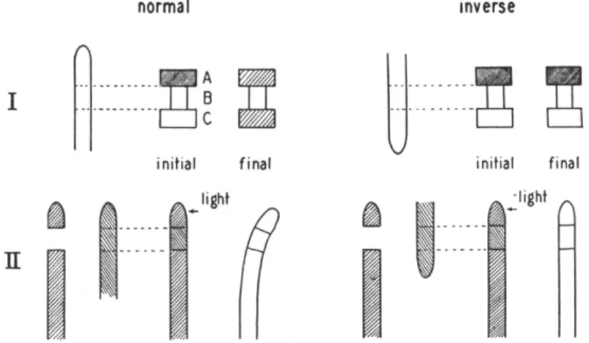
One of the most remarkable properties of living plant tissue is the strictly polar way in which auxin is transported in it. The polarity of shoots, particularly in regard to bud development and root formation, has been recognized from very early times, and the polar transport of auxin provides an explanation for at least many such phenomena. The earlier work on polar transport of auxin has been so fully reviewed (360, Chap. 6) that it needs only the briefest recapitulation here.
In seedlings, phototropism is detected by the tip and the stimulus conducted toward the base; movement in the reverse direction doer not
normal inverse
Λ
s
1
B ] C
initial final
^ light
\J
as
initial finalm
-light A
FIG. 5.—I. Diagram of transport experiment. Auxin is transported from agar block A through coleoptile section B to receiving block C. Left, normal transport;
right, section inveited, no transport. Degree of shading indicates auxin concentra- tion in agar.
II. Transmission of phototropic stimulus through normal (left) and inverted (right) section of coleoptile introduced between tip and base of another coleoptile.
(From Went and Thimann, 360.)
occur. Interposition between the tip and base of a section of inverted tissue prevents the movement (see Fig. 5, II), which is therefore strictly basipetal.. Auxin'will be transported directly through a short section of Avena coleoptile in the apex-to-base direction, but not inversely (Fig.
5, I). The process is not one of diffusion, as was proved by the experi- ments of van der Weij (346), which were carried out as shown in Fig. 5,1, the auxin in the blocks being determined by the Avena test. The main results can be summarized as follows:
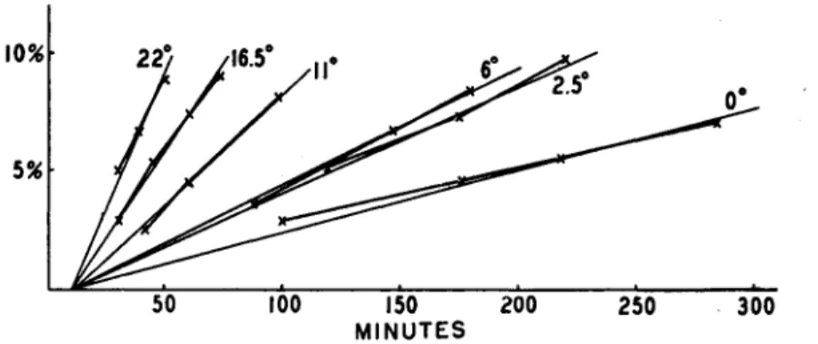
(ί) The temperature coefficient of the amount transported per unit time between 0° and 30°C. is about 3, i.e., that of a chemical reaction.
(#) The velocity, however, as measured by the time taken for the first auxin to appear at the basal end of the conducting tissue, is about 12 mm./hour in Avena and is independent of temperature. This is deter- mined by extrapolation (see Fig. 6). (8) The concentration of auxin in the agar block at the receiving end soon equals that in the donating block, and subsequently exceeds it, so that auxin must be actively trans- ferred against its gradient. (4) By etherizing the sections, polarity
FIG. 6.—Auxin transported, as per cent of the amounts applied, through 2-mm.
sections of Avena coleoptiles as a function of time in minutes. Extrapolation of the curves of different temperatures indicates that they all cross the x axis at about 10 minutes, showing that transport velocity is independent of temperature between 0°
and 22°C. (From van der Weij, 346.)
disappears and with it also disappears the "active" nature of the trans- port; it now becomes essentially a diffusion process.
Auxin transport is thus like that of "objects along a moving band; the band goes at constant speed so that the number of objects arriving at the end per unit time is independent of the length; the time required for the first object to reach the end is proportional to the length of the band;
if not removed from the end the objects continue to pile up" (Went and Thimann, 360). Stems (see Beal, 21), petioles, hypocotyls, and leaf veins behave like coleoptiles so far as they have been studied.4 Tissue cultures, especially of carrot and endive, demonstrate the polarity of auxin transport in many ways (Gautheret, 98, pp. 161-166). Other auxins than indoleacetic acid move both more slowly and in smaller quantities per unit time. The data of Went and White (361) yield the following rates in millimeters per hour through Avena coleoptiles:
4 Unpublished experiments of W. P. Jacobs show that the polarity is far from strict in young Phaseolus hypocotyls.
Indoleacetic acid 9.0 Indolebutyric acid 6.6 Anthraceneacetic acid 5.4 Napthaleneacetic acid 3.9 cw-Cinnamic acid Not detectable
It should be added that longitudinal transport of auxin is not affected by light (226) ; this is important for the understanding of phototropism (see Section V).
The mechanism by which this active transport is achieved is not understood. Accumulation of solutes against a gradient, as by roots or by algae growing in very dilute nutrient solutions, must involve a com- parable type of active transport (352), but in this case in the lateral rather than the longitudinal direction. Arisz has recently brought to light (10) a similar transport of amino acids through the tentacles of Drosera, and Schumacher (265) described polar movement of fluorescein in stem hairs of cucurbits. The polarity of auxin transport is therefore not an entirety isolated phenomenon.
Attempts have been made to relate the transport to the electrical polarity of the plant. The apex of shoots is in general negative to the base, as shown by the early work of Lund (see 200) with nonpolarizable electrodes. This apical negativity is still present in short sections of stems or coleoptiles, and is largely abolished by etherization (64). The anion of a weak acid such as auxin would, of course, be transported from apex to base under such a potential. Koch (153) showed that plant auxin in agar does in fact move toward the anode, and Clark (63) con- firmed this for pure indoleacetic acid. Kögl et al. (167) showed essen- tially the same thing by making the agar block in the Avena test negative to the plant, and passing a small current, which had the effect of increas- ing the resulting curvature, doubtless by increasing the movement of auxin from the agar into the plant. Then, too, coleoptiles and shoots placed in air or water between oppositely charged poles curve toward the positive pole (6,49,153); such curvature implies more growth on the side toward the negative pole. Electrolytic movement of auxin has even been produced directly in plant tissue by Koch (153), by inserting elec- trodes into opposite sides of sunflower hypocotyls, which were subse- quently halved and tested for auxin (by applying them to roots). The hypocotyls here curved toward the negative pole and the convex half gave the greater curvature on the test roots. These experiments all show that electrolytic movement is possible, and takes place in the right direction. But here the parallel ends, for the following reasons: (1) a potential gradient of 50 volts/cm. was needed for detectable transport—
far higher than the electrical gradients observed in plants, (j!) externally
applied potentials do not affect the polarity of auxin transport through coleoptile sections, even though they may reverse the electric polarity, (8) inverting the section with respect to gravity inverts the electrical gradient but does not affect the auxin transport (63), and (J) treatment with 10-100 p.p.m. of sodium glycocholate completely abolishes the transport but does not affect the electrical polarity, or indeed any other observable property of the coleoptile section (see Table II; from Clark, 64).
The absence of any effect of low glycocholate concentrations on respiration, while auxin transport is wholly prevented, is of interest since
TABLE II
EFFECT OF GLYCOCHOLATE ON AUXIN TRANSPORT AND ELECTRICAL POLARITY OF COLEOPTILE SECTIONS
Sections infiltrated
with
Water
Na glycocholate 10 p.p.m lOOp.p.m 1000 p.p.m
Units auxin transported in 2 hr.
11.4 0 0 0
Emf be- tween apex
(—) and base (+),
mv.
10 10 10 0
Proto- plasmic streaming
+ + +
Qot (in separate
expts.)
1.21 1.22
Appearance of tissue
Turgid Turgid Turgid Flaccid
normal respiration is apparently essential for transport of auxin into the section (33). The absence of any inhibiting effect on streaming suggests that transport does not take place in the streaming protoplasm. Simi- larly, Schumacher (265) could observe protoplasmic cyclosis going on simultaneously with polar movement of fluorescein in the cells of the cucurbit hair.
As will be shown in the following section, curvatures induced by gravity involve a movement of auxin laterally across the coleoptile or stem. Here also it has been thought that an electrical gradient, resulting from gravity, might be responsible, and long ago Brauner (47,48) showed that indeed the under side of a stem placed horizontal becomes electro- positive to the upper side (the "geoelectric effect"). The potential difference due to gravity is established before any curvature occurs, and there are several very suggestive relations between the potential and the subsequent auxin transport brought out by Schrank (264). No causal relationship has as yet been established, however.
It can only be concluded that auxin transport is not directly related to electric polarity; it is in some way related to respiratory processes but the link can readily be broken without damaging these processes.
B. UPWARD TRANSPORT
There are two conditions under which auxin is transported upward, i.e., from base to apex. The first is when it is applied to the upward- moving transpiration stream, as by pouring a solution on the soil (137) or adding auxin to a nutrient solution in which stem cuttings (138) or roots (272) are immersed. In such cases, so long as transpiration occurs, the auxin is passively carried upward in the xylem in the same way as salts or dyes and the amount absorbed parallels the absorption of water.
It is a function of the transpiration rate but is also influenced by the con- centration of salts in solution. Skoog has, however, shown (272) in extensive experiments with tomato stems that auxin taken up in this way then moves laterally into the surrounding living tissues and is re-exported downward by the normal polar transport.
The other condition is when very high concentrations are applied.
Went and White (361), taking every precaution to avoid leakage along surfaces, still obtained inverse transport in the coleoptile when concen- trations of 1000 mg./l. indoleacetic acid were used. Snow (282, see also 284) obtained curvatures apical to the point of application by using fairly high concentrations in lanolin; the effect was more marked when the application was close to the vascular bundle, so that it probably involved movement in the transpiration stream also. Stewart (298) showed by Avena tests that auxin moved upward when very strong (2%) paste was applied to the first interno<Je of a young bean plant.
It is probable that these effects are due to the toxicity of high auxin concentrations.
Finally mention may be made of the interesting case of inverted cuttings, i.e., cuttings rooted at the apex, budding from the base, and planted inversely. In such cuttings there is a gradual development of a new series of cells from the shoots to the roots, opposite in polarity to those originally present, and correspondingly Went (357) found that at first the auxin transport is apex-to-base polar, but gradually base-to-apex transport appears as well. Normal cuttings show no such change. This phenomenon only serves to emphasize the strictly polar nature of auxin transport under normal physiological conditions.
V. Role of Auxin in Tropisms
Although it was through tropisms· that the role of the "growth sub- stance " was first discovered (see Section I) interest in the past ten years