Examination of Thyrotropin-Releasing Hormone (TRH)-synthesizing System in the Rodent
Hypothalamus
Ph.D. Thesis
Andrea Kádár
Semmelweis University
János Szentágothai Ph.D. School of Neuroscience
Tutor: Csaba Fekete D.Sc.
Opponents: Dóra Reglődi D.Sc.
Alán Alpár Ph.D.
Chairman of committee: György M. Nagy D.Sc.
Members of committee: Katalin Halasy D.Sc.
Zsuzsa Várnainé Tóth Ph.D.
Budapest
2014
2 I. Table of contents
I. Table of contents ... 2
II. List of abbreviations ... 5
III. Introduction ... 8
1.Thyroid hormones: metabolism, signaling and effects ... 8
1.1. The metabolism of THs ... 8
1.2. The transport of THs ... 10
1.3. TH receptors and signalization ... 11
1.4. Effects of THs ... 12
2. Hypophysiotropic TRH neurons ... 13
3. The negative feedback regulation of the hypophysiotropic TRH neurons by THs ... 14
4. Tanycytes: location in the ME and role in the regulation of the HPT axis ... 17
4.1. The structure of the ME ... 17
4.2. Tanycytes ... 18
4.3. The role of tanycytes in the regulation of hypothalamic T3 homeostasis ... 19
5. Neuronal inputs of hypophysiotropic TRH neurons ... 21
5.1. Catecholaminergic neurons of the brainstem ... 22
5.2. Arcuate nucleus ... 22
5.3. The hypothalamic dorsomedial nucleus... 26
6. Regulation of the HPT axis during fasting and refeeding ... 27
7. Melanocortin resistance of the hypophysiotropic TRH neurons during the early phase of refeeding ... 28
8. Leptin-induced synaptic rearrangement in the regulation of feeding-related hypothalamic neuronal groups ... 30
9. Non-hypophysiotropic TRH neurons ... 31
10. Identification of the activated elements of neuronal networks ... 32
IV. Specific aims ... 33
V. Materials and methods ... 34
1. Animals ... 34
2. Tissue preparation and labeling ... 35
3
2.1. Methods used for mapping the distribution of hypophysiotropic TRH neurons in the PVN of
mice ... 35
2.2. Methods to identify the route of T3 transport between the thyroid hormone activating tanycytes and the hypophysiotropic TRH neurons ... 39
2.3. Methods used for studies focusing on the mechanism of the melanocortin resistance of the hypophysiotropic TRH neurons during refeeding ... 41
2.4. Method for combined use of immunocytochemistry and Nissl-staining ... 42
3. Image analyses ... 44
4. Statistical analyses ... 47
5. Specificity of the primary antisera ... 47
VI. Results ... 50
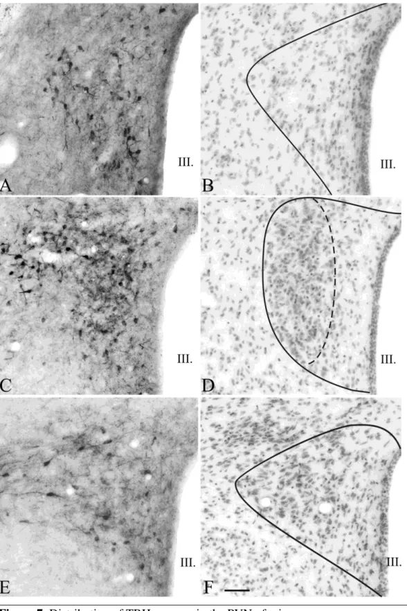
1. Distribution of hypophysiotropic TRH neurons in the mouse hypothalamus ... 50
1.1 The organization of the PVN in mice ... 50
1.2. Distribution of TRH-IR neurons in the PVN ... 51
1.3. Distribution of hypophysiotropic TRH neurons in the PVN ... 51
1.4. Comparative localization of hypophysiotropic TRH neurons and vasopressin and oxytocin neurons in the PVN ... 54
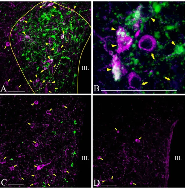
1.5. Co-localization of CART and TRH in the neurons of the PVN ... 58
2. Identification of the transport route the thyroid hormone activating tanycytes and hypophysiotropic TRH neurons ... 61
2.1. Presence of MCT8 in the hypophysiotropic axon terminals in the median eminence ... 61
2.2. Co-localization of MCT8 and TRH in hypophysiotropic axon terminals ... 61
3. Clarification of the mechanism causing the melanocortin resistance of hypophysiotropic TRH neurons during refeeding ... 64
3.1. AGRP- and α-MSH-IR innervation of TRH neurons ... 64
3.2. The effect of fasting on the number of α-MSH- and AGRP-IR boutons on TRH neurons ... 64
3.3. Effect of 2h vs. 24h of refeeding on the number α-MSH- and AGRP-IR boutons in contact with TRH neurons ... 64
3.4. The effect of leptin treatment on the fasting-induced rearrangement of α-MSH- and AGRP- inputs to TRH neurons ... 65
4. Development of an improved method to combinate immunocytochemistry with Nissl- staining ... 71
4.1. Effects of RNase-free immunocytochemical method on immunostaining ... 71
4
4.2. Analysis of the juxtaposition of varicosities and neurons using standard immunocytochemical
method combined with Nissl-counterstaining ... 71
4.3. Analysis of the juxtaposition of varicosities and neurons in the vPVN after refeeding using RNase-free immunocytochemical method combined with Nissl-counterstaining ... 71
VII. Discussion ... 74
1. Distribution of hypophysiotropic TRH neurons in the mouse PVN ... 74
2. A new mechanism regulating thyroid hormone availability of hypophysiotropic TRH neurons ... 76
3. Fasting-induced alterations in the α-MSH- and AGRP-IR innervation of TRH neurons: a possible mechanism of melanocortin resistance during the early phase of refeeding ... 78
4. The effect of RNase-free immunocytochemical treatment on the cytoplasmic staining with Nissl-dyes ... 81
VIII. Conclusions ... 84
IX. Summary ... 86
X. Összefoglalás ... 87
XI. References ... 88
XII. List of publications ... 115
1. List of publications underlying the thesis ... 115
2. List of publications related to the subject of the thesis ... 115
XIII. Acknowledgements... 116
5 II. List of abbreviations
α-MSH - Alpha-melanocyte stimulating hormone
ABC - Avidin-biotin-peroxidase complex
AGRP - Agouti-related protein
ARC - Arcuate nucleus
AVP - Vasopressin
BAT - Brown adipose tissue
BBB - Blood-brain barrier
BSA - Bovine serum albumin
BT - Biotinylated tyramide
CART - Cocaine- and amphetamine regulated transcript
CNS - Central nervous system
CRE - cAMP-response-element
CREB - cAMP-response-element binding protein
CRH - Corticotropin-releasing hormone
CSF -Cerebrospinal fluid
D1 - Type 1 deiodinase
D2 - Type 2 deiodinase
D3 - Type 3 deiodinase
DAB - Diaminobenzidine
DEPC - Diethylpyrocarbonate
DMN - Dorsomedial nucleus
EPSC - Excitatory postsynaptic current
ERK - Extracellular signal-regulated kinase
GFP - Green fluorescent protein
GHRH - Growth hormone-releasing hormone
GnRH - Gonadotropin-releasing hormone
HPT axis - Hypothalamus-pituitary-thyroid axis
icv. - Intracerebroventricular
i.p. - Intraperitoneal
IPSC - Inhibitory postsynaptic current
6
IR - Immunoreactive
KO - Knock out
MC3-R - Melanocortin receptor type 3
MC4-R - Melanocortin receptor type 4
MCT8 - Monocarboxylate transporter type 8
MCT10 - Monocarboxylate transporter type 10
ME - Median eminence
mTOR - Mammalian target of rapamycin
NCoR1 - Nuclear receptor co-repressor 1
NHS - Normal horse serum
NP - Neurophysin
NPY - Neuropeptide Y
OATP1C1 - Organic anion transporting polypeptide 1c1
OT - Oxitocin
PBS - Phosphate buffered saline
P-CREB - Phosphorylated cAMP-response-element binding protein
PFA - Paraformaldehyde
PI3K - Phosphatidylinositol-3-kinase
PIF - Prolactin-inhibiting factor
POMC - Proopiomelanocortin
P-STAT3 - Phosphorylated signal transducer and activator of transcription 3
PVN - Paraventricular nucleus
rT3 - Reverse triiodothyronin
RXR - Retinoic X receptor
SCN - Suprachiasmatic nucleus
SRC1 - Steroid receptor co-activator 1
SRIF - Somatostatin
STAT3 - Signal transducer and activator of transcription 3
T2 - Diiodothyronin
T3 - Triiodothyronin
T4 - Thyroxine
TB - Tris buffer
7
TH, THs - Thyroid hormone, thyroid hormones
TR, TRs - Thyroid hormone receptor, thyroid hormone receptors
TRα - Thyroid hormone receptor alpha
TRα1 - Thyroid hormone receptor alpha type 1
TRβ - Thyroid hormone receptor beta
TRβ1 - Thyroid hormone receptor beta type 1
TRβ2 - Thyroid hormone receptor beta type 2
TRE - Thyroid hormone responsive element
TRH - Thyrotropin-releasing hormone
TSH - Thyroid stimulating hormone
UCP-1 - Uncoupling protein 1
VMN - Ventromedial nucleus
vPVN - Ventral paraventricular nucleus
WAT - White adipose tissue
WT - Wild type
Y1 - Neuropeptide Y receptor type 1
Y5 - Neuropeptide Y receptor type 5
8 III. Introduction
The hypophysiotropic TRH neurons are the main central regulators of the hypothalamus-pituitary-thyroid (HPT) axis [1]. These TRH neurons are located in the hypothalamic paraventricular nucleus (PVN) [2-4], integrate neuronal and humoral signals and serve as a final common pathway in the regulation of the hypothalamic- pituitary-thyroid axis [5]. The axons of the hypophysiotropic TRH neurons terminate outside the blood-brain-barrier (BBB) in the external zone of the median eminence (ME) where TRH is secreted to the portal capillary system of the anterior pituitary [6]. From these blood vessels, TRH enters to the extracellular space in the pituitary, and stimulates the synthesis and secretion of thyroid-stimulating hormone (TSH) [5], the hormone that regulates the hormone production of thyroid gland. Thyroid hormones (THs) have widespread biological actions including the regulation of energy metabolism through influencing the basal metabolic rate, adaptive thermogenesis and feeding behavior and regulation of the development and normal functioning of the central nervous system (CNS) [7-11].
1.Thyroid hormones: metabolism, signaling and effects 1.1. The metabolism of THs
Thyroid hormones - 3,3’,5-triiodothyronin (T3) and thyroxine (T4) - are synthesized in the follicles of thyroid gland by iodination of tyrosine residues of thyroglobulin [12].
Although T4 is the predominant product of the thyroid gland, T3 is the form of thyroid hormones that can bind to thyroid hormone receptors with high affinity [13]. The activation of thyroid hormones by conversion of T4 to T3 and the degradation of these hormones are catalyzed by deiodinase enzymes [13, 14]. These enzymes are critical for the regulation of thyroid hormone levels in the circulating blood, but also for the tissue specific, local regulation of thyroid hormone availability [15].
All three known deiodinase enzymes (D1-3) are selenoproteins containing selenocysteine residue in their active site [16].
Type 1 deiodinase (D1) can remove iodine from both the inner and outer rings of THs, forming either T3 or the inactive reverse T3 (rT3) from T4 or convert T3 to the inactive diiodothyronin (T2) [14, 16]. Since this enzyme can both activate and inactivate thyroid
9
hormones, it has the least clear physiological role in the deiodinase protein family.
Because D1 is able to convert T4 to the considerably active T3, initially it was thought to be the main source of extra-thyroidal T3 [16, 17]. Recently, however, increasing evidence indicates that D1 contributes significantly to the circulating T3 concentration only in hyperthyroid patients, but not in healthy subjects [18, 19]. Therefore, currently D1 is considered a thyroid hormone inactivating enzyme in euthyroid conditions by producing inactive rT3 or T2 [14]. The liver and kidney, where D1 participates in the clearance of TH derivatives, contains a relatively large concentration of this enzyme, but D1 is also present in the anterior pituitary, intestine, placenta and thyroid gland [16, 20]. In rats, D1 is also present in the cerebral cortex with relatively low activity, but in humans, D1 is absent from the CNS [21, 22].
The main source of extra-thyroidal T3 is type 2 deiodinase (D2) that converts T4 to T3 by 5’-monodeiodination of the outer ring of the molecule [14, 18]. In addition to regulating circulating T3 concentration, this deiodinase enzyme also plays critical role in the organ specific regulation of T3 availability [15]. Although the adequate level of T3 is essential for the sufficient neuronal activity, T3 can be transported through the BBB remarkably less efficiently than T4 [23]. More than 75% of T3 in the cortex is generated locally by D2 catalyzed conversion of T4 to T3 [24]. In human D2 is the only 5’
deiodinase in the brain [15]. In accordance with this cardinal role, D2 has a widespread expression in the brain [15, 16]. D2 is expressed predominantly in glial elements of the neuronal tissue, such as tanycytes and astrocytes [25]. These glial cells have crucial role in the maintenance of the adequate T3 level in the CNS [25]. Tanycytes are specialized ependymal cells located in the basal and lateral walls of the third ventricle in the hypothalamus [26]. Hypothalamic D2 expression is concentrated in the median eminence and arcuate nucleus, where D2 is expressed primarily in tanycytes [14]. D2 activity is absent from most hypothalamic regions including the PVN where the hypophysiotropic TRH neurons reside [27]. This distribution of D2 is in contrast with the homogenous D2 expression of astrocytes in other brain regions [27-29]. D2 can also be found at high levels in the pituitary, brown adipose tissue (BAT) and placenta and also occurs in the gonads, pineal gland, thymus, mammary gland, coronary artery and smooth muscle cells of the aorta [15, 16].
10
In contrast to the role of D2, the primarily function of the type 3 deiodinase (D3) is the local inhibition of TH signalization by inactivation of THs. D3 performs inner ring deiodination converting T4 to rT3 and T3 to T2 [15, 16]. This deiodinase enzyme is primarily expressed in neurons in the CNS where D3 plays an important role by protecting neurons from the excess of T3 [14, 30]. In the brain, D3 mRNA can be observed widely in the cerebral cortex, hippocampal pyramidal cells, granule cells of the dentate and pyriform cortex. In addition, lower level of D3 expression can be found in hypothalamic neurons [15, 16, 31]. Although the main function of D3 is to protect tissues from excess THs, the systemic administration of thyrotoxic doses of T3 induces a surprisingly modest increase in D3 expression of the hypothalamus. This finding is in contrast with observations in other brain regions such as the cortex, the hippocampus and the olfactory bulb [31]. Hence, hypothalamic deiodinase enzymes appear to convert the changes of peripheral THs to changes of hypothalamic T3 concentration allowing T3 to serve as a regulatory signal for neuroendocrine functions [14]. Besides its presence in the CNS, a remarkable amount of D3 is also expressed in most of the fetal organs and placenta, confirming the essential role of this enzyme in the precise regulation of the TH level during embryonic development [32].
1.2. The transport of THs
Although THs are lipid soluble molecules, their passage through the cell membranes and the BBB requires specific transporter molecules [33]. The transport of THs is facilitated by at least three transporter molecules: organic anion transporting polypeptide 1c1 (OATP1C1), monocarboxylate transporter 8 (MCT8) and 10 (MCT10) [34].
OATP1C1, a member of the organic anion transporting family, is highly specific for T4, rT3 and T4 sulfate [35, 36]. It binds T3 with much lower affinity [35, 36]. The presence of OATP1C1 has been described in Leydig cells of the testis and in the capillaries of the brain [37-39]. In the rodent brain, OATP1C1 is highly expressed in cerebral microvessels underlining its cardinal role in the TH transport through the BBB [40]. OATP1C1 is also expressed in hypothalamic tanycytes [40].
The most studied TH transporter is MCT8, a member of the monocarboxylate transporter family with 12 helical transmembrane domains [34]. This transporter molecule has strong affinity for T3, T4 and rT3 [41]. MCT8 is expressed in various
11
tissues including heart, liver, kidney, adrenal and thyroid gland [42, 43]. MCT8 is also present in most areas of the CNS, such as hypothalamus, choroid plexus, amygdala, hippocampus, olfactory bulb and cortex [40, 44-46]. Although MCT8 is the main TH transporter of neurons it can also be found in tanycytes [45]. The importance of MCT8 in normal neurological function is underlined by the demonstration of the mutation of this gene in families with inherited mental retardation and underdevelopment [47, 48]. The syndrome caused by this mutation is called Allan-Herndon-Dudley syndrome, which has typical endocrine abnormalities in addition to the strong neurological phenotype: elevated serum T3 and reduced T4 and rT3 levels [49]. Although Mct8 knock out (KO) mice have the same alteration in the level of THs, interestingly no major neurological abnormalities were found in these animals [50, 51] that suggests species-specific differences in the neuronal transport of THs and raises the possibility that in rodents, MCT8 is not the only neuronal thyroid hormone transporter.
The amino acid sequence of MCT10 shows great degree of homology with MCT8 [34]. Although data about its physiological function is limited, cell culture studies demonstrate that MCT10 prefers T3 instead of T4 [52]. This TH transporter molecule is expressed in the intestine, kidney, liver, muscle and placenta, but there is no data that confirms its presence in the CNS [53-56].
1.3. TH receptors and signalization
The thyroid hormone receptors (TRs) are located in the nucleus of cells and act on thyroid hormone response elements (TRE) of DNA as ligand-dependent or independent transcription factors [57, 58]. All the known types of TRs are encoded by two genes:
THRA and THRB [58]. Alternative splicing of the THRA transcript results in different TRα isoforms, but only TRα1 acts as a functional receptor [59]. The other TRα variants seem to have inhibitory effect on TH induced gene expression in in vitro studies [59]. The THRB gene also has two products: TRβ1 and TRβ2 [58]. Both of these receptors are functional receptors and differ in the transcription initiation site, which results in different amino end of the two TRβ isoforms [60]. The distribution of TRα1 and TRβ1 is widespread, whereas TRβ2 is expressed predominantly in TRH neurons of the PVN and in thyrothrop cells in the anterior pituitary [60-63]. Examination of TRβ KO and TRβ2
12
KO animals confirms that the TRβ2 receptor plays a crucial role in mediating the negative feedback effect of THs on TRH and TSH synthesis [64, 65].
The thyroid hormone regulated genes can be divided into two main categories:
positively and negatively regulated genes. The mechanisms of positive and negative regulation of thyroid hormone responsive genes is markedly different. The effect of T3 on the positively regulated genes is mainly mediated by the heterodimer complex of a TR and retinoic X receptor (RXR) [66]. The TR-RXR complex is fully active when not only T3 binds to TR, but 9-cis retinoic acid also binds to the RXR component of the heterodimer [67]. The transcriptional initiation of positively regulated genes is facilitated by the recruitment of co-activator molecules to the ligand bound TR-RXR complex [58, 60]. In the absence of T3, the unliganded TR-RXR heterodimer complex is also associated with TRE, however, in the absence of T3, TRs recruit co-repressor proteins mediating the basal, ligand independent repression of the target genes [68, 69].
In contrast, the transcriptional activity of genes negatively regulated by T3 is stimulated by the complex of the unliganded receptor and the co-repressor molecules, while the liganded receptor-co-activator complex suppresses these genes [65]. Although, it was debated for years, the action of TRs on the negatively regulated genes also appears to require direct binding of the receptor to the TRE of genes [70-72]. The typical co- activator molecule, steroid receptor co-activator 1 (SRC1) has inhibitory effect on the HPT axis [73] while the co-repressor molecule, nuclear receptor co-repressor 1 (NCoR1) is required for the ligand independent activation of the HPT axis [74, 75].
1.4. Effects of THs
Thyroid hormones can influence a variety of tissue types. They regulate the embryonic neurogenesis, the migration of pyramidal cells in the cerebral cortex [11, 76-79], granule cells in the hippocampal gyrus dentatus [80-82] and Purkinje cells in the cerebellum [83, 84]. Furthermore, they have effects affect on axonal growth [85] and dendritic arborization [86, 87]. THs also influence learning and memory in adults [88, 89] and influence wakefulness [90] and mood [91]. In addition, THs influence energy homeostasis via increasing the basal metabolic rate [10], stimulating the diet-induced [92]
and adaptive thermogenesis [93], facilitating lipogenesis [94, 95] and increasing food intake [95].
13
One of the major effects of THs on energy expenditure is the stimulation of basal metabolic rate. In cells regulated by THs, the basal metabolic activity is stimulated by the increased number and size of the mitochondria, the increased expression of the respiratory chain components, and the increased membrane permeability for Na+ and K+, and the increased concentration of the Na+/K+ ATP-ase in the membrane [96].
Thyroid hormones also increase the energy expenditure by stimulating the thermogenesis in the BAT [97, 98]. The heat production of BAT requires sympathetic activation [97, 98], thyroid hormones [93] and local activation of T4 by D2 [99]. The synergistic effect of the adrenergic stimulation via β adrenergic receptors and the locally generated T3 result in increased uncoupling protein 1 (UCP-1) synthesis and thermogenesis [99, 100]. Without thyroid hormones or D2, sympathetic activity is unable to increase UCP-1 synthesis and thermogenesis [99]. During cold exposure, the marked increase of D2 expression also plays a critical role in the local increase of T3 concentration in the BAT. This virtually saturates all TRs in this tissue, allowing thus the cold induced thermogenesis of the BAT to be fully active even if circulating thyroid hormone levels are slightly above normal [99]. The increased UCP-1 level interrupts the connection between the respiratory chain and ATP synthesis. This way, most of the energy released by substrate oxidation dissipates as heat [8].
2. Hypophysiotropic TRH neurons
The primary central regulator molecule of thyroid hormone synthesis is TRH, a tripeptide amide (pGlu-His-ProNH2) [1]. In the anterior pituitary, TRH stimulates the synthesis and release of TSH by acting on the G protein coupled type 1 TRH receptor [101]. In addition, TRH increases the posttranslational glycosylation of TSH, which has an important effect on the biological activity of the molecule [102, 103]. The secreted TSH then acts on the thyroid gland and regulates its hormone production [104].
Neutralization of endogenous TRH by administration of anti-TRH serum induces transient reduction of circulating level of TSH and THs [105, 106]. In newborn TRH KO mice, no difference can be observed neither in the morphology of anterior pituitary nor in the synthesis of THs at birth compared to wild type (WT) mice [107, 108]. However, the gradual increase of the TH levels observed during the first postnatal days in WT animals is absent in the TRH KO mice [107, 108]. Furthermore, the lack of TRH causes a
14
dramatic decrease in the number of the TSH-immunoreactive (-IR) cells in the anterior pituitary and also in the TH levels after the 10th postnatal day [107, 108]. These observations suggest that TRH has an essential role in the early postnatal development of the HPT axis and also in the maintenance of the activity of this neuroendocrine axis in adult animals. The critical importance of TRH in the regulation of the HPT axis is also demonstrated in some human patients with idiopathic central hypothyroidism [109]. In these patients the release of biologically inactive TSH is accompanied by low circulating levels of THs [109]. This clinical picture can be cured with chronic intravenous TRH administration [109], further demonstrating the key role of TRH in the central regulation of the HPT axis.
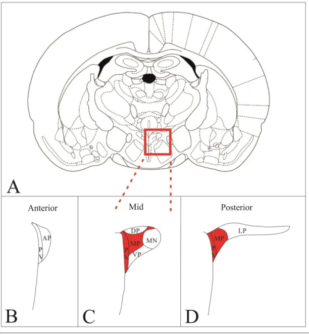
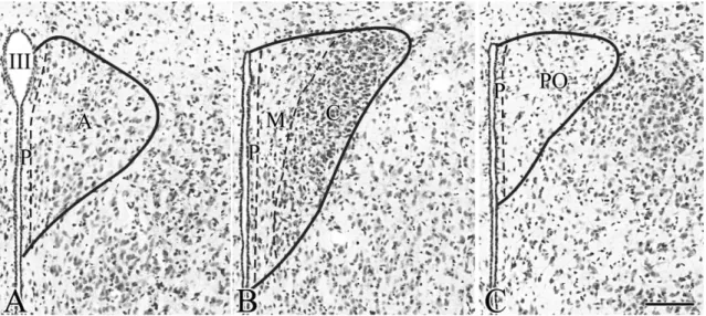
TRH is widely synthesized throughout the CNS [5], but only the so called hypophysiotropic TRH neurons that project to the external zone of the median eminence and secret the TRH into the portal capillary are involved in the regulation of HPT axis [3, 6]. These neurons are located in the PVN, a bilateral triangle-shaped nucleus in the dorsal margin of the third ventricle. The PVN can be divided into two main divisions named based on the size of their neurons: the parvocellular and magnocellular divisions. In rats, the parvocellular division can be further divided into six subdivisions: the anterior, medial, dorsal, periventricular, ventral and lateral subdivisions (fig.1.) [110]. TRH- synthesizing neurons can be found in all parvocellular subdivisions in rats [111], but the hypophysiotropic TRH neurons are located exclusively in the periventricular and medial parvocellular subdivisions (fig.1.) [2, 3, 6, 111]. In rats, a distinctive feature of the hypophysiotrophic TRH neurons is their cocaine- and amphetamine regulated transcript (CART) expression; this peptide has not been identified in any other TRH-expressing neuron population of the brain [2, 3, 6, 111]. Although the localization of the hypophysiotropic TRH neurons is precisely determined in rats, limited information is available currently about the distribution of these neurons in mice, the most frequently used animal model of the current days.
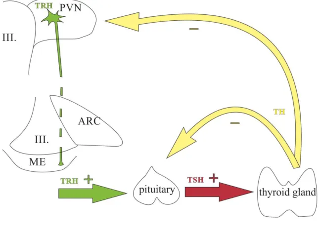
3. The negative feedback regulation of the hypophysiotropic TRH neurons by THs Since the maintenance of normal thyroid hormone levels is critical for the appropriate functioning of the organism, the activity of both the hypophysiotropic TRH-producing neurons and the TSH-expressing cells of the anterior pituitary are tightly regulated by the
15
negative feedback effect of THs [60] (fig.2.). Hence, under basal conditions the rise of TH levels inhibits TRH and TSH expression. In contrast, the fall in circulating TH levels has stimulatory effect on TRH and TSH secretion. [112]. In the CNS, the TRH gene expression is controlled by the negative feedback effect of thyroid hormones only in the hypophysiotropic TRH neurons [113]. The observation that T3 administration to the PVN of hypothyroid animals results in a decrease of the TRH expression exclusively in the side of the implantation verifies that THs have direct effect on TRH neurons [114]. The negative feedback effect of THs on TRH expression is mainly mediated by TRβ2 [64], although results of studies using siRNA mediated TRβ1 gene silencing suggest that TRβ1 also contributes to the ligand independent activation of TRH gene expression [115].
Interestingly, hypophysiotropic TRH neurons also express TRα1 which has stimulatory effect on TRH expression in the presence of T3 [65]. However, this TRα1-mediated effect is masked by the inhibitory effect of TRβ receptors in adult animals [65, 116]. In addition to the direct effect of THs on the TRH gene, THs also modulate TRH production by the downregulation of prohormone convertases type 1/3 and 2, resulting in accumulation of immature proTRH derivatives and a decrease in the production of mature TRH molecules [117].
16
Figure 1. Schematic drawing illustrates the organization of the PVN in rats and the localization of the hypophysiotropic TRH neurons. The higher magnification maps based on the figures of Fekete et al. [112] show the subdivisions of rat PVN in three different antero-posterior levels (B-D). Red areas indicate the presence of hypophysiotropic TRH neurons. TRH neurons with hypophysiotropic function are absent from the anterior level (B), but present in the medial and periventricular subdivisions of the mid (C) and posterior (D) levels of PVN.
MN – magnocellular division Parvocellular subdivisions:
PV – periventricular; AP – anterior; VP – ventral; MP – medial; DP – dorsal; LP - lateral
17
Figure 2. Schematic illustration of the hypothalamus-pituitary-thyroid axis.
Cell bodies of the hypophysiotropic TRH neurons are located in the PVN and their axons terminate in the ME where TRH is released to the circulation. TRH reaches the anterior pituitary through the hypophysial portal system and stimulates the TSH secretion. TSH stimulates the T3/T4 production of the thyroid gland. THs exert negative feedback effect both on the hypophysiotropic TRH neurons and the thyrotrop cells of the anterior pituitary.
4. Tanycytes: location in the ME and role in the regulation of the HPT axis 4.1. The structure of the ME
The release of the hypophysiotropic hormones into the circulation occurs in the BBB- free ME, where the axons of the hypophysiotropic neurons terminate [118]. The ME can be divided into three parts: ependymal layer, internal and external zones [119].
Hypophysiotropic neurosecretory cells, such as TRH-, corticotropin-releasing hormone (CRH)-, gonadotropin-releasing hormone (GnRH)-, prolactin-inhibiting factor- or dopamine (PIF)-, growth hormone-releasing hormone (GHRH)- and somatostatin
18
(SRIF)-producing neurons release their substances in the densely vascularized external zone of the ME [3, 6, 118, 120-124]. The hormones secreted by hypophysiotropic neurons to the capillary loops are transported directly to the anterior pituitary by its long portal vascular system [125]. The internal zone of the ME contains the axons of the magnocellular neurosecretory cells, these vasopressin (AVP)- and oxytocin (OT)- containing axons pass through the ME and terminate in the posterior pituitary where the hormones are secreted to the circulation [126, 127]. The ependymal layer that forms the floor and ventrolateral walls of the third ventricle contains the cell bodies of specialized glial cells called tanycytes [26]. These glial cells has essential role in the regulation of HPT axis [5].
4.2. Tanycytes
Tanycytes are elongated glial cells with cell body located in the ependymal layer [26].
Tanycytes contact the cerebrospinal fluid (CSF) with their surface covered with microvilli and has a long basal process to the median eminence or to discrete hypothalamic areas [26].
Four subtypes of tanycytes (α1-2, β1-2) can be distinguished based on their location, morphology and function [26, 128].
α-tanycytes line the lateral walls of the third ventricle [26, 128]. The basal process of α1-tanycytes project to the hypothalamic ventromedial (VMN) and dorsomedial nuclei (DMN) where they terminate on neurons while α2-tanycytes send their process to the neurons and/or capillaries of the arcuate nucleus (ARC) and the lateral part of the tuberoinfundibular sulcus [26, 128].
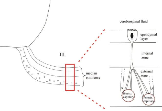
β-tanycytes line the floor of the third ventricle; β1-tanycytes are located in the lateral envaginations of the third ventricle and project to the tuberoinfundibular sulcus terminating on the surface of the pars tuberalis of the pituitary [26, 128]. β2-tanycytes which form the ventral wall of the third ventricle send their basal process to the portal capillary system in the median eminence [26, 128] where the axons of the hypophysiotropic neurons terminate to release their neurohormones into the hypophysial portal circulation [118] (fig. 3.).
A characteristic property of β-tanycytes is their involvement in barrier formation. β1- tanycytes form a barrier between the arcuate nucleus and the BBB free median eminence
19
[129] while β2-tanycytes establish a barrier between CSF and the neuropil of the median eminence [26]. β1-tanycytes are linked together by zonulae adherentes and maculae adherentes, whereas β2-tanycytes are joined together by tight junctions and zonulae adherentes [26].
An important feature of tanycytes is that they establish connection with the CSF and also with blood vessels [26]. Therefore, the tanycytes are in the anatomical position to sense the alterations in the composition of the CSF, but also the changes in the level of blood-born substances [26].
4.3. The role of tanycytes in the regulation of hypothalamic T3 homeostasis
Increasing amount of data suggest that the tanycytes are not only supportive and BBB forming cells, but they also play a major role in the regulation of neuroendocrine axes [26]. The β2 tanycytes can inhibit the secretion of neuroendocrine hormones by blocking the access of hypophysiotropic terminals to the fenestrated capillaries of the median eminence [130]. The coverage of the portal capillaries by the end feet of β2-tanycyte processes is variable and depends on the physiological conditions [130, 131]. By this plasticity tanycytes can separate the axon terminals from the capillaries or, to the opposite, they can allow access of the terminals to the surface of blood vessels [130]. The most studied example for the tanycyte remodeling is the regulation of the access of GnRH nerve terminals to the portal capillaries during the different phases of the estrous cycle.
In proestrus, GnRH axons terminate in the pericapillary space, resulting in massive GnRH release [130]. In contrast, in diestrus, tanycyte end feet separate the GnRH axon terminals from the portal capillaries, contributing to a decreased secretion of GnRH [130]. Changes in peripheral TH levels also induce remodeling of the end feet of tanycyte processes around the capillaries [131]. This observation suggests that tanycytes may also regulate the access of the hypophysiotropic TRH neurons to the capillaries and the release of TRH to the circulation.
The importance of tanycytes in the regulation of the HPT axis is further supported by the data that these glial cells express a number of proteins which are involved in the hypothalamic TH metabolism and signaling and have regulatory effect on the HPT axis.
These proteins include TRβ2 [132], the TH transporter OATP1c1 and MCT8 [40, 133], D2 that has capability to convert T4 to the active TH form [27, 29, 134], T3, and D2
20
inactivating WSB-1 and reactivating USP-33 enzymes [135]. The role of D2 in the regulation of HPT axis is underlined by the fact that the restoration of peripheral T3 level in hypothyroid rats by administration of T3 without addition of T4 is unable to decrease TRH gene expression to euthyroid level [136], although T3 is the predominant form of THs that acts on TRs [13]. This observation can be explained by the fact that T3 is poorly transported to the brain [35, 36], and the majority of hypothalamic T3 is produced locally from T4 by D2 [16]. Although the main hypothalamic sources of D2 are tanycytes, D2 can also be found in some hypothalamic astrocytes [25]. Interestingly, astrocyte specific D2 knock out mice show no disturbance in the negative feedback regulation of TRH, suggesting that astrocytes do not play a role in the negative feedback effect of THs on TRH neurons [25]. In addition, in the hypothalamus, hypothyroidism induces only slight increase of D2 expression and no rise of D2 activity [27, 137], suggesting that hypothalamic D2 does not compensate the changes in the circulating level of THs.
Therefore, the changes of peripheral T4 levels are converted into changes of hypothalamic T3 concentration that is critical for the feedback regulation of TRH neurons. [14].
Although it is clear that the D2-containing tanycytes are the principal source of T3 acting on hypophysiotrophic TRH neurons [14], it has not been clarified yet how T3 is transported from the tanycytes to the nucleus of the hypophysiotropic neurons in the PVN.
The perikarya of hypophysiotropic TRH neurons are located far from the tanycytes;
therefore, volume transmission between the tanycytes and the TRH neurons would be a relatively slow process. In contrast, the tanycyte end feet and the terminals of the TRH neurons are closely associated in the external zone of the ME [26]. We have recently hypothesized that T3 produced by tanycytes may be taken up by axon terminals of these neurosecretory neurons in the ME and transported retrogradely to the nucleus of these cells.
21
Figure 3. Schematic drawing presents the location of β2-tanycytes in the median eminence.
The cell bodies of β2-tanycytes can be found in the ventral and ventro-lateral walls of the third ventricle, but they send processes to the capillary loops in the external zone of median eminence. Tanycytes are able to take up substances from both the blood flow and the CSF and actively influence the composition of the CSF.
III – third ventricle; T – β2-tanycyte; A – axon terminal of hypophysiotropic neurons.
5. Neuronal inputs of hypophysiotropic TRH neurons
In addition to receiving humoral inputs, the TRH expression of hypophysiotrophic neurons is also regulated by neuronal inputs to adapt the HPT axis to the changing internal and external milieu. The hypophysiotropic TRH neurons integrate neuronal inputs from various areas of the central nervous system [5]. The most extensively studied and best- known sources of this innervation are the catecholaminergic cell groups of the brainstem, the hypothalamic arcuate nucleus (ARC) and the DMN [5].
22 5.1. Catecholaminergic neurons of the brainstem
Approximately 20% of the synapses on TRH neurons originate from the catecholaminergic neurons of the brainstem [138], indicating the critical importance of this input in the regulation of HPT axis. These axons mainly form asymmetric synapses on the surface of hypophysiotropic TRH neurons which is indicative of the excitatory function of these inputs [139]. Two types of catecholaminergic neurons innervate the hypophysiotropic TRH neurons which can be distinguished by their neurotransmitter content: adrenergic and noradrenergic neurons [140]. Approximately two thirds of the catecholaminergic innervation is adrenergic and one third is noradrenergic [140].
Adrenergic inputs originates from the C1-3 regions of the medulla whereas the noradrenergic innervation of TRH neurons arrives primarily from the A1 and A6 areas of the brainstem [141]. The catecholaminergic input of TRH neurons has been shown to mediate the cold induced upregulation of the HPT axis [5]. In cold environment, TRH gene expression is rapidly and transiently increased in the PVN, which, in turn, causes increased peripheral TSH and TH levels [142]. This activation of the HPT axis can be important in the stimulation of thermogenesis in the brown adipose tissue to prevent the drop of body temperature [5]. The cold induced activation of the hypophysiotropic TRH neurons is mediated by the catecholaminergic neurons of the medulla, as it can be inhibited by intraventricular administration of α-adrenergic antagonists [143].
Furthermore, the cold induced activation of the HPT axis is absent in rats during the first ten postnatal days when the catecholaminergic innervation of TRH neurons is immature [144]. The activation of the catecholaminergic inputs results in a shift in the set point of the negative feedback regulation of the hypophysiotropic TRH neurons in a way that higher thyroid hormone levels are necessary to suppress the TRH gene expression [5].
These changes lead to the development of central hyperthyroidism in cold environment.
5.2. Arcuate nucleus
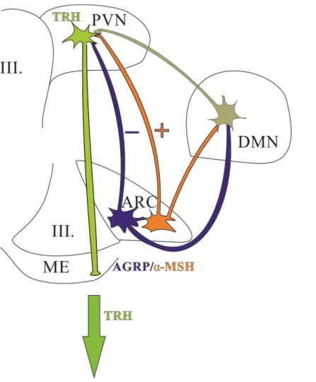
At least two separate neuron populations of the ARC send projections to TRH neurons to mediate metabolic signals from the blood to the PVN (fig.4). These two neuron populations can be distinguished by their neuropeptide content: while one of these neuronal populations synthesizes α-melanocyte stimulating hormone (α-MSH) and cocaine- and amphetamine regulated transcript (CART), the other neuronal group
23
produces agouti-related protein (AGRP) and neuropeptide Y (NPY) [5, 145]. While both cell groups are capable of detecting satiety signals, they are regulated oppositely by these signals and they exert antagonistic effects on the hypophysiotropic TRH neurons [5, 145].
5.2.1. The anorexigenic neuronal population of the ARC
One of the feeding related neuronal groups of the arcuate nucleus synthesizes two anorexigenic peptides, α-MSH and CART [146, 147]. These neurons are primarily located in the lateral part of the arcuate nucleus and have crucial role in the regulation of energy homeostasis [145]. Intracerebroventricular (icv.) administration of α-MSH or CART markedly inhibit food intake and stimulate energy expenditure [148, 149]. α-MSH exerts its central effects on the energy homeostasis via two G protein coupled receptors, the melanocortin 3 and 4 receptors (MC3-R and MC4-R) [150, 151]. Binding of α-MSH to either MC3-R or MC4-R induces cAMP mediated signaling [152] which results in rapid phosphorylation of the cAMP-response-element binding protein (CREB) [153]. The phosphorylated CREB (P-CREB) binds to the cAMP response element (CRE) in the promoter of the regulated genes [153].
The signaling pathway of CART is less understood, because of the failure of previous studies to identify the CART receptor. However, several second messenger systems were identified that might mediate the effects of CART on the target cells. In CRH neurons, CART can induce CREB phosphorylation [154]. In the hippocampus, CART inhibits voltage-dependent intracellular Ca2+ signaling probably through the inhibition of L-type voltage-gated Ca2+ channel activity via a G protein dependent pathway [155]. In addition, in cell culture of AtT20 and PC12 cells, CART induces the phosphorylation of Extracellular Signal-Regulated Kinase (ERK) [156, 157]. It seems possible that CART also acts via a G-protein coupled signaling pathway.
In addition to the anorexigenic effect of the centrally administered peptides, the role of α-MSH and CART in the regulation of energy homeostasis is also supported by the phenotype of transgenic animals. The lack of α-MSH caused by the mutation of its precursor proopiomelanocortin (POMC) gene results in hyperphagia and morbid obesity in transgenic mice [158, 159]. This phenotype is caused by the combined effect of the increased food intake and the decreased energy expenditure of these animals [158, 159].
A highly similar phenotype was also discovered in humans due to the mutation of the
24
POMC gene [160]. Mutation of the MC4-R also results in morbid obesity in both mice and humans [161]. Indeed, the most frequent monogenic cause of morbid obesity is the mutation of this receptor [162]. The absence of CART in KO mice results in less profound phenotype in young animals, but these animals also develop obesity when they are older [163]. The importance of the α-MSH/CART neurons in body weight regulation gains further support from the observation of obesity following the ablation of POMC neurons in adult animals [164].
The α-MSH/CART neurons express receptors for circulating metabolic signals including glucose, insulin and leptin, and the activity of these neurons is regulated by nutritional conditions [165]. The activity of the α-MSH/CART neurons and the synthesis of α-MSH in these cells are markedly stimulated by both insulin and leptin [165]. Leptin also increases the synthesis of CART [165]. During fasting, when circulating levels of leptin and insulin fall, α-MSH neurons are inhibited and the α-MSH and CART synthesis is repressed [165]. In contrast, leptin administration can prevent this fasting induced inhibition of the α-MSH/CART neurons [165].
In the rat PVN, about 70% of TRH neurons in the periventricular parvocellular subdivision and 34% in the medial parvocellular subdivision are innervated by α- MSH/CART neurons [166]. The axon terminals of anorexigenic neurons of the ARC typically form asymmetric synapses on the surface of TRH neurons, suggesting that these neurons have stimulatory effect on the TRH neurons [2]. Indeed, central administration of both α-MSH and CART have stimulatory effect on the TRH gene expression in the PVN [2, 149, 166, 167]. Icv. administration of α-MSH or CART to fasted rats stimulates TRH synthesis [2, 166].
In the hypophysiotropic TRH neurons, α-MSH acts predominantly on type 4 melanocortin receptor (MC4-R) [168] and induces a cascade that results in P-CREB binding to the CREB response element (CRE) of the TRH gene promoter [169]. This P- CREB binding contributes to the stimulation of TRH gene expression in hypophysiotropic neurons [169, 170]. Indeed, mutation of the CRE element in the TRH promoter can block the stimulatory effect of α-MSH on this promoter [170].
CART has a very similar stimulatory effect on hypophysiotropic TRH neurons [2].
However, the effect of CART seems to be mediated by different second messenger
25
pathway as central CART administration does not induce CREB phosphorylation in hypophysiotropic TRH neurons [154].
5.2.2. The orexigenic neuronal population of the ARC
The medially located feeding related neuron population of the ARC expresses two orexigenic peptides, neuropeptide Y (NPY) and agouti-related protein (AGRP) [5]. This neuron population stimulates food intake and decreases energy expenditure [171]. The central administration of AGRP or NPY has potent orexigenic effect [171]. In the hypothalamus NPY acts on Y1 and Y5 receptors [172] which have Gi-protein mediated inhibitory effect on adenylyl-cyclase and converges with α-MSH induced signalization [173]. AGRP is an endogenous antagonist of α-MSH on the MC3-R and MC4-R [174, 175] therefore, AGRP can also influence the cAMP-dependent pathway in the target cells.
Toxin-mediated elimination of the AGRP/NPY neuron population in adult transgenic mice produces dramatically hypophagic animals with increased energy expenditure [164].
Interestingly, neither AGRP- and NPY-KO animals nor the double KO mice have abnormally hypophagic feeding behavior [176-178], suggesting the development of compensatory mechanisms.
Similarly to the anorexigenic neuronal group, the orexigenic cell population also expresses receptors for leptin, insulin and glucose [165], which confirms that these cells also play role in the detection of metabolic signals. Leptin, insulin and glucose inhibit the gene expression of NPY, though AGRP synthesis is only influenced by leptin and not by insulin or glucose [165]. All these metabolic signals inhibit the activity of these cells [165]. During fasting when the circulating level of leptin is low the AGRP/NPY neurons are active and the level of both AGRP and NPY expression is elevated [165]. Leptin administration to fasted rats can decrease the AGRP and NPY synthesis to fed levels, indicating that leptin is a key regulator of the activity of AGRP/NPY neurons [165].
The AGRP/NPY neurons densely innervate the hypophysiotropic TRH neurons of the PVN, but as opposed to α-MSH/CART neurons, they form inhibitory synapses [179]. In addition, the chronic icv. administration of either NPY or AGRP to fed rats can markedly decrease TRH gene expression, similarly to the effect of fasting [180, 181].
In MC4-R KO mice AGRP has no effect on TRH gene expression, which confirms that the predominant melanocortin receptor for the regulation of hypophysiotropic TRH
26
neurons is MC4-R [182]. NPY can influence the TRH gene expression via both the Y1 and Y5 receptors [183].
5.3. The hypothalamic dorsomedial nucleus
The hypothalamic dorsomedial nucleus (DMN) also innervates densely the hypophysiotropic TRH neurons of the PVN [5]. Neurons of the DMN predominantly form symmetric type synapses on the surface of TRH neurons, suggesting an inhibitory effect [184]. However, the DMN seems to contain both orexigenic and anorexigenic neuron populations [185]. While neurons of the DMN can directly detect metabolic signals such as leptin, insulin, ghrelin and glucose [185-188], they also receive massive innervation from both α-MSH- and AGRP- producing neurons of the ARC, indicating that the DMN may also relay indirect metabolic information sensed by the ARC neurons to the TRH system [189, 190]. Since the chemical ablation of the ARC results in the lack of the regulation of TRH neurons by fasting or leptin [191], it is likely that the regulatory effect of the DMN on the hypophysiotropic TRH neurons is not independent from the ARC inputs. Therefore, the ARC-DMN-PVN pathway may serve as an alternative pathway in the mediation of metabolic signals to hypophysiotropic TRH neurons.
In addition, the DMN also receives input from the suprachiasmatic nucleus (SCN), the key regulator of the circadian rhythms [112]. Lesions of the DMN reduces circadian rhythms of wakefulness, locomotor activity, feeding and serum corticosteroid levels [192]. The DMN has also been shown to play critical role in the food entrainable circadian regulation of endocrine functions [193]. These observations suggest that the DMN may integrate the ARC and SCN inputs and contribute to the synchronization of light- and energy homeostasis mediated regulation of the HPT axis.
27
Figure 4. Schematic drawing illustrates the connection between the neuron populations of the ARC and the hypophysiotropic TRH neurons of the PVN.
The ARC contains two antagonistic neuron populations that project to the TRH neurons in the PVN. α-MSH-immunoreactive neurons form excitatory synapses on the surface of TRH neurons and also stimulate the TRH expression by at least two neuropeptides: α- MSH and CART. To the opposite, AGRP neurons form inhibitory synapses on TRH neurons and these neurons inhibit the TRH expression by AGRP and NPY release. DMN receives robust innervation from both α-MSH and AGRP neurons and project to TRH neurons in the PVN.
6. Regulation of the HPT axis during fasting and refeeding
During longer periods of restricted access to environmental resources, the rapid and precise adaptation to energy availability has a critical role in survival. Fasting induces a marked decrease in circulating levels of TH which is accompanied by lower TSH levels and a significant reduction of TRH gene expression [194-196]. This status referred to as
28
central hypothyroidism promotes energy conservation by decreasing basal metabolic rate and reducing thermogenesis [5]. The fasting induced central hypothyroidism can be prevented by the chemical ablation of the ARC [191]. Furthermore the lack of ARC inhibits the stimulatory effect of leptin on TRH gene expression, and also on TSH and TH release in fasting animals [191]. After 24 hours of starvation when the mass of white adipose tissue (WAT) is still not reduced significantly, the circulating level of leptin already falls because of its decreased release from the pool stored in adipocytes [197].
The low circulating level of leptin causes the loss of the activation of α-MSH/CART neurons which results in decreased stimulatory tone on TRH gene expression [165]. To the opposite, during fasting the decreased blood-level of leptin stimulates NPY and AGRP gene expression and the activity of these cells, which, in turn, inhibit TRH neurons in the PVN [165] (fig.5.).
Leptin administered icv. to rats can induce the phosphorylation of signal transducer and activator of transcription 3 (STAT3), an intracellular component of leptin receptor mediated signalization machinery, in the TRH neurons in the PVN [198]. This suggests that leptin can directly act on TRH neurons. Although the distribution of phosphorylated STAT3 (P-STAT3)-IR TRH neurons regulated directly by leptin and the pattern TRH neurons that are activated by α-MSH after leptin administration overlap only partially.
This observation suggests that TRH neurons which are directly regulated by leptin differs from the ones receiving information about leptin concentration indirectly from the ARC [198]. The relevance of the ARC-independent leptin signalization pathway is unclear yet, but the role of this direct leptin signaling pathway seems to be insignificant during fasting as the inhibitory effect of fasting on the HPT axis is absent in ARC ablated rats [191] and also in double-transgenic mice lacking both melanocortin and NPY signaling [199].
7. Melanocortin resistance of the hypophysiotropic TRH neurons during the early phase of refeeding
When animals are refed after fasting, they are satiated 2h after the onset of food intake [200], but their energy expenditure is increased only after 24h coincidently with the normalization of TRH gene expression [201]. A quick satiety response of the animals is accompanied by the presence of c-Fos, a member of the immediate early gene family and a marker of neuronal activation, in 90% of the α-MSH neurons in the ARC and in the α-
29
MSH activated neurons of the ventral parvocellular subdivision of the PVN [200]. In contrast, only a modest increase in the presence of c-Fos can be observed in TRH neurons and accordingly, TRH gene, TSH and TH levels are not influenced by this short period of refeeding [201]. The TRH mRNA expression in the PVN returned to fed level only 24 hours after the start of refeeding [201]. However, the activation of the α-MSH neurons of the ARC plays critical role in the determination of meal size during the first two hours of refeeding. During this early phase of refeeding the hypophysiotropic TRH neurons seem to be resistant to the stimulatory effect of these ARC neurons [201]. The background of the melanocortin resistance of TRH neurons during the early phase of refeeding is still unidentified.
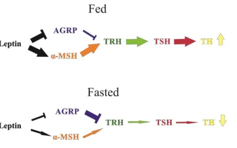
Figure 5. Schematic illustration of the regulatory effect of leptin on the HPT axis.
In fed animals the high circulating level of leptin facilitates the stimulatory effect of α- MSH neurons on TRH synthesis and represses the inhibitory effect of AGRP neurons on TRH neurons. These actions result in the release of THs. During fasting reduced circulating levels of leptin induce the inactivation of α-MSH neurons and cancel the inhibition of AGRP neurons. The altered activity of neuron populations of ARC results in the repression of TRH-synthesis which causes the fall of circulating TH levels.
30
8. Leptin-induced synaptic rearrangement in the regulation of feeding-related hypothalamic neuronal groups
Increasing evidence suggests that leptin can induce the synaptic rearrangement of the hypothalamic feeding-related neuronal circuitry. In the ARC, leptin-related alteration has been described in the innervation of both feeding related neuron populations that regulate the TRH neurons [202]. In leptin deficient ob/ob mice, a significant increase in the number of excitatory postsynaptic currents (EPSC) can be observed onto NPY neurons compared to WT mice, while the frequency of inhibitory postsynaptic currents (IPSC) onto the α-MSH-producing POMC neurons was significantly higher in ob/ob mice than in WT animals [202]. These observations were confirmed by the electron microscopical comparison of ob/ob and WT mice, which revealed alterations in the total number and the ratio of excitatory and inhibitory synapses on the surface of both NPY and POMC neurons in ob/ob mice compared to WT mice [202]. The lack of leptin increases the total number of synapses on the perikarya of NPY neurons [202]. Although a slight fall can be observed in the number of inhibitory synapses in ob/ob mice, the dominant effect of the lack of leptin is the marked increase of excitatory synapses on these cells when compared to WT animals [202]. In contrast, when studying the perikaryal surface of α-MSH neurons in ob/ob mice, a remarkable decrease can be observed in the number of excitatory synapses and a slight, but not significant reduction in the number of inhibitory connections [202]. In addition, leptin seems to induce changes not only in the incidence of synaptic inputs to α-MSH/CART and AGRP/NPY neurons but it also contributes to altered axonal growth of these neurons during development [203]. In the PVN of 10-day- old ob/ob mice, 10 times less fibers of ARC origin can be observed compared to WT littermates [203]. Although this disruption in the fiber density decreases in adult mice, a 3 to 4 fold lower density of axons originating from the ARC can still be detected in the medial parvocellular subdivision of the PVN of leptin deficient mice [203]. The chronic intraperitoneal (i.p.) leptin injection to 4-days-old ob/ob mice can prevent the disruption of axonal growth from the ARC to the PVN, corroborating the essential role of leptin in the stimulation of the development of the hypothalamic feeding-related neuronal network [203]. Although leptin treatment of adult ob/ob mice does not reverse the decreased ARC innervation of the PVN [203], transcriptional-profiling studies suggest that in the PVN of adult mice leptin regulates genes like basigin (Bsg), apolipoproteinE (ApoE), growth
31
associated protein 43 (Gap43), synuclein-γ and GABAA receptor associated protein (Gabarap); these genes are involved in synapse formation and neuronal growth [204].
These data about role of leptin in the regulation of hypothalamic synaptic plasticity suggest that leptin may also regulate TRH gene expression by influencing the synaptic plasticity of the arcuato-paraventricular pathway.
9. Non-hypophysiotropic TRH neurons
Although the first described and most studied role of TRH is its hypophysiotropic function, TRH is also released as a neurotransmitter modulating synaptic transmission within the central nervous system. TRH synthesizing neurons are located in many areas of the CNS, such as the olfactory bulb [205], septum [206], brainstem [207], amygdala [208], cerebellum [209], cortex [210] and the hypothalamic PVN [5, 211], periventricular nucleus [111, 211], DMN [111, 211], perifornical area [111] and the lateral hypothalamus [211]. The function of the non-hypophysiotropic TRH-expressing neuron populations is less well understood although TRH is known to have wide non-hypophysiotropic effects, like analgesia, anticonvulsant activity, increase of arousal, memory facilitation, increase of locomotor activity, organ specific increase of blood flow and intensified gastrointestinal activity [5]. In addition, increasing evidence indicates that some of the non-hypophysiotropic TRH neuron populations in the hypothalamus may also play role in the regulation of energy homeostasis; non-hypophysiotropic TRH-synthesizing neurons of the anterior PVN and perifornical area seem to be involved in the appetite regulation [212, 213].
Examination of TRH knock out (TRH-KO) mice indicates that the lack of TRH induces several alterations that are related to the non-hypophysiotropic roles of TRH [108]. In these mice behavioral abnormalities like depressive mood and alcohol intolerance can be observed [108]. In accordance, TRH expression is often altered in psychiatric disorders [214]. Although centrally administered TRH has well known anorexigenic effect [215], the data about the body weight and development of the TRH- KO animals is controversial. Interestingly, at the age of 4 weeks the TRH-KO mice were smaller than WT animals, but no significant difference was observed between the two groups at postnatal week 8 [107].
32
10. Identification of the activated elements of neuronal networks
In the studies applying c-Fos immunolabeling for the identification of the activated neurons, the question of the neuronal input of identified cells is always difficult.
Generally, double labeling immunohistochemical methods are used for the visualization of the connection between axons and perikarya [166]. Although the observation of axon varicosities on the surface of a neuronal cell body cannot verify the synaptic communication, but it indicates, at least, the anatomical connectivity of the two neuronal structures. Therefore, analyses of the presence of immunoreactive boutons on the surface of perikarya at light microscopic level can be helpful to estimate the extent of the interaction of the two systems, if the presence of synapses is validated by electron microscopy [166].
Althought c-Fos labeling can identify the activated neurons, the analysis of their inputs is not possible with this technique in the absence of cytoplasmic labeling in the activated neurons.
The Nissl-staining method is a widely used counterstaining method which labels the cytoplasm of neurons based on the interaction of basic dyes and the nucleic acid content of the cells [216, 217]. These dyes, such as cresyl violet, toluidine blue, anylin, methylen blue and thionine bind to the DNA with strong affinity and visualize primarily the cell nuclei, but also label the cytoplasmic RNA content of the cells [216]. Since remarkable amount of protein is continuously synthesized in neurons, a high concentration of rough endoplasmatic reticulum can be observed in the cytoplasm of these cells [218, 219]. Nissl- staining is often used in combination with immunohistochemical methods as a counterstaining approach to facilitate the mapping of the labeled cell populations [220- 222]. Unfortunately, after the immunolabeling procedure Nissl-dyes often visualize only the cell nuclei, but not the cytoplasm, making the counterstaining less valuable [220-222].
In this case the individual determination of the contacts of the c-Fos labeled neurons is not possible. We hypothesized that the lack of cytoplasmic labeling after immohistochemical processes is caused by the digestion of the RNA content of the neurons by RNase enzyme contamination and that the application of RNase-free conditions and RNase inhibitors during the immunohistochemical processes could improve the labeling of cytoplasm by the subsequent Nissl-staining.
33 IV. Specific aims
1. To map the distribution of hypophysiotropic TRH neurons in the mouse hypothalamus
2. To determine the route of T3 transport between the thyroid hormone activating tanycytes and the hypophysiotropic TRH neurons
3. To clarify the mechanism of the melanocortin resistance of the hypophysiotropic TRH neurons during refeeding
4. To develop an improved method for the combined use of immunocytochemistry and Nissl-staining
34 V. Materials and methods
1. Animals
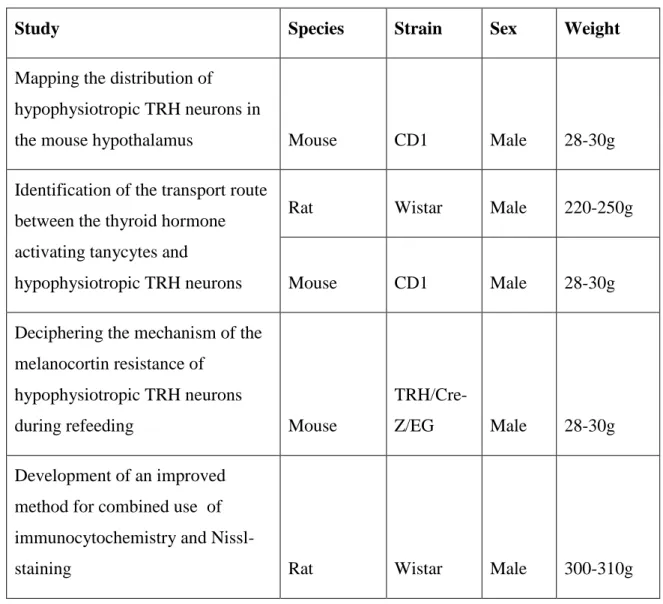
The rodent species and strains used for the studies of the thesis are summarized in Table 1. The animals were housed under standard environmental conditions (light between 6:00 A.M. and 6:00 P.M., temperature 22°C, food and water ad libitum) in the animal facility of the Institute of Experimental Medicine of the Hungarian Academy of Sciences. All experimental protocols were reviewed and approved by the Animal Welfare Committee at the Institute of Experimental Medicine of the Hungarian Academy of Sciences and carried out in accordance with legal requirements of the European Community.
Table 1. Summary of animals used in different experiments
Study Species Strain Sex Weight
Mapping the distribution of hypophysiotropic TRH neurons in
the mouse hypothalamus Mouse CD1 Male 28-30g
Identification of the transport route between the thyroid hormone activating tanycytes and
hypophysiotropic TRH neurons
Rat Wistar Male 220-250g
Mouse CD1 Male 28-30g
1. Deciphering the mechanism of the melanocortin resistance of
hypophysiotropic TRH neurons
during refeeding Mouse
TRH/Cre-
Z/EG Male 28-30g
Development of an improved method for combined use of immunocytochemistry and Nissl-
staining Rat Wistar Male 300-310g
35 2. Tissue preparation and labeling
2.1. Methods used for mapping the distribution of hypophysiotropic TRH neurons in the PVN of mice
2.1.1. Flouro-Gold injection and tissue preparation for immunocytochemistry
To detect hypophysiotropic neurons, 4 mice received intravenous injection of Fluoro- Gold (15g/g BW in 100µl 0.9% saline, Fluorochrome. Llc., Denver, CO., USA). Two days later, the mice were injected intraperitoneally with the same dose of Fluoro-Gold to further enhance the labeling in the hypophysiotropic cell bodies. Fluoro-Gold is a retrograde tracer which is not able to pass through the BBB. Therefore, only the axons terminating outside of the BBB, including the axon terminals of hypophysiotropic TRH neurons in the median eminence can take up Fluoro-Gold from the circulation and transport it to their perikarya where the accumulation of this tracer can be detected. Since non-hypophysiotropic TRH neurons have no direct connection with the circulation, they are not able to concentrate Fluoro-Gold. Therefore, the presence of Fluoro-Gold uptake in TRH-IR perikarya is indicative of the hypophysiothropic nature.
Five days after the second tracer injection, the deeply anesthetized animals (ketamine:
50 g/g; xylazine: 10 g/g body weight, i.p.) were icv. treated with colchicine, an inhibitor of axonal transport (1 g/g body weight) by injection through a 26 G needle placed into the lateral ventricle under stereotaxic control (coordinates from Bregma:
antero-posterior –0.2 mm, lateral –1mm, dorsoventral -2.5mm) [223]. The colchicine treatment was applied to facilitate the detection of TRH-IR perikarya. In intact animals, only a few TRH-IR perikarya are detectable with immunocytochemistry in the PVN, because TRH is rapidly transported to the axons. In contrast, the colchicine induced inhibition of axonal transport markedly increases the TRH-content of cell bodies and therefore, the detectability of the perikara of TRH neurons. Control animals were not injected with Fluoro-Gold, but received icv. colchicine treatment as described above.
Twenty hours after the colchicine treatment, the animals were deeply anesthetized with ketamine-xylazine and perfused through the ascending aorta with 10 ml phosphate buffered saline (PBS; pH 7.4), followed by 30 ml 1% acrolein + 3% paraformaldehyde (PFA) in PBS, and finally 20 ml 3% PFA in PBS. Both PFA and acrolein form irreversible cross-links between the primary amino groups of proteins, resulting in the fixation of tissues. PFA is used in most routine immunocytochemical detections. However, TRH is
36
a very small peptide and the antibody used for the detection of TRH was produced against TRH conjugated to bovine serum albumin (BSA) with acrolein, which markedly modifies the structure of TRH. Therefore, the antiserum raised this way can only recognize TRH if the tissue is also perfused with an acrolein-containing tissue fixative.
After perfusion, the brains were removed and cryoprotected in 20% sucrose containing PBS at 4°C overnight, to prevent frezeing artefacts, and then, frozen on dry ice. Twenty- five micrometer thick, coronal sections through the rostro-caudal extent of the PVN were cut on freezing microtome (Leica Microsystems, Weltzar, Germany), collected in four identical sets in cryoprotective solution (30% ethylene-glycol; 25% glycerol; 0.05 M phosphate buffer) and stored at -20°C until used.
2.1.2. Pretreatment for single-, double- and triple labeling immunocytochemistry
The sections were treated with the strong reducing agent sodium borohydride (1% in distilled water for 30 min) to extinguish the free aldehyde activity of acrolein by reducing it to hydroxyl group. Then the sections were incubated in a mixture of 0.5% Triton X- 100, a detergent applied to increase the permeability of cell membranes, and 0.5% H2O2
to inhibit the endogenous peroxidase activity (in PBS for 15 min). Nonspecific antibody binding was blocked with 2% normal horse serum (NHS) in PBS for 15 min. The sections were then processed for single-, double- or triple labeling immunocytochemistry as described below.
2.1.3. Single-labeling immunocytochemistry for TRH
The sections were incubated in sheep anti-TRH serum (1:4000, generated in our laboratory, # 08W2) in serum diluent (2% NHS, 0.2% PhotoFlo, 0.2% sodium azide in PBS) for two days at 4°C, followed by incubation in biotinylated donkey anti-sheep IgG (1:500, Jackson ImmunoResearch Laboratories, West Grove, PA, USA) for two hours at room temperature. Then the sections were immersed in Avidin-Biotin-Peroxidase complex (ABC) (1:1000 Vector Laboratories Inc., Burlingame, CA, USA) in 0.05 M Tris buffer (TB) (pH 7.6) for 1 hour. The peroxidase activity of the antibody-ABC complex was visualized by Ni-DAB developer [mixture of 0.05% diaminobenzidine (DAB), 0.15% Nickel-ammonium-sulfate, 0.005% H2O2 in 0.05 M Tris buffer] that resulted a dark blue precipitate in the TRH-IR neurons. Then the sections were mounted onto glass