underlying thyroid hormone mediated effects
Ph.D. Thesis
Anna Kollár D.V.M.
Semmelweis University
János Szentágothai Doctoral School of Neurosciences
Institute of Experimental Medicine Hungarian Academy of Sciences
Consultant: Balázs Gereben, D.V.M., D.Sc.
Official reviewers: Péter Gálfi, D.V.M., D.Sc.
Zsuzsanna Tóth, Ph.D.
Head of the final examination committee: Alán Alpár, M.D., D.Sc.
Members of the final examination committee: Attila Patócs, M.D., Ph.D.
Emília Madarász, D.Sc.
Budapest
2017
TABLE OF CONTENTS
TABLE OF CONTENTS ... 2
1. LIST OF ABBREVIATIONS ... 4
2. INTRODUCTION ... 7
2.1. Thyroid hormones are crucial factors of development ... 7
2.2. The structure and function of the HPT axis ... 9
2.3. Regulation of tissue TH action by deiodination ... 13
2.4. Regulation of thyroid hormone availability in the brain ... 15
2.5. Machinery of nuclear thyroid hormone action: thyroid hormone receptors and thyroid hormone response elements ... 17
2.5.1. Thyroid hormone receptor isoforms ... 19
2.6. Assessment of thyroid hormone action ... 22
2.6.1. Positive regulation of gene expression by thyroid hormones ... 22
2.6.2. Negative regulation of gene expression by TH ... 23
2.7. Posttranscriptional mechanisms regulating biological activity ... 26
2.7.1. Alternative splicing ... 26
2.7.2. The 5’ untranslated region of the D2 mRNA ... 27
2.8. Measurement of TH-dependent gene expression with promoter assays ... 27
2.8.1. Luciferase assays in general ... 27
2.8.2. Investigation of thyroid hormone response elements ... 28
3. OBJECTIVES ... 30
4. METHODS ... 31
4.1. Animals ... 31
4.2. Generation of expression constructs... 31
4.2.1. Constructs prepared for the analysis of RNA-dependent post- transciptional regulation of D2 ... 31
4.2.2. Generation of expression constructs for the analysis of luciferase reporters ... 31
4.3. DNA transfections ... 33
4.4. RNA isolation and RT-PCR ... 33
4.5. Northern blots ... 34
4.6. In situ hybridization... 34
4.7. Assays ... 36
4.7.1. Deiodinase assay ... 36
4.7.2. Luciferase assay ... 37
4.7.3. SEAP assay ... 37
4.8. Statistics... 37
4.9. Sequences ... 37
5. RESULTS ... 38
5.1. Investigation of thyroid hormone availability in the developing chicken hypothalamus... 38
5.1.1. Assessment of D2 mRNA expression in the developing chicken brain using RT-PCR and Northern blot... 38
5.1.2. D2 activity in the developing chicken brain (E7-E15) ... 40
5.1.3. Distribution of D2 mRNA in the brain of developing chicken ... 42
5.1.4. Distribution of D2 mRNA in the brain of adult chicken ... 44
5.2. Understanding the RNA-dependent post-transcriptional regulation of the type 2 deiodinase (D2) encoding dio2 gene ... 46
5.2.1. Cloning and characterization of an alternatively spliced chicken D2 encoding transcript ... 46
5.2.2. Investigation of the functional role of the 5’UTR of chicken D2 mRNA ... 49
5.3. Identification of authentic reporter proteins for studies on T3-dependent gene transcription ... 51
6. DISCUSSION ... 54
6.1. Investigation of thyroid hormone availability in the developing chicken hypothalamus... 54
6.2. Understanding the RNA-dependent post-transcriptional regulation of the type 2 deiodinase (D2) encoding dio2 gene ... 57
6.3. Identification of authentic reporter proteins for studies on T3-dependent gene transcription ... 60
7. CONCLUSIONS ... 63
8. SUMMARY ... 65
9. ÖSSZEFOGLALÁS ... 66
10. BIBLIOGRAPHY ... 67
11. BIBLIOGRAPHY OF THE CANDIDATE’S PUBLICATIONS ... 88
11.1. List of publications the thesis based on ... 88
11.2. Other publications ... 88
12. ACKNOWLEDGEMENTS ... 89
1. LIST OF ABBREVIATIONS
ANOVA analysis of variance AP-1 activator protein 1
bp base pair(s)
BSA bovine serum albumin
cAMP cyclic adenosine monophosphate
CBP CREB-binding protein
cDNA complementary deoxyribonucleic acid
cD2 chicken type 2 deiodinase
CpG cytidine-phosphateguanosine
CREB cAMP response element binding protein cRNA complementary ribonucleic acid
CSF cerebrospinal fluid
D1 type 1 deiodinase
D2 type 2 deiodinase
D3 type 3 deiodinase
DBD DNA binding domain
DIG digoxigenin
DMEM Dulbecco’s Modified Eagle Medium
DNA deoxyribonucleic acid
DRIP/TRAP vitamin D receptor interacting protein/thyroid receptor associated protein
DTT dithiothreitol
E embryonic day, day of incubation
EDTA ethylenediaminetetraacetic acid
EFsec selenocysteine specific elongation factor
ER estrogen receptor
FBS fetal bovine serum
fmol femtomole(s)
FSH follicle stimulating hormone GFAP glial fibrillary acidic protein HAT histone acetyltransferase
HDAC histone deacetylase
HPT hypothalamo-pituitary-thyroid
HRE hormone response element
kb kilobase(s)
Km Michaelis-Menten constant
LBD ligand binding domain
LH luteinizing hormone
MCT monocarboxylate anion transporter
µl microliter
µM micromole(s), micromolar
mRNA messenger ribonucleic acid mTRα mouse thyroid receptor α NCoR nuclear receptor co-repressor
NGF nerve growth factor
NLS nuclear localization signal
nM nanomole, nanomolar
nTRE negative thyroid response element
OATP1C1 organic anion transporting polypeptides 1
PBS phosphate buffered saline
PCAF p300/CBP associated factor PCR polymerase chain reaction
PTU propyl thiouracil
PVN paraventricular nucleus
RNA ribonucleic acid
rT3 reverse T3, 3,3’,5’-triiodothyronine RTH resistance to thyroid hormone
RT-PCR reverse-transcription-polymerase chain reaction
RXR retinoid X receptor
SBP selenocysteine binding protein SEAP secretory alkaline phosphatase SEM standard error of the mean
SECIS selenocysteine inserting sequence
SMRT silencing mediator of retinoid and thyroid receptors sORF short open reading frame
SRC steroid receptor co-activator
SSC standard sodium citrate
SUN-CoR small ubiquitous nuclear co-repressor
SV simian virus
T2 diiodothyronine
T3 triiodothyronine, 3,5,3’-triiodothyronine T4 thyroxine, 3,5,3’,5-tetraiodothyronine
TBP TATA-binding protein
TH thyroid hormone
TK thymidine kinase
TRE thyroid hormone response element TRH thyrotropin releasing hormone
TRα thyroid receptor α
TRβ thyroid receptor β
tRNA transfer ribonucleic acid
TSH thyroid stimulating hormone, thyrotropin
UTR untranslated region
2. INTRODUCTION
Thyroid hormone (TH) plays a fundamental role in the development and function of various organ systems. TH is especially important for the development and function of the brain (Bernal et al., 2003). Thyroxine (T4) is the most abundant (~80%) secretory product of the human thyroid gland. However, the presence of 3,5,3’-triiodothyronine (T3) was also revealed in the human plasma by Gross and Pitt-Rivers (1952). T3 is a deiodinated form of T4 and it was established that T3 is the compound responsible for most known TH-dependent biological effects despite its less than 20% presence in the thyroidal secretory output. Importantly, the existence of T4 to T3 conversion was proved by Sterling et al. (1976) indicating that not only the thyroid but also the prohormone T4 can give rise to T3.
The regulation of circulating TH levels is governed by the hypothalamo-pituitary- thyroid (HPT) axis (Fekete and Lechan 2014). The HPT axis is programmed to keep serum T3 levels in the physiological range that is predominantly achieved by controlling the release of TH from the thyroid gland, especially via regulating the output of the long-lived T4 prohormone. However, TH action occurs in tissue/cell compartments and its regulation requires quick and tissue-specific costumization (Gereben et al., 2015).
Thus, the impact of the HPT axis on tissue TH levels is limited. According to the current consensus, a complex and tissue-specific regulatory system is responsible for the control of tissue TH action. The crucial players of this system are the members of the deiodinase enzyme family that catalyze TH activation and inactivation, the TH transporters, and the nuclear machinery of TH action (Gereben et al., 2008; Visser 2000).
2.1. Thyroid hormones are crucial factors of development
TH is essential for the development and differentiation of various cell types and exerts a striking impact on the developing central nervous system. A classical study established the correlation between iodine deficiency and cretinism (Ord 1888). It is well-known that congenital hypothyroidism is accompanied with detrimental effects. Therefore serum thyroid stimulating hormone (TSH) levels are subjected to routine screening in human neonates to ensure that TH supplementation will be performed in the critical
time window after birth to avoid the devastating and irreversible consequences (Fisher et al., 1979).
It took decades to accumulate data allowing a deeper understanding of the underlying mechanisms. TH was found to be crucial in the regulation of later neuron differentiation events: proper myelination (Balázs et al., 1969a and 1969b), dendrit and axon development and synaptogenesis. It was also demonstrated that suboptimal TH levels affect the expression of genes controlling myelin formation in rats, e.g. the ones encoding myelin basic protein, proteolipid protein and myelin associated glycoprotein (Farsetti et al., 1991; Rodriguez-Pena et al., 1993). Under prolonged hypothyroidism, the number of myelinated axons decreased and axons of lower diameter were found to be not myelinated in neonatal rats. These changes were also observed in cortical regions involved in visual, auditory and motoric activities. THs are inevitable for proper cell migration and formation of the layers of the cerebral cortex. Purkinje cells in the cerebellum and the pyramidal cells in the cortex were found highly sensitive to appropriate TH availability. Purkinje cells are unable to develop their dentritic tree in hypothyroidism (Legrand 1967a and 1967b) and this was underlain by affected cytoskeletal organization (Silva and Rudas, 1990) while the late migration of granular cells in the cerebellum was also severely affected. THs also affect glial cell differentiation (oligodendrocytes, astrocytes and microglia) (Gharami and Das 2000;
Lima et al., 2001). The underlying mechanisms involve biochemical changes related to glucose-amino acid conversion, glutamine-dehidrogenase activity, decreased activity of oxidative enzymes (Balázs et al., 1971; Cocks et al., 1970) and also yet not fully revealed mechanisms governed by cell-cycle modulators such as cyclin D1, E2F-1 or p27 (Garcia-Silva et al., 2002). It was shown that the number of matured astrocytes and oligodendrocytes is reduced in the white matter tracts of hypothyroid rats (Schoonover et al., 2004; Martínez-Galán et al., 1997). Furthermore, THs also impact the development of the cytoskeleton via the upregulation of glial acidic fibrillary protein (GFAP) and F-actin, as demonstrated both in animal and cell culture models (Paul et al., 1996). THs enhance the secretion of different extracellular matrix proteins like laminin and fibronectin via growth factor secretion (Trentin et al., 1995 and 2001). Importantly, nerve growth factor (NGF) secreted by astrocytes has a crucial role in the control of neurit growth (Lindsay 1979; Charrasse et al., 1992). The abovementioned effects of
TH on glia cells also impact neurons. The role of TH played in neural development is summarized in Figure 1.
Figure 1. Role of thyroid hormones during different stages of brain development (based on Bernal 2015).
2.2. The structure and function of the HPT axis
The HPT axis is governed by hypophysiotropic thyrotropin releasing hormone (TRH) expressing neurons of the hypothalamic paraventricular nucleus (PVN). Axons of these neurons project to the external zone of the median eminence, located below the floor of the third ventricle where TRH is released from axon varicosities (Lechan and Fekete 2006). TRH reaches the anterior pituitary via the portal system and binds to the 7 transmembrane domain containing Gq/11 protein coupled TRH receptors of thyrotropes followed by the increase of thyrotropin (TSH) production and secretion. TSH is a glycoprotein that is formed by an -subunit consisting of 92 amino acids (Salvatore et al., 2011). This subunit is very similar to those of the follicule stimulating hormone (FSH) and luteinizing hormone (LH) (Gray 1988). In contrast, the 110-amino acid-long
-subunit of TSH is specific for this molecule. TSH reaches the thyroid gland by the circulation and binds to the 7 transmembrane domain containing TSH receptors on thyrocytes. In contrast to the TRH receptor, the TSH receptor is coupled to Gs protein and acts via the activation of adenylate cyclase and consequently evokes cyclic adenosine monophosphate (cAMP) release. The capacity of a given amount of TSH to induce cAMP release is regulated by TSH bioactivity, a phenomenon dependent on TRH-promoted glycosylation of the TSH molecule (Salvatore et al., 2011). TSH promotes various steps of TH formation and release. These include the iodide uptake of the thyrocytes in the thyroid gland, synthesis and iodination of the thyroglobulin,
endocytosis of the colloid and activation of peroxidase. TSH is also able to increase the rate of cell division in the thyroid gland (Portulano et al., 2014).
TH regulates the HPT axis by negative feedback. Increased serum T4 and T3 levels decrease TSH and TRH secretion, while a decrease of circulating TH stimulates the hypothalamus and the anterior pituitary to release TRH and TSH, respectively.
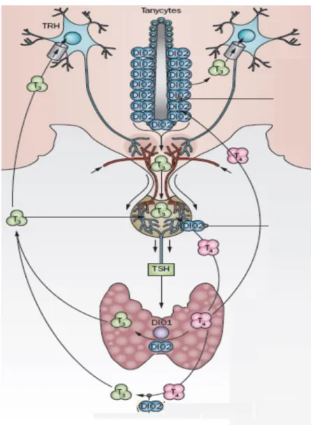
Tanycytes of the mediobasal hypothalamus play a very important role in the feedback mechanism. These specialized glial cells are located in the floor and the ventrolateral walls of the third ventricle (Bruni 1974, Krisch et al., 1978). Numerous tanycyte processes, especially those of -tanycytes located in the floor of the third ventricle end in the median eminence and contact capillaries and axon terminals of neurosecretory neurons (Lechan and Fekete 2007). Importantly, the median eminence is located outside of the blood-brain barrier and its TH content originates both from local, tanycyte- generated sources and from the periphery (Kakucska et al., 1992). TRH expression in the PVN of hypothyroid rats could be normalized with exogenous T3 only with a dose evoking hyperthyroidism in the periphery. This finding clearly underlines the functional importance of hypothalamic T4 to T3 activation in the regulation of TRH (Kakucska et al., 1992). Tanycytes are able to uptake T4 either from the fenestrated capillaries of the median eminence, the intercellular space of the basal hypothalamus or from the cerebrospinal fluid (CSF). They actively convert T4 to T3 since these cells contain high amounts of deiodinase type 2 (D2) enzyme (for more details of D2 see Section 2.3). The generated T3 is released either to the mediobasal hypothalamus, the CSF or the median eminence into the microenvironment located between tanycyte processes and axonal segments of parvocellular neurosecretory neurons. Hypophysiotropic TRH neurons contain monocarboxylate anion transporter 8 (MCT8) protein in the plasmamembrane of their axons that enables them to uptake T3 in the median eminence and consequently this allows the regulation of TRH expression by T3 of the median eminence (Kalló et al., 2012). This could work via an anticipated retrograde axonal transport of T3 to reach the PVN but this remains to be proved. This is an important point to be revealed since TRH neurons in the PVN rely on external T3 due to their inability to generate T3 is underlain by the lack of D2 in these cells. Furthermore, a significant portion of TRH neurons do not contain the TH degrading deiodinase 3 enzyme (D3), thus their ability to actively regulate their intracellular T3 content is limited, indicating that these cells are
programmed to accurately translate the hypothalamic T3 levels into TRH expression (Kalló et al., 2012). Thus, in the mediobasal hypothalamus, D2-mediated local regulation of T3 availablity functionally interacts with the regulation of the HPT axis.
See Figure 2 for summary.
Figure 2. The hypothalamo-pituitary-thyroid axis and its interaction with local D2-mediated T3
generation. TRH: thyrotropin releasing hormone, TSH: thyroid stimulating hormone, T4: thyroxine, T3: triiodothyronine, DIO1: deiodinase type 1, DIO2: deiodinase type 2 (modified from Gereben et al., 2015).
While TH is a major regulator of hypophysiotropic TRH neurons, these cells also receive afferents from different brain regions to allow proper response of the axis to the changing environment. Metabolic signals are transmitted from the arcuate nucleus,
circadian signals come from the suprachiasmatic nucleus while catecholaminergic afferents from the brainstem transmit information on changes of the external temperature (Fekete and Lechan 2014).
Set-point formation of negative feedback during ontogeny is a fundamental process for the regulation of the HPT axis and its consequences persist through the entire lifespan.
However, only limited information is available on the onset of the negative feedback mechanism.
Both the development of the brain and the ontogeny of the HPT axis show marked species-specific differences. Rats are altricial animals, their brain is not well developed at birth and their hypothalamo-pituitary-thyroidal gland axis is not fully matured (Schwartz 1983, Legrand 1986). In contrast, the precocious sheep (Fisher 1991) and chickens have well developed HPT axes at the date of birth or hatching (Oppenheimer and Schwartz, 1997). Children at birth represent an intermediate state between these examples with an immature central nervous system but a fully developed axis. Thus rodents do not serve as appropriate model for the regulation of the development of the human HPT axis while the related mechanisms of hypothalamic feedback cannot be studied in humans due to ethical reasons. Therefore, chickens represent an invaluable model for these studies, since the developmental kinetics of the chicken HPT axis is rather similar to that of humans. In addition, the chicken embryo allows to study developmental phenomena in the absence of interfering maternal regulatory circuits. In contrast, data obtained from rats should be handled with care because of the significant differences exisiting between the development of the HPT axes of rodents and humans (Taylor et al., 1990).
In birds THs control piping, hatching, thermogenesis and growth, but also play a role in the neurulation of the chicken embryo (Flamant and Samarut 1998; Decuypere et al., 1990; Beckett and Arthur 1994). The onset of the thyroid function in chickens starts from embryonic day 9.5 (E9.5), T4 and T3 can be detected in the yolk and serve as maternal thyroid hormone supply (Prati et al., 1992). The thyroid gland starts to secrete its hormones in increasing amounts, but the TSHβ mRNA is increasing further until E19 as the negative feedback is not yet functional at this developmental stage (Gregory et al., 1998).
2.3. Regulation of tissue TH action by deiodination
Many organs can customize their TH action independently from circulating TH levels and deiodinase enzymes play a striking role in this event (Gereben et al., 2008).
Thyroxine can be activated by 5’ deiodination by removing iodine from the outer phenolic ring of thyroxine (activation pathway) while inner ring 5 deiodination results in the inactivation of T4 or T3 (Figure 3). Historically, the tissues were categorized based on their capability of utilizing TH. Tissues contributing to serum TH levels were considered as TH exporters while those tissues which do not, were considered as TH importers (Crantz et al., 1982). While this nomenclature properly recognized the importance of tissue-specific differences in TH economy, in light of recent data it falls short to address that i) tissue specific contribution to systemic TH levels is not constant but dependent on TH status and ii) in organs with complex cellular composition it also depends on cell-type specific events. For example, the rodent heart is not capable of producing T3 locally and it is fully dependent on the circulating T3 in the serum. The liver is TH importer in euthyroidism and TH exporter in hyperthyroidism. Importantly, the brain generates most of its T3 locally, but does not contribute significantly to the T3 level of the serum. Furthermore, striking compartmentalization exists inside the brain, as the glial compartment performs T3 generation and the neuronal compartment consumes T3. Thus, cell-type specific events ensure the cellular export and import of TH within the same tissue (see Section 2.4). In humans, T3 originates predominantly from D2-mediated outer ring deiodination of the prohormone thyroxine in tissues (Maia et al., 2005). While biochemically both D2 and type 1 deiodinase (D1) can catalyse this process, due to its high Km, D1 is not capable of generating T3 under euthyroid conditions. Consequently, in vivo, D2 is the predominant activating deiodinase.
These enzymes belong to the selenodeiodinase enzyme family and contain the rare amino acid selenocysteine in their active center. In the deiodinase encoding mRNA selenocysteine is encoded by an in-frame UGA codon that serves otherwise as a stop codon in proteins that do not belong to the selenoprotein family. The translational readthrough of UGA is ensured by a specific mRNA secondary structure, the selenocysteine inserting sequence (SECIS) element, located in the 3’ untranslated region (3’UTR) of the selenoprotein encoding mRNA (Berry et al., 1991a; Gereben et al., 1999). The SECIS binds the SECIS binding protein (SBP2) that interacts with
selenocysteine specific elongation factor (EFSec). The latter binds selenocysteine transfer RNA (tRNA) allowing cotranslational selenocysteine incorporation at the UGA on the ribosome (Bianco et al., 2002). Although this process is very complex and energy consuming, the resulting selenocysteine-containing deiodinase enzyme has much higher substrate affinity than the cysteine-containing version. This is because selenium can be ionized more easily at physiological pH than the sulphur of cysteine, turning the formed protein into a much more powerful oxido-reductase. On the other hand, this comes to the cost of the efficiency of translation that is ~50-100 times lower for the selenodeiodinase D2 than for its cysteine mutant version (Steinsapir et al.; 2000).
Therefore, selenocysteine incorporation results in a low level of selenoenzyme that is highly efficient (Berry et al., 1991b).
D1 is capable of removing the iodine from both the outer and inner rings, while D2 is an exclusive outer ring deiodinase. An important difference between the two enzymes is that the in vitro Km of D1 for T4 deiodination is much higher (1-2 µM) compared to D2 (1-2 nM). D1 has low affinity and high capacity, at the same time D2 has high affinity and low capacity for T4. While D1-mediated deiodination is strongly inhibited by 6n- propyl-thiouracil (PTU), D2 deiodination is not. This feature helped to discover the PTU-resistant 5’ deiodination and as a consequence to identify D2 in the pituitary of hypothyroid rats (Silva and Larsen, 1977). D2 is the enzyme that can efficiently generate T3 in vivo, while D1-mediated T3 generation can only occur under hyperthyroid conditions. Although D1 predominantly inactivates reverse T3 in vivo, the special kinetics of D2 enables this protein to activate thyroxine even at low substrate levels.
D1 was identified in several tissues. In rats liver, kidney, thyroid gland and the pituitary contain D1. Its 2.1 kb long cDNA was cloned by Berry et al. (1991a). D1 is anchored to the plasmamembrane (Baqui et al., 2000).
D2 is present in the glial compartment of various brain regions and in the pituitary (Silva and Larsen, 1977). It can also be found in brown adipose tissue, placenta, keratinocytes, myocardium, skeletal muscle and in the thyroid gland (Salvatore et al., 1996b; Croteau et al., 1996, Bianco et al., 2002). The first full length D2 cDNA of higher vertebrates was isolated from chicken (Gereben et al., 1999). The D2 mRNA
contains a SECIS element close to the end of 3’UTR. The coding region is of average size (840 bp) but the whole RNA is unusually long (6094 bp) due to the long untranslated regions and the distance between the SECIS and the UGA of the active center is almost 5 kb. D2 is localized in the endoplasmic reticulum (Gereben et al., 2008).
D3 represents the third known member of the family. This enzyme removes iodine from the inner ring of TH and its main role is to inactivate T4 and T3. D3 is expressed in the brain, palcenta, skin and fetal liver. D3-mediated T3 inactivation is crucial in the regulation of TH availability in neurons and also prevents the fetus from high levels of maternal T3 in specific phases of ontogeny (Tu et al., 1999; Gereben et al., 2008). D3 is localized in the plasmamembrane and in the dense core vesicles of neurosecretory neurons (Baqui et al., 2003; Kalló et al., 2012).
Figure 3. Deiodinase-mediated metabolism of iodothyronines. T4: thyroxine, T3:
triiodothyronine, T2: diiodothyronine, reverse T3: reverse triiodothyronine (Gereben et al., 2008).
2.4. Regulation of thyroid hormone availability in the brain
T4 and T3 concentrations in the brain are strictly governed by complex, locally controlled mechanisms. This process is tightly regulated by deiodination-dependent TH
metabolism and neuro-glial TH transport (Freitas et al., 2010; Gereben et al., 2008). It was demonstrated that in the brain of rat fetuses T3 is exclusively generated from local T4 deiodination and the circulating T3 virtually does not reach the brain and a high proportion of T3 (>80% in the cerebral cortex) was shown to be locally generated in the adult rat brain (Crantz et al., 1982). As it was discussed in details in Section 2.2, the median eminence is a unique region of the brain which lies outside of the blood-brain barrier, so T3 from the periphery can reach this area. In contrast, other tissues like the liver are readily accessed by serum T3 (Calvo et al., 1990; Grijota-Martinez et al., 2011).
Due to the presence of D3 in the axonal compartment of the external zone of the median eminence, T3 availability is subjected to cell-type specific regulation in this crucial sensor region (Kalló et al., 2012).
In the brain, D2 and D3 are responsible for the regulation of TH availability. D2 is expressed in astrocytes in various brain regions and in tanycytes located in the floor and infralateral wall of the mediobasal hypothalamus (Tu et al., 1997). In contrast, D3 is expressed in neurons (Tu et al., 1999). Astrocytes generate T3 from the T4 by taking it up from the brain capillaries and provide activated TH to neurons (Freitas et al., 2010).
This cell compartment regulates its intracellular TH levels via D3-mediated degradation of T4 to rT3 and T3 to and T2.
As it was discussed in details in Section 2.2, the highest D2 expression in the brain can be found in the tanycytes and this phenomenon has a crucial role in the feedback regulation of the hypophysiotropic TRH neurons. Cell-type specific ablation of D2 activity in astrocytes and in the pituitary of transgenic mice revealed that the astrocytic D2 pool exerts no impact on the regulation of the HPT axis. It also shed light on the complex functional interactions allowing the hypothalamus to keep the systemic TH economy in balance even in the absence of pituitary D2 (Werneck de Castro et al., 2015). Perivascular glial cells take up thyroxine from the blood vessels while a smaller fraction of TH is transported through the choroid plexus and the cerebrospinal fluid. TH passes the plasmamembrane via membrane transporters like the monocarboxylate anion transporter 8 and 10 and the natrium independent organic anion transporting polypeptides 1 (OATP1C1). MCT8 transporters are specific to iodothyronines and transport both T3 and T4. The transporter is expressed in the blood-brain barrier, choroid plexus, neurons, tanycytes and astrocytes (Heuer et al., 2005). OATP1C1 transports
mainly T4 and it can be found in the endothelial cells of the blood-brain barrier and choroid plexus and in the astrocytes (Roberts et al., 2008). L-type amino acid transporters were also described in the astrocytes and neurons (Jansen et al., 2005).
Mutations in the MCT8 transporter protein are manifested in an X chromosome-linked psychomotor retardation, the Allan-Herndon-Dudley syndrome that is hallmarked in neonates by neurological symptoms, global developmental delay and mental deficiency due to the defects in the T3 transport in critical development phases (Bernal 2005).
However, MCT8-deficient mice show only negligible neurological impairments (Liao et al., 2011). This is underlined by a compensatory effect of OATP1C1 that was elegantly demonstrated by the severe phenotype of the double MCT8/OATP1C1 double knock- out (Mayerl et al., 2014). In parallel, it was demonstrated that the level of the OATP1C1 transporter is much lower in humans compared to rodents allowing less compensation for MCT8 deficiencies (Heuer et al., 2005).
2.5. Machinery of nuclear thyroid hormone action: thyroid hormone receptors and thyroid hormone response elements
In principle, hormones act in two different ways on their target cells: a group of hormones (polypeptides, monoamines, prostaglandins) do not enter the cells and bind to receptors on the cell surface, while another large group of hormones (small lipophilic molecules) enter the target cells and display their effects through intracellular receptors (Lazar 2011). TH receptors (TRs) belong to the nuclear hormone receptor superfamily and represent the cellular homologues of the v-erbA oncogen of the avian erythroblastosis retrovirus (Weinberger et al., 1986; Sap et al., 1986). Other members of this superfamily are receptors of
other classical hormones (e.g.: glucocorticoid, mineralocorticoid, estrogen, androgen)
vitamins (vitamin D, retinoic acid, retinoic X)
metabolic intermediates and products (that ligand peroxisome proliferator- activated, liver X, bile acid receptor, Rev-Erb receptor)
xenobiotics (that ligand pregnane X and constitutive androstane receptor) (Mangelsdorf et al., 1995)
Orphan receptors with unknown ligand were also described. A good example of this is the Rev-erb A receptor (Yen 2001).
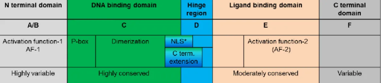
The nuclear hormone receptors are hormone dependent transcription factors having a molecular weight in the range of ~100 kDa. For nuclear trafficking, a nuclear localization signal (NLS) is required to be attached to the receptor upon translation (Lazar 2011). Members of the nuclear receptor superfamily share a similar domain structure and contain separated domains for DNA binding and ligand binding (see Figure 4). The DNA binding domain (DBD) and the ligand binding domain (LBD) are located at the amino and carboxy-terminal portion of the protein, respectively. Despite the structural similarity of this domain (with 12 α helical segment), slight molecular differences in the region account for the ligand specificity. The DNA binding domain is able to recognize specific DNA sequences (hormone response elements, HREs) in the promoter region of a responsive target gene. The DBD contains zinc fingers that contact the DNA, the region responsible for the recognition of the specific DNA hexamer is the P box that is basically a small stretch of amino acids (Lazar 2011). TR binds the AGGTCA hexamer, also called as half-sites (see Figure 5), while glucocorticoide receptors bind to AGAACA. In some cases the receptor binds to extended half sites with C-terminal extension of the DNA-binding domain. TR predominantly binds the DNA heterodimerized with Retinoid X receptor (RXR). The dimers bind to the two half sites and the number of bases and the sequence between the half sites determine the target gene specificity. TRs usually bind to direct repeats separated by 4 bases between them (Umesono et al., 1991), but in some cases the orientation of the half-sites and their spacing is different (Williams and Brent 1995). RXR and TR bind to the 5’ and 3’ half site sequences, respectively. Upon ligand binding, TR undergoes a conformational change which alters its capacity to recruit coactivators or corepressors. This event is a prerequisite of the TR-dependent alteration of gene transcription. Whether the resulting effect will be manifested in activation or repression is primarily dependent on the TRE characteristics of a specific gene (see Section 2.6).
*nuclear localisation signal Figure 4. General structure and functions of the nuclear receptors
Figure 5. Binding of thyroid hormone receptors to DNA. Thyroid (TR) and retinoid X receptor (RXR) heterodimers bind to DNA half site sequences called TH response element (TRE).
2.5.1. Thyroid hormone receptor isoforms
Two TR isoforms have been identified. TRα was first isolated from chicken liver (Sap et al., 1986) and at the same time also from rat brain (Thompson et al., 1987). The TRβ was isolated from human placenta (Weinberger et al., 1986), chicken (Forrest et al., 1990 and 1991) and rat (Obregon et al., 1986). TRα and TRβ are encoded by two separate genes: TRα on the human chromosome 17, while TRβ on chromosome 3. TRα has two main subtypes, α1 and α2. TRα2 is an alternatively spliced variant, which is unable to bind thyroid hormones because some critical amino acids are replaced in the carboxy-terminal (Lazar et al., 1989b) and the dimerization properties of this receptor isoform are also changed. Some further protein products encoded by the TRα gene were also described such as TRα3 and the truncated ΔTRα1 and ΔTRα2 proteins. The function of the isoforms that do not bind the hormone is not clear, although these might have a role in attenuating the physiological effects of T3 by competing with the T3 binding variants (Lazar et al., 1989a).
The TRβ gene encodes four different types of proteins: TRβ1, TRβ2, TRβ3 and ΔTRβ3.
All of them are able to bind T3 but the truncated ΔTRβ3variant cannot bind to the DNA.
The A and B domains of the TRβ receptors are different from each other but their DNA and ligand binding domains are very similar.
The tissue distribution of TR isoforms shows tissue-specific differences and their physiologic role depends on the given tissue in which they are expressed. It was e.g.
demonstrated that TRα is important in the regulation of cardiac function, body temperature, intestinal and lymphocyte development. This receptor is also widely expressed in the brain (Morte et al., 2002). Some studies in mice confirmed the role of TRα in behaviour as it is responsible for the specification of the hippocampal neural circuits (Guadano-Ferraz et al., 2003). TRβ can be found in the pituitary, liver and cochlea. TRβ2 is the predominant TR isoform in the PVN (Bernal et al., 2003; Abel et al, 2001) and this subtype is responsible for the differentiation of the Purkinje cells in the cerebellum. For the different receptor types and their functions see Table 1.
Table 1. Thyroid hormone receptor isoforms, their tissue distribution and functions
(Based on Ortiga-Carvalho et al., 2014)
TRs are expressed in humans and in sheep well before the formation of the thyroid gland and the onset of thyroid hormone synthesis. In the human fetus the receptors can be detected from the 10th week of gestation while in sheep from the 50th day. TRα1 isoform is responsible for the vast majority of the total T3 binding in the fetal brain (Schwartz et al., 1992; Strait et al., 1991). In the adult rat brain distribution of T3
binding in the brain is approximately 60% α1, 30% β1 and 10% β2 (Schwartz et al., 1994).
Several types of thyroid hormone receptor mutations were described and TR knockout murine models provided novel insights into the physiological roles of the different receptor types (Ortiga-Carvalho et al., 2014). It was shown in mice that in the absence of TRβ neither behavioural nor neuroanatomical abnormalities were observed (Forrest et al., 1996b). The TRβ knockout mice are deaf, their colour vision is impaired indicating the role of this receptor type in the development of the cochlear cell and the retinal photoreceptor. Human patients without TRβ have normal mental development (Takeda et al., 1992), but the cochleo-vestibular development may be affected (Forrest et al., 1996a; Refetoff et al., 1967). Resistance to thyroid hormone syndrome (RTH) is typically caused by mutations in the TRβ gene (Jones et al., 2003). Patients with mutated TRβ have severe learning difficulties and reduced intelligence quotient. Their TSH and thyroid hormone levels are increased, goitre is developed accompanied with an impaired negative feedback of the HPT axis. The vision and hearing of these patients are also affected and heart defects, such as tachycardia, exacerbate the homeostatic functions (Refetoff and Dumitrescu 2007, Pazos-Moura et al., 2000). In general, mutations in TRβ manifest in more severe clinical signs than the complete absence of the receptor.
More recently, TRα1 mutant RTH patients have also been identified without presenting major disturbances in parameters of TH economy; in these cases TSH and circulating thyroid hormones are only mildly affected. On the other hand, the syndrome is represented by growth retardation, delayed bone development, cognitive deficits, severe constipation and impaired neuronal development (Van Mullem et al., 2012; Vennström et al., 2008).
It needs to be mentioned that the lack of TR function and hypothyroidism are manifested in different consequences since the deletion of TRs in knock-out mice do not lead to brain hypothyroidism. This is supported by the fact that the unliganded TRs can also act on their target gene, either in a positive or a negative manner, depending on the positive or negative regulation of the gene by T3. This removal of TR function also abolishes the regulatory function of unliganded TRs (Chassande 2003).
2.6. Assessment of thyroid hormone action
Binding of TH to TR exerts either activating or repressory effect on TH responsive target genes (Glass and Rosenfeld 2000).
2.6.1. Positive regulation of gene expression by thyroid hormones
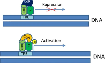
In contrast to the other nuclear homone receptors, e.g. the estrogen receptor (ER), the TR-RXR heterodimers are predominantly located in the nucleus even in the absence of ligand and even the unliganded TR can bind to TRE. Under uninduced conditions of a positively regulated TH sensitive gene the TR/RXR heterodimers bind co-repressor molecules. Binding of T3 triggers a conformational change of TR that will release co- repressors and bind co-activators. This TH-mediated activation of a positively regulated gene consists of derepression followed by activation. See Figure 6 for a schematic depiction of positively regulated genes (Lazar 2003).
Figure 6. Activation and repression of positively regulated genes by thyroid hormone receptors.
(Based on the model described by Lazar, 2003). (RXR: retinoid X receptor, TR: thyroid hormone receptor, CoR: co-repressor, CoA: co-activator, TRE: thyroid hormone response element, TH: thyroid hormone).
Several co-activators were identified that enhance the transcriptional activation of TH- responsive genes after TR binds to its ligand. Two important members of TH co- activators are the steroid receptor co-activator complex (SRC) and the vitamin D receptor interacting protein/thyroid receptor associated protein complex (DRIP/TRAP complex). SRCs have three subtypes: SRC-1, SRC-2 and SRC-3. Beside their capability to associate with TR they can also interact both with the CREB-binding protein (CBP) and p300. The CBP/p300 complex is able to interact with p300/CBP associated factor
(PCAF) that possesses histon acetylase (HAT) activity. Histon acetylases promote loosening the chromatin structure allowing transcription of the TH-responsive gene (McKenna et al., 1999). PCAF and CBP are able to interact with TATA-binding protein (TBP) associated factors and RNA polymerase II. Co-activators are able to bind to the helices 3, 5 and 6 of the ligand binding pocket of TR with their consensus LXXLL amino acid sequence. The components of the DRIP/TRAP pathway are able to associate with RNA polymerase II but they do not possess histone acetylase activity.
Major co-repressors of TH action are represented by the nuclear receptor co-repressor (N-CoR) and the silencing mediator of retinoid and thyroid receptors (SMRT). The latter is capable of anchoring further large multiprotein complexes that contain histon deacetylase (HDAC) activity. Histon deacetylases play a crucial role in maintaining the chromatin structure in a form that does not favour the basal transcription. DNA methylation may also attribute to basal repression since methyl-CpG-binding proteins associate with Sin3 containing co-repressor complex and HDAC (Nan X et al., 1998;
Wade et al., 1999). Small ubiquitous nuclear co-repressor (SUN-CoR) was also described (Zamir et al., 1997). The nuclear receptor co-repressor (NCoR) and the SMRT are approximately 50% homologue based on their amino acids and have similar structural domains. In the interaction domain of NCoR and SMRT consensus sequences (I/L)xx(I/V)I were found, similarly to that of the co-activators described above (Lazar 2011).
2.6.2. Negative regulation of gene expression by TH
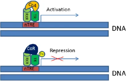
Mechanisms underlying TH-dependent negative regulation of gene expression are still incompletely resolved and different models have been worked out to explain the phenomenon. According to one of these models, TR binds to a negative TRE (nTRE). It means that in the case of hormone binding the receptor anchors to co-repressors (with HDAC activity), on the other hand in unliganded state the receptor binds to co- activators (with HAT activity). One of the in vivo examples for this model is represented by the negative effect of the TH bound receptor on TSH expression in the pituitary (Shibusawa et al., 2003) and on the regulation of TRH (see Figure 7).
Figure 7. Activation and repression of negatively regulated genes by thyroid hormone receptors (Model 1). (Based on the model described by Shibusawa et al., 2003) (RXR: retinoid X receptor, TR: thyroid hormone receptor, CoR: co-repressor, CoA: co-activator, nTRE: negative thyroid hormone response element, TH: thyroid hormone).
It was also suggested that on negatively regulated target genes co-repressor complexes are recruited in the absence of TH. After T3 binding co-activators are recruited, but at the same time the transcription is repressed (Astapova and Hollenberg 2013) (Figure 8).
Figure 8. Activation and repression of negatively regulated genes by thyroid hormone receptors (Model 2). (Based on the model described by Astapova and Hollenberg 2013). (RXR: retinoid X receptor, TR: thyroid hormone receptor, CoR: co-repressor, CoA: co-activator, TRE: negative thyroid hormone response element, TH: thyroid hormone).
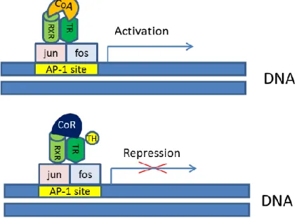
It was also suggested, that TR binding to DNA is not a prerequisite of the TH-mediated negative regulation (Zhang et al., 1991; Pfahl 1993). In this case, an activator protein 1 (AP-1) heterodimer (consisting of Jun and Fos) is involved in the process. Upon AP-1 binding TR recruits co-activators in the absence of the ligand and binds to co-repressor if T3 is present (Figure 9).
Figure 9. Activation and repression of negatively regulated genes by thyroid hormone receptors (Model 3). (Based on the model described by Zhang et al., 1991 and Pfahl, 1993). (RXR:
retinoid X receptor, TR: thyroid hormone receptor, CoR: co-repressor, CoA: co-activator, AP-1 site: activator protein 1 binding site, TH: thyroid hormone).
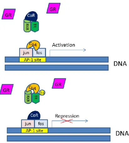
This model can also involve regulation by non-T3 dependent pathways, indicating that TR can compete with other nuclear receptors for co-activators or co-repressors (Figure 10). Without the ligand they bind (“steal”) co-repressors, while in the presence of TH they bind to co-activators (Kamei et al., 1996 and Tagami et al., 1997).
Figure 10. Activation and repression of negatively regulated genes by thyroid hormone receptors (Model 4) (Based on the model described by Kamei et al., 1996 and Tagami et al., 1997). (RXR: retinoid X receptor, TR: thyroid hormone receptor, CoR: co-repressor, CoA: co- activator, AP-1 site: activator protein 1 binding site, TH: thyroid hormone, GR: glucocorticoid receptor).
2.7. Posttranscriptional mechanisms regulating biological activity 2.7.1. Alternative splicing
Alternative splicing is a phenomenon in eukaryotes which increases the biodiversity of proteins encoded by the genome (Black 2003). Precursor messenger RNAs (or primary transcripts) can be modified by post-transcriptional processes. Alternative splicing can generate different mRNAs and as a consequence, different protein subtypes can be encoded by the same gene. There are two distinct mechanisms of alternative splicing, the exon skipping/switching and the intron slippage (Habener 2011). During exon skipping some exons are included while some of them may be excluded from the
primary transcript. Intron slippage is a process in which part of an intron is used in an exon, thus part of the intron becomes a coding region. Various examples are known when alternative splicing affects TH signaling. As it has been already highlighted in Section 2.5.1 the TRα2 is an alternatively spliced variant of the TRα1 isoform which is not able to bind T3 (Lazar et al., 1989b).
2.7.2. The 5’ untranslated region of the D2 mRNA
The 5’ untranslated region (5’UTR) of a specific mRNA is located between the 5’
proximity of the mRNA and the translational initiator ATG of the protein encoding region. The 5’UTR of the chicken D2 mRNA is unususally long (~600 bp). It contains short open reading frames (sORF) (Gereben et al., 1999). The sORFs are small alternative coding regions flanked by start and stop codons separated by an RNA segment consisting of bases in a number divisible by 3. Presence of the “Kozak consensus sequence” is the prerequisite of an efficient translational initiation of an eukaryotic open reading frame, represented by purine nucleotides (adenine or guanine) in the -3 position, where +1 is the first base of the ATG start codon (Kozak 1986).
According to the scanning model of translation, the 40S ribosomal subunit moves 5’ to 3’ along the mRNA and upon reaching an ATG embedded into a Kozak consensus gets charged with methionine and by the help of additional factors recruits the 60S subunit in order to initiate translation (Alberts et al., 1994). Thus, sORFs in the 5’UTR of D2 may be subjected to translation.
2.8. Measurement of TH-dependent gene expression with promoter assays 2.8.1. Luciferase assays in general
Accurate measurement of gene transcription is a critically important tool to study genomic events underlying modulation of cell function. This is especially important for studies on TH-mediated events since most of the known effects of this hormone involve TH-induced transcriptional events. Taking advantage of bioluminescence is a state of the art approach to study transcriptional events. Bioluminescence, by definition, is the production and emission of visible light by a living organism as a result of a natural chemical reaction (McElroy et al., 1969). Several organisms are capable of producing light such as bacteria, insects, fungi and different marine organisms. During the
chemical reaction, the photon emitting luciferin gets oxidized by the luciferases.
Luciferins are conserved but the luciferase enzymes are species-specific. The best established firefly luciferase originates from the North American firefly Photinus pyralis and is a 61 kDa protein. Renilla luciferase originating from Renilla reniformis represents another luciferase protein which was isolated from corals of coastal waters of North America (Lorenz et al., 1991). Recently, a novel luciferase enzyme Nano luciferase has been isolated from deep sea shrimp Oplophorus gracilirostris. It contains two smaller 19kDa and two larger 35 kDa subunits. This luciferase was used to develop the engineered luciferase NanoLuc in which the furimazine is used as a coelenterazine analogue. For cellular analytical assays this new luciferase has the advantage that it is more stable, showing brighter luminescence with sustained signal duration, has greater thermal, pH and urea stability, smaller size (19kDa) and monomeric structure. Its emission maximum is at 460 nm (Hall et al., 2012). Luciferase assays are rapid, sensitive and require non-radioactive substrates. In general, they are approximately 30- 1000 times more sensitive than the classical chloramphenicol acetyltransferase reporter system (de Wet et al., 1987). It was also confirmed that luciferin is able to enter the cells enabling to conduct the luciferase expression studies in intact cells.
2.8.2. Investigation of thyroid hormone response elements
It is a crucial requirement of accurate gene expression studies that the reporter itself should not be affected by the factor used to modulate the promoter and its flanking region. However, in CV1 mammalian cells expression of the classical firefly luciferase was down-regulated by T3 in a TR-dependent but promoter-independent manner (Tillman et al., 1993). Consequently, the CV1 cells cannot be reliably used for TRE structure or activity studies. This phenomenon was further evaluated with the analysis of unliganded thyroid hormone receptors on the Luc expression in HEK-293, COS-7 and JEG-3 cells (Maia et al., 1996). Among these cell lines the choriocarcinoma cell line JEG-3 was found to be the most sensitive for these studies. The authors suggested that an unidentified negative TRE should be present in the luciferase coding region and concluded that firefly luciferase cannot accurately measure the T3-dependent gene expression.
Later, a cautionary note has been issued by Chan et al. (2008) regarding the use of the pBi-L (Clontech, Mountain View, CA, USA) dual expression plasmid for the generation
of transgenic mice. In these studies the expression of both TRα1 and luciferase were negatively regulated by T3. This was independent of both the cis introduction of TR in the vector and the trans expression of TR from another vector. The negative regulation could be observed only in the presence of TRs and only in those vectors that contained the luciferase reporter. All these data above demonstrate that the use of firefly luciferase has its limitations in the T3-dependent gene expression studies.
3. OBJECTIVES
Regulatory factors of thyroid hormone mediated effects were studied using molecular and cell biological approaches. We focused on the following issues.
I. Investigation of thyroid homone availability in the developing chicken hypothalamus
II. Understanding the RNA-dependent post-transcriptional regulation of the type 2 deiodinase (D2) encoding dio2 gene
III. Identification of authentic reporter proteins for studies on T3-dependent gene transcription
We intended to address the following specific questions:
1. What is the distribution pattern of the D2 mRNA in the developing and adult chicken brain?
2. Do alternative splicing and the D2 5’UTR play a role in the post- transcriptional regulation of D2 activity?
3. Are novel luciferase reporters more accurate to assess T3-mediated transcriptional changes than the classical firefly luciferase?
4. METHODS
4.1. Animals
Eight-week-old specific pathogen-free White Leghorn chickens and chicken embryos on the embryonic day (E)7, E8, E9, E10, E11, E13, E15 and E17 were obtained from the Central Veterinary Institute and Ceva-Phylaxia (Budapest, Hungary). The incubation was started at E0. Animal tissue samples were collected in accordance with the legal requirements of the Animal Care and Use Committee of the Institute of Experimental Medicine (Hungarian Academy of Sciences, Budapest).
4.2. Generation of expression constructs
4.2.1. Constructs prepared for the analysis of RNA-dependent post-transciptional regulation of D2
The backbone of the chicken D2 (cD2) reporter construct contained a cD2 coding region between EcorI-HindIII and a rat D1 minimal SECIS element between HindIII- NotI (Gereben et al., 1999). Constructs were prepared with different UTR fragments cloned between the SacII site of the D10 vector and EcoRI. To generate the cD2 5’UTR construct, SacII-EcoRI fragment of the full-length cD2 cDNA was inserted between the corresponding sites of the cD2 reporter. The cORF(Wt)-cD2 construct was prepared by using oligonucleotides: sense tccccgcggG CCGAGAAACA ATGGGATAGC GCgaattcc and antisense, ggaattcGCG CTATCCCATT GTTTCTCGGC ccgcgggga.
Oligonucleotides were annealed to generate double stranded DNA. For the cORF(Mut- ATG)-cD2 construct, the following oligonucleotides were used for annealing (sense, tccccgcggG CCGAGAAACA tTGGGATAGC Gcgaattcc; antisense, ggaattcGCG CTATCCCAaT GTTTCTCGGC ccgcgggga). The resulting inserts were cloned after SacII-EcoRI digestion into the cD2 reporter. The cDNA encoding the Δ77cD2 protein was isolated as described in Section 4.4. The spliced cDNA was inserted between SacII and NotI of the D10 mammalian expression vector using the same approach as described before for the wild-type cD2 mRNA (Gereben et al., 1999). The generated constructs were confirmed by automated sequencing.
4.2.2. Generation of expression constructs for the analysis of luciferase reporters The thymidine kinase-luciferase (TK-Luc) construct was generated by removing the TRE triplet of pTRE-TK-Luc by digestion with BamHI and BglII followed by religation
and confirmation of the final construct by sequencing. The original plasmid backbone, pTRE-TK-Luc contains the thymidine kinase minimal promoter of the herpes simplex virus and was kindly provided by Dr. AM Zavacki (Boston, MA, USA).
The TK-(dCpG)Luc was prepared using the TK-Luc plasmid backbone as follows. The pMOD Luc-ShS v02 plasmid (InvivoGen, San Diego, CA, USA) was used as a template to amplify the (dCpG)Luc coding region with Vent PCR (oligonucleotides: sense,
catgcc ATGGAGGATGCCAAGAATATTAAGAA; antisense, ggaattc
TTATTTGCCACCCTTCTTGGCCTTGATCA). The amplicon was cut with NcoI and inserted into the NcoI - and the blunted EcoNI sites of TK-Luc. The construct was confirmed by sequencing.
The TK-NanoLuc was prepared by isolating the TRE lacking the minimal TK promoter from the pTRE-TK-Luc through digestion with BglII and HindIII and subsequent cloning of the released fragment into the corresponding sites of the pNL1.1 vector (Promega, Madison, WI, USA) followed by confirmation of the final construct by sequencing.
The TK-Renilla-Luc was generated by truncating the 760 bp-long TK promoter of pRL- TK (Promega) using BglII and EcoRI digestion followed by blunting with Klenow polymerase and subsequent religation. This resulted in a minimal TK promoter between EcoRI and HindIII that is 31 bp shorter than the 128 bp-long minimal TK promoter of TK-Luc. 3’ to the TK promoter, this construct also contains a 136 bp chimeric intron originating from pRL-TK. The construct was confirmed by restriction mapping.
The constructs are shown in Figure 22.
The mouse TRα (mTRα) expression construct was generated using the TRαCDM plasmid (Prost et al., 1998) (kindly provided by Dr. AM Zavacki, Boston, MA, USA) as a template to amplify mTRα coding region with Vent PCR (oligonucleotides sense:
ggaattccat tATGGAACAG AAGCCAAGCA AGGT, antisense: ataagaatgc ggccgcTTAG ACTTCCTGAT CCTCAAAGA). The amplicon was cut with EcoRI and NotI and inserted into these sites of a pCI-Neo vector (Promega) and confirmed by sequencing.
The secreted embryonic alkaline phosphatase (SEAP) encoding pSEAP2-Promoter plasmid (Clontech) was used for transfection control.
4.3. DNA transfections
For studies on the post-transcriptional regulation of D2 HEK-293 cells were transfected with calcium phosphate precipitation as described earlier (Brent et al., 1989). 10 micrograms of D10 based vector encoding the deiodinase was transiently transfected in the presence of 4 µg D15 helper vector required for the transcriptional activation of the promoter of D10 (Gossen and Bujard 1992). Results are given as the mean SEM of D2 activities of duplicate plates of at least three separate experiments as the percentage of the cD2 control.
For the investigation of the luciferase reporters, JEG-3 human choriocarcinoma cells (kindly provided by Dr. J. Szekeres, Pécs, Hungary) were cultured in 24-well plates in Dulbecco’s Modified Eagle Medium (DMEM) supplemented with 10% fetal bovine serum (FBS). When ~70% confluency was reached, cells were transfected with 800 ng DNA/well (including 200 ng Luciferase reporter, 100 ng mouse TRα, 10 ng pSEAP2 and 490 ng pUC as inert DNA) using Lipofectamine® 2000 (Life Technologies/Thermo, Carlsbad, CA, USA). After ~6 hours the transfection media was replaced with DMEM containing 10% hormone-free fetal bovine serum (FBS) and incubated for 40 hours (Egri and Gereben, 2014). Briefly, 100 mg charcoal (Sigma, St.
Louis, MO, USA) and 50 mg dextran (Sigma) were preincubated overnight in 0.01 mol/l Tris buffer (pH=7.6). After centrifugation 40 ml FBS was added and incubated for 1 hour. The suspension was recentrifugated and the supernatant was used to supplement DMEM. The media was replaced with DMEM with 10% hormone-free FBS containing either 50 nM 3,5,3’-triiodothyronine (+T3) or NaOH vehicle (-T3). After 24 hours, the culture media was collected for SEAP measurement (see Section 4.7.2). The cells were washed with phosphate buffered saline (PBS) and harvested in 100 µl Passive lysis buffer (Promega).
4.4. RNA isolation and RT-PCR
For the investigation of the ontogenic redistribution of D2, brain samples of E7, E8, E9, E10, E11, E13, and E15 chicken embryos were dissected in duplicates, and total RNA was isolated with Trizol (Life Technologies, Inc.). E13 and E15 brains were separated for telencephalon+diencephalon (A) and brainstem+cerebellum (B) parts. RNA was subjected to first strand cDNA synthesis using an oligonucleotide-dT primer and
amplified with D2-specific primers, as described earlier (Gereben et al., 2002). The cD2 oligonucleotides were as follows (5’-3’): sense, CTG AAT TCA TCC GGC AGA AGA GAG; antisense, AGC TTC TCC TCC AAG TTT GA. The nonquantitative D2 amplification was performed using the following program: 94 °C for 2 min; 35 cycles of 94 °C for 30 sec, 58 °C for 30 sec, and 72 °C for 1 min; and then 72 °C for 4 min.
For the studies on post-transcriptional regulation of D2 adult chicken telencephalons and livers were used. Total RNAs isolated from the telencephalon and liver were reverse transcribed using the antisense oligonucleotide CTCACCAGAA GGCCTGAAGA G and amplified by Taq polymerase (Sigma) by oligonucleotides, sense: CTGAATTCAT CCGGCAGAAG AGAG; antisense: AGCTTCTCCT CCAAGTTTGA. The amplicons generated on the brain cDNA were cloned into pGEM-T and subjected to automated sequencing. The amplifications were performed in two separate reactions.
4.5. Northern blots
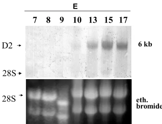
The Northern blot for the ontogenic D2 distribution study was performed as previously described (Gereben et al., 1999). Briefly, total RNA was isolated with Trizol from the brains of E7, E8, E9, and E10 and hemispheres of E13, E15 and E17 chicken embryos as described in Section 4.4. A digoxigenin (DIG)-labelled single stranded cDNA probe complementer to 450 bp of the cD2 coding region was used to detect D2 in 30 µg of total RNA. The probe was labelled by linear PCR using the (5’-3’) TGCACAATGCACACTCGCTC antisense oligonucleotide and DIG-deoxyuridine 5- triphosphate. As denominator for densitometry, the density of the 28S subunit of ethidium bromide stained gels was used.
4.6. In situ hybridization
The technique was used for the analysis of D2 mRNA ontogenic redistribution of the brains of E8 and E15 chicken embryos and 8-week-old chickens. The heads of three E8 embryos and the brains of three E15 embryos and three 8-week-old chickens were quickly frozen on dry ice and stored at –80 C until used. Serial 12-µm-thick coronal sections were cut on cryostat, mounted on gelatine-coated slides, and dried at 42C
overnight. The sections were fixed with 4% paraformaldehyde in PBS (pH 7.4) for 30 minutes washed in 2-fold concentration of standard sodium citrate (2X SSC), acetylated with 0.25% acetic anhydride in 0.9 % triethanolamine for 20 min; and then treated in graded solutions of ethanol (70, 80, 96, 100%), chloroform and a descending series of ethanol (100, 96%) for 5 minutes each and hybridized with an approximately 840 bp single-stranded DIG-11-uridine 5-triphosphate (Roche Diagnostics GmbH, Mannheim, Germany)-labelled cRNA probe for the entire coding region of cD2. The hybridizations were performed under plastic coverslips in a buffer containing 50% formamide, 2-fold concentration of standard sodium citrate, 10% dextran sulfate, 0.5% sodium dodecyl sulfate, 250 µg/ml denatured salmon sperm DNA, and the DIG-labelled probe, diluted at 1:100 for 16 h at 56C. The slides were washed in 1X SSC for 15 min and then treated with RNase (25 µg/ml) for 1 h, at 37C. After additional washes in 0.1X SSC (2 X 30 min) at 65C, sections were washed in PBS and treated with the mixture of 0.5%
Triton X-100 and 0.5% H2O2 for 15 min and then with 2% bovine serum albumin (BSA) in PBS for 20 min to reduce the nonspecific antibody binding. The sections were incubated with a mixture of sheep anti-DIG-alkaline phosphatase Fab fragments (1:1000, Roche Diagnostics) overnight at 4C. The alkaline phosphatase signal was detected using 5-bromo-4-chloro-3-indolyl-phosphate/4-nitroblue tetrazolium chromogen system (Roche Diagnostics) according to the manufacturer’s instructions.
The reaction was developed for 6 hours, and then, the sections were rinsed in Tris buffer (pH 7.6). The sections were coverslipped using Aquatex mounting medium (Merck, Darmstadt Germany), and the images were taken with an Axiophot microscope (Carl Zeiss Inc, Göttingen, Germany) equipped with real-time spot digital camera (Diagnostic Instruments Inc., Sterling Heights, MI, USA). For semiquantitative analyses all samples were treated simultaneously. Three 20X field of the hypothalamus of each E15 and adult brain from three different anterior-posterior levels were analysed using ImageJ software (public domain from National Institutes of Health, USA). Background density points were removed by thresholding the image. The sum of integrated density values (density X area) was calculated for each animal. The specificity of hybridization was confirmed using a sense cD2 coding region probe, that resulted in the total absence of specific hybridization signal in the brain at all stages studied.