Molecular regulation of thyroid hormone activation
Ph.D. Thesis
Péter Egri
Semmelweis University
János Szentágothai Ph.D. School of Neuroscience
Tutor: Dr. Balázs Gereben, D.Sc.
Opponents: Dr. Endre Nagy, D.Sc.
Dr. Árpád Dobolyi, D.Sc.
Chairman of committee: Dr. Miklós Tóth, D.Sc.
Members of committee: Dr. Krisztina Kovács, D.Sc.
Dr. Andrea Tamás, Ph.D.
Budapest
2016
1. T
ABLE OF CONTENTS1. TABLE OF CONTENTS ... 2
2. LIST OF ABBREVIATIONS ... 3
3. INTRODUCTION ... 5
3.1. Significance and action of thyroid hormones ... 5
3.2. Deiodinase enzyme family ... 16
3.3. The ubiquitin-proteasome system ... 28
4. SPECIFIC AIMS ... 35
5. MATERIALS AND METHODS ... 36
5.1. DNA constructs and RT-PCR ... 36
5.2. Cell culture and transfection ... 39
5.3. Reagents and treatments ... 40
5.4. Luciferase promoter assay ... 41
5.5. Quantitative PCR ... 41
5.6. Western blot ... 42
5.7. Deiodinase assay ... 42
5.8. Secreted alkaline phosphatase (SEAP) assay ... 43
5.9. Fluorescent resonance energy transfer (FRET) ... 44
5.10. Animals and surgery ... 45
5.11. Thyroid hormone measurement ... 46
5.12. TSH bioactivity measurement... 46
5.13. Immunohistochemistry ... 46
5.14. Statistical analysis ... 47
6. RESULTS ... 48
6.1. Interaction between PACAP and thyroid hormone signaling ... 48
6.2. Characterization of MARCH6 as D2 ubiquitin ligase ... 56
6.3. Structural background of the ubiquitination of deiodinases ... 66
7. DISCUSSION ... 73
7.1. Interaction between PACAP and thyroid hormone signaling ... 73
7.2. Characterization of MARCH6 as D2 ubiquitin ligase ... 77
7.3. Structural background of the ubiquitination of deiodinases ... 84
8. CONCLUSIONS ... 90
9. SUMMARY ... 92
10. ÖSSZEFOGLALÁS ... 93
11. REFERENCES ... 94
12. PUBLICATION LIST ... 118
12.1. List of publications the thesis is based on ... 118
12.2. Other publications ... 118
2. L
IST OF ABBREVIATIONSIn accordance with HUGO Gene Nomenclature Committee and Mouse Genome Database guidelines, proteins are written in upper case, human gene symbols and transcripts are indicated upper case and italicized while rodent gene symbols and transcripts are indicated first-letter upper case and italicized.
(a)CSF (artificial) cerebrospinal fluid BAT brown adipose tissue
CDS coding DNA sequence CRE cAMP response element
CREB cAMP response element-binding protein D1 type 1 iodothyronine deiodinase protein D2 type 2 iodothyronine deiodinase protein D3 type 3 iodothyronine deiodinase protein
DIO1/Dio1 gene or mRNA of human/rodent type 1 iodothyronine deiodinase DIO2/Dio2 gene or mRNA of human/rodent type 2 iodothyronine deiodinase DIO3/Dio3 gene or mRNA of human/rodent type 3 iodothyronine deiodinase E1 ubiquitin activating enzyme
E2 ubiquitin conjugating enzyme E3 ubiquitin ligase enzyme
ECFP enhanced cyan fluorescent protein EERI Eeyarestatin I
ERAD endoplasmic-reticulum-associated degradation EYFP enhanced yellow fluorescent protein
FRET fluorescence (Förster) resonance energy transfer HPT axis hypothalamo-pituitary-thyroid axis
KM Michaelis-Menten constant LPS lipopolysaccharide
MARCH6 Membrane-Associated Ring Finger (C3HC4) 6
mCherry monomeric modified red fluorescent protein
MG132 proteasome inhibitor (carbobenzoxy-L-leucyl-L-leucyl-L-leucinal) NF-κB nuclear factor κ-light-chain-enhancer of activated B cells
p65 subunit of the NF-κB transcription factor
PACAP pituitary adenylate cyclase-activating polypeptide PKA protein kinase A
PVN paraventricular nucleus Sec selenocysteine
SHH Sonic hedgehog
SP1 specificity protein 1 T3 3,3’,5-triiodo-L-thyronine rT3 3,3’,5’-triiodo-L-thyronine
T4 thyroxine, 3,3’,5,5’-tetraiodo-L-thyronine
TH thyroid hormone
TR thyroid hormone nuclear receptor TRE thyroid hormone response element TSS transcriptional start site
Ub ubiquitin
UPS ubiquitin-proteasome system UTR untranslated region
WSB1 WD40 SOCS-box containing protein
3. I
NTRODUCTION3.1. SIGNIFICANCE AND ACTION OF THYROID HORMONES
3.1.1. General aspects of thyroid hormone action in the brain
Thyroid hormones (THs) play essential role in the regulation of a wide range of biological phenomena and fundamentally affect the metabolic and developmental processes. THs exert a major impact on brain development and function, discussed in section 3.1.3 in more detail [1]. Furthermore, the level of TH in specific brain regions has far-reaching consequences on various peripheral organ systems [2]. Thus, understanding cellular and molecular mechanisms regulating TH levels in the brain is a great importance both for brain-related and peripheral processes.
Specific features of multiple regulatory levels need to be taken into account when the complex framework of TH economy is discussed. First, the primary product of human thyroid gland is the stable prohormone thyroxine (T4) that cannot be efficiently bound by the thyroid hormone nuclear receptors (TRs) on the canonical ligand-binding pocket [3].
Therefore T4 is not able to modulate gene expression via the canonical, TR-mediated pathway of TH action [4]. The regulatory capacity of hypothalamo-pituitary-thyroid (HPT) axis is restricted predominantly to the synthesis and release of the T4 prohormone.
Second, mostly T4 can be transported via the blood-brain and CSF-brain barrier while the transcriptionally active, circulating serum T3 has poor access to most of the areas of the central nervous system due to the selective affinity of TH transporters (Fig. 1) [5]. Taken together, the HPT axis has obvious limitations and is insufficient to control TH action in the brain. Third, and as a consequence, TH action in the brain requires the local conversion of T4 to T3 that is critical in the TH-dependent modulation of gene expression.
The metabolism of THs is catalyzed by members of the deiodinase enzyme family allowing both the activation and inactivation of THs. Exclusively the type 2 deiodinase (D2) is expressed as activating deiodinase in the human brain and D2 is the main source of the T3 in the central nervous system [6, 7].
Figure 1. Limitations of the HPT axis in the regulation of local thyroid hormone action
The thyroid hormone receptor binds T3 with high affinity but thyroid hormone transporters of the blood- brain barrier have low permeability for T3 that makes local activation essential for thyroid hormone action in the brain.
Importantly, TH metabolism in the brain is highly compartmentalized. While D2 is expressed exclusively in glial cells – in astrocytes and hypothalamic tanycytes – the neurons are not able to activate THs, the glial-derived T3 affects the neuronal transcriptome on a paracrine manner. However, neurons are able to modulate their intracellular TH level via type 3 deiodinase (D3) catalyzed inactivation [8]. Therefore the control of TH mediated gene expression in the brain requires the coordinated actions of TH transport and deiodinase-mediated TH metabolism (Fig. 2). These processes are especially significant in the regulation of the HPT axis as they result not only in local but also systemic changes in TH economy via the control of negative feedback of TRH neurons, see in section 3.1.4.
Figure 2.Thyroid hormone metabolism
Activation and inactivation pathways are catalyzed by the members of deiodinase enzyme family via removal of iodine from the outer or inner ring of thyroid hormone derivatives, respectively.
In conclusion, the interaction between deiodinase-mediated TH activation in the brain and the HPT axis represents a core mechanism of controlling TH economy thorough the body. Thus, we focused our studies to better understand the molecular regulation of TH activation and its impact on the HPT axis. The following sections provide a brief overview on different regulatory levels of TH action including the mechanism of TH transport and TRs followed by the introduction of the role THs in the central nervous system (CNS) and the periphery. The second part will focus on the deiodinase-mediated metabolism of THs – especially the activation by D2 – and the regulation of the D2 enzyme.
3.1.2. Mechanism of thyroid hormone action: transporters and receptors
TRs – exerting the canonical TH effect – selectively bind T3 as ligand therefore the precise control of intracellular availability of T3 directly affects the exerted effects of TH.
This regulation requires the contribution of thyroid hormone transporters (both at the blood- brain barrier and at paracrine transport between glial and neuronal cells) and thyroid hormone metabolizing enzymes, deiodinases. The coordinated action of transporters and deiodinases also provides the opportunity of fine-tuning of T3 availability at the cellular level and determining the liganded state of TRs (Fig. 3) [9].
Figure 3.Thyroid hormone metabolism in the brain
Schematic depiction of the thyroid hormone availability in the brain based on the neuro-glial compartmentalization of thyroid hormone transport and metabolism.
According to the old dogma, THs as lipophilic molecules were thought to undergo passive membrane transport upon their passage through the plasmamembrane into the cytosol without the involvement of active transport. However, this idea was disproved by the identification of different types of thyroid hormone transporters that allowed the specific transport of TH through the plasmamembrane [10, 11]. The importance of active TH transport is clearly demonstrated in the Allan-Herndon-Dudley syndrome (AHDS) leads to a mixed thyroid phenotype with mental retardation, central hypotonia and impaired auditory development [12]. AHDS is caused by mutations in thyroid hormone transporter SLC16A2 (MCT8) belongs to the monocarboxylate transporter (MCT) family along with SLC16A10 (MCT10). SLC16A2 is capable to transport both T4 and T3 while SLC16A10 has selective binding for T3 [5]. SLC16A2 is widely expressed in the CNS including neurons, glial cells and capillaries [13].
The second class of thyroid hormone transporters is the large family of organic anion-transporting polypeptides (OATPs). The selectivity of SLCO1C1 for T4 over T3
and its enriched expression in endothelial cells of capillaries could underline the unequal permeability of the blood-brain barrier for TH derivatives [13]. Additionally, while the SLCO1C1 knock-out mouse has a mild phenotype [14], its combined deletion with SLC16A2 resembles the developmental abnormalities of AHDS in human [15]. L-amino acid transporter (LAT) family is also capable for TH transport SLC7A2 (LAT1) and SLC7A8 (LAT2) are identified as TH transporters however their in vivo importance in TH transport are much less characterized compared to the above mentioned transporters [5].
The canonical and most studied pathway of TH action is mediated by transcriptional events via the TRs [4]. In contrast to other nuclear receptors, e.g. the estrogen receptor and glucocorticoid receptor, TRs are located predominantly in the nucleus even in unliganded form. The general TR structure contains the following domains in order from N-terminus to C-terminus: N-terminal activator (A/B), DNA-binding (C), hinge region (D), ligand-binding and dimerization (E), C-terminal activator (F) (Fig. 4) [16]. Two TR encoding genes were identified (THRA and THRB) each of these has three transcript variants [17]. TRα1, TRα2 and TRα3 are transcribed from THRA gene however only TRα1 has classical receptor functions while TRα2 and TRα3 lack the T3-binding ability due its alternatively spliced C-terminus and abolished T3-
Figure 4. Schematic structure of thyroid hormone nuclear receptors
Amino acid position refers for human orthologues; note the alternative splicing of the C-terminus of TRα2 and TRα3 results in abolished ligand binding.
binding pocket [17, 18]. Additionally, a downstream alternative transcriptional start site within THRA gene results in a truncated transcripts similarly to TRα2 lacking the T3- binding capacity. TRs bind one T3 molecule prior to activation. While a recent study indicated an additional ligand-binding surface within TRα1 that shows selectivity for T4
however the importance of this site is remained to be confirmed [19]. The THRB gene has three transcript variants: TRβ1, TRβ2 and TRβ3. Based on crystal structure data a second ligand binding surface could be formed by TRβ at least for the TRβ-specific T3-analogue GC-24 [20]. Beside the canonical genomic effects of TRs TRβ was demonstrated to modulate the phosphatidylinositol-3-kinase (PI3K) pathway and affect HIF-1α level via non-translational manner. The importance of this pathway is poorly understood yet.
The model of TH action suggests that TR-mediated regulation of gene expression is based on the altered interaction profile for cofactors upon T3-binding. This current model is based on the activation of well conserved positive thyroid hormone response elements (TREs) in the genome while the negative action of TH’s is much less understood. The prediction of the TR binding sites for negative regulation is not conclusive. In the absence of T3, TRs bind to TRE and recruit histone deacetylases e.g.
nuclear receptor corepressor (NCoR1) and silencing mediator of retinoic acid and thyroid hormone receptors (SMRT, NCoR2) [21]. The binding of T3 alters the equilibrium between the monomeric-dimeric states and allows the heterodimerization with retinoid X-receptor (RXR). This transition leads to the release of repressor complex and harboring activator proteins including histone acetyltransferases (steroid receptor coactivator-1 and 2, SRC1, SRC2 or NCoA1/2) and p300 (Fig. 5) [22]. In case of a negative TRE the T3
induced binding of TR represses promoter activity however the precise mechanism of action of THs on negative TRE is poorly characterized.
TRα is expressed in most tissues including the brain, cardiac and skeletal muscle, intestine and bone. TRβ1 has also wide expression profile e.g. in the brain and liver while TRβ2 is the predominant isoform in the hypothalamus and pituitary, consequently plays a crucial in the TH-feedback of HPT axis [23]. The differential expression has also clinical significance providing the opportunity of tissue-specific targeting of TH action using TRα- or TRβ-selective agonists or antagonists. Mutations in TR proteins can lead to the manifestation of the Resistance to thyroid hormone (RTH) syndrome hallmarked by altered TH-binding capacity causing symptoms of hyperactivity, emotional alterations and mental deficit on different levels of severity. Most known cases involve mutation(s) in TRβ [24] but recently TRα mutant patients have been also revealed [25-27].
Extranuclear, non-canonical TRs have been also discovered. Mitochondrial processes are major target of TH action and the p43 protein was identified as a mitochondrial TR [28, 29]. Under specific conditions the kinetics and the cell permeability independent nature of TH action suggested the existence of thyroid hormone membrane receptors that could govern genome-independent action. Integrin αVβ3 was identified as a cell membrane located receptor showing specific binding for T4 [30]. This cascade activates the MAPK signalization that results in phosphorylation of TRβ and increasing its transcriptional activity [31].
Figure 5. Schematic model of the activation of thyroid hormone receptors by ligand binding TR: thyroid hormone receptor; TRE: thyroid hormone response element (DR4 type); HDAC: histone deacetylase; NCoR: nuclear corepressor; NCoA: nuclear coactivator; H: histone; Ac: acetyl group.
3.1.3. The biological significance of thyroid hormones 3.1.3.1. Brain
THs are crucial regulators of developmental programs and their role in neural development is clearly demonstrated by symptoms caused by various defects of different events of TH action. Congenital hypothyroidism requires on-time TH supplementation to avoid irreversible developmental brain deficits. Promoting the exit from cell cycle and affecting differentiation programs THs are major regulators of progenitor cell development. Altered TH synthesis, metabolism or transport result in dramatic effects on neuronal differentiation and maturation manifested in significant loss in cognitive functions [1, 32]. Numerous genes with specific roles played in brain development are regulated by THs. Expression of crucial factors that organize the migration and survive of precursor cells like reelin, brain-derived neurotropic factor (BDNF) and nerve growth factor (NGF) are under the regulation of THs [33-35]. Oligodendrocyte differentiation, myelination and axonal growth are proved to be sensitive to TH status via the involvement of crucial genes like myelin basic protein (MBP) and myelin-associated glycoprotein (MAG) that are transcriptionally upregulated by THs [36, 37]. Expression of synaptotagmin – a protein involved in the docking and fusion of synaptic vesicles – is also affected by THs [38] suggesting a general mechanism how THs are able to influence neural communication.
Cerebellar development, foliation, maturation and migration of cerebellar neurons are strongly affected by THs via TRα [39, 40]. Dendritic arborization of Purkinje cells is also controlled by THs [39]. Delayed maturation and abnormal migration of granular cells was observed while the maturation of Bergmann glia and GABAergic interneurons were also affected in TRα dominant negative mutant mice [41]. These developmental processes reflect to the mechanistic background of impacted motoric phenotype in hypothyroidism observed both in human and animal models with deficiencies on different levels of TH signaling [40]. Normal cerebellar development requires the rapid supplementation of TH in congenital hypothyroidism.
Decreased mental capacity by hypothyroidism is associated with impaired learning and memory as broad range of hippocampal functions are controlled by THs. Synaptic remodeling, excitability, associative learning and inhibitory inputs for hippocampal cells
are also affected by THs [42]. Timing of the developmental phases of new-born neurons is controlled by coordinated TR expression [43]. Affected mood and behavior in patients with altered TH state is underlined by the connection between THs and the molecular elements of serotoninergic system [44]. THs are in tight interaction with other neurosecretory systems both in the hypothalamus and the pituitary. Reproductive functions are targeted by local TH metabolism in the hypothalamus by the regulation of seasonal activity [45, 46] and lactation [47, 48]. TH levels also affect stress response and anxiety, CRH expression is stimulated by THs [49-51].
Proper TH signaling is also essential in the sensory system. THs are crucial in the development of auditory system as found in congenital hypothyroidism or in Allan- Herndon-Dudley syndrome with severe deficits or complete loss of hearing [12]. Both the morphogenesis and function of the auditory system requires precise control of local TH transport and metabolism [52-55]. Improper TH level results in delayed program of eye development, eye opening and retina morphogenesis probably via altered mitochondrial biogenesis [56]. THs are also important regulators of opsin expression and patterning, defects in TH signaling results in reprogramming of M-opsin cones to S type [57, 58].
3.1.3.2. Peripheral organs
THs increase mitochondrial activity and elevate the metabolic rate both on cellular and systemic level [59, 60]. Alterations in TH actions directly affect energy homeostasis and could serve as a source of several clinical symptoms. In hypothyroidism, the basal metabolism is decreased and consequently energy consumption is reduced that can be manifested in weight gain while hyperthyroidism has the opposite effect on energy homeostasis. Liver has crucial function in chemical energy storage, conversion and transport therefore hepatic transcriptome is a major target of TH actions. THs are also crucial factors in the central regulation of energy balance. Genes involved in lipolysis, lipogenesis, fatty acid transport and gluconeogenesis were shown to be under TH control.
The clinical significance of this regulation is demonstrated by impaired liver metabolism in altered TH status [61]. THs also affect the cardiovascular system and cardiac metabolism increasing heart rate and volume while hyperthyroidism leads to cardiac muscle hypertrophy. THs contribute to the regulation of skeletal muscle metabolism and
substrate preference of chemical energy production while hypothyroidism manifested in decreased muscle tone [62].
The brown adipose tissue (BAT) is a well-documented target of THs involved in the maintenance of body temperature in hypothermic condition, especially in rodents [63]. While this function is also important in human neonates, until the last decade it was thought that thermogenesis by BAT is absent in human adults due to the documented regression of BAT depos. However, a few years ago the presence of functionally active BAT islands was identified in the skeletal muscle by PET imaging [64-66]. Recent studies suggest that these depots belong to the beige adipose tissue derived from different progenitors compared to BAT [67-69]. Human BAT or beige fat could have clinical significance since experimental data demonstrated the induction of beige adipose tissue by drugs used for treatment of type 2 diabetes [68]. Therefore in adult humans these cells could play an important role in maintaining body energy balance rather than protecting body temperature.
The thermogenesis is an alternative route in mitochondria to use the electrochemical energy of proton gradient between the two sides of the inner membrane. In this process the protons are not transferred through the ATP synthase but carried by the uncoupling protein 1 (UCP1 or thermogenin) into the mitochondrial matrix. Therefore the electrochemical energy generated by oxidative phosphorylation is converted to thermal energy instead of storage in chemical energy by ATP. After the discovery of UCP1 homologue proteins have been also identified and shown to be expressed in several tissues including the hypothalamus however the exact function and uncoupling capacity of these UCP’s are remained to be clarified similarly to the exact mechanism of heat generation by shuffling H+-ions into the mitochondrial matrix.
The BAT is under the control of the sympathetic nervous system by noradrenergic stimulation predominantly via β3-adrenergic receptor [70]. The selective agonists of this receptor evoke the activation of BAT and induction of the differentiation of beige cells in white adipose tissue [71]. Induction of heat production is controlled by the noradrenergic stimulus-driven activation of cAMP second messenger system resulting in upregulation of Ucp1 transcription and lipolytic enzymes. The release of fatty acids by lipoprotein lipase from triglycerides is also increased by elevation of intracellular cAMP and serves both as fuel and also as cofactor for UCP1. TH contributes to elevated UCP1 level and T3
production is elevated by the cAMP-mediated induction of the Dio2 gene, see also in 3.2.2.1. As a consequence, hypothyroidism results in severe deficits in adaptive thermogenesis. [72].
3.1.4. The hypothalamo-pituitary-thyroid (HPT) axis
The synthesis and release of THs are controlled by the HPT axis. The HPT axis consists of hypophysiotropic TRH neurons of the paraventricular nucleus, thyroid- stimulating hormone (TSH) secreting cells of the adenohypophysis and the thyroid gland itself. The precise control is supported by multilevel negative feedback of the axis (Fig.
6).
Hypophysiotropic TRH neurons are located in the paraventricular nucleus (PVN) of the hypothalamus sending axon terminals to the portal vessels of the median eminence and regulating the TSH synthesis and secretion of the adenohypophysis. We summarize below how THs are able to modulate TRH expression along with distinct peptidergic inputs on TRH neurons. TRH gene encodes a precursor protein (preproTRH) containing five copies of TRH. The precursor is processed by endopeptidases (prohormone convertase, PC) cleaving the three amino acid-long TRH peptide. TRH contains N- terminal pyroglutamate and its C-terminus is amidated (pGlu-His-Pro-NH2). These modifications are obligatory for the active peptide. PC1 expression and pyroglutamyl peptidase (PPII) – cleaves the pyroglutamate at the N-terminus – is regulated by THs [73- 76]. The TRH gene contains several TREs therefore directly responsive to TH via TRβ2 receptors [77-80].
Hypothalamic T3 concentration is controlled by local activation and inactivation of TH but TH economy in this brain region is more complex than in the rest of the brain.
The reason is that the blood-brain barrier in the median eminence – the region below the floor of the third ventricle – is incomplete. Therefore hypothalamic TH levels are affected both by local TH metabolism, tanycytic D2, neuronal D3 and by circulating T3 (Fig. 6).
Thus T3 input of TRH neurons are coming from both local and peripheral sources. The balance between these two T3 sources can be shifted towards locally generated T3 by tanycytic D2 under specific circumstances as clearly demonstrated in the infection- evoked rodent model of non-thyroidal illness. In this model, despite falling
Figure 6. Schematic depiction of thyroid hormone feedback of the HPT axis Involvement of thyroid hormone metabolism in the regulation of HPT axis.
serum TH levels, the upregulated tanycytic D2 activity results in local hypothalamic hyperthyroidism that leads to the suppression of HPT axis and hypothyroid peripheral status [81]. Therefore D2 activity in tanycytes is under strict control that includes the posttranslational ubiquitin-proteasome system (UPS), see in section 3.3. Factors that modulate tanycytic D2 activity have the potential to directly affect the HPT axis [82].
Beside their crucial role of T3-production tanycytes are capable to affect TRH neurons by several ways. As mentioned above PPII inactivates TRH and tanycytes express PPII that is regulated by THs [75, 76]. Tanycyte processes terminate on the capillaries of the median eminence and form a dynamic barrier for the axon terminals that release hormone- secreting neurons as observed in case of TRH [83] and GnRH neurons [84].
The TRH promoter cAMP-response element (CRE), glucocorticoid response element (GRE), STAT3 and SP1 binding sites [85, 86]. These pathways transmit crucial peptidergic signals involved in the regulation of energy homeostasis. TRH cells receive information from the orexigenic NPY/AgRP- and anorectic αMSH/CART-containing neurons in the arcuate nucleus sensing humoral signals including insulin, leptin, glucose and ghrelin [87]. Recent studies revealed that cells coexpressing TRH and pituitary
adenylate cyclase-activating peptide (PACAP) in the PVN send stimulatory inputs to the inhibitory AgRP cells of the arcuate nucleus providing a short feedback loop for TRH cells [88]. TRH neurons are innervated by the hypothalamic dorsomedial nucleus and the adrenergic nuclei of the brainstem; afferents from the last region contain CART, NPY and PACAP peptides [89-91].
The axons of TRH neurons project to the median eminence where release to the portal system and stimulate the TSH expression and secretion of thyrotropic cells in the adenohypophysis (Fig. 6). This occurs via the type I TRH receptor [92] and by the involvement of the activation of phospholipase C pathway inducing Ca2+-release and activation of CAMK-mediated CREB phosphorylation [93]. TSH is a heterodimer of thyrotropin-specific TSHβ and TSHα (CGA) that is a common subunit of chorionic gonadotropin (CG), luteinizing hormone (LH), follicle-stimulating hormone (FSH) and TSH. Both α and β subunit expression are stimulated by TRH via CRE sites and inhibited by TH via negative TREs that results in a short feedback-loop [94, 95]. Importantly, these hormones are glycoproteins and the amount and pattern of carbohydrates highly affects their stability and bioactivity that posttranslational modifications are sensitive for TRH [96, 97]. TSH glycosylation was also shown to be sensitive for T3 and its secretion and bioactivity is also affected by deletion of Dio2 gene [98, 99] indicating that the short feedback loop of HPT axis also requires the local T3 generation via D2.
In the thyroid gland the activated TSH receptor (TSHR) induces cAMP production and PI3K activation [100, 101] stimulating the transcription of genes involved in TH synthesis including I- uptake by Na+/I- symporter (NIS) [102], thyroglobulin [103] thyroid peroxidase (TPO) [104] and I--recycling by iodotyrosine dehalogenase (DEHAL1) [105].
3.2. DEIODINASE ENZYME FAMILY
3.2.1. Structure and biochemistry of deiodinases
THs are metabolized by a specialized oxidoreductase enzyme family called iodothyronine deiodinases. Deiodinases are different both in structure, catalytic mechanism and function from iodotyrosine deiodinases involved in the recirculation of iodine in the thyroid gland. As a common feature, deiodinases contain a rare amino acid;
mechanism is not completely understood it has been established that based on larger atomic radius, Sec is much more efficiently ionized at physiological pH than cysteine that represents a major advantage to catalyze oxidoreductive processes, like deiodination. As a result, the presence of Sec increases the affinity for substrate robustly. Its replacement with Cys results in a ~1000-fold elevated KM of D2 for T4 along with a highly increased translation [106]. Thus the presence of Sec in deiodinases results in high substrate affinity and low protein level. Deiodinases has the ability to catalyze the loss of iodine both of the outer and inner ring of thyronine backbone (Fig 2.). The incorporation of Sec is carried out by a complex apparatus during the translation (Fig. 7) [107]. Unlike the common 20 amino acids, Sec is encoded by the UGA STOP codon that is subjected to read-through in selenoproteins. This process requires the presence of a specific mRNA secondary loop structure called selenocysteine insertion sequence (SECIS) element in the 3’-untranslated region of the selenoprotein encoding mRNA. The SECIS-loop is bound by SECIS binding protein 2 (SBP2) that interacts with the special elongation factor eEFSec allowing the recruitment of the tRNA specific for Sec. In contrast to the common amino acids Sec is synthetized on its transfer RNA (tRNASec). The tRNASec is loaded with a serine then the hydroxyl-group of serine is edited to selenol-group by enzymatic cascade [108].
Available clinical data demonstrated that mutations in SBP2 affects the TH metabolism and reported impaired response for T4 but not for T3 [109]. In summary, this sophisticated system allows the cotranslational incorporation of a special amino acid Sec into selenoproteins by a complex and energy-dependent manner.
Figure 7. Synthesis and cotranslational insertion of selenocysteine into selenoproteins EFSec: Selenocysteine-specific elongation factor; SBP2: SECIS-binding protein 2.
3.2.1.1. Type 1 iodothyronine deiodinase (D1)
D1 is encoded by the DIO1 gene located on the chromosome 1p32 in human and by the homologous Dio1 gene in mouse on the chromosome 4. DIO1 gene contains 4
exons; exon 1 contains the translational start ATG codon after a short, 25 nucleotide- lengths 5’-untranslated region (UTR) [110]. Exon 2 encodes the catalytic selenocysteine and exon 4 carries the SECIS region within the 3’-UTR which is crucial in the insertion of selenocysteine amino acid during the translation.
The product of the DIO1 gene is a 249 amino acid-lengths type I membrane protein located in the plasmamembrane, with 26-30 kDa molecular weight. As a common feature of deiodinases D1 forms dimer which is obligatory for active conformation. The three dimensional structure of D1 has not been resolved yet therefore the domain structure and catalytic mechanism are based on predictions and sequence homologies. D1 shows highly conserved amino acid sequence between species. The N-terminus of D1 contains a short extracellular-tail composed from approximately the first 10 amino acids followed by the transmembrane helix located between amino acid position 10 and 40. Despite of the fact that D1 is highly conserved between species the first region of the cytosolic globular domain between amino acid position 40 and 70 shows variability (Fig. 8) [111].
Importantly, within this region there is a short deletion in feline and canine D1, two species where the biochemical properties of D1 are slightly different compared to other mammals [112, 113]. The globular domain contains the active center and the catalytic selenocysteine in the amino acid position 126. The region spans the active center is highly conserved between homologues and orthologue deiodinases.
Figure 8. Alignment of human D1, D2 and D3 proteins and the predicted domain structure
D1 has the ability to catalyze both outer- (ORD) and inner-ring deiodination (IRD) that is unique compared to the other members of deiodinase enzyme family. Therefore both the activation an inactivation of thyroxin (ORD and IRD of T4) are catalyzed by D1 with the affinity for substrate KM ≈ 10-6 M in both cases which is slightly higher compared to the same values of D1 for the IRD of T. However the affinity for IRD of rT is one
order of magnitude lower (KM ≈ 10-7 M) suggesting this derivative is the preferred substrate of D1. While the catalytic mechanism of deiodinases is poorly understood, interestingly the conserved deletion in case of feline and canine D1 revealed the importance of this region in the IRD of rT3. In case of this species the KM is elevated to
~10-5 M. 6-n-propyl-2-thiouracil (PTU) competes with the reducing agent of D1 while has a minimal effect on the other two deiodinases this effect has both clinical and methodological importance.
D1 is expressed in various tissues and cell types but importantly not in the human central nervous system. The most abundant D1 expression was found in liver, kidney and intestine while its activity is also present in the thyroid gland, pituitary and other tissues depending on the developmental stage. It is important to note that in contrast to the other two deiodinases D1 is not restricted to either the activation or inactivation of HTs therefore its expression and activity has to be evaluated in accordance with thyroid status and the available substrates. Hepatic D1 contributes to circulating T3 in hyperthyroidism while under euthyroid conditions D1 deiodinates predominantly rT3 as demonstrated in the Dio1 KO mice that shows slightly elevated T4 level while rT3 level is increased 3-fold [114].
Available data are limited on the regulation of the DIO1 gene however it was demonstrated that its transcription is positively regulated by T3 [115]. This attribute of D1 explains the relatively larger contribution of liver D1 to the circulating T3 in hyperthyroid conditions which is sensitive to PTU. Thyroidal but not the liver D1 was shown to be modulated by TSH-driven cAMP however it is unlikely that this effect is driven by the cAMP-sensitivity of DIO1 gene [116]. Similar asymmetry was observed in case of selenium-deficiency which results in decreased hepatic and renal D1 activity while thyroidal and pituitary D1 is slightly affected [117]. The energy balance, steroids and circadian rhythm are also showed to affect D1 activity however it is unlikely to be a specific and direct effect on DIO1 gene. There is no evidence to have posttranslational modification on D1 protein. In contrast with D2 that is targeted by the ubiquitin- proteasome system D1 is not ubiquitinated and has longer half-life [118].
3.2.1.2. Type 2 iodothyronine deiodinase (D2)
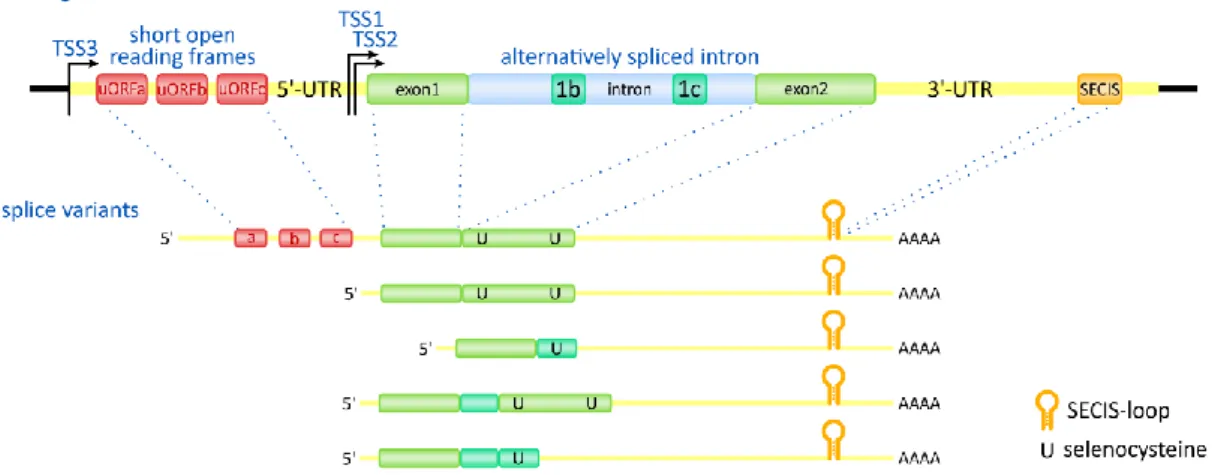
Type 2 deiodinase (D2) is encoded by the DIO2 gene in human and located on chromosome 14q24 while Dio2 gene on chromosome 12 in mouse. DIO2 gene contains two exons separated by a ~7.4 kb intron. Three transcription start sites (TSS) [119] were found in DIO2 gene affecting the unusually long 5’-UTR of the mRNA. Five splice variants of human DIO2 gene were identified that show differences in the insertion of two portions of the intron and the termination of translation. The alternatively spliced mRNA forms encode four different putative proteins [120]. The 5’-UTR of DIO2 mRNA contains upstream/short open reading frames (uORF or sORF) in different species, and not their number (3-5) but their existence is conserved between species (Fig. 9). The uORF-A affects the translation efficiency of the DIO2 coding sequence (CDS) and helps to keep D2 protein level low due to represent a translational roadblock during ribosomal scanning [121]. The DIO2 mRNA has a 5 kb 3’-UTR containing the SECIS-loop at the 3’-end. The 3’-UTR segment between the D2 CDS and SECIS-loop contributes to the instability of the DIO2 mRNA [121] however the accurate mechanism of this phenomenon is not yet resolved.
Figure 9. Structure of DIO2 gene and splice variants
uORF: upstream open reading frame; UTR: untranslated region; TSS: transcription start site; SECIS:
selenocysteine insertion sequence.
Despite the unusual length of the DIO2 mRNA (6-7.5 kb in vertebrates) the D2 protein is encoded by an only ~800 kb long coding region generating a 31 kDa type I membrane protein, localized in the ER in stable retention. Similarly to the other members of deiodinase family, D2 also forms homodimers and this structure is obligatory for the
active conformation of the enzyme. D2 has a short ER-lumen localized N-terminal tail followed by the transmembrane domain between amino acid position 20 and 40. The transmembrane domain plays an important role in the dimerization of D2 via ionic interaction between helixes. Based on homology and hydrophobicity predictions the TM domain is followed by a cytosolic linker region and thioredixin-fold βαβ motif between amino acid 122 and 163 [122]. The catalytic selenocysteine amino acid is localized within this domain in position 133 of human D2 which is followed by iodothyronine-deiodinase active center motif localized between amino acid 164 and 192. The C-terminus of the protein contains a variable and ββα thioredoxin motif between 193-224 and 225-273 amino acids, respectively. Unlike D1 and D3, D2 contains a second UGA close to its C- terminus at codon position 266, however the second Sec residue is not required for catalytic activity of the enzyme (Fig. 8) [123].
Point mutations that alter amino acid in the D2 protein were identified but their significance is still poorly understood [124-126]. These polymorphisms include L4H, T92A and T102I amino acid changes. L4H and T102 seem to have identical properties compared to abundant allele of D2 [125]. Interestingly, T92A is suggested to be in correlation with numerous symptoms e.g. type 2 diabetes mellitus, Graves’ disease, mental abnormalities, affected bone and muscle metabolism etc. however most of these studies are controversial and restricted to clinical data [124]. It is also remained to be clarified whether the statistical correlation is underlined by the effect of T92A polymorphism via modified biochemical characteristics of D2 or this polymorphism is a linked genetic marker of other mutations in same region of chromosome [124, 127-130].
While the precise background of the effect T92A polymorphism is poorly understood it is important to note that the 92th amino acid position is within the previously identified instability-loop of D2 involved in its posttranslational regulation by ubiquitin-proteasome system.
In contrast to D1, the catalytic activity of D2 is restricted to ORD of TH derivatives.
The mechanism of deiodination and the structural basis of the difference between D2 and D1 are incompletely understood. However, there are major differences between the biochemical properties between the two T4 activating deiodinases, D2 and D1. First, the primary substrate of D2 is T4 and has three orders of magnitude lower, ~10-9 M in vitro KM for this molecule, than that of D1. D2 is also effective in the deiodination of rT3
indicating that the structural differences between D1 and D2 result in higher catalytic efficiency. The importance of selenocysteine played in the catalytic mechanism of deiodinases was demonstrated clearly for D2; the mutation of this residue to cysteine results three orders of magnitude higher KM for T4 [106]. The homologue mutation in D1 leads to only one order of magnitude increase in KM for the preferred substrate rT3 [131].
Importantly, the studies targeting the biochemical characteristics of deiodinases were performed in cell lysates not using purified enzymes therefore the comparisons between different studies should be read carefully.
D2 is widely distributed in different tissues and its function is primarily the local T3 generation compared to D1. Importantly, D2 is the activating deiodinase in central nervous tissue and its expression is confined to glial cell types. High D2 activity was described in pituitary [98] while high DIO2 mRNA level in thyroid [132]. Another important target organ is the brown adipose tissue (BAT) where the D2-driven T3
increases the noradrenergic signal stimulated UCP1 expression. Probably due the common lineage, similarly to BAT, D2 refers for T3 generation in skeletal muscle, and seems to be important in tissue regeneration after injury [133]. D2 was also found in cardiac muscle, lung [134], perinatal liver and skin.
The complex regulation of D2 is summarized in section 3.2.2.
3.2.1.3. Type 3 iodothyronine deiodinase (D3)
D3 is encoded by the DIO3 gene located on chromosome 14q32 in human. The homologue chromosome region belongs to the Dlk1-Dio3 imprinted locus in mouse on chromosome 12 and D3 is preferentially expressed from the paternal allele [135], at least at the periphery, while in the brain Dio3 imprinting is region specific [136]. DIO3 gene contains one exon and the predominant DIO3 transcript is 2.1 kb however there were identified alternative transcription start sites that result in different transcript sizes [137- 139]. These forms do not affect the coding sequence however in low abundance an alternative translation initiation methionine containing transcript was identified resulting 26 amino acid elongation of the N-terminal extracellular tail. The abundance of alternative transcripts is dependent on thyroid status. Additionally, the DIO3 locus is also transcribed in antisense orientation named as DIO3OS pseudogene however its translation to protein is controversial [139].
D3 is a 32 kDa type I membrane protein located in the plasma membrane with its C-terminus in the cytosol forming homodimers [140, 141]. Similarly to D1 and D2, D3 has a short N-terminal tail followed by transmembrane region that was suggested between amino acids 29 and 49. The predicted domain structure of D3 shows high similarity to the other two member of the enzyme family. The first crystallized data on deiodinase structure was recently obtained on the D3 globular domain fragment [142]. These results confirm the thioredoxin- and peroxiredoxin-fold homology and the insertion of iodothyronine deiodinase-specific helix-loop-β-sheet organization with critical function in the TH binding. D3, similarly to D1, contains one selenocysteine residue in amino acid position 162 of human D3 (Fig. 8). Posttranslational modification of D3 is not indicated by the presently available data.
The catalytic action of D3 is restricted to IRD therefore the inactivation of THs. D3 has nearly equal affinity for T3 and T4 (KM=1-2 nM and 4-5 nM, respectively) therefore it has the ability to reduce directly the thyroid prohormone compound without its activation [122]. The recently obtained structural data allowed to constitute the model of catalytic mechanism of D3 and – based on the conserved structure of deiodinases – also helped to better understand the principle of deiodination catalyzed by D1 or D2. This study revealed the presence of conserved amino acids with a predicted proton-shuttle function and importance in the protonation of carbonyl atom after elimination of iodoninium by selenate. It has been suggested that the regeneration of active center requires a two-step mechanism: in the first phase it is reduced by the formation of an intramolecular disulfide-bond which is reduced by the endogenous cofactor of deiodinases remained to be identified. This mechanism seems to be non-functional for D2 since this enzyme lacks the required C-terminal cysteine [142]. D3 is also insensitive for PTU [110].
D3 is expressed in neurons in the CNS cells where it inactivates T3 generated by glial D2. In Dio3 KO mice the abolished clearance of T3 by D3 results in severe thyrotoxicosis in perinatal life leading to central hypothyroidism via suppressed HPT axis [143]. Placental and uterine D3 is crucial in supporting the independent thyroid state of embryos from the maternal environment [144]. D3 is also expressed in liver and intestine especially in the embryonic days involved in the control of fetal TH environment. High D3 activity could be detected in skin and reproductive organs [145].
In the CNS the DIO3 transcription shows high sensitivity for the thyroid state which serves as a negative feedback toward the transcriptionally active TH [138]. In the placenta and uterus DIO3 is strongly correlated with estrogen and progesterone levels these hormones have a synergistic effect on DIO3 translation at least within this region [146].
The DIO3 is directly upregulated by HIF-1α in response of hypoxic condition to locally decrease the energy and oxygen consumption via inactivating T3 [147].
3.2.2. Regulation of type 2 deiodinase (D2) 3.2.2.1. Transcriptional regulation of D2
The regulation of D2 is the best characterized among deiodinases (Fig. 10). D2 is sensitive to intracellular cAMP level and a functional CRE was identified within the 5’
flanking region (5’ FR) of both the human DIO2, and the rat and mouse Dio2 genes showing that D2 expression is directly targeted by cAMP-driven CREB phosphorylation (Fig. 11) [119, 148, 149]. Beside the induction of DIO2 promoter, the cAMP/PKA pathway is also an important regulator of the basal promoter activity of DIO2 gene demonstrated by the mutation of cAMP response element (CRE) that resulted in decreased basal promoter activity by one order of magnitude [119]. Despite the crucial role of cAMP/PKA pathway in the regulation of DIO2 transcription only a few upstream factors have been revealed elevating D2 activity via this pathway. During the induction of adaptive thermogenesis in brawn adipose tissue (BAT) the noradrenergic stimulus via β3 adrenergic receptors promotes intracellular cAMP production and increases D2 expression. The elevated T3 generation contributes to the induction and increase of UCP1 level [150]. The adrenergic stimulus also targets D2 in the pineal gland regulating the photoperiodic alterations in TH activation [151]. In birds
Figure 10.Regulatory levels of D2 activity
Schematic depiction of elements involved in the transcriptional, posttranscriptional and posttranslational regulation of D2 activity.
and mammals, the hypothalamic TH metabolism is in tight correlation with the seasonal regulation of gonads and reproductive axis and D2 expression is under the control of TSHβ derived from pars tuberalis [152-154]. However, the importance of cAMP- mediated regulation of D2 in tanycytes is poorly understood especially in aspects of HPT axis.
The human DIO2 gene was shown to be a positive target for NF-κB pathway as the overexpression of p65 subunit of NF-κB results robust increase of DIO2 promoter activity (Fig. 11) [155]. This pathway plays a major role in the bacterial lipopolysaccharide (LPS) infection-induced suppression of the HPT axis via the induction of D2 activity in tanycytes that results in the inhibition of TRH neurons. This mechanism generates local hypothalamic hyperthyroidism and uncouples local TH levels from the peripheral TH economy in non-thyroidal illness [81, 156]. In cultured tanycytes LPS was able to induce D2 and the inhibition of the NF-κB pathway completely abolished this effect [157].
In contrast to the conserved responsiveness to cAMP and NF-κB, the DIO2 promoter shows species-specific response to other factors e.g. the human DIO2 but not the rat Dio2 gene was found to be responsive to TTF1 (Nkx2.1) [149] and this could underline the strikingly different D2 levels in the human vs rat thyroid gland (Fig. 11). A similar phenomenon was observed regarding GATA-4 and Nkx2.5. Human DIO2 is
Figure 11.Transcriptional regulation of D2
Transcription factor binding sites in the promoter of the human DIO2 gene.
affected by these factors while rat Dio2 is not sensitive. This mechanism is suggested to explain the difference in D2 expression in case of cardiac muscle of these two species [158].
D2 expression was showed to be inversely regulated by THs in the cortex however direct transcriptional effect is remained to be elucidated because the lack of evidences for negative TRE in the promoter region of DIO2 gene [159].
These examples reveal the complex transcriptional regulation of the DIO2 gene encoding D2 that is affected by several pathways. However little known how these signaling cascades and factors affect D2 in vivo, especially in hypothalamus where the local T3 generation has crucial impact on the HPT axis.
3.2.2.2. Posttranscriptional regulation of D2
The 5’-UTR of DIO2 mRNA contains uORFs and these are able to decrease the efficiency of D2 translation that allows keeping D2 protein at low level that contributes to the tight regulation of D2 activity (Fig. 10) [121]. The 5’-UTR structure can be subjected to alternative splicing that provides an additional mechanism to regulate the efficiency of DIO2 translation but the physiological significance of this mechanism remains to be understood (Fig. 9) [119]. The DIO2 mRNA has very long 3’-UTR (4-5 kb) and contains RNA instability motifs that contribute to the short half-life of DIO2 mRNA [121].
3.2.2.3. Posttranslational regulation of D2
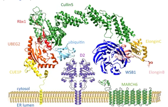
As a unique feature among deiodinases, D2 activity is subjected to complex posttranslational control by the ubiquitin-proteasome system. The mechanism of this regulatory pathway is summarized in section 3.3. The short half-life of D2 protein is caused by ubiquitin-mediated proteasomal degradation (Fig. 10) [118]. The D2 is processed by an E3 ligase complex organized by the Cullin5 E3 ligase backbone and Elongin B and C adaptor proteins. The WSB1 SOCS-box protein binds to Elongin B and C and refers for the recognition of D2 by this complex (ECSWSB1). The ubiquitin is carried by the UBE2J (Ubc6) and UBE2G (Ubc7) E2 enzymes [160, 161]. This machinery is strongly associated to D2 homodimers on the cytosolic surface of ER and allows a precise control of D2 activity on T4-dependent manner therefore the D2 is also targeted by TH control on the posttranslational level (Fig. 12). The substrate induced ubiquitination of D2 represented the first example of this type of regulation of hormonal activity via an ER resident enzyme [118].
Figure 12. Schematic model of ubiquitinating apparatus in close association with D2 in the ER Cullin5: scaffold protein; Rbx1, Elongin B and C: adaptors; WSB1: SOCS-box substrate-binding subunit;
UBEG2 (Ubc7): E2 ubiquitin conjugating enzyme; CUE1P: anchoring and activator protein of UBEG2.
Importantly, this system is capable to downregulate the D2 activity without degradation of the protein which is favorable in case of a selenoprotein that requires an energy-dependent insertion of the rare amino acid selenocysteine. The ubiquitination of D2 protein results a conformational change that abolishes the enzyme activity [162]. The ubiquitinated inactive state of D2 could be reversed by deubiquitinating enzymes via the cleavage of ubiquitin chain resulting regained activity. Previous studies identified the USP33 (VDU1) and USP20 (VDU2) proteins with the capability to deubiquitinate the D2 [163]. The recognition of D2 by ECSWSB1 is facilitated by T4, the
substrate of D2, demonstrating a unique regulation of enzyme activity. Moreover the T4- dependent reversible inactivation of D2 allows a rapid and efficient way to fine-tune the TH activation capacity. Beside the ECSWSB1 complex, recent studies based on yeast two hybrid screen identified the MARCH6 (TEB4, mammalian orthologue of Doa10 in yeast) ERAD ubiquitin ligase as potential E3 enzyme for D2 [164].
As previously mentioned, the ubiquitination is a unique feature of D2 among deiodinases however the background of the difference is not clearly understood. Previous studies identified critical elements involved in the short half-life and ubiquitination of D2 protein. It is also remained to be elucidated how ubiquitin ligases contributes to the regulation of D2. Specific features of D2 ubiquitination has been suggested in the hypothalamus with major impact on TH replacement therapy [82, 165]. Therefore detailed molecular characterization of D2 ubiquitination is essential to better understand the regulation of hypothalamic TH activation. The crucial components of the ubiquitin- proteasome system will be summarized below.
3.3. THE UBIQUITIN-PROTEASOME SYSTEM
3.3.1. Mechanism of ubiquitin-conjugation
The ubiquitin-proteasome system (UPS) was discovered as a complex machinery dedicated to targeted intracellular proteolysis that impacts a wide range of cellular processes e.g. cell division, signal transduction, gene regulation and ER quality control (endoplasmic reticulum-associated degradation, ERAD) [166]. Further studies on the UPS revealed that it is more accurate to distinguish between the ubiquitin tagging and proteasomal degradation since only a subclass of ubiquitinated proteins is degraded by
proteasome. The ubiquitin is an evolutionary conserved small, 8.5 kDa protein containing 76 amino acid residues containing seven lysines and C-terminal glycine-glycine residues.
It is important to note these features serve as the structural basics of the diversity of ubiquitin signal as described below.
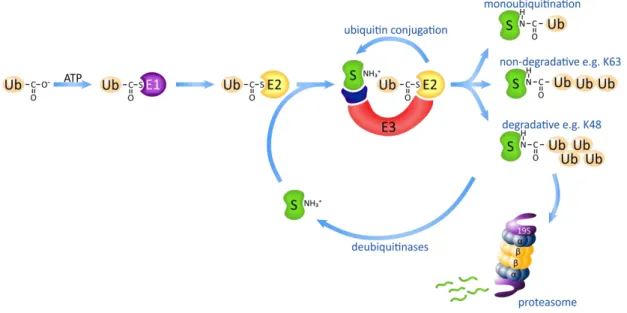
In the first step the ubiquitin-activating enzyme (E1) catalyzes the formation of an ester linkage between adenosine-monophosphate from ATP and the C-terminal glycine of ubiquitin followed by direct conjugation of ubiquitin to a cysteine residue of the E1 enzyme by thioester linkage resulting a high-energy reactive bound (Fig. 13). In the most of the species only a single gene encodes E1 suggesting that the role of the E1 enzyme is restricted to the universal activation step of the ubiquitin moiety and has a limited effect on its further processing or target selection [167].
The action of E1 is followed by the transfer of ubiquitin to a cysteine residue of ubiquitin-conjugating enzyme (E2) via formation of a thioester linkage. In contrast to E1 there are multiple E2 enzymes with specific expression, localization and different mechanisms of action (Fig. 13). The core catalytic domain of E2’s is highly conserved but some of them contain N- or C- terminal extension that affects their localization or interaction with E3 ligases [167]. While the function of E1 enzymes is restricted to supporting the energetic background of ubiquitin conjugation some of E2 sterically determinates or restricts the ubiquitin conjugation process.
Figure 13.Mechanism of ubiquitin conjugation
Ub: ubiquitin; E1: ubiquitin activating enzyme; E2: ubiquitin conjugating enzyme; E3: ubiquitin ligase (complex); S: substrate protein.
The most diverse component of the ubiquitinating apparatus consists of E3 ligases;
there are hundreds of proteins with ubiquitin ligase function. E3 enzymes catalyze the formation of isopeptide-bound between the C-terminal carboxyl-group of ubiquitin moiety and ε-amino-group on one or more lysine residues of target protein, the second often referred as multiubiquitination. Importantly, the E3 ligases have the ability to extend the initial ubiquitin by the addition of further ubiquitin moieties result polyubiquitin chain formation. The quantity and quality of these chains substantially determinate the way of downstream processing of the target protein (Fig. 13).
The E3 ligases can be grouped into three classes based on the mechanism of ubiquitin-transfer and structural properties. HECT-type (homologue to E6-AP carboxyl- terminus) ligases form a covalent bond between one of their cysteine residue and ubiquitin therefore the ubiquitin is not directly attached to the target protein. The other two groups similarly bind the ubiquitin-carrier E2 and transfer ubiquitin directly to the substrate protein. The vast majority of E3 enzymes is RING-type (homologue to the Zn-finger of
“really interesting new gene”) coordinating two Zn2+-ion which are involved in the stabilization of RING-domain. In contrast, the U-box E3 ligases have a structurally similar domain to RING but lack the metal-binding ability.
Most of the RING-finger E3 ligases have modular multiprotein structure splitting the major functions (scaffolding the complex, binding E2 and recognizing substrate) between different protein chains [168]. In case of Cullin-based ligases these functions are carried out separately. The core unit of the complex is the Cullin lacking the ability to bind to ubiquitin-loaded E2 that is carried out by Rbx proteins. Another part of the complex refers for the substrate recognition e.g. F-box and SOCS-proteins which are connected to the Cullin-organized complex via Skp1 or Elongin B and C adaptor proteins [169]. This modular scheme allows the precise combination of target selection with the type of conjugated ubiquitin chain.
The fused ubiquitin or ubiquitin chain determinate the fate of the substrate protein and its degradation in the proteasome represents only one possibility. The exact understanding of ubiquitin code is one of the most important issues related to the UPS function. The single ubiquitin or ubiquitin chain and in the last case the structure of the chain result a major difference in the acceptability for ubiquitin-binding proteins which stands in the structural background of the linkage between ubiquitin signal synthesis and
function. Other theories raised the possibility that the difference is based on molecular dynamic aspects which are determined by the orientation of proximal and distal ubiquitin within the ubiquitin chain [170].
While the soluble proteins in the cytosol could reach the proteasome directly after tagged by degradative ubiquitin signal, the ER membrane proteins have to undergo a complex process to access to the proteasome. This includes the cleavage of degradative ubiquitin signal partial unfolding and transport to the cytosol where new degradative signal is synthetized for these proteins. The core member to organize this machinery is the p97/VCP complex. The importance of this system is to support the accessibility of proteasome for ER-proteins [171].
3.3.2. Cleavage of ubiquitin: the deubiquitinases
The ubiquitin chain synthetizing apparatus allows the selective labelling of proteins resulting precise targeting of their action that is fine-tuned by the reversibility of ubiquitin-mediated modification. This system allows a reversible posttranslational modification of protein function, localization, stability and importantly the recircularization of ubiquitin. The latter is critical within the cell demonstrated by the inhibition of the proteasome (e.g. by MG132) that contains subunits with deubiquitinase activity for cleavage of polyubiquitin chain from substrates. Inhibition of this process leads to ubiquitin “depletion” and interferes with ubiquitin-conjugation [172]. Because de novo ubiquitin synthesis is not able to compensate the inhibition of ubiquitin recycling this condition has severe toxicity for the cell. Importantly, de novo ubiquitin synthesis is also requires the cleavage of ubiquitin because the four genes encode ubiquitin in repeated copies of ubiquitin or ubiquitin is translated in fusion to other proteins and in all of the cases ubiquitin moiety is released after cleavage [173, 174]. Deubiquitination is also involved in the extraction of ER-localized proteins to the cytosol ensuring the unfolding of target proteins which are reubiquitinated after reaching the cytosol.
Deubiquitinases (ubiquitin-specific proteases, USPs) are cysteine- or metalloproteases with the ability to cleave ubiquitin or ubiquitin chain at different sites.
The cleavage site could be determined by the docking surface on deubiquitinases, linkage type of ubiquitin chain and also by the substrate [175]. These capabilities allow a broad
range of action of deubiquitinases including the rescue from degradation, allowing chain- editing or transport.
3.3.3. Importance of ubiquitin-ubiquitin linkage
All of the seven lysine residues of ubiquitin and also the N-terminal amino-group could be involved in the formation of a polyubiquitin chain but only the K48- and K63- linked chains are well characterized. The following section briefly addresses the diversities of protein processing by different ubiquitin linkages.
Despite of lacking evidences on the exact role of K6 ubiquitination this type of ubiquitin chain seems to exert non-degradative functions. K6-linked polyubiquitin chain synthesis is involved in the recognition of DNA damage and the recruitment of repair machinery recognized by Werner helicase interacting protein 1 (WRNIP1) [176, 177].
Cytoskeletal dynamics and remodeling are affected by K6-mediated ubiquitination of α- tubulin [178].
K11-linked ubiquitin chain possesses proteolytic functions and its essential role was demonstrated in endoplasmic reticulum associated degradation (ERAD). In other cases, K11 ubiquitin chain formation interferes with K48-linked ubiquitination rescuing the target protein from degradation [179] or resulting partial proteolysis as in the case of Ci (insect orthologue of mammalian Gli proteins involved in Sonic hedgehog signalization).
K11 ubiquitin chain synthesis is demonstrated in signaling pathways and cell cycle [180, 181] Interestingly, Doa10 – the yeast orthologue of mammalian MARCH6 – is suggested to be involved in K11-linkage formation in combination with Ubc6 E2 enzyme [182].
Importantly, both of them are involved in the regulation of D2.
The K27-linked polyubiquitin chain is predominantly a non-degradative tag, involved in DNA damage response, histone-modification [183]. K27-linked ubiquitin chain is synthesized by Parkin ubiquitin ligase and shown to be involved in the turnover of mitochondria [184].
Recently, only very limited data are available on processes modulated by K29- linked ubiquitin chain exclusively and this type of action is often reported in mixed or not characterized ubiquitin chains. K29-chain is involved in the activation and regulation of the stability of β-catenin transcription factor modulator in Wnt pathway [185, 186].
The function of K33-linked chains is also poorly characterized but it was suggested to work as a non-degradative signal. The K33 signal affects actin assembling and cellular trafficking in trans-Golgi [187] and has a role in the regulation of TCR-ζ phosphorylation [188].
There is no doubt that the K48-linked ubiquitin chain is the most characterized and understood linkage type. While other linkage types also can target proteins for proteasomal degradation, the K48-chain is the major functional linker of ubiquitin conjugation to proteasomal degradation, referred together as the ubiquitin-proteasome system [166, 189].
The K63-linked ubiquitin chain is the canonical example of non-degradative ubiquitin signals. The activation of NF-κB pathway requires K63-linked ubiquitin chain formation involved in IKK activation [190, 191]. This chain type is also involved in the response for double-strand break of DNA by the recruitment of the repair machinery [192]
and play a role in intracellular trafficking and endocytosis [193].
3.3.4. The proteasome
The proteasome is a multicatalytic protease complex with a fundamental role in several cellular functions. The subunits are organized into 4 rings forming a barrel structure. While the potential ancestral prokaryotic proteasome the HslU–HslV complex show hexameric symmetry [194] the eukaryotic proteasomes are common in the formation of heptameric rings [195]. The 20S core proteasome is built up by two outer rings formed by the α-subunits and two inner rings which are constituted by the β- subunits. The α-rings have a gate keeper function – especially α3-subunit is critical in this aspect – and contribute to the conveying of unfolded proteins [196]. The β-rings carry the protease activities: β1 shows caspase-like, β2 trypsin-like and β5 chymotrypsin-like substrate preference. The proteasome has an important role in the process of antigen presentation by MHCI proteins via the generation of short oligopeptides. In case of immunoproteasome, the catalytic subunits are changed to β1i (LMP2), β2i (MECL) and β5i (LMP7) therefore the generated oligopeptides are optimized in the critical amino acid positions for binding into antigen pocket of MHCI [197]. Interestingly, genes encoding
β1i and β5i subunits are located in the MHC locus and deletion of these three immunoproteasome subunits leads to severely altered antigen presentation [198].
The 20S core proteasome carries the catalytic activity however the mature proteasome usually contains additional cap complexes. The most abundant lid structure is the 19S (PA700) subunit incorporating multiple functions. Similarly to 20S complex the 19S regulator has also ring-shape structure and it contains multiple subunits that can be divided into two major subclasses: ATPase (RPT) and non-ATPase (RPN) proteins.
19S complex refers for the recognition and docking of polyubiquitin chain on target protein, unfolds the protein, and involved in the gating mechanism of 20S core proteasome. The complex of 19S and 20S is referred as 26S proteasome.
The 11S (PA28) subunit increases proteasomal activity although it restricts its acceptability to unfolded substrates. It is suggested that the 11S subunit is involved in MCHI antigen presentation by forming hybrid proteasomes with 19S subunit and docking it to the TAP1/2 proteins in the ER.