R E S E A R C H A R T I C L E Open Access
Co-occurrence of thyroid and breast cancer is associated with an increased oncogenic SNP burden
Bence Bakos1*† , András Kiss1†, Kristóf Árvai1†, Balázs Szili1, Barbara Deák-Kocsis1, Bálint Tobiás1, Zsuzsanna Putz1, Richárd Ármós1, Bernadett Balla1, János Kósa1, Magdolna Dank1, Zsuzsanna Valkusz2, István Takács1,
Ádám Tabák1,3,4†and Péter Lakatos1†
Abstract
Background:Epidemiological evidence suggests that synchronous or metachronous presentation of breast and thyroid cancers exceeds that predicted by chance alone. The following potential explanations have been
hypothesized: common environmental or hormonal factors, oncogenic effect of the treatment for the first cancer, closer follow-up of cancer survivors, shared underlying genetic risk factors. While some cases were found to be related to monogenic disorders with autosomal inheritance, the genetic background of most cases of co-occurring breast and thyroid cancer is thought to be polygenic.
Methods:In this retrospective case-control study we compared the genetic profile of patients with a history of breast cancer (n= 15) to patients with co-occurring breast and thyroid cancer (n= 19) using next generation sequencing of 112 hereditary cancer risk genes. Identified variants were categorized based on their known association with breast cancer and oncogenesis in general.
Results:No difference between patients with breast and double cancers was observed in clinical and pathological characteristics or the number of neutral SNPs. The unweighted and weighted number of SNPs with an established or potential association with breast cancer was significantly lower in the group with breast cancer only (mean difference−0.58, BCa 95% CI [−1.09,−0.06],p= 0.029, and mean difference−0.36, BCa 95% CI [−0.70,−0.02],p= 0.039, respectively). The difference was also significant when we compared the number of SNPs with potential or known association with any malignancy (mean difference−1.19, BCa 95% CI [−2.27,−0.11],p= 0.032 for unweighted, and mean difference−0.73, BCa 95% CI [−1.32,−0.14],p= 0.017 for weighted scores).
Conclusion:Our findings are compatible with the hypothesis of genetic predisposition in the co-occurrence of breast and thyroid cancer. Further exploration of the underlying genetic mechanisms may help in the identification of patients with an elevated risk for a second cancer at the diagnosis of the first cancer.
Keywords:Thyroid cancer, Breast cancer, Metachronous cancer, Oncogenesis
© The Author(s). 2021Open AccessThis article is licensed under a Creative Commons Attribution 4.0 International License, which permits use, sharing, adaptation, distribution and reproduction in any medium or format, as long as you give appropriate credit to the original author(s) and the source, provide a link to the Creative Commons licence, and indicate if changes were made. The images or other third party material in this article are included in the article's Creative Commons licence, unless indicated otherwise in a credit line to the material. If material is not included in the article's Creative Commons licence and your intended use is not permitted by statutory regulation or exceeds the permitted use, you will need to obtain permission directly from the copyright holder. To view a copy of this licence, visithttp://creativecommons.org/licenses/by/4.0/.
The Creative Commons Public Domain Dedication waiver (http://creativecommons.org/publicdomain/zero/1.0/) applies to the data made available in this article, unless otherwise stated in a credit line to the data.
* Correspondence:bakos.bence@med.semmelweis-univ.hu
Bence Bakos, András Kiss, Kristóf Árvai, Ádám Tabák and Péter Lakatos participated equally in the study
1Department of Internal Medicine and Oncology, Semmelweis University Faculty of Medicine, 1098 Korányi S. u. 2/a, Budapest, Hungary Full list of author information is available at the end of the article
Background
An increased co-occurrence of breast cancer (BC) and differ- entiated thyroid cancer (TC) compared to chance alone has been found repeatedly in recent decades [1, 2]. Numerous studies have shown a bidirectional association between these cancers: a more pronounced risk of thyroid cancer in breast cancer survivors and a less pronounced increase in the inci- dence of breast cancer after thyroid cancer [2–4].
A number of hypotheses have been suggested to ex- plain these findings [3, 4]. The one, most frequently cited, describes a common (yet unknown) hormonal etiologic factor. This hypothesis is supported by the ob- servation of a strong female predominance of both tumor types. Estrogen has been implicated in the patho- genesis of both breast and, to a lesser degree, thyroid cancer [5]. Furthermore, thyroid dysfunction has also been tentatively linked to carcinogenesis [6–8]. It is also possible that the hormonal milieu of pregnancy (hCG, increased TRH and prolactin) would increase the thyroid and the estrogen-like effects of thyroid hormones, hence the risk of both cancer types [9]. Endocrine disrupting chemicals affecting estrogen receptors have also been theoretically implicated [10].
Another set of hypotheses emphasize carcinogenic ef- fects of previous cancer treatment. Radio- and hormone therapy for breast cancer and I131treatment for thyroid tumors have both been implicated in carcinogenesis [11–13]. It is also possible that the increased surveillance of cancer survivors could lead to earlier recognition or even overdiagnosis of a second primary malignancy.
A shared genetic background of these cancers is also a po- tential explanation for the observed association. Cowden and Cowden-like syndromes are characterized by the presence of hamartomas and an increased risk of, among others, both thyroid and breast cancer. These conditions are mostly monogenic disorders with an autosomal dominant inherit- ance. Mutations in the PTEN, SDHB, SDHD, MTHFR and PARP4 genes, and hypermethylation of KLLN have all been implicated [14–16]. Much less is known about the genetic background of sporadic cases of metachronous or synchron- ous breast and thyroid cancer. However, an increased famil- ial risk of other primary malignancies of affected patients points towards underlying germline mutations [3,17].
In the present paper, we investigated the potential for a shared genetic background of thyroid and breast can- cers. The recognition and elucidation of such genetic risk factors could aid in the identification, follow-up, and potential preventive treatment of high-risk individuals with a first primary malignancy.
Methods
Patients and setting
We analyzed the genetic polymorphisms in 112 heredi- tary cancer risk genes in a case-control study of patients
with a history of either breast or both breast and thyroid cancer.
Patients were selected from a pool of n= 274 thyroid cancer patients who had received I131 treatment at our institution between January 2014 and October 2018.
Twenty-one of these individuals received treatment for breast cancer before or after the diagnosis of a thyroid cancer. Two patients could not have been reached for consent, resulting in a final sample of 19. Given that the risk of thyroid cancer development after breast cancer is larger than vice versa, the control group consisted of 15 subjects who received treatment for breast cancer at our institution at least 12 years before the present study. Pa- tients with a personal or a family history of thyroid can- cer were excluded from this group. These criteria were specified to minimize contamination of the control group with patients with an increased genetic risk for thyroid cancer development. No other inclusion or ex- clusion criteria were set.
We collected the following clinical characteristics from hospital and outpatient records: age at cancer diagnosis, tumor size, histology, staging and grading, presence or absence of lymph node and distant metastases, details of surgical-, radiation- chemothera- peutic-, hormonal and I131 treatment. Cancer diagno- sis for all cases was based on histological data using the WHO classification of breast and endocrine tu- mors (4th and 3/4th edition respectively). Staging was based on the TNM system (7th edition). Informed consent was obtained from all subjects before any study related procedures were performed. The study was approved by Semmelweis University Regional and Institutional Committee of Science and Research Ethics.
Genetic analysis
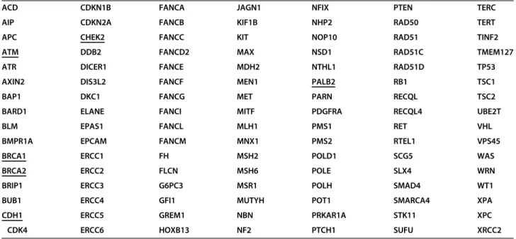
We have aimed to cover all the well-established heredi- tary cancer risk loci. The final list of the 112 investigated genes was compiled based on literature data [18–20], and is shown in Table1.
Exome amplicon library was prepared using the Ion AmpliSeq Library Kit Plus combined with the Ion AmpliSeq Exome RDY kit (ThermoFisher, MA, USA).
Briefly, 100 ng of genomic DNA was added to dried down, ultra-high multiplexed primer pairs (12 pools) in a 96-well plate and amplified with the following PCR conditions: at 99 °C for 2 min; at 99 °C for 15 s and at 60 °C for 16 min (10 cycles) and holding at 10 °C.
Primers were partially digested using a FuPa reagent, and then sequencing adapters and barcodes were ligated to the amplicons. The library was purified using the Agencourt AMPure XP Reagent (Beckmann Coulter, CA, USA). The concentration of the final library was de- termined by Ion Library TaqMan Quantitation Kit
(ThermoFisher, MA, USA) on an ABI 7500 qPCR instru- ment with absolute quantification method.
Template preparation was performed with Ion 540 OT2 Kit (ThermoFisher, MA, USA) on semi-automated Ion OneTouch 2 instrument using emPCR method.
After breaking the emulsion, the non-templated beads were removed from the solution during the semi- automated enrichment process on Ion OneTouch ES (ThermoFisher, MA, USA) machine. Later adding the sequencing primer and polymerase, the fully prepared Ion Sphere Particles (ISPs) were loaded into an Ion 540 chip, and the sequencing runs were performed using the Ion S5 Sequencing kit (ThermoFisher, MA, USA) with 500 flows.
Sequence data from the Ion Torrent run were analyzed using the platform-specific pipeline software Torrent Suite v5.10 for base calling, trim adapter and primer se- quences, filtering out poor quality reads, and de- multiplex the reads according to the barcode sequences.
Briefly, TMAP algorithm was used to align the reads to the hg19 human reference genome, and then, the variant caller plug-in was executed to search for germline vari- ants in the targeted regions. Integrative genomics viewer (IGV) was used for visualization of the mapped reads.
Variants were annotated using the Ion Reporter (Ther- moFisher, MA, USA) software. Variant interpretation was restricted to the selected genes with known connec- tion to oncology diseases. Variants were stratified by gene association with breast cancer and by variant asso- ciation with cancer (no/potential/known) based on avail- able data from the ClinVar database at the time of writing [21]. Synonymous mutations were not excluded from the analysis.
Statistical analysis
Descriptive data are given as means ± standard deviation (SD) for continuous data, and frequencies (percentages) for categorical variables. Between-group differences in clinical parameters were assessed using 2-sample t tests for continuous variables, and χ2 tests or Fisher exact tests for categorical variables.
When comparing the number of different variants across groups, the assumptions of normality and homo- geneity of variances were assessed using the Shapiro–
Wilk test and Levene’s test respectively. Given that in some cases these assumptions were not met, Welch’s t- test was used to compare the number of variants with known, potential or no association with breast cancer and with any cancer. We also used the bootstrap proced- ure to get robust confidence intervals and p-values. To increase statistical power, we lumped together variants with potential and known associations using both weighted and unweighted scores. To generate the weighted scores, we gave double weight for variants with known association compared to those variants with po- tential association. To correct for multiple testing we ap- plied the Benjamini-Hochberg procedure targeting a false discovery rate < 15%. A corrected two-tailedp-value less than 0.05 was considered statistically significant. Ef- fect sizes were computed as adjusted standardized mean differences (Hedge’s g). Analyses were conducted using IBM SPSS Statistics for Windows, Version 23.0 (IBM Corp. Released 2015 Armonk, New York, U.S.A.).
Results
Table2displays patients’clinical characteristics. BC only patients had a slightly younger age at the time of breast Table 1List of cancer risk genes assessed. BC associated genes are underlined
ACD CDKN1B FANCA JAGN1 NFIX PTEN TERC
AIP CDKN2A FANCB KIF1B NHP2 RAD50 TERT
APC CHEK2 FANCC KIT NOP10 RAD51 TINF2
ATM DDB2 FANCD2 MAX NSD1 RAD51C TMEM127
ATR DICER1 FANCE MDH2 NTHL1 RAD51D TP53
AXIN2 DIS3L2 FANCF MEN1 PALB2 RB1 TSC1
BAP1 DKC1 FANCG MET PARN RECQL TSC2
BARD1 ELANE FANCI MITF PDGFRA RECQL4 UBE2T
BLM EPAS1 FANCL MLH1 PMS1 RET VHL
BMPR1A EPCAM FANCM MNX1 PMS2 RTEL1 VPS45
BRCA1 ERCC1 FH MSH2 POLD1 SCG5 WAS
BRCA2 ERCC2 FLCN MSH6 POLE SLX4 WRN
BRIP1 ERCC3 G6PC3 MSR1 POLH SMAD4 WT1
BUB1 ERCC4 GFI1 MUTYH POT1 SMARCA4 XPA
CDH1 ERCC5 GREM1 NBN PRKAR1A STK11 XPC
CDK4 ERCC6 HOXB13 NF2 PTCH1 SUFU XRCC2
cancer diagnosis compared to the synchronous/meta- chronous cancer group (47.7 vs 54.4, p = 0.079). All other clinical parameters evaluated were similar in the BC and BC-TC groups. It is worth noting that no differ- ence was found in the frequency of the different BC treatment modalities (irradiation, hormone or chemotherapy).
The results of the genetic analyses for the BC-TC pa- tients and the control group are presented in Tables 3 and 4 respectively. Details of the statistical analysis are reported in Table5.
We found a significantly lower number of SNPs with potential or known association with breast cancer in the BC only group (mean difference−0.58, BCa 95% CI [−
1.09, −0.06], t (27) =−2.30,p = 0.029, g = 0.74 for un- weighted and mean difference−0.36, BCa 95% CI [−
0.70, −0.02], t(27.26) =−2.17, p = 0.039, g = 0.70 for weighted scores).
The individual number of SNPs with no known gen- etic association with cancer was nominally higher in the BC only group (mean difference 0.46, BCa 95% CI [−
0.27, 1.20], t(25.56) = 1.30, p= 0.206, g = 0.39), while the number of potentially associated SNP or those with a known association was nominally lower in the BC only group (mean difference−0.92, BCa 95% CI [−2.01, 0.17], t(30.98) =−1.72, p= 0.096, g = 0.59 and mean dif- ference−0.27, BCa 95% CI [−0.63, 0.09], t(31.37) =− 1.55,p= 0.131, g = 0.51, respectively).
When we lumped together SNPs with potential or known association with carcinogenesis to increase statis- tical power, we found significantly higher values in the synchronous/metachronous cancer group compared to Table 2Patient characteristics at BC diagnosis and at follow-up
BC only BC and TC p-
value Mean / No of cases SD / % Mean / No of cases SD / %
No. of patients 15 19
Age at follow-up 67.3 9.2 62.3 10.5 0.16
BC clinical characteristics
Age at BC diagnosis 47.7 10.2 54.4 11.2 0.079
BC grade 0.363
1 3 37.5% 3 20% 1
2 1 12.5% 6 40% 0.104
3 4 50% 6 40% 1
BC T stage 0.393
1 4 36.4% 7 58.3% 0.715
2 6 54.5% 5 41.7% 0.475
4 1 9.1% 0 0% 0.441
Lymph node metastasis 5 33.3% 8 42.1% 0.728
Vascular invasion 2 22.2% 2 15.4% 1
HER2-positivity 6 40% 4 21% 0.276
ER-positivity 11 73% 10 52% 0.296
PR-positivity 8 53% 8 42% 0.730
Invasive ductal carcinoma 13 86.7% 16 84.2% 1
Distant metastasis 3 20% 2 10.5% 0.634
BC radiation therapy 14 93.2% 17 89.5% 1
BC chemotherapy 10 66.7% 9 60% 1
BC hormone therapy 11 73.3% 10 62.5% 0.704
BC targeted therapy 1 6.7% 2 12.5% 1
BC relapse 4 26.7% 3 15.8% 0.672
TC clinical characteristics
Age at TC diagnosis – – 53.5 15 –
Papillary histology – – 18 94.% –
Follicular histology – – 1 5.3% –
Lymph node metastasis – – 4 21% –
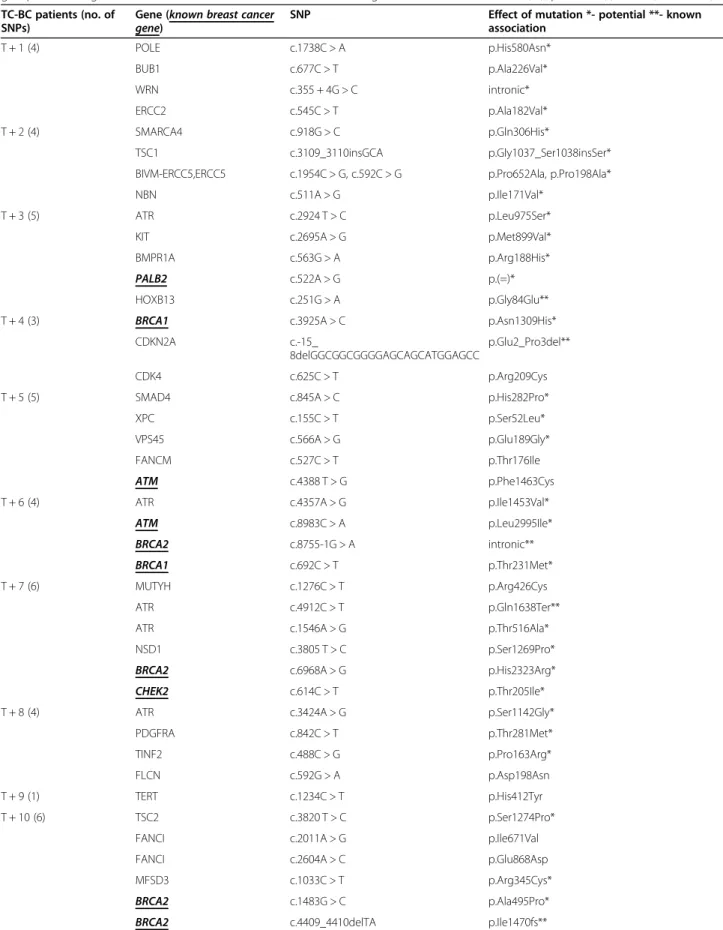
Table 3List of any cancer (and breast cancer) related genes and unique variants identified by individual patients in the BC-TC group. The strength of known association of each variant with oncogenesis is marked as known (**), potential (*) or no (no marker) TC-BC patients (no. of
SNPs)
Gene (known breast cancer
gene) SNP Effect of mutation *- potential **- known
association
T + 1 (4) POLE c.1738C > A p.His580Asn*
BUB1 c.677C > T p.Ala226Val*
WRN c.355 + 4G > C intronic*
ERCC2 c.545C > T p.Ala182Val*
T + 2 (4) SMARCA4 c.918G > C p.Gln306His*
TSC1 c.3109_3110insGCA p.Gly1037_Ser1038insSer*
BIVM-ERCC5,ERCC5 c.1954C > G, c.592C > G p.Pro652Ala, p.Pro198Ala*
NBN c.511A > G p.Ile171Val*
T + 3 (5) ATR c.2924 T > C p.Leu975Ser*
KIT c.2695A > G p.Met899Val*
BMPR1A c.563G > A p.Arg188His*
PALB2 c.522A > G p.(=)*
HOXB13 c.251G > A p.Gly84Glu**
T + 4 (3) BRCA1 c.3925A > C p.Asn1309His*
CDKN2A c.-15_
8delGGCGGCGGGGAGCAGCATGGAGCC
p.Glu2_Pro3del**
CDK4 c.625C > T p.Arg209Cys
T + 5 (5) SMAD4 c.845A > C p.His282Pro*
XPC c.155C > T p.Ser52Leu*
VPS45 c.566A > G p.Glu189Gly*
FANCM c.527C > T p.Thr176Ile
ATM c.4388 T > G p.Phe1463Cys
T + 6 (4) ATR c.4357A > G p.Ile1453Val*
ATM c.8983C > A p.Leu2995Ile*
BRCA2 c.8755-1G > A intronic**
BRCA1 c.692C > T p.Thr231Met*
T + 7 (6) MUTYH c.1276C > T p.Arg426Cys
ATR c.4912C > T p.Gln1638Ter**
ATR c.1546A > G p.Thr516Ala*
NSD1 c.3805 T > C p.Ser1269Pro*
BRCA2 c.6968A > G p.His2323Arg*
CHEK2 c.614C > T p.Thr205Ile*
T + 8 (4) ATR c.3424A > G p.Ser1142Gly*
PDGFRA c.842C > T p.Thr281Met*
TINF2 c.488C > G p.Pro163Arg*
FLCN c.592G > A p.Asp198Asn
T + 9 (1) TERT c.1234C > T p.His412Tyr
T + 10 (6) TSC2 c.3820 T > C p.Ser1274Pro*
FANCI c.2011A > G p.Ile671Val
FANCI c.2604A > C p.Glu868Asp
MFSD3 c.1033C > T p.Arg345Cys*
BRCA2 c.1483G > C p.Ala495Pro*
BRCA2 c.4409_4410delTA p.Ile1470fs**
the BC only group using both unweighted (mean differ- ence−1.19, BCa 95% CI [−2.27, −0.11], t(31.88) =− 2.24, p= 0.032, g = 0.76) and weighted (mean difference
−0.73, BCa 95% CI [−1.32,−0.14], t(31.80) =−2.51,p= 0.017, g = 0.83) scores.
Discussion
In our study, we found a significantly higher burden of established and potential germline cancer risk variants among subjects with a history of metachronous thyroid and breast cancer compared to patients with a history of breast cancer only. The trend was similar and effect sizes were still considerable when we assessed the number of SNPs with known and potential cancer risk separately.
However, in these cases, probably due to lack of power, statistical significance was not reached. Our results are compatible with the hypothesis that the sporadic co- occurrence of thyroid and breast cancer is of
multigenetic origin and probably related to the burden of the carcinogenic SNPs rather than an individual gene alteration.
Comparison of clinical features of breast cancer, such as onset, stage, histology and treatment also yielded no significant differences. There was a non-significant ten- dency for later age of breast cancer onset in the study group which is contrary to previous findings [22].
The metachronous and synchronous occurrence of breast and thyroid cancer has been first described nearly four decades ago [1]. The relationship between the two tumor types has since been found to be bidirectional. In recent meta-analyses, the odds ratio for thyroid cancer following breast cancer treatment was 1.55 while the odds ratio of developing BC after TC was reported be- tween 1.18–1.32 [3, 4]. Studies exploring the cause be- hind this association emphasize either the role of common underlying hormonal and environmental Table 3List of any cancer (and breast cancer) related genes and unique variants identified by individual patients in the BC-TC group. The strength of known association of each variant with oncogenesis is marked as known (**), potential (*) or no (no marker) (Continued)
TC-BC patients (no. of SNPs)
Gene (known breast cancer
gene) SNP Effect of mutation *- potential **- known
association
T + 11 (2) PTCH1 c.4324C > T p.Arg1442Trp
CDH1 c.32 T > C p.Leu11Pro*
T + 12 (5) PMS1 c.2783 T > C p.Leu928Pro*
ATM c.1273G > A p.Ala425Thr*
ATM c.1300C > T p.Pro434Ser*
BRCA2 c.9038C > T p.Thr3013Ile
STK11 c.413A > G p.Glu138Gly*
T + 13 (2) MUTYH c.536A > G p.Tyr179Cys**
SLX4 c.2359G > A p.Glu787Lys
T + 14 (3) FANCG c.634G > A p.Ala212Thr*
CHEK2 c.1556G > T p.Arg519Leu*
KIF1B c.2680G > A p.Val894Met*
T + 15 (2) RECQL c.1360C > T p.Arg454Cys*
CHEK2 c.444 + 1G > A intronic**
T + 16 (4) TSC1 c.2418G > A p.Met806Ile*
POLD1 c.835_837delGAG p.Glu279del*
PDGFRA c.1099G > A p.Val367Met*
DIS3L2 c.1447C > G p.Arg483Gly*
T + 17 (3) RECQL4 c.3062G > A p.Arg1021Gln
SLX4 c.3890G > A p.Gly1297Glu
SLX4 c.179A > C p.Gln60Pro
T + 18 (4) MSH6 c.3226C > T p.Arg1076Cys**
ATM c.7290 T > G p.His2430Gln*
MAX c.25G > T p.Val9Leu*
BRCA1 c.181 T > G p.Cys61Gly**
T + 19 (0) – –
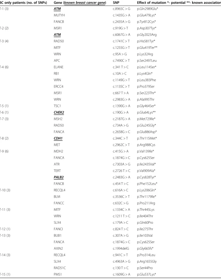
Table 4List of any cancer (and breast cancer) related genes and unique variants identified by individual patients in the control group. The strength of known association of each variant with oncogenesis is marked as known (**), potential (*) or no (no marker) BC only patients (no. of SNPs) Gene (known breast cancer gene) SNP Effect of mutation *- potential **- known association
T-1 (3) ATM c.8965C > G p.Gln2989Glu*
MUTYH c.1435G > A p.Glu479Lys*
FANCB c.2435A > G p.Tyr812Cys*
T-2 (2) MSR1 c.919G > T p.Asp307Tyr*
ATM c.6067G > A p.Gly2023Arg
T-3 (4) RAD50 c.1741C > T p.His581Tyr*
MITF c.1255G > T p.Glu419Ter**
WRN c.95A > G p.Lys32Arg
APC c.7490C > T p.Ser2497Leu
T-4 (6) ELANE c.341 T > C p.Leu114Ser*
RB1 c.10A > C p.Lys4Gln*
WRN c.1149G > T p.Leu383Phe
ERCC4 c.1135C > T p.Pro379Ser
MSR1 c.667 T > A p.Ser223Thr*
WRN c.2983G > A p.Ala995Thr
T-5 (1) TSC1 c.1390G > A p.Gly464Ser*
T-6 (1) CHEK2 c.190G > A p.Glu64Lys**
T-7 (3) MSH2 c.2187G > A p.Met729Ile*
RAD50 c.734A > G p.Glu245Gly*
FANCA c.2658G > C p.Glu886Asp*
T-8 (2) CDH1 c.344C > T p.Thr115Met*
MET c.2962C > T p.Arg988Cys
T-9 (6) MDH2 c.415G > A p.Val139Ile*
FANCA c.1874G > C p.Cys625Ser
ATR c.7303A > G p.Ile2435Val*
TERT c.2726 T > C p.Val909Ala*
PALB2 c.2483G > A p.Cys828Tyr*
FANCB c.454 T > C p.Phe152Leu*
T-10 (3) RECQL4 c.616A > C p.Lys206Gln*
BLM c.3536C > T p.Thr1179Ile*
FANCC c.632C > G p.Pro211Arg
T-11 (3) MITF c.1334C > A p.Thr445Lys
WRN c.1211 T > C p.Ile404Thr
SLX4 c.179A > C p.Gln60Pro
T-12 (1) FANCI c.824 T > C p.Ile275Thr
T-13 (3) BUB1 c.307A > G p.Ile103Val
FANCA c.1874G > C p.Cys625Ser
AXIN2 c.1994delG p.Gly665fs*
T-14 (3) RECQL4 c.941C > T p.Pro314Leu
SLX4 c.4963A > G p.Arg1655Gly
RAD51C c.130 T > C p.Ser44Pro
T-15 (1) PMS1 c.1609G > A p.Glu537Lys*
factors, or the role of the first malignancy in the devel- opment of the second. The potential role of cancer treat- ment and the increased surveillance of cancer survivors fall in this latter category. With breast cancer being the most common type of malignancy in women [23] and with the increasing incidence of thyroid cancer world- wide [24,25], the importance of this topic is clear.
One potential hypothesis explaining this bidirectional relationship implicates common hormonal factors and is rooted in the overwhelming predominance of both can- cer types in reproductive-aged women [4]. Estrogen, progesterone and androgen receptors have been shown to be overexpressed not only in breast cancer but also in thyroid neoplasms [26]. In vitro and animal studies also point towards the potential oncogenic effect of estrogens in thyroid cells [26, 27]. An increased TSH secretion in response to estrogens has also been suggested as a po- tential pathomechanism for thyroid cancer development [3], as serum TSH levels have been shown to correlate with thyroid cancer risk [28]. TSH and thyroid hor- mones also have been implicated in breast carcinogen- esis by in vitro animal and observational studies. T3 seems to stimulate proliferation in breast cancer cells in vitro at least in part through interactions with the es- trogen signalling system [29, 30]. Tumor suppressor pathways downstream of the TRβnuclear thyroid recep- tor and oncogenic pathways downstream of the mem- brane receptorαVβ3 have been suggested to mediate the role of thyroid hormones in carcinogenesis [31–33].
While some observational studies in humans found a de- creased breast cancer incidence in hypothyroid and an increased incidence in hyperthyroid patients [7,8], other studies have failed to replicate these findings [34,35].
Endocrine disrupting chemicals (EDC) have been linked to numerous types of hormone dependent malig- nancies including both thyroid and breast cancer [36, 37]. In addition to EDCs, obesity, a shared risk factor for both tumor types has also been postulated to increase the risk of synchronous/metachronous cancer
development [38,39]. Selective estrogen receptor modu- lators (SERMs) are widely used in breast cancer treat- ment. While there is some evidence that these drugs due to their partial estrogen effect increase TSH secretion and thyroid cell proliferation, their potential role in thy- roid cancer development has not yet been studied [13].
The similar frequency of hormone therapy in the BC only, and the BC-TC groups in our study point toward a limited role of sex hormones in the development of co- occurring breast and thyroid cancer.
Other theories suggest the role of prior cancer treat- ment in the development of the second primary malig- nancy. Despite several improvements minimizing radiation scatter to surrounding tissues, there is a defin- itely increased risk for certain malignancies following ex- ternal beam radiation for breast cancer [40]. While some reports are conflicting [41], most available data do not substantiate such a connection between thyroid cancer and previous adjuvant breast irradiation [42–45]. Simi- larly, radioactive iodine treatment given for thyroid can- cer does not seem to play a role in subsequent breast cancer development [46–48]. Our data with similar fre- quency of radiation therapy for breast cancer in the in- vestigated groups also argue against the role of external or internal radiation in the development of synchron- ous/metachronous breast and thyroid cancer.
The increased co-occurrence of these tumor types could also be related to surveillance bias. The higher compliance with, and higher rate of screening efforts among cancer survivors could also affect the time of diagnosis and could lead the substantial overdiagnosis of clinically irrelevant second primary malignancy [49].
This seems to be especially true for differentiated thy- roid cancer [50]. However, as there are no screening programs for breast cancer among thyroid cancer survi- vors or vice versa, surveillance bias is unlikely to account for the preferential association of these tumor types.
Cowden syndrome (CS) and Cowden-like syndromes (CLS) are characterised by hamartomas and an Table 5Comparison of the two patient groups by the number of genetic variants
BC only BC and TC p-
value Mean Std. error Mean Std. error Genes associated with BC
No. of SNPs with potential/ known association with BC 0.27 0.12 0.84 0.22 0.029
No. of SNPs with potential/ known association with BC(weighted scores) 0.17 0.08 0.53 0.15 0.039 112 cancer risk genes
No. of SNPs with no known association with oncogenesis 1.20 0.30 0.74 0.20 0.206
No. of SNPs with a potential association with oncogenesis 1.40 0.39 2.32 0.37 0.096
No. of SNPs with a known association with oncogenesis 0.20 0.11 0.47 0.14 0.131
No. of SNPs with potential/ known association with oncogenesis 1.60 0.36 2.79 0.39 0.032
No. of SNPs with potential and known association with oncogenesis(weighted scores) 0.90 0.18 1.63 0.23 0.017
extremely increased risk for several types of malignan- cies including breast cancer, thyroid cancer, endometrial cancer, colorectal cancer, and melanoma with standard- ized incidence ratios for breast and thyroid cancer in the range of 6 to 9 [14]. CS and most forms of CLS have an autosomal dominant inheritance. However, these condi- tions, referred to as PTEN hamartoma tumor syn- dromes, make up only a small fraction of synchronous/
metachronous thyroid and breast cancer cases. This is consistent with our finding that no patient in our sam- ples had a mutation in the PTEN gene. Thus, the exist- ence of other shared genetic risk factors is highly probable. To the best of our knowledge, genetic analyses exploring this notion have not been conducted and gen- etic factors underlying sporadic metachronous cancer cases are yet to be elucidated.
The main strengths of this study were the state-of-the- art genetic analysis using NGS technology, and the fact that both cases and controls came from the same source population. The main limitation of our research was the relatively low number of patients limiting statistical power even in analyses of genetic scores. Furthermore, our study was unable to identify or investigate any single genetic risk locus behind synchronous/metachronous cancer development. Two types of bias could also have affected our findings. First, participants were recruited years after diagnosis potentially leading to survivor bias.
Second, the exclusion of controls with less than 12 years of follow-up lead to an age difference between our study groups that is contrary to previous findings [22], and probably reflects selection bias. However, both of these selections were deemed to be necessary to minimize contamination of the control group with thyroid cancer.
Conclusions
In conclusion, we reported an increased burden of car- cinogenic SNPs in people with both thyroid and breast cancer compared to individuals with breast cancer only, based on whole exome sequencing of 112 known heredi- tary cancer risk genes. We found no differences in clini- copathologic parameters between the groups suggesting that both groups have similar presentation at the time of diagnosis of breast cancer. While our study was not powered to identify specific risk loci for metachronous cancer development, our findings further support the multigenetic etiology of co-occurring breast and thyroid cancer. Our findings do not directly contradict any of the other, previously detailed theories explaining the as- sociation between these tumor types. Nevertheless, our results do underline the need for further genetic re- search in this field, which are lacking at the moment.
Abbreviations
BC:Breast cancer; BCa 95% CI: Bias-corrected and accelerated 95% bootstrap confidence interval; CLS: Cowden-like syndromes; CS: Cowden syndrome;
EDC: Endocrine disrupting chemical; hCG: Human chorionic gonadotropin;
IGV: Integrative genomics viewer; ISP: Ion sphere particles; SD: Standard deviation; SERM: Selective estrogen receptor modulator; TC: Thyroid cancer;
TRH: Thyrotropin-releasing hormone
Acknowledgements Not applicable.
Authors’contributions
BB1, AK, PL, and IT conceived of the presented idea. All authors were involved in planning and supervising the work. BB1, BB2, AK, PL, MD, ZsV supervised sample and data collection. Genetic data collection, analysis and interpretation were carried out by KÁ, BB2, BT, and JK. Statistical analysis was done by ÁT, BSz, BDK, and RÁ. Manuscript was drafted by BB1, AK, PL, ZsP, and RA, and was substantively revised by MD and ZsV. All authors discussed the results and contributed to the final manuscript. All authors read and approved the final manuscript.
Funding
The research was financed by the FIKP and the Thematic Excellence Program (Tématerületi Kiválósági Program, 2020–4.1.1.-TKP2020) of the Ministry for Innovation and Technology in Hungary, within the framework of the Molecular Biology thematic program of Semmelweis University. The funding body had no role in the design of the study and collection, analysis, and interpretation of data and in writing the manuscript.
Availability of data and materials
All data generated or analysed during this study are included in this published article.
Declarations
Ethics approval and consent to participate
This research was approved and licenced by the Semmelweis University Regional and Institutional Committee of Science and Research Ethics, and was conducted in accordance with the World Medical Association’s Declaration of Helsinki. Written informed consent was obtained from all participants before entering the study and prior to any study related procedures. This consent extended to the study team accessing their relevant medical history and genetic data. All participants were 18 years or older at the time of the study.
Consent for publication Not applicable.
Competing interests
The authors state that they have no competing interests.
Author details
1Department of Internal Medicine and Oncology, Semmelweis University Faculty of Medicine, 1098 Korányi S. u. 2/a, Budapest, Hungary.2First Department of Medicine, University of Szeged Faculty of Medicine, Szeged, Hungary.3Department of Epidemiology and Public Health, University College London, London, UK.4Department of Public Health, Semmelweis University Faculty of Medicine, Budapest, Hungary.
Received: 25 October 2020 Accepted: 18 May 2021
References
1. McTiernan A, Weiss NS, Daling JR. Incidence of thyroid cancer in women in relation to known or suspected risk factors for breast cancer. Cancer Res.
1987;47(1):292–5.
2. An JH, Hwangbo Y, Ahn HY, Keam B, Lee KE, Han W, et al. A possible association between thyroid Cancer and breast Cancer. Thyroid. 2015;25(12):
1330–8.https://doi.org/10.1089/thy.2014.0561.
3. Nielsen SM, White MG, Hong S, Aschebrook-Kilfoy B, Kaplan EL, Angelos P, et al. The breast-thyroid Cancer link: a systematic review and meta-analysis.
Cancer Epidemiol Biomark Prev. 2016;25(2):231–8.https://doi.org/10.1158/1 055-9965.EPI-15-0833.
4. Joseph KR, Edirimanne S, Eslick GD. The association between breast cancer and thyroid cancer: a meta-analysis. Breast Cancer Res Treat. 2015;152(1):
173–81.https://doi.org/10.1007/s10549-015-3456-6.
5. Moleti M, Sturniolo G, Di Mauro M, Russo M, Vermiglio F. Female Reproductive Factors and Differentiated Thyroid Cancer. Front Endocrinol (Lausanne). 2017;8:111.
6. Hardefeldt PJ, Eslick GD, Edirimanne S. Benign thyroid disease is associated with breast cancer: a meta-analysis. Breast Cancer Res Treat. 2012;133(3):
1169–77.https://doi.org/10.1007/s10549-012-2019-3.
7. Cristofanilli M, Yamamura Y, Kau SW, Bevers T, Strom S, Patangan M, et al.
Thyroid hormone and breast carcinoma. Primary hypothyroidism is associated with a reduced incidence of primary breast carcinoma. Cancer.
2005;103(6):1122–8.https://doi.org/10.1002/cncr.20881.
8. Shi XZ, Jin X, Xu P, Shen HM. Relationship between breast cancer and levels of serum thyroid hormones and antibodies: a meta-analysis. Asian Pac J Cancer Prev. 2014;15(16):6643–7.https://doi.org/10.7314/APJCP.2014.15.16.
6643.
9. Kurata A. Differentiated thyroid cancer: why does it affect predominantly women during the reproductive period and have higher incidence of mutual association with breast cancer? Med Hypotheses. 2019;122:5–7.
https://doi.org/10.1016/j.mehy.2018.10.008.
10. Hoffman K, Lorenzo A, Butt CM, Hammel SC, Henderson BB, Roman SA, et al. Exposure to flame retardant chemicals and occurrence and severity of papillary thyroid cancer: a case-control study. Environ Int. 2017;107:235–42.
https://doi.org/10.1016/j.envint.2017.06.021.
11. Tunio MA, Al Asiri M, Bayoumi Y, Stanciu LG, Al Johani N, Al Saeed EF. Is thyroid gland an organ at risk in breast cancer patients treated with locoregional radiotherapy? Results of a pilot study. J Cancer Res Ther. 2015;
11(4):684–9.https://doi.org/10.4103/0973-1482.167613.
12. Zhang Y, Liang J, Li H, Cong H, Lin Y. Risk of second primary breast cancer after radioactive iodine treatment in thyroid cancer: a systematic review and meta-analysis. Nucl Med Commun. 2016;37(2):110–5.https://doi.org/10.1 097/MNM.0000000000000419.
13. Zidan J, Rubenstein W. Effect of adjuvant tamoxifen therapy on thyroid function in postmenopausal women with breast cancer. Oncology. 1999;
56(1):43–5.https://doi.org/10.1159/000011928.
14. Ngeow J, Stanuch K, Mester JL, Barnholtz-Sloan JS, Eng C. Second malignant neoplasms in patients with Cowden syndrome with underlying germline PTEN mutations. J Clin Oncol. 2014;32(17):1818–24.https://doi.org/10.1200/
JCO.2013.53.6656.
15. Ni Y, He X, Chen J, Moline J, Mester J, Orloff MS, et al. Germline SDHx variants modify breast and thyroid cancer risks in Cowden and Cowden-like syndrome via FAD/NAD-dependant destabilization of p53. Hum Mol Genet.
2012;21(2):300–10.https://doi.org/10.1093/hmg/ddr459.
16. Bolf EL, Sprague BL, Carr FE. A linkage between thyroid and breast Cancer: a common etiology? Cancer Epidemiol Biomark Prev. 2019;28(4):643–9.
https://doi.org/10.1158/1055-9965.EPI-18-0877.
17. Goldgar DE, Easton DF, Cannon-Albright LA, Skolnick MH. Systematic population-based assessment of cancer risk in first-degree relatives of cancer probands. J Natl Cancer Inst. 1994;86(21):1600–8.https://doi.org/10.1 093/jnci/86.21.1600.
18. Bailey MH, Tokheim C, Porta-Pardo E, Sengupta S, Bertrand D, Weerasinghe A, et al. Comprehensive characterization of Cancer driver genes and mutations. Cell. 2018;174(4):1034–5.https://doi.org/10.1016/j.cell.2018.07.034.
19. Foulkes WD. Inherited susceptibility to common cancers. N Engl J Med.
2008;359(20):2143–53.https://doi.org/10.1056/NEJMra0802968.
20. Shah PD, Nathanson KL. Application of panel-based tests for inherited risk of Cancer. Annu Rev Genomics Hum Genet. 2017;18(1):201–27.https://doi.
org/10.1146/annurev-genom-091416-035305.
21. Landrum MJ, Lee JM, Benson M, Brown G, Chao C, Chitipiralla S, et al.
ClinVar: public archive of interpretations of clinically relevant variants.
Nucleic Acids Res. 2016;44(D1):D862–8.https://doi.org/10.1093/nar/gkv1222.
22. Kuo JH, Chabot JA, Lee JA. Breast cancer in thyroid cancer survivors: An analysis of the surveillance, epidemiology, and end Results-9 database.
Surgery. 2016;159(1):23–9.https://doi.org/10.1016/j.surg.2015.10.009.
23. Siegel RL, Miller KD, Jemal A. Cancer statistics, 2017. CA Cancer J Clin. 2017;
67(1):7–30.https://doi.org/10.3322/caac.21387.
24. Kilfoy BA, Zheng T, Holford TR, Han X, Ward MH, Sjodin A, et al.
International patterns and trends in thyroid cancer incidence, 1973-2002.
Cancer Causes Control. 2009;20(5):525–31.https://doi.org/10.1007/s10552- 008-9260-4.
25. Davies L, Welch HG. Current thyroid cancer trends in the United States.
JAMA Otolaryngol Head Neck Surg. 2014;140(4):317–22.https://doi.org/10.1 001/jamaoto.2014.1.
26. Yane K, Kitahori Y, Konishi N, Okaichi K, Ohnishi T, Miyahara H, et al.
Expression of the estrogen receptor in human thyroid neoplasms. Cancer Lett. 1994;84(1):59–66.https://doi.org/10.1016/0304-3835(94)90358-1.
27. Rajoria S, Suriano R, Shanmugam A, Wilson YL, Schantz SP, Geliebter J, et al.
Metastatic phenotype is regulated by estrogen in thyroid cells. Thyroid.
2010;20(1):33–41.https://doi.org/10.1089/thy.2009.0296.
28. Haymart MR, Repplinger DJ, Leverson GE, Elson DF, Sippel RS, Jaume JC, et al. Higher serum thyroid stimulating hormone level in thyroid nodule patients is associated with greater risks of differentiated thyroid cancer and advanced tumor stage. J Clin Endocrinol Metab. 2008;93(3):809–14.https://
doi.org/10.1210/jc.2007-2215.
29. Hall LC, Salazar EP, Kane SR, Liu N. Effects of thyroid hormones on human breast cancer cell proliferation. J Steroid Biochem Mol Biol. 2008;109(1–2):
57–66.https://doi.org/10.1016/j.jsbmb.2007.12.008.
30. Silva JM, Dominguez G, Gonzalez-Sancho JM, Garcia JM, Silva J, Garcia- Andrade C, et al. Expression of thyroid hormone receptor/erbA genes is altered in human breast cancer. Oncogene. 2002;21(27):4307–16.https://doi.
org/10.1038/sj.onc.1205534.
31. Kim WG, Cheng SY. Thyroid hormone receptors and cancer. Biochim Biophys Acta. 2013;1830(7):3928–36.https://doi.org/10.1016/j.bbagen.2012.
04.002.
32. Martinez-Iglesias O, Garcia-Silva S, Tenbaum SP, Regadera J, Larcher F, Paramio JM, et al. Thyroid hormone receptor beta1 acts as a potent suppressor of tumor invasiveness and metastasis. Cancer Res. 2009;69(2):
501–9.https://doi.org/10.1158/0008-5472.CAN-08-2198.
33. Carr FE, Tai PW, Barnum MS, Gillis NE, Evans KG, Taber TH, et al. Thyroid hormone receptor-beta (TRbeta) mediates runt-related transcription factor 2 (Runx2) expression in thyroid Cancer cells: a novel signaling pathway in thyroid Cancer. Endocrinology. 2016;157(8):3278–92.https://doi.org/10.1210/
en.2015-2046.
34. Muller I, Kilburn LS, Taylor PN, Barrett-Lee PJ, Bliss JM, Ellis P, et al. TPOAb and thyroid function are not associated with breast Cancer outcome:
evidence from a large-scale study using data from the Taxotere as adjuvant chemotherapy trial (TACT, CRUK01/001). Eur Thyroid J. 2017;6(4):197–207.
https://doi.org/10.1159/000460246.
35. Fang Y, Yao L, Sun J, Yang R, Chen Y, Tian J, et al. Does thyroid dysfunction increase the risk of breast cancer? A systematic review and meta-analysis. J Endocrinol Investig. 2017;40(10):1035–47.https://doi.org/10.1007/s40618-01 7-0679-x.
36. Gore AC, Chappell VA, Fenton SE, Flaws JA, Nadal A, Prins GS, et al. EDC-2:
the Endocrine Society's second scientific statement on endocrine-disrupting chemicals. Endocr Rev. 2015;36(6):E1–E150.https://doi.org/10.1210/er.2015-1 010.
37. Soto AM, Vandenberg LN, Maffini MV, Sonnenschein C. Does breast cancer start in the womb? Basic Clin Pharmacol Toxicol. 2008;102(2):125–33.
https://doi.org/10.1111/j.1742-7843.2007.00165.x.
38. Harvie M, Howell A, Evans DG. Can diet and lifestyle prevent breast cancer:
what is the evidence? Am Soc Clin Oncol Educ Book. 2015;(35):e66–73.
https://doi.org/10.14694/EdBook_AM.2015.35.e66.
39. Ma J, Huang M, Wang L, Ye W, Tong Y, Wang H. Obesity and risk of thyroid cancer: evidence from a meta-analysis of 21 observational studies. Med Sci Monit. 2015;21:283–91.https://doi.org/10.12659/MSM.892035.
40. Matesich SM, Shapiro CL. Second cancers after breast cancer treatment.
Semin Oncol. 2003;30(6):740–8.https://doi.org/10.1053/j.seminoncol.2003.08.
022.
41. Grantzau T, Overgaard J. Risk of second non-breast cancer among patients treated with and without postoperative radiotherapy for primary breast cancer: a systematic review and meta-analysis of population-based studies including 522,739 patients. Radiother Oncol. 2016;121(3):402–13.https://doi.
org/10.1016/j.radonc.2016.08.017.
42. Li CI, Rossing MA, Voigt LF, Daling JR. Multiple primary breast and thyroid cancers: role of age at diagnosis and cancer treatments (United States).
Cancer Causes Control. 2000;11(9):805–11.https://doi.org/10.1023/A:1008942 616092.
43. Huang J, Walker R, Groome PG, Shelley W, Mackillop WJ. Risk of thyroid carcinoma in a female population after radiotherapy for breast carcinoma.
Cancer. 2001;92(6):1411–8.https://doi.org/10.1002/1097-0142(20010915)92:
6<1411::AID-CNCR1464>3.0.CO;2-9.
44. Lal G, Groff M, Howe JR, Weigel RJ, Sugg SL, Lynch CF. Risk of subsequent primary thyroid cancer after another malignancy: latency trends in a population-based study. Ann Surg Oncol. 2012;19(6):1887–96.https://doi.
org/10.1245/s10434-011-2193-2.
45. Kim SS, Kim SJ, Bae YT, Lee JY, Kim BH, Kim YK, et al. Factors associated with the development of new onset diffuse thyroid F18-fluorodeoxyglucose uptake after treatment of breast cancer in patients without a history of thyroid disease or thyroid dysfunction. Thyroid. 2012;22(1):53–8.https://doi.
org/10.1089/thy.2011.0013.
46. Brown AP, Chen J, Hitchcock YJ, Szabo A, Shrieve DC, Tward JD. The risk of second primary malignancies up to three decades after the treatment of differentiated thyroid cancer. J Clin Endocrinol Metab. 2008;93(2):504–15.
https://doi.org/10.1210/jc.2007-1154.
47. Sawka AM, Thabane L, Parlea L, Ibrahim-Zada I, Tsang RW, Brierley JD, et al.
Second primary malignancy risk after radioactive iodine treatment for thyroid cancer: a systematic review and meta-analysis. Thyroid. 2009;19(5):
451–7.https://doi.org/10.1089/thy.2008.0392.
48. Verkooijen RB, Smit JW, Romijn JA, Stokkel MP. The incidence of second primary tumors in thyroid cancer patients is increased, but not related to treatment of thyroid cancer. Eur J Endocrinol. 2006;155(6):801–6.https://doi.
org/10.1530/eje.1.02300.
49. Ito Y, Miyauchi A, Oda H. Low-risk papillary microcarcinoma of the thyroid: a review of active surveillance trials. Eur J Surg Oncol. 2018;44(3):307–15.
https://doi.org/10.1016/j.ejso.2017.03.004.
50. Ahn HS, Kim HJ, Welch HG. Korea's thyroid-cancer "epidemic"--screening and overdiagnosis. N Engl J Med. 2014;371(19):1765–7.https://doi.org/10.1 056/NEJMp1409841.
Publisher’s Note
Springer Nature remains neutral with regard to jurisdictional claims in published maps and institutional affiliations.