Gut inflammation: current update on
pathophysiology, molecular mechanism and pharmacological treatment modalities
.Gyires K., Tóth V.E., Zádori Z.S.
Department of Pharmacology and Pharmacotherapy, Faculty of Medicine, Semmelweis University, Nagyvárad tér 4., 1089 Budapest, Hungary
Corresponding author: Klára Gyires, Department of Pharmacology and Pharmacotherapy, Semmelweis University, Nagyvárad tér 4., 1089, Budapest, Hungary.
Telephone: 36-1-210-4416 FAX: 36-1-210-4412 e-mail: gyirkla@net.sote.hu
Abstract
Inflammatory bowel disease (IBD) is a chronic and relapsing inflammatory condition of the gastrointestinal tract. The two main forms of IBD are Crohn's disease and ulcerative colitis. According to the recent concept the disease is caused by a combination of factors, including genetics, immune dysregulation, barrier dysfunction and the change in microbial flora. Environmental factors, such as changes in diet, antibiotic use, smoking or improved domestic hygiene (e.g. eradication of intestinal helminths) probably contribute to the development and increased prevalence of IBD. Dysregulation of mucosal immunity in IBD causes an overproduction of inflammatory cytokines resulted in uncontrolled intestinal inflammation. Based on extensive research over the last decade, besides the conventional therapy, there are several novel pathways and specific targets, on which focus new therapeutics. New therapeutics aim 1./ to correct genetic susceptibility by stimulating NOD2 expression, TLR3 signaling or inhibition of TLR4 pathway, 2./ to restore the immune dysregulation by inhibition of pro-inflammatory cytokines (TNF-α, IL-6, IL-13, IL-17, IL-18, IL-21), Th1 polarisation (IL-2, IL-12, IL-23, IFN-γ), T-cell activation, leukocyte adhesion, as well as by immunostimulation (GM-CSF, G-CSF) and anti-inflammatory cytokines (IL-10, IL-11, IFN-β-1a), 3./ to restore mucosal barrier function and stimulate mucosal healing by different growth factors, such as GH, EGF, KGF, TGF-β, VEGF, 4./ to restore the normal bacterial flora by antibiotics, probiotics. However, in spite of these numerous potential targets, the true value and clinical significance of most of the new biologics and molecules are not clear yet.
1. Introduction
Inflammatory bowel disease (IBD), a chronic and relapsing inflammatory condition of the gastrointestinal (GI) tract.The two main forms of IBD are Crohn's disease (CD) and ulcerative colitis (UC), though other forms are also known, which are also classified as not typical IBD (e.g. collagenous colitis, lymphocytic colitis, ischaemic colitis, diversion colitis, Behçet's disease, indeterminate colitis).
The cause of IBD is not exactly known. The recent consensus is that IBDs are initiated and perpetuated by an impaired immune response against the gut microbiota in genetically susceptible individuals [1, 2] and the disease is caused by a combination of factors, including genetics, immune dysregulation, barrier dysfunction, change in microbial flora and environmental influences (see reviews [3-7]).
Though UC and CD share some common clinical symptoms, the two diseases possess very distinct features. First of all, the location of the inflammation is different; CD can develop at any part of the intestine, though most of the cases are localized at the terminal ileum. In contrast, in UC the inflammatory process is restricted to the colon and the rectum.
Moreover, the pathological changes in CD affect the whole bowel wall and manifested as transmural lesions, while in UC the inflammation is restricted to the mucosa (epithelial lining of the gut). Also differences in immunological response of intestinal mucosa have been described. CD is associated with the activation of types 1 and 17 T-helper (Th) cells in response to interleukin (IL)-12, IL-18, IL-23 and transforming growth factor β (TGF-β), and activation of these cells results in increased secretion of the pro-inflammatory cytokines IL-2, IL-17, interferon (IFN)-γ and TNF-α [8, 9]. In patients with UC the mucosal inflammation of the colon is mainly associated with a Th2 cell activation mediated by IL-4, IL-5 and IL-13 that results in an increased level of IL-13 [10, 11]. However, in both cases T-cells are also activated by direct contact with antigens [12].
As regards the clinical symptoms, body weight loss and fever more common in CD.
Ulcerations, granulomas, and bowel fistulas are characteristic for CD, in contrast, UC affects the mucosa in a continuous manner. Moreover, smoking was found to be protective against UC and might improve its course, but seems to increase the risk of developing CD and worsens its course [13].
On the other hand, extra-intestinal manifestations (liver problems, arthritis, skin manifestations and eye problems) can develop in both CD and UC. Some patients have an extra-intestinal manifestation as their first symptom of the disease, while they still have only
mild gastrointestinal manifestation, or none at all. Anemia is the most prevalent extraintestinal complication of both IBDs [14, 15].
The chronic inflammation of the gut causes wide-ranging clinical symptoms in both forms, like nausea, diarrhea (which is often porridge-like in CD, while mucus-like with blood in UC) or constipation [16, 17].
UC or CD patients have increased risk for colorectal carcinoma (CRC). Patients with UC and Crohn’s ileocolitis have an elevated risk of developing colon cancer, while patients with CD and enteritis have an elevated risk of developing small-bowel cancer [18, 19]. The cumulative risk for developing colorectal cancer was 8% at 22 years from onset of symptoms for Crohn’s colitis and 7% at 20 years from onset of symptoms for UC, as it accounts for one in six of all deaths in IBD patients [20].
The high incidence and prevalence of IBD (worldwide incidence of UC and CD varies between 0.5-24.5 and 0.1-16 individuals per 100.000 inhabitants, respectively) [21], and the costs of the long-term and only symptomatic treatment of the patients place a significant burden on the healthcare system: the expenses exceed 1.7 billion dollars per year in the United States [22], and are in similar range in European countries.
Although in the last decade our knowledge about the pathomechanism of IBDs greatly expanded (which is also clearly demonstrated by the continuously rising number of publications in this field), and several important milestones have been achieved, distinction between causing events and secondary consequences is still challenging.
The aim of this review is to shortly summarize the current knowledge and newest findings in the pathomechanism of IBD as well as to overview some of the therapeutic targets and strategies (convential and novel ones) for the treatment of IBD.
2. Patomechanism
2.1. Genetic susceptibility
The familial aggregation of IBD has already been observed several decades ago and studies conducted on twins also confirmed the importance of hereditary factors in the pathogenesis of IBD (especially for CD), though they also highlighted the role of environmental trigger factors [23-27].
Genome wide association studies (GWASs) performed during recent years provided better insight into the genetic background of IBD. They revealed 163 genomic susceptibility
loci associated with IBD so far, 110 with both disease phenotypes, and further 30 and 23 associated selectively either with CD or UC, respectively [28, 29]. The considerable overlap of susceptibility loci in CD and UC indicates that these two IBD phenotypes share several common factors in their pathogenesis.
Although the exact functional role of several IBD susceptibility genes still remains to be established, many of them are associated with host immune functions, including both innate and adaptive immunity.
The first susceptibility gene identified for CD was NOD2/CARD15, which brought the role of innate immunity and pattern recognition receptors (PRRs) in the pathogenesis of IBD to the fore. As described in the next section, PRRs play an essential role in the host microbial interaction by sensing conserved microbial structures (pathogen-associated molecular patterns, PAMPs). Binding of PAMPs results in the activation of multiple signaling pathways including nuclear factor-κB (NF-κB) and mitogen activated protein kinases (MAPKs), which in turn induce the production of inflammatory mediators and also initiate multiple cellular processes, including cell proliferation and differentiation [30-32]. NOD2, a member of the nucleotide-binding oligomerization domain (NOD)-like receptor (NLR) family, recognizes muramyl dipeptide (MDP), a component of peptidoglycan (PGN) in nearly all bacteria [30].
The three most common NOD2 mutations, a frame-shift insertion mutation (3020insC) and two missense mutations (R702W and G908R) result in impaired recognition of MDP and in loss of NF-κB activation in response to lipopolysaccharide (LPS) and PGN [33-36]. Several groups confirmed the association of NOD2 with CD, but interestingly, no such connection was found in Japanese individuals [37]. This is in line with other findings (see below) indicating that genetic determinants can differ significantly between populations.
In the last 2 decades several other PRR genes have been associated with IBD. A british group reported that a complex insertion/deletion polymorphism in NOD1/CARD4 (+32656) may contribute to the development of IBD and can result earlier onset and extra-intestinal manifestations [38], but other groups could not reproduce these findings in German [39], Scottish and Swedish patients [40].
Single nucleotide polymorphisms (SNPs) in Toll-like receptor (TLR) TLR1 and TLR2 genes (R80T and R753Q) were found to increase the risk of pancolitis in UC, but did not increase the susceptibility to disease development [41].
Several studies have been conducted to identify the role of the TLR4 gene in the pathogenesis of IBD, but similarly to NOD2, substantial heterogeneity was found between populations. The D299G SNP was associated with IBD in Belgian [42], German [43, 44],
Greek [45] or Australian [46], but not in Southern Italian [47], Hungarian [48] or New Zealand patients [49, 50]. Nevertheless, meta-analyses have provided evidences for an association between D299G and IBD [49, 50]. Similar discrepancies were observed with the T399I SNP, because significantly increased allele and carrier frequencies for this mutation were observed in patients with UC in a German cohort [44], while other groups could not demonstrate such association [45, 47, 49, 50].
The importance of PRR mutations in the development of IBD is further supported by the findings that a TLR9 polymorphism (-1237T/C) was significantly higher in patients with CD [50], while polymorphism in the CARD9 gene (rs10870077), which encodes an adaptor molecule of PRR signaling, was associated with both CD and UC [51, 52].
Beside PRR genes GWASs have identified several other susceptibility genes, which has led to better understanding of the pathomechanism. The identification of IBD associated polymorphisms in autophagy-related 16-like 1 (ATG16L1) and immunity-related GTPase family M protein (IRGM) genes has revealed that impaired autophagy, and the consequent defects in innate immune responses to intracellular pathogens may be critical components of the chronic inflammation in IBD [53-56]. IBD associated alterations in X-box-binding protein 1 (XBP1) and orosomucoid-like 3 (ORMDL3) genes imply that changes in the unfolded protein response (UPR) and the failure to manage endoplasmic reticulum stress may also contribute to the pathogenesis, for example due to increased apoptosis of Paneth cells [57, 58]. Mutations in the mucin genes (e.g. MUC1, MUC19) or in the prostaglandin receptor EP4 gene (PTGER4) can lead to impaired mucosal barrier functions [51, 59], while genetic variations in the IL18RAP [52], IL23R [60, 61], STAT3 [62] or SMAD3 [51] genes highlight the importance of failures in the adaptive immune responses in IBD.
2.2. Immune dysregulation
The intestinal mucosa is continuously exposed to a vast number of antigens (both dietary and microbial), which are recognized by the mucosal immune system. Under normal circumstances it distinguishes between beneficial and pathogenic microbes - it tolerates normal commensal bacteria, while eliminates invading pathogens. Today it is widely accepted that abnormal immune regulation is a key factor in the pathomechanism of IBD and alterations in both innate and adaptive immunity have been observed. This section shortly overviews the key players of the immune system and their contribution to IBD. For more comprehensive recent immunological reviews see e.g. [63-66].
The pattern recognition receptors (PPRs) play a key role in the bacteria-host interaction.
These innate immune receptors are expressed by different cells of the intestinal mucosa (like dendritic cells (DCs), macrophages and intestinal epithelial cells (IECs)), and recognize conserved microbial structures (PAMPs). Probably the best-characterized family of PRRs are the Toll-like receptors (TLRs). This family comprises 13 members in mammals, ten in humans (TLR1-10) and 12 in mice (TLR1-9, TLR11-13) [67]. Most TLRs (TLR1, 2, 4, 5, 6 and 11) are localized on the cell surface, where they recognize mainly bacterial cell wall components (LPS of Gram negative bacteria by TLR4, lipoproteins from Gram-positive bacteria by TLR1, 2 and 6, flagellin by TLR5), while some members in this family are localized intracellularly in the endosomes (TLR3, 7, 8 and 9) and recognize viral or bacterial nucleic acids [68, 69].
The family of NOD-like receptors (NLRs) includes 23 members in humans and 34 in mice [31, 69]. These cytoplasmic receptors contain a leucine-rich repeats (LRR) domain, which senses bacterial ligands, a central NOD domain (also called NATCH domain) required for activation and an N-terminal effector domain that mediates interactions with other signaling proteins. Based on the effector domain five subfamilies can be distinguished, these are NLRA (also called CIITA, which contains an acidic domain), NLRB (or NAIP, which contains baculovirus inhibitor repeats (BIR)), NLRC (or NOD, which possesses a caspase recruitment domain (CARD)), NLRP (or NALP, contains a pyrin domain (PYD)) and NLRX (contains an unidentified domain) [30, 69, 70].
Further PRRs are the retinoic acid-inducible gene-I (RIG-I)-like receptors (RLRs), like RIG-I, MDA5 and LGP-2, which are also intracellularly localized and sense primarily viral RNAs [71], and the C-type lectin receptors (CLRs), including Dectin-1, Dectin-2, mannose receptor, C-type lectin receptor DC-SIGN and Mincle, which play an essential role in antifungal immunity [72, 73].
As mentioned above, PRRs are expressed by various cells in the intestinal mucosa and their activation modulates inflammatory processes at various levels. At first, they regulate the barrier function of the mucosa, which is the front line of defense against intestinal pathogens.
The damage of IECs and their barrier function leads to an increased penetration of the microbes in the gut wall, which in turn activates immune cells and causes inflammation.
Several studies demonstrate barrier disturbance in both animal colitis models and in patients with CD and UC [74-77]. In IBD IEC permeability increases both transcellularly (in which TNF-α has a major role) [78] and paracellularly via junctional complexes. For instance, an impaired tight junction sealing due to upregulation of claudin 2 and downregulation of claudin
5, claudin 8 and occludin was reported in patients with active CD [79]. PRRs are involved in the modulation of epithelial integrity. Activation of TLR2 enhances the transepithelial resistance in vitro (through redistribution of the tight junction protein ZO-1) and increases tight junction-associated IEC barrier integrity in vivo [80, 81], and recent evidence suggest that NOD2 potentiates the TLR2-induced improvement of mucosal barrier [82]. Beside TLR2 also TLR9 has been shown to enhance transepithelial resistance [83], while the action of TLR4 is still not clear, since both improvement and disruption of the mucosal barrier have been described upon stimulation with LPS [82, 84].
PRRs are also able to enhance mucosal barrier functions via stimulating the secretion of antimicrobial peptides (AMPs), like cathelicidin and defensins. These peptides are secreted into the gut lumen by leukocytes and epithelial cells and regulate host microbial interaction and the composition of the commensal microbiota [74, 85]. The expression of α-defensins by Paneth cells is associated with NOD2 signaling [86] and decreased α-defensin production was observed in CD patients with NOD2 mutations [87, 88] although it has also been raised that reduced α-defensin production is only the consequence, and not the cause of the inflammation [89]. Beside regulating the α-defensin production, PRRs stimulate also the secretion of other AMPs. The activation of TLR4- and TLR2 increased β-defensin-2 expression by human IECs [90] and the expression of cathelicidin by mucosal macrophages was connected with TLR9 signaling in mice [91]. Taking the manifold effects of PRRs on AMP production into consideration, it is not unexpected that PRR signaling can also shape the structure of the gut microbial community. For example in TLR2 KO mice the proportion of Firmicutes and Proteobacteria was significantly higher and lower, respectively, in the gut microbiota [92], while NLRP6 deficiency was associated with increased representation of Prevotellaceae [93].
Hence, not only microbes can influence the host immune response via PRRs, but vice versa, PRRs can also influence the make up of the microbiota and control the load of commensal bacteria.
The healing of the intestinal mucosa in case of epithelial injury is essential to restore barrier functions. The controlled migration, proliferation and functional differentiation of IECs is regulated by various growth factors (including epithelial growth factor (EGF), TGF-α, TGF-β and fibroblast growth factor (FGF)), chemokines, regulatory cytokines (e.g. IL-6, IL- 22) and trefoil peptides [94]. Several results suggest that epithelial restitution is influenced by PRRs. TLR2 induces gap junctional intercellular communication via connexin-43, and controls IEC restitution during acute and chronic inflammatory damage [95]. Similarly to TLR2, TLR4 is also likely to improve mucosal healing. Decreased epithelial proliferation was
found in TLR4 deficient mice [96] and antibody directed against TLR4 impaired mucosal healing due to reduced expression of cyclooxygenase-2, prostaglandin E2 (PGE2) and amphiregulin [97]. Beside TLRs also NLRs are able to modulate the regeneration of the IECs.
NLRP3 (also known as NALP3 or cryopyrin), which is one of the best-characterized NLRPs, recruits ASC (apoptosis-associated speck-like protein) and pro-caspase-1 into a large protein complex (inflammasome) to mediate the secretion of IL-1β and IL-18 [66]. Zaki et al. [98]
demonstrated that NLRP3-, ASC- and caspase-1 KO mice are highly susceptible to DSS- colitis, which is due to the decreased maturation and secretion of IL-18, and the consequent reduction of epithelial regeneration.
Beside regulating mucosal barrier functions, PRRs on DCs and macrophages are key factors in innate immunity.
DCs express a wide range of PRRs and interpret microbial patterns to direct other immune cells towards immunity or tolerance. They are able to sample luminal antigens directly by forming transepithelial dendrites [99]. Upon encounter with pathogens, DCs undergo rapid maturation characterized by upregulated expression of major histocompatibility complex (MHC) and co-stimulatory molecules (like CD80, CD86, CD40) and production of pro-inflammatory cytokines (IL-1, IL-6, IL-12, IL-23, TNF-α). Then they migrate to the draining lymph nodes, where promote the proliferation and differentiation of naïve CD4 T- cells to Th1, Th2 or Th17 subsets. On the other hand, DCs are also important for the maintenance of homeostasis and tolerance against the commensal microbes via the production of anti-inflammatory cytokines such as IL-10 and TGF-β and the production of tolerogenic regulatory T (Treg) cells [63, 100, 101].
Several studies demonstrate the active involvement of DC in the pathogenesis of IBD.
The number of DCs expressing the maturation markers CD80, CD83, CD86 and CD40 is elevated in CD and UC [102-105]. DCs express also higher levels of TLR2 and TLR4 [102]
and accordingly, show exaggerated response to LPS in IBD [106]. This may result in false recognition of commensal bacteria and induction of pro-inflammatory immune responses.
Beside secreting higher amounts of pro-inflammatory cytokines, DCs are also less able to induce tolerogenic Foxp3+ Treg cells in CD [107].
The role of intestinal macrophages in IBD has also been intensively studied [63].
Similarly to DCs, macrophages also present antigens to T-cells and induce their differentiation to pro- or anti-inflammatory subsets [108, 109]. M1 macrophages produce pro- inflammatory cytokines TNF-α, IL-12 and IL-23, and promote a polarized Th1 response, while M2 macrophages are characterized by production of IL-10 [109]. Thus, M2
macrophages may have important role in maintaining intestinal homeostasis and alterations in the levels of macrophages and their cytokines can contribute to the pathomechanism of colitis in animal models and in IBD. Accordingly, Smith et al. [110] found different cytokine profiles released by macrophages in healthy controls and CD patients after stimulation with heat-killed Escherichia coli or with the TLR2 ligand Pam3CSK4.
It is noteworthy, that Kamada et al. [111] observed the infiltration of unique CD14+
intestinal macrophages in the mucosa of CD patients at both inflamed and non-inflamed sites.
These macrophages produced larger amounts of pro-inflammatory cytokines, such as IL-6, IL-23 and TNF-α, than typical intestinal resident macrophages, and the authors raised the possibility that CD14+ macrophages may play a key role in the predominance of Th1 immune response found in CD [111].
The role of macrophage migration inhibitory factor (MIF), an other pro-inflammatory cytokine originating from both T-cells, innate immune cells and epithelial and endothelial cells in the pathomechanism of IBD is emerging [112, 113]. MIF promotes the recognition of LPS and Gram-negative bacteria by upregulating the basal expression of TLR4 in the macrophages, thus serves as a key factor in the initiation of innate immune response [114].
The release of MIF stimulates the release of inflammatory cytokines, potentiates the recruitment of neutrophils to the inflammatory site and triggers metalloprotease (e.g. MMP- 13) expression, leading to inflammation and tissue damage [112]. The expression of MIF was increased in DSS-induced colitis and administration of anti-MIF antibody significantly improved the DSS-induced symptoms [115]. Moreover, the levels of MIF in the sera of UC patients were significantly higher [116], which suggests that anti-MIF therapy may be a new therapeutic approach in IBD. Since increased MIF-expression is also associated with tumorigenesis [117], it is tempting to speculate that inhibition of MIF may also reduce the risk of colon cancer related to chronic inflammation.
As depicted above, the activation of PRRs expressed on innate immune cells and the consequent release of inflammatory cytokines results in different T-cell pattern. In CD mainly the Th1 cytokines (IL-12, TNF-α, IFN-γ), while in UC predominantly Th2-associated cytokines (like IL-5 and IL-13) are dominating [118-120], and recognition of the importance of these cytokines led to the development of anti-cytokine biologic agents in the therapy of IBD. However, it has to be emphasized that the cytokine profile of these diseases is much more complex and even individual cytokines may possess diverse or opposing action in different clinical and immunological settings [64].
Furthermore, the recent discovery of the Th17 lineage has raised a new paradigm. Th17 cells differentiate from naïve CD4 T-cells, which process is induced and regulated by various cytokines, like IL-1β, IL-6, IL-21, IL-23 and TGF-β [121]. It has been revealed that Th17 cells, producing IL-17 and other pro-inflammatory cytokines play an essential role in the development of colitis in both mice and humans [122-124].
The activation of PRRs (both TLRs and NLRs) by the intestinal microbiota is essential for the development of Th17 cells, which is clearly demonstrated by the marked reduction of the Th17 cell number in germ-free mice [125, 126]. Accordingly, stimulation of TLR5 on lamina propria DCs promoted the differentiation of Th17 cells [127], while TLR9-deficient mice had decreased number of lamina propria Th17 cells [128]. Moreover, the stimulation of DCs with MDP has been shown to enhance NOD2-mediated production of IL-1β and IL-23, which in turn promoted the IL-17 production by memory T-cells [129]. These results highlight the importance of PRRs also for adaptive immunity. In addition, although originally PRRs were thought to regulate innate immunity and only indirectly the adaptive responses, it turned out that also T- and B-cells express PRRs, and TLR or NLR agonists are able to directly influence their functions [32, 130, 131].
In summary, disruption of the mucosal barrier, increased and altered activation of DCs and macrophages, impaired balance between pro- and anti-inflammatory cytokines and polarisation of the adaptive immune response towards the effector T-cells all contribute to the pathomechanism of IBD, and PRRs are an important link between the participants of this complex system.
2.3. The microbiota
The human gut is inhabited by ~ 100 trillion bacteria, which can consist of more than 1000 species overall and at least 160 species in each individual [132]. This indigenous bacterial community (the microbiota) is influenced by several factors (like diet, age or health status) and varies between individuals, although mainly only at the levels of strains and species, while most of the bacteria are members of the Bacteroidetes and Firmicutes phyla [133, 134]. The microbiota plays a fundamental role in energy metabolism and immunity, however, its role has also been implicated in several diseases, e.g. obesity, insulin resistance and IBDs (recently reviewed e.g. by [135-137]).
In the last decades considerable efforts have been made to identify a specific pathogen in IBD, and some bacteria (e.g. Mycobacterium avium spp. paratuberculosis or adherent-
invasive Escherichia coli (AIEC)) have been proposed as causative agents, mainly in CD [138-140]. Moreover, various clinical studies were conducted to analyze the potential therapeutic effect of different antibacterial regimens (usually with the agents clarithromycin, metronidazole, ciprofloxacin or rifaximin), but the results are conflicting [141, 142]. Now it is generally assumed, that instead of one (or few) distinct pathogen(s) an altered composition of gut flora, resulting in dysbiosis, and an overactive immune response may lead to chronic intestinal inflammation [143].
It is well established that the microbiota in IBD patients differs significantly from that of healthy people. One main difference is the decreased representation of the Firmicutes phylum.
A reduction of the Clostridium leptum and coccoides groups (also known as Clostridium cluster IV and XIVa, respectively), and in particular the decreased amount of Faecalibacterium prausnitzii was observed by several groups [144-148]. Indigenous Clostridia may possess anti-inflammatory properties by inducing IL-10 expressing Foxp3+ CD4+ Treg cells in the colon [149]. Moreover, the loss of these bacteria may result in a decreased butyrate production, which is one of the most important bacterial products in the gut and exerts various anti-inflammatory effects via regulating the migration of neutrophils, inhibiting the production of pro-inflammatory cytokines and increasing the expression of tight junction proteins in colon epithelia [136, 150].
Another important genus in this phylum is the Lactobacillus, which contains several probiotic strains. Some of them (e.g. the Lactobacillus rhamnosus GG strain) may possess beneficial effects in IBD [151] and recent evidences indicate that Lactobacillus salivarius Ls33 is able to inhibit experimentally induced colitis in a NOD2-dependent manner [152].
Furthermore, von Schillde et al. identified a Lactobacillus paracasei prtP-encoded protease named lactocepin, which degrades the pro-inflammatory chemokine IP-10 (interferon gamma- induced protein 10) and consequently alleviates colonic inflammation [153]. Thus, a reduced amount of Lactobacillus may contribute to the pathogenesis of IBD. Indeed, such alterations in the microbiota were observed by several authors [93, 145], though increased amounts [144]
or no change of Lactobacillus levels [147] have also been reported.
Alterations in the Bacteroidetes phylum are also likely to contribute to the chronic inflammation. An increased representation of the Prevotellaceae has been documented in IBD patients [154, 155] and recently also in the intestines of mice with an impaired innate immunity [93]. It is assumed that these bacteria are colitogenic via producing sulfatases, that degrade mucus oligosaccharides and disrupt mucosal barrier function [156].
In contrast, Bacteroides fragilis is supposed to exert anti-inflammatory effects. Its Polysaccharide A (PSA) component has been shown to inhibit Helicobacter hepaticus- induced experimental colitis in mice [157] and to directly induce the anti-inflammatory function of Foxp3+ Treg cells by acting on TLR2 [158]. Although early observations suggested a higher abundance of Bacteroides fragilis in IBD patients [159], recent studies found lower levels of this commensal [146, 147].
Similarly to Lactobacilli, Bifidobacteria from the Actinobacteria phylum also contain several probiotic strains and exert anti-inflammatory effects. Bifidobacteria had beneficial effect in both animal models [160] and in patients with UC and CD [161-163], while lower count of Bifidobacteria was measured in IBD [147]. One potential protective mechanism is the activation of Tregs, as it has been observed in the case of Clostridia and B. fragilis (see above). The probiotic Bifidobacterium breve induced IL-10-producing Treg type 1 cells by activating intestinal CD103+ dendritic cells via the TLR2/MyD88 pathway [164]. Another strain, Bifidobacterium lactis significantly decreased the colonic expression of various pro- inflammatory, dendritic and T-cell markers, like IL-6, TNF-α, COX-2, CD40-L or IFN-γ in mice [160].
In summary, there is growing evidence that an imbalance between colitogenic (e.g.
AIEC) and tolerogenic bacteria (like Clostridia, Lactobacilli and Bifidobacteria) has a major role in the pathomechanism of IBD. Colitogenic bacteria can damage the epithelial barrier functions either directly or via producing toxins. For instance AIEC strain LF82 disrupts the tight junction protein zonula occludens-1 [165] and through binding to the cell adhesion molecule CEACAM 6 can lead to abnormal expression of claudin 2 [166], while enteropathogenic Escherichia coli (EPEC) induces epithelial cell apoptosis by producing a bacterial toxin called cycle inhibiting factor [167]. Both results in an increased intestinal permeability, which permits the penetration of luminal antigens and microbes, that can stimulate pro-inflammatory responses.
However, the altered microbial flora can also induce inflammation indirectly, via reduced production of anti-inflammatory bacterial metabolites, like butyrate and other short- chain fatty acids (see above) [136] or via an altered metabolism of bile acids. Primary bile acids cholic acid and chenodeoxycholic acid are transformed by intestinal bacteria to their secondary forms, deoxycholic acid and lithocholic acid, which can disrupt intestinal barrier functions [168, 169]. The recent results of Duboc et al. provide direct evidence of the connection between dysbiosis, bile acid dysmetabolism and chronic colonic inflammation
[144]. They found a marked decrease in bacteria of the Firmicutes phylum and altered levels of secondary and conjugated bile acids in the faeces of patients with IBD.
2.4. Environmental factors
Environmental factors may also play a role in the pathogenesis of IBD. As mentioned earlier, the composition of the microbiota is substantially influenced by diet and other life style factors [170, 171], and accordingly, dietary changes may contribute to the pathomechanism of IBD. Indeed, it was demonstrated that consumption of a diet high in saturated (milk derived)-fat promoted colitis in IL-10 knock out mice [172]. The triggering factor was presumably the increased taurine-conjugation of bile acids, which increased the availability of organic sulfur and the amount of the sulfite-reducing microbe Bilophila wadsworthia, which in turn induced Th1 immune responses. Moreover, Kim et al. [173]
reported that high fat diet altered the microbiota leading to an increased lumenal LPS content in the colon, which increased intestinal permeability and induced inflammation through a TLR4 signaling pathway.
In addition, vitamin D deficiency [174], as well as active and passive tobacco smoking [13, 175], air pollution [176] or improved domestic hygiene and sanitation [177] are additional factors that may modify the homeostasis of the intestinal mucosa. The observations, that IBD is common in Western countries, while uncommon in less developed areas, raised the intriguing hypothesis that improved hygiene and the consequent loss of routine exposure to parasitic worms (helminths) may play an important role in the pathomechanism of IBD [178].
Helminth infections may have several beneficial effects on the immune system (reviewed by [179, 180]). They induce the formation of regulatory DCs, regulatory Treg cells and the production of anti-inflammatory cytokines (IL-10, TGF-β), while decreasing the formation of effector T-cells and pro-inflammatory cytokines (like IFN-γ, IL-17 or IL-12/23).
Moreover, they seem to alter the composition of the microbiota and promote the growth of probiotic Lactobacillaceae [181].
3. Therapy
The traditional therapeutic concept of CD is based on the so-called "step-up" approach:
less toxic drugs (but often less effective), are given in mild disease, whereas more effective
(but potentially more toxic) agents are given in severe disease or in patients who are unresponsive to first-line therapy. Common conventional medications currently start with 5- aminosalicylic acid drugs, corticosteroids (prednisolone, methylprednisolone, budesonide), immunosuppressive agents (azathioprine, 6-mercaptopurine, methotrexate, cyclosporin, tacrolimus) [182]. The more effective biological therapies are usually considered as a last option and only in case of refractory diseases, because they often cause severe adverse effects [183, 184]. This strategy is recommended by current guidelines [185].
However, the natural course of the disease is not likely to be modified by conventional treatment [186]. Since anti-TNF-α therapy can induce and maintain clinical remission and mucosal healing, early administration of anti-TNF-α biological agents may prevent late complications [187]. Consequently, the question has been raised recently: whether to maintain or to reverse the traditional therapeutic pyramid [188]. On the other hand, it also has to be kept in mind that majority of CDs have benign course and immune modulators and biologics have severe adverse effects, that may result in increased risk of infections and malignant diseases (lymphoma) [189]. Consequently, treatment of the patients, who have a mild, benign course of the disease with highly potent biologics, such as TNF-α antagonists is not a good therapeutic choice, because of the risk of adverse effects.
The basic therapy of IBD has involved aminosalicylic formulations and glucocorticoids for UC as well as CD. Immunosuppressive agents such as azathioprine and 6-mercaptopurine were shown to be effective in both CD and UC, while methotrexate proved to be effective as a steroid-sparing agent in CD. On the other hand, cyclosporine induced a pronounced therapeutic effect in severe, active UC [190, 191].
A milestone in the therapy of CD was the introduction of infliximab, the first monoclonal antibody against TNF-α, proved to be effective in induction of remission in CD patients who had been refractory to other therapeutic agents [192]. Later, it was demonstrated that both adalimumab, and certolizumab, monoclonal antibodies against TNF-α, maintained the clinical remission [193, 194].
However, surprisingly, etanercept (another anti-TNF-α agent, that fuses the TNF receptor to the constant end of the IgG1 antibody) failed to exert similar beneficial effect in CD [190, 195].
As mentioned above, anti-TNF-α agents may induce several side effects. Moreover, among the primary responders only a third of patients will maintain remission after 1 year [196], and the therapy is often limited by a loss of efficacy. Therefore, finding novel targets and the development of novel therapeutic strategies became an urgent need.
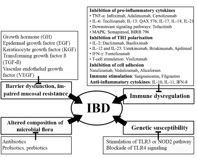
The strategy to find new targets is based on the main pathological alterations that characterize IBD. Since the disease is caused by a combination of factors, including genetic susceptibility, immune dysregulation, barrier dysfunction, and the change in microbial flora, the new therapeutics aim to correct or restore 1./ the genetic alterations, 2./ the immune dysregulation, 3./ the barrier dysfunction and mucosal resistance 4./ the altered composition of gut flora (Fig. 1.).
3.1. Correction of genetic alterations
Under normal, healthy condition the intestinal mucus layer prevents exposure of IECs to luminal bacteria. Several mechanisms are involved in protection of intestinal epithelium against luminal microbiota, e.g. defensins (secreted by Paneth cells) and the production of immunoglobulin A (IgA). PRRs are involved in the production of cytokines necessary for the development of immunity and have crucial role in innate microbial sensing by IECs, DCs and macrophages [30]. Their activation initiates NF-κB and MAPK signaling pathways resulting in the production of pro-inflammatory cytokines and antimicrobial peptides [5, 197]. In addition, autophagy has a crucial role in the maintenance of intracellular homeostasis preventing abnormal accumulation of protein aggregates, intracellular components, such as organelles, apoptotic bodies, and microbes [198]. One of the key proteins involved in the execution of the autophagic process is ATG16L1 [199].
Though a large number of genetic loci have been found to be associated with CD, the polymorphism of two genes, NOD2/CARD15 [200] and ATG16L1 seem to have particular importance in the development of the disease [201]. While in healthy subjects ATG16L1 encodes threonine at amino acid position 300 (ATG16L1*300T), ATG16L1 encoding alanine (ATG16L1*300A) instead of threonine at the same position increases the risk of the development of CD, due to impairement in bacterial capture by autophagy [202].
Moreover, patients with CD showed decreased expression of mucosal TLR3 and increased expression of TLR4, which results in downstream release of inflammatory modulators, for example TNF-α and IL-1 [203]. Consequently, stimulation of the TLR3 or NOD2 pathway may represent a new approach of the therapy of IBD. Experimental data suggest that activation of TLR3 (e.g. by synthetic viral RNA) or NOD2 were effective for prevention of dextran sodium sulphate (DSS)-induced acute colitis in the mouse [204-206], which implies that stimulation of TLR3 or NOD2 signaling may represent a new, successful therapeutic strategy for the treatment of CD in those patients who show reduced expression of
TLR3 or carry NOD2 mutations [207]. On the other hand, blocking TLR signaling may represent another approach for IBD treatment, because TLR2 and TLR4 are up-regulated in IBD [208]. Experimental data suggest that TLR4 blockade decreased inflammation in DSS- induced colitis in mice, but also interfered with colonic mucosal healing, since anti-TLR4 antibody treatment during recovery from DSS colitis resulted in defective mucosal healing (as described above) [209].
Attempts have been made to develop new TLR4 signaling inhibitors. Recently, arylidenemalonate derivatives were found to suppress LPS-induced production of NF-κB, TNF-α, IL-1β and nitric oxide [210], suggesting its potential therapeutic value for various inflammatory diseases. However, their real role in IBD remains to be clarified.
3.2. Restoration of immune dysregulation
Today it is widely accepted that abnormal immune regulation is a key factor in the pathomechanism of IBD and alterations in both innate and adaptive immunity have been observed. Dysregulation of mucosal immunity in IBD leads to an overproduction of inflammatory cytokines and trafficking of effector leukocytes into the intestinal mucosa, thus resulting in an uncontrolled intestinal inflammation. Under homeostatic condition there is a balance between regulatory (Treg) and effector T-cells (Th1, Th2 and Th17). Mucosal inflammation is induced either by an increase in the effector T-cell population and an increased production of pro-inflammatory cytokines (such as TNF-α, IFN-γ, IL-1, IL-6, IL- 12, IL-17, IL-23) or a reduced function of Treg cells and decreased level of anti-inflammatory cytokines (e.g. TGF-β, IL-4, IL-10 or IL-11) produced by different immune cells located in the lamina propria of the intestinal mucosa. Cytokines may therefore be targets for IBD therapy [211]. Moreover, elimination of intestinal inflammation may be achieved either by reduction of effector T-cell populations or by increasing regulatory T-cell activity [5, 212].
Several reviews have been published recently on the potential therapeutic agents in IBD that target the immune dysregulation [7, 12, 188, 190, 211, 213-218].
Restoration of the immune dysregulation may be achieved by several mechanisms, such as inhibition of 1./ pro-inflammatory cytokines, 2./ Th1 polarisation and proliferation, 3./ T- cell stimulation (anti-CD3 therapy), 4./ cell adhesion, as well as 5./ by immune stimulation (Granulocyte Macrophage Colony Stimulating Factor /GM-CSF)/ and Granulocyte Colony Stimulating Factor /G-CSF/ and 6./ by increase of anti-inflammatory cytokines.
3.2.1. Inhibition of pro-inflammatory cytokines
Inhibitors of TNF-α
TNF-α is a fundamental mediator of this abnormal immune response. TNF-α has two forms, a transmembrane and soluble form, and its action is mediated by 2 receptors, TNF-R1 (TNF receptor type 1, CD120a or p55/60) and TNF-R2 (TNF receptor type 2, CD120b or p75/80) [219] resulting in complex and differential actions. While the pro-inflammatory effects of TNF-α are mediated by TNF-R1, the immunoregulatory functions are independent from this receptor. Consequently, selective inhibitors of TNF-R1 that reduce the pro- inflammatory function of TNF-α without affecting its immunoregulatory effects, may represent a new therapeutic approach of IBD.
In the last 15 years, biological agents targeting TNF-α have significantly improved the therapy of IBD refractory to conventional drugs. The efficacy of this therapy alone reflects the pleiotropic effects of TNF-α.
Infliximab, a monoclonal chimeric antibody, targeting human TNF-α, became the first monoclonal antibody available for the treatment of CD and UC. The potency of this agent in moderate-to-severe CD and UC has been one of the most important advances in the treatment of IBD.
Infliximab also induces T-cell apoptosis, that contributes to its therapeutic effect [220].
Namely, in the gut, there is a tight control of activation and expansion of T-cells. T-cell expansion is limited by apoptosis, and T-cell resistance to apoptosis with consequent T-cell expansion was observed in patients with IBD, which may contribute to the pathomechanism [221]. Hence, it may be raised that induction of apoptosis in T-cells and other effector cells may have therapeutic importance in the treatment of IBD and pro-apoptotic signaling might be a target for drug development [5].
It has been shown that some anti-TNF-α agents induce apoptosis in monocytes and lymphocytes both in vitro and in vivo [222, 223]. Consequently, apoptosis-induction seems to be an important part of the therapeutic action of TNF-α antagonists and differences in their apoptotic efficacy might contribute to the differences found in their clinical efficacy. In addition to infliximab, several other therapeutic agents that are effective in the treatment of IBD, including corticosteroids, sulfasalazine, azathioprine, 6-mercaptopurine and anti-IL-12 antibody, induce apoptosis of activated T-cells [222, 224-226].
Adalimumab, a fully human IgG1 monoclonal antibody to TNF-α, was found to be
effective in patients with CD refractory to conventional therapy and in patients with an attenuated response to infliximab [211, 227, 228]. Recent study showed that adalimumab could induce and maintain clinical remission in patients with moderate-to-severe UC as well, who did not have a satisfactory response to steroids or immunosuppressive agents [229].
Certolizumab and certolizumab pegol (its PEGylated Fab' fragment with increased plasma half-life) are humanized TNF-α monoclonal antibodies. Majority of the human studies demonstrated their effectiveness in maintenance of response and remission in CD [230, 231].
However, Sandborn et al. failed to confirm the effectiveness of certoluzimab after 6 weeks treatment [232].
Etanercept is a genetically engineered fusion protein consisting of two recombinant human TNF p75 receptors linked to an Fc portion of human IgG1 fragment. It was found to be ineffective for the treatment of patients with moderate to severe CD in the same dose range that was effective in rheumatoid arthritis [233].
CDP571, an immunoglobulin G4 humanized monoclonal anti-TNF-α antibody showed a slight and short lived reduction in clinical activity of UC [234]. It proved to be less effective than infliximab and further clinical development of CDP571 for the treatment of CD has been discontinued (see review [194]).
Novel TNF inhibitors have been developed, such as golimumab, dersalazine, HMPL- 004 and ozoralizumab (ATN-103). These compounds are in various phases of the clinical trial process, and their real therapeutic values have to be determined [12]. In addition, recently a vaccine against TNF-α has been developed (TNF-α kinoid, Debio-01512), as a new mechanism for inhibition of TNF-α [196], and phase I/II clinical trials in patients with moderate to severe CD were found to be promising [235, 236].
Inhibition of IL-6
IL-6 has a fundamental role in immune regulation and inflammation. The IL-6 receptor (IL-6R) system has both a membrane-bound (IL-6R) and a soluble form (sIL-6R).
Increased serum concentrations of IL-6 and sIL-6R have been shown to correlate to clinical activity of CD, and animal models have strongly suggested the therapeutic potential of anti- IL-6R monoclonal antibody. For example, anti-IL-6R monoclonal antibody reduced the symptoms of colitis in Th1 cell-mediated murine colitis model. Similarly, blockade of sIL-6R in vivo by a newly designed gp130-Fc fusion protein resulted in reduction of colitis activity and induction of apoptosis, indicating that sIL-6R suppresses mucosal T-cell apoptosis.
Accordingly, it was shown recently that IL-6 induces the anti-apoptotic genes BCL2 and BCL-XL [237]. These results suggest the therapeutic potential of anti-IL-6R monoclonal antibody in CD [237, 238].
Tocilizumab (also known as MRA) is a humanized IL-6 receptor antibody, recognizes both the membrane-bound and the soluble form of IL-6R and specifically blocks IL-6-induced actions [239]. Tocilizumab is expected to ameliorate the autoimmune inflammatory diseases characterized by IL-6 overproduction and has been developed as a therapeutic agent for rheumatoid arthritis (see review [240]). Tocilizumab treatment of patients with active CD induced reduction of the disease activity index compared to the placebo group, however, remission of the disease was archived only at very low proportion of the patients [241]. The beneficial effect of tocilizumab was confirmed by Nishimoto [242].
Inhibition of IL-13
IL-13 has been shown to be pathogenic in IBD, particularly in UC. It impairs the function of the epithelial barrier and also causes apoptosis of epithelial cells [10, 11, 243, 244]. IL-13 overexpression in the inflamed mucosa is particularly characteristic for UC and IL-13 is considered as the major effector cytokine in UC [245].
The anti-IL-13 antibody anrukinzumab (IMA-638) as well as tralokinumab (CAT-354), a fully human anti-IL-13 antibody are under clinical studies (phase IIa) in patients with mild to moderate UC. QAX576, another fully human antibody against IL-13, is in phase I/II trials in patients with CD, the final results are pending [246, 247].
Inhibition of mitogen activated protein kinases (MAPKs)
Expression of pro-inflammatory cytokines, which are critical in the pathogenesis of IBD, is regulated by one or more MAPK pathways. Accordingly, activation of several MAPK members was found in biopsies from the inflamed mucosa of CD patients [248].
CNI-1493 (semapimod), inhibitor of JNK/p38MAP kinases, showed significant clinical improvement of severe CD, confirming the potential role of inflammatory MAPKs in the pathogenesis of CD [249]. In contrast, a highly potent inhibitor of p38 MAPK, BIRB 796 (doramapimod) was studied in chronic active CD in a multicenter, multinational trial with placebo control, and the results failed to show evidence for clinical efficacy [250].
Concerns with the use of MAPK inhibitors have been raised, for example inhibition of p38 and 42/44 MAPK reduces normal bactericidal activity of neutrophils [251]. Moreover, ubiquitous presence of the MAPK pathways and their involvement in several processes suggests that MAPK inhibition should be selective for an isoform, rather than general.
New avenues against pro-inflammatory cytokines or downstream signaling pathways
Currently, several novel agents have been developed to target either pro-inflammatory cytokines or downstream signaling pathways. Pro-inflammatory cytokines IL-17, IL-18 and IL-21 were found to be elevated in the inflamed intestinal mucosa of patients with IBD [252- 254]. Monoclonal antibodies developed against these cytokines are currently in phase I/II clinical trials in patients with CD. Unexpectedly, secukinumab (AIN 457) targeting IL-17 in double-blind, placebo controlled study in patients with CD worsened the disease compared with placebo [255]. In contrast, inhibitor of the release of IL-17, vidofludimus (4SC- 101/SC12267) showed beneficial effect in a single-arm, open-label study, indicating that it may be useful in maintaining clinical remission both in patients with CD and UC [256].
Further investigation is needed to clarify the modulatory role of IL-17 in IBD.
On the other hand, gut inflammation can also be restricted by blocking the downstream signaling pathways mediated by cytokines. Signaling molecules that interact with cytokine receptors are the Janus kinases (JAK), JAK1, JAK2 and JAK3, which play a fundamental role in the development and differentiation of immune cells.
Tofacitinib, inhibitor of JAK3 was developed recently [257], but various clinical efficacies have been experienced. A multicentre, double-blind, placebo controlled study with tofacitinib in patients with moderate to severe active CD showed no clinically significant response following 4 weeks of treatment compared with placebo, but in patients with moderate to severe UC it showed improvement in both clinical response and remission rates [258, 259].
3.2.2. Inhibition of TH1 polarisation and proliferation
Inhibition of IL-2
Inhibition of the binding of IL-2 to the IL-2 receptor results in inhibition of the growth, proliferation and differentiation of T-cells to 'effector' T-cells.
Basiliximab (chimeric monoclonal antibody) and daclizumab (humanized monoclonal antibody) are targeted against the α-chain of the IL-2 receptor (CD25), and inhibit the binding of IL-2 to the IL-2 receptor. However, daclizumab failed to induce a significant action in UC [260]. Neither basiliximab increased the effect of corticosteroids in the induction of remission in patients with corticosteroid-resistant moderate to severe UC [261].
Inhibition of IL-12 and IL-23
CD is associated with an enhanced Th1 cytokine response, which results in increased production of IL-12, a pro-inflammatory cytokine, that stimulates the production of IFN-γ and TNF-α. IL-12 is also involved in the differentiation of naïve T-cells into Th1 cells. It is a heterodimeric protein with 2 subunits: the p35 and p40. The structurally similar cytokine IL- 23 (its p40 subunit is identical to IL-12p40, and its p19 subunit shows certain similarities to IL-12 p35) has also an important role in intestinal inflammation, in conjunction with IL-6 and TGF-β1. IL-23 stimulates naïve CD4+ T cells to differentiate to Th17 cells. The highly aggressive immune response together with IL-12/IL-23 could have a determining role in initiation and perpetuation of chronic intestinal inflammation in CD [262, 263].
Recently p40 peptide-based vaccines have been developed. Pretreatment of rats with the vaccines induced specific antibodies to IL-12 and IL-23, which was associated with improvement of intestinal inflammation and fibrosis, indicating that the vaccine may provide a potential approach for the long-term treatment of CD [264].
A human study showed that treatment with a human monoclonal antibody against IL- 12 may induce clinical responses and remissions in patients with active CD. This treatment is associated with decreased Th1-mediated inflammatory cytokines at the site of disease [265].
Ustekinumab, and briakinumab are human monoclonal antibodies against IL-12 and IL-23, both targeting the p40 subunit. Ustekinumab significantly increased the rates of response and remission as maintenance therapy, and it may be particularly useful in patients who previously did not respond to anti-TNF therapy [266]. Briakinumab, however, in patients with moderate to severe CD was not effective for the induction or maintenance of remission ([267].
Apilimod is an inhibitor of the transcription of IL-12 and IL-23. In a randomised controlled trial apilimod failed to induce significantly greater effect than placebo treatment [268].
Interferon-γ
IFN-γ was found to be elevated in all genetic animal models of IBD and seems to have determining role in the development of Th1 responses. If the target of IBD therapy is to reduce Th1 responses, then inhibition of IFN-γ represents one potential therapeutic approach.
Fontolizumab, a humanized monoclonal antibody to IFN-γ was shown to have a beneficial effect on disease activity [12, 211, 269]. An other study suggested that though a strong clinical response was not induced by fontolizumab, a significant decrease in C-reactive protein levels was observed. Further studies are necessary to determine its efficacy [270].
3.2.3. Inhibition of T-cell stimulation
Since intestinal inflammation may be resulted by an increased activity of the effector T-cell population with excessive inflammatory responses and CD3 is required for T-cell activation, drugs that target T-cell activation may exert therapeutic effect in IBD.
Visulizumab, a humanized anti-CD3 monoclonal antibody was demonstrated to induce apoptosis in activated T-cells selectively, and enhance the production of IL-10, a potent anti- inflammatory cytokine [271]. It proved to be effective against T-cell transfer colitis [272].
The results of an open-label phase I human study with visilizumab in patients with severe corticosteroid-refractory UC suggested that it may be clinically beneficial [273].
Another study confirmed this beneficial effect of visulizumab in a pilot randomized phase I/II study. Visulizumab improved both symptomatic and clinical responses in severe, steroid-refractory UC, though, all patients experienced adverse reactions [274]. However, in a randomised, double-blind, placebo-controlled study visilizumab was not effective for severe, corticosteroid-refractory UC [275]. The same conclusion was drawn by Wood [276].
CD40 is also involved in T-cell activation. Humanized monoclonal antibody against CD40L has been developed, however in a phase II trial for CD due to side effect (thromboembolism) the trial was halted [277].
3.2.4. Inhibition of cell adhesion
Lymphocyte trafficking to the gut is a basically important step in the initiation and maintenance of intestinal inflammation in patients with IBD. Alpha 4 integrin, a cell-surface glycoprotein involved in the adhesion, migration and activation of immune cells, is expressed on most of the lymphocytes, and combined with either a β1 subunit (that interacts
predominantly with the endothelial ligands, vascular cellular adhesion molecule 1 /VCAM-1/) or a β7 subunit (that interacts predominantly with the mucosal addressin cellular adhesion molecule 1 (Mad-CAM-1)) [278]. The interaction between α4β7 integrin and Mad-CAM-1 is important in mediating leukocyte homing to gut mucosa [279]. Several therapeutic agents have been developed that specifically target the α4β7 subunit of integrin.
Natalizumab, a humanized monoclonal antibody against the cell adhesion molecule α4-integrin subunit, was proved to be effective in experimental colitis both in the mouse and the rat [280, 281]. Moreover, the effect of natalizumab has been studied also in humans, and it was shown to block the adhesion and migration of white blood cells into the gut and to reduce chronic inflammation associated with CD [282]. Reviews based on relevant literature and meta-analysis of the controlled trials of natalizumab suggested that the therapy was superior to placebo in inducing remission of CD [283, 284]. However, natalizumab therapy is associated with an increased risk of reactivation of latent John Cunningham (JC) virus, which causes the potentially fatal progressive multifocal leukoencephalopathy (PML). The risk to develop PML is increased by the presence of anti-JC virus antibodies, and previous or concomitant treatment with immunosuppressive (IF-β1 or azathioprine) agents [285].
Vedolizumab (MNL-0002) is gut-specific, α4β7-integrin-neutralizing monoclonal antibody, that appears to lack systemic effects. It was particularly effective in UC and also in CD, indicating that vedolizumab might be therapeutic option for the treatment of therapy- refractory patients [286]. A recent review evaluated the safety and efficacy of vedolizumab for the treatment of CD, and it was concluded that vedolizumab is an effective and well- tolerated drug [287]. Though it does not increase the risk of infection, it was demonstrated that vedolizumab may reduce the number of Treg cells and consequently their suppressive effect on colonic inflammation (see review [288]).
Alicaforsen (ISIS 2302) is an antisense oligodeoxynucleotide to ICAM-1 [289].
Experimentally, it inhibited the DSS-induced colitis both in mice and rats [289, 290], but in human studies both improvement of clinical symptoms and lack of effect have been shown in CD (see reviews [12, 211, 291, 292]). In patients with active UC alicaforsen enemas induced a beneficial effect, and the drug was well tolerated [291].
3.2.5. Immune stimulation
Though granulocyte macrophage colony stimulating factor (GM-CSF) and granulocyte colony stimulating factor (G-CSF) are hematopoietic growth factors, they
stimulate cells of the innate immune system (neutrophils, macrophages and DCs). They have also been shown to be produced within Paneth cells of intestinal mucosa, and their receptors are expressed within IECs [293]. Moreover, GM-CSF has been demonstrated to promote proliferation within these cells [294].
GM-CSF has been shown to reduce DSS-induced colitis and decrease the levels of pro-inflammatory cytokines (TNF-α and IL-1β) in colonic tissue samples [295, 296]. These results were confirmed recently; GM-CSF decreased the DSS-induced colitis and the expression of pro-inflammatory cytokines. In addition, the duration of ulcer healing was shorter and epithelial regeneration was facilitated in GM-CSF-treated mice [295]. Based on these preclinical results, a potential role for GM-CSF in the therapy of patients with IBD has been raised.
Sargramostim (recombinant human GM-CSF) and filgrastim (recombinant human G- CSF) have been examined in several human studies and majority of them suggested a significant improvement and remission of CD ([297] and reviews [12, 298]). However, sargramostim in a phase III multicentre double-blind, placebo controlled study in patients with active CD failed to induce a significant difference in clinical efficacy compared to placebo. Further human studies are necessary to reveal their role in the treatment of IBD [211].
3.2.6. Anti-inflammatory cytokines
IL-10
IL-10 reduces the production of pro-inflammatory cytokines IL-1α, IL-6 and TNF-α, downregulates and controls the acute inflammation and thereby may improve the course of IBD. IL-10 polymorphisms have been shown to be associated with IBD and mutations in IL- 10 and IL-10 receptor (IL-10R) in patients with very early onset IBD was observed [299].
However, Buruiana et al. [300] on the basis of a systematic review of the literature concluded that IL-10 does not appear to provide any benefit for the treatment of active CD.
IL-11
IL-11 besides its thrombocytopoietic properties improves the mucosal barrier function and inhibits the inflammatory reaction by reducing expression of NF-κB and in turn IL-1,
TNF-α and other proinflammatory peptides [301]. Therefore IL-11 was examined on experimental colitis in the rat induced by trinitrobenzene sulfonic acid (TNBS), where IL-11 exerted protective effect [302]. Recent findings confirmed the beneficial action of IL-11:
administration of exogenous IL-11 was found to be protective against lethal colitis in TLR2- deficient mice (TLR2 is involved in maintaining epithelial barrier function) [303]. However, in a human study recombinant human IL-11 was less effective than prednisolone in the treatment of CD [304].
Interferon-β-1a (IFN-β-1a)
Data of the literature reflect conflicting results on the effect of IFN-β-1a in IBD. Some studies indicate that IFN-β-1a exerts a therapeutic effect, while others found that IFN-β-1a did not produce a significant therapeutic action compared with placebo. Recent clinical trial in patients with CD came to the conclusion that there was no difference between the effects of the administration of IFN-β-1a and placebo [305, 306].
3.3. Restoration of barrier dysfunction and stimulation of mucosal healing and resistance
Repeated intestinal epithelial damage and the consequent disruption of the intestinal barrier function is a key mechanism of IBD [3, 6]. Namely, the alteration of intestinal barrier function may result in translocation of commensal bacteria into the intestinal wall, leading to uncontrolled T-cell activation and inflammation. Damage of barrier integrity, with increased antigen and bacterial uptake is believed to be important in the pathophysiology of CD [4].
An intact barrier function of the intestinal epithelium prevents translocation of commensal bacteria into the mucosa. Consequently, though mucosal healing has been considered as a sign of complete healing of gut inflammation, it should be emphasized that mucosal healing can be considered as an initial step in suppression of inflammation [307]. In a systematic review Neurath and Travis [307] analyzed the influence of conventional therapeutic agents as well as the biologics on the healing of intestinal mucosal injury in IBD, and they concluded that while corticosteroids have little or no positive effects on induction/maintenance of mucosal healing in CD (but induced healing in patients with UC), azathioprine, in a lesser extent methotrexate, as well as natalizumab, infliximab, adalimumab and certolizumab pegol could induce mucosal healing [307].
Following injury of intestinal mucosa, IECs migrate into the damaged area to restore barrier integrity [298, 308], followed by proliferation of IECs to correct the epithelial defect.
Finally, differentiation of IECs is necessary to restore mucosal barrier and epithelial function.
These consecutive events (restitution, proliferation and differentiation) are mediated by regulatory proteins (such as chemokines), defensins, as well as by growth factors [298].
Growth factors have crucial role in cell restitution/proliferation/differentiation, and in angiogenesis. Moreover, TGF-β inhibits the differentiation of naïve T-cells to Th1/Th2 subtypes.
Growth hormone (GH)
GH, a regulatory peptide that stimulates aminoacid and electrolyte uptake by the intestine, decreases intestinal permeability and stimulates collagen synthesis by induction of the expression of insulin-like growth factor (IGF). IGF was shown to promote epithelial repair in intestinal inflammation in preclinical animal models. For example, recombinant human growth hormone induced protective effects in TNBS-induced colitis [309]. A preliminary study showed that human growth factor was effective in CD, it improved the clinical symptoms (the number of liquid/soft stools, severity of abdominal pain and overall well being) and a statistically significant increase in circulating levels of IGF-1 was observed [310]. Further, placebo controlled human studies should be performed to determine the real place of growth factor in the therapy of IBD.
Epidermal growth factor (EGF)
Human recombinant EGF was shown to be effective in experimental colitis induced by TNBS given prophylactically, before the induction of colitis [311]. In UC patients EGF enemas administered parallel with mesalamine resulted in a significant improvement of symptoms [312], however, the effect of EGF enema alone (with placebo) has not been studied.
Keratinocyte growth factor (KGF)
KGF-1 and -2 are ligands of the fibroblast growth factor (FGF) family. KGFs may have role in wound healing and maintaining epithelial homeostasis. Expression of KGF-1 was