Intestinal alkaline phosphatase in the colonic mucosa of children with inflammatory bowel disease
Kriszta Molnár, Ádám Vannay, Beáta Szebeni, Nóra Fanni Bánki, Erna Sziksz, Áron Cseh, Hajnalka Győrffy, Péter László Lakatos, Mária Papp, András Arató, Gábor Veres
Kriszta Molnár, Áron Cseh, András Arató, Gábor Veres, First Department of Pediatrics, Semmelweis University, H-1083 Buda- pest, Hungary
Ádám Vannay, Beáta Szebeni, Erna Sziksz, Research Group for Pediatrics and Nephrology, Semmelweis University and Hun- garian Academy of Sciences, H-1083 Budapest, Hungary Nóra Fanni Bánki, SE-MTA “Lendulet” Diabetes Research Group, H-1083 Budapest, Hungary
Hajnalka Győrffy, Second Department of Pathology, Semmel- weis University, H-1091 Budapest, Hungary
Péter László Lakatos, First Department of Medicine, Semmel- weis University, H-1083 Budapest, Hungary
Mária Papp, Second Department of Medicine, University of De- brecen, H-4032 Debrecen, Hungary
Author contributions: Veres G, Vannay Á and Molnár K de- signed the research; Arató A and Veres G included the patients;
Molnár K, Szebeni B, Bánki NF and Cseh Á performed the analyses; Sziksz E and Győrffy H analyzed the histological data;
Molnár K, Vannay Á and Veres G wrote the paper; Lakatos PL, Papp M and Arató A critically reviewed the paper.
Supported by Grants OTKA-76316, OTKA-K81117, and ETT- 028-02 (Veres G and Vannay Á are holders of the János Bolyai Research grant); János Bolyai Research Scholarship of the Hun- garian Academy of Sciences
Correspondence to: Gábor Veres, MD, PhD, First Depart- ment of Pediatrics, Semmelweis University, H-1083 Budapest, Hungary. veres.gabor@med.semmelweis-univ.hu
Telephone: +36-208-258163 Fax: +36-1-3036077 Received: November 2, 2011 Revised: April 17, 2012 Accepted: April 21, 2012
Published online: July 7, 2012
Abstract
AIM: To investigate intestinal alkaline phosphatase (iAP) in the intestinal mucosa of children with inflammatory bowel disease (IBD).
METHODS: Colonic biopsy samples were taken from 15 newly diagnosed IBD patients and from 10 healthy controls. In IBD patients, specimens were obtained
both from inflamed and non-inflamed areas. The iAP mRNA and protein expression was determined by reverse transcription-polymerase chain reaction and Western blotting analysis, respectively. Tissue localiza- tion of iAP and Toll-like receptor (TLR) 4 was investi- gated by immunofluorescent staining.
RESULTS: The iAP protein level in the inflamed muco- sa of children with Crohn’s disease (CD) and ulcerative colitis (UC) was significantly decreased when compared with controls (both P < 0.05). Similarly, we found a significantly decreased level of iAP protein in the in- flamed mucosa in CD compared with non-inflamed mucosa in CD (P < 0.05). In addition, the iAP protein level in inflamed colonic mucosa in patients with UC was decreased compared with non-inflamed mucosa in patients with CD (P < 0.05). iAP protein levels in the non-inflamed mucosa of patients with CD were similar to controls. iAP mRNA expression in inflamed colonic mucosa of children with CD and UC was not significant- ly different from that in non-inflamed colonic mucosa with CD. Expression of iAP mRNA in patients with non- inflamed mucosa and in controls were similar. Co-local- ization of iAP with TLR4 showed intense staining with a dotted-like pattern. iAP was present in the inflamed and non-inflamed mucosa of patients with CD, UC, and in control biopsy specimens, irrespective of whether it was present in the terminal ileum or in the colon.
However, the fluorescent signal of TLR4 was more pro- nounced in the colon compared with the terminal ileum in all groups studied.
CONCLUSION: Lower than normal iAP protein levels in inflamed mucosa of IBD patients may indicate a role for iAP in inflammatory lesions in IBD. Based on our results, administration of exogenous iAP enzyme to pa- tients with the active form of IBD may be a therapeutic option.
© 2012 Baishideng. All rights reserved.
BRIEF ARTICLE
doi:10.3748/wjg.v18.i25.3254 © 2012 Baishideng. All rights reserved.
Key words: Intestinal alkaline phosphatase; Toll-like recep- tor; Colonic biopsy; Children; Inflammatory bowel disease Peer reviewer: Dr. Limas Kupcinskas, Professor, Department of Gastroenterology, Kaunas University of Medicine, Mickeviciaus 9, LT 44307 Kaunas, Lithuania
Molnár K, Vannay Á, Szebeni B, Bánki NF, Sziksz E, Cseh Á, Győrffy H, Lakatos PL, Papp M, Arató A, Veres G. Intes- tinal alkaline phosphatase in the colonic mucosa of children with inflammatory bowel disease. World J Gastroenterol 2012;
18(25): 3254-3259 Available from: URL: http://www.wjgnet.
com/1007-9327/full/v18/i25/3254.htm DOI: http://dx.doi.
org/10.3748/wjg.v18.i25.3254
INTRODUCTION
The etiology of inflammatory bowel disease (IBD), in- cluding ulcerative colitis (UC) and Crohn’s disease (CD), remains unclear. It is hypothesized that, in genetically sus- ceptible individuals, inappropriate and ongoing activation of a mucosal immune response against luminal antigens is a major cause of the inflammation[1,2]. In active IBD, the tolerance towards the resident intestinal flora is de- creased. The balance between protective and commensal luminal bacterial species is lost and, due to increased mu- cosal permeability and insufficient mucosal clearance, the commensal flora and pathogenic bacteria enter into the lamina propria and destructive inflammatory responses are unavoidable[3-6]. This prompts an exaggerated immune response with the activation of the two arms of mucosal immune system, the innate and adaptive elements[7,8].
The activation of the innate immune system heav- ily depends on the recognition of microbes by pattern recognition receptors such as Toll-like receptors (TLRs).
The TLR family consists of 13 members, and each has different type of ligands. One of them is TLR4, which is responsible for recognition of lipopolysaccharide (LPS), a principal component of the bacterial outer membrane.
Uncontrolled activation of TLR4 may lead to the loss of mucosal barrier integrity, aggravation of the inflamma- tory response within the gut epithelial mucosa, increased expression of TLR-ligands and tumorigenesis[9-13]. Pre- viously, we found increased TLR4 protein and mRNA levels in the inflamed mucosa of children with IBD and celiac disease[14,15].
An increasing body of evidence also supports the regulatory role of intestinal alkaline phosphatase (iAP) in TLR activation. iAP is expressed on the apical surface of enterocytes and exists in membrane-bound and soluble forms[16]. iAP plays an essential role in the inactivation of LPS through dephosphorylation of its lipid A moiety, thus generating a non-toxic monophosphoryl section.
This dephosphorylated monophosphoryl lipid A is not able to form a complex with TLR4[17,18].
There is only one human study where expression of iAP (mRNA) in adult IBD patients was analyzed, and lower than normal iAP mRNA expression was found
in epithelial specimens[19]. It should be noted, however, that no data on the level of iAP protein in IBD mucosa is available. The aim of our study was to investigate iAP protein and mRNA levels in affected and non-affected colon mucosa of children with newly diagnosed IBD. In addition, our secondary aim was to determine the local- ization of iAP enzyme with TLR4.
MATERIALS AND METHODS
Patients and colonic biopsies
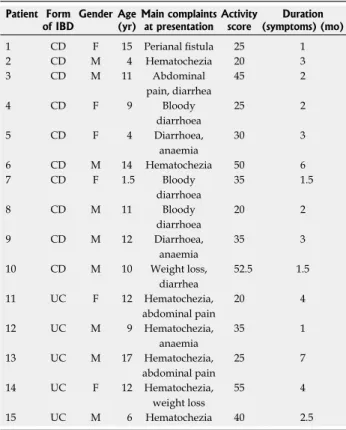
Ten children (7 boys, 3 girls; median age: 10.5 years, range: 1.5-15 years) with newly diagnosed CD, 5 children (3 boys, 2 girls; median age: 11 years, range: 6-17 years) with newly diagnosed UC, and 10 control children (5 boys, 5 girls; median age: 9.5 years, range: 1.5-16 years) were enrolled in the study (Table 1). IBD was diagnosed according to the Porto criteria[20,21]. The presenting symp- toms in CD were perianal fistula, hematochezia, abdomi- nal pain, diarrhea-bloody diarrhea, or anemia. All of the patients later diagnosed with UC had hematochezia, and some had abdominal pain and weight loss. Colonic bi- opsy samples were taken from macroscopically inflamed and non-inflamed sites of the colonic mucosa in children with CD. As each UC children had pancolitis, only in- flamed mucosa was obtained from UC patients (Table 1).
The activity score was calculated by means of the Pediat- ric Crohn’s Disease Activity Index (PCDAI) and Pediatric Ulcerative Colitis Activity Index (PUCAI)[22,23]. Measuring disease activity in pediatric CD is based on disease history (abdominal pain, stools per day and general well-being), laboratory findings, weight, abdominal and perianal ex- amination, extra-intestinal manifestations, and growth rate[24]. PUCAI requires no laboratory measurements[25]. The mean PCDAI of our patients was 33.75, and the mean PUCAI was 35. This means that both groups had moderate-to-severe disease activity. Control children were referred to the outpatient clinic with rectal bleeding, con- stipation or weight loss. Colonoscopy was part of their diagnostic procedure and the biopsy specimens showed normal macroscopic appearance and histology. Written informed consent was obtained from parents prior to the procedure, and the study was approved by the Semmel- weis University Regional and Institutional Committee and Research Ethics.
RNA isolation and real-time polymerase chain reaction Total RNA was isolated from the colonic biopsy samples by RNeasy Total RNA Isolation Kit (Qiagen GmbH, Hilden, Germany), according to the instructions of the manufacturer. One μg of total RNA was reverse-tran- scribed and iAP mRNA expressions were determined by real-time polymerase chain reaction (PCR) on Light Cy- cler480 (Roche Diagnostics, Mannheim, Germany). PCRs were performed containing RealTime ready Catalog As- say primer (Roche Diagnostics), Light Cycler 480 Probes Master (Roche Diagnostics, Mannheim, Germany), and cDNA. Conditions for iAP mRNA measurements: one
cycle, 95 ℃, 10 min (denaturation), followed by several cycles at 95 ℃, 10 s and 30 s, 72 ℃ 1 s (annealing and extension). The mRNA expression of glyceraldehyde- 3-phosphate dehydrogenase (GAPDH) as internal control was determined using Brillant Ⅱ Fast SYBR Green quan- titative polymerase chain reaction Master Mix (Stratagene, Cedar Creek, TX, United States), PCR primers (Forward:
5’-CAC CAC CAT GGA GAA GGC TG-3’; Reverse:
5’-GTG ATG GCA TGG ACT GTG-3’, Invitrogen, CA, United States) and cDNA. Conditions for GAPDH:
one cycle, 95 ℃, 2 min, 50 cycles at 95 ℃ 20 s and 60 ℃, 40 s. Results were analyzed by Light-Cycler software 480 (Roche Diagnostics).
Protein isolation and Western blotting
Colonic biopsy specimens were homogenized in lysing solution, and protein concentrations were determined by DC Protein Assay (Bio-Rad Laboratories, Hercules, CA, United States); 0.5 µg protein from each sample was separated by 10% sodium dodecyl sulfate-polyacrilamide gel electrophoresis (120 V, 40 mA, 120 min) (PenguinTM Dual-Gel Water Cooler Systems, Owl, NH, United States) and transferred to nitrocellulose membrane (GE Health- care, Little Chalfont, United Kingdom) (70 V, 220 mA, 120 min) (MiniTankTM electroblotter, Owl). Membranes were blocked in 1% non-fat dry milk solution (1 h) and
incubated with iAP specific rabbit polyclonal antibody (1:1000, 1 h) (AbCam, Cambridge, United Kingdom).
Equal protein loading was confirmed by β-actin specific (C-11) goat polyclonal IgG antibody (1:100) (Santa Cruz Biotechnology Inc., Santa Cruz, CA, United States). Per- oxidase-conjugated secondary anti-rabbit IgG or donkey anti-goat IgG antibodies (1:2000, 30 min) (Santa Cruz Biotechnology Inc.) were used. Immunoreactive bands were visualized using the enhanced chemiluminescence Western blotting detection protocol (GE Healthcare).
Bands were analyzed with software Image J. 1.42q (Na- tional Institutes of Health, United States). The values were expressed as relative optical density.
Immunofluorescent staining
Biopsy samples were snap-frozen, embedded in Shan- don cryomatrix (ThermoElectron Co., Waltham, United States), cut to 3-4 μm slides and double incubated with TLR4 specific goat polyclonal antibody and iAP specific rabbit polyclonal antibody (1:100, 1 h) (Abcam Plc). Sec- ondary antibodies were Alexa Fluor 488 donkey anti-goat and Alexa Fluor 568 goat anti-rabbit antibodies (Invit- rogen). Zeiss LSM 510 Meta confocal laser scanning mi- croscope (Carl Zeiss, Jena, Germany) was used with 20 × Plan Apochromat (NA = 0.8) and 63 × Plan Apochromat oil immersion differential interference contrast objectives (NA = 1.4).
Statistical analysis
Data were analyzed using Statistica 7.0 software (StatSoft Inc., Tulsa, OK, United States). After testing the nor- mality with Shapiro-Wilk’s test, non-parametric Mann- Whitney U test was used. Data were considered statisti- cally significant if P ≤ 0.05, and expressed as mean ± standard deviation.
RESULTS
iAP protein levels
Western blotting analysis revealed one distinct band at 60 kDa. The iAP protein level in the inflamed mucosa of children with CD and UC was lower by 22% and 20%, respectively, compared with controls (P < 0.05). We found a lower iAP protein level in the inflamed mucosa in CD compared with non-inflamed mucosa in CD (P
< 0.05). The iAP protein level in the inflamed colonic mucosa in UC patients was decreased by 24% compared with non-inflamed mucosa in CD patients (P < 0.05) (Figure 1). iAP protein levels in the non-inflamed mucosa of patients with CD were normal.
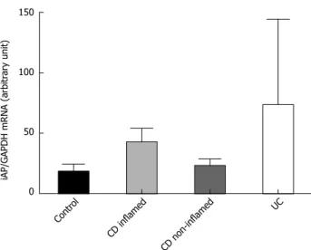
iAP mRNA expression
iAP mRNA expression in inflamed and non-inflamed colonic mucosa in IBD was comparable with that in con- trols (Figure 2).
Mucosal localization of iAP and TLR4
The distribution of iAP was restricted to the epithelial
Table 1 Clinical characteristics of newly diagnosed patients with Crohn's disease and ulcerative colitis
Patient Form
of IBD Gender Age
(yr) Main complaints at presentation Activity
score Duration (symptoms) (mo)
1 CD F 15 Perianal fistula 25 1
2 CD M 4 Hematochezia 20 3
3 CD M 11 Abdominal
pain, diarrhea
45 2
4 CD F 9 Bloody
diarrhoea
25 2
5 CD F 4 Diarrhoea,
anaemia
30 3
6 CD M 14 Hematochezia 50 6
7 CD F 1.5 Bloody
diarrhoea
35 1.5
8 CD M 11 Bloody
diarrhoea
20 2
9 CD M 12 Diarrhoea,
anaemia
35 3
10 CD M 10 Weight loss,
diarrhea
52.5 1.5
11 UC F 12 Hematochezia,
abdominal pain
20 4
12 UC M 9 Hematochezia,
anaemia
35 1
13 UC M 17 Hematochezia,
abdominal pain
25 7
14 UC F 12 Hematochezia,
weight loss
55 4
15 UC M 6 Hematochezia 40 2.5
The activity score was calculated by means of pediatric Crohn’s disease activity index and pediatric ulcerative colitis activity index. IBD:
Inflammatory bowel disease; CD: Crohn’s disease; UC: Ulcerative colitis;
M: Male; F: Female.
mucosa and LPS derived from the bacterial flora[28,29]. In the present study, we obtained data regarding the protein level, mRNA expression and localization of iAP in the intestinal mucosa of children with IBD. Lower than normal iAP levels were observed in the inflamed mucosa of CD and UC patients.
Previously it was hypothesized that the altered LPS- dephosphorylating activity may be a consequence of decreased iAP activity. We think that, in accordance with Tuin et al[19], our observations also suggest that iAP has a role in the pathogenesis of IBD. Decreased iAP levels in the inflamed mucosa may be associated with decreased LPS detoxification and, consequentially, with increased TLR4 activation. On the other hand, we found no signifi- cant difference in iAP mRNA expression that may indi- cate a possible role of posttranscriptional regulation.
Tuin et al[19]demonstrated decreased iAP mRNA ex- pression in pretreated CD patients compared with con- trols. However, it should be noted that, in this study, more than half of the patients received immunosuppressive drugs such as infliximab, methotrexate, corticosteroids, and thiopurine at the time of sample collection, which may influence iAP mRNA synthesis[30]. The unique feature of our study is the investigation of children without prior immunomodulatory therapy, hence, our results can be considered as characteristic for IBD.
Previously, we have demonstrated increased TLR4 mRNA expression and protein levels in the inflamed colonic mucosa of children with IBD[14]. Therefore, the finding that iAP and TLR4 are co-localized, is particularly important from two aspects. First, it supports a linked role of iAP in the maintenance of mucosal integrity both in healthy and in diseased subjects. Second, the lower than normal iAP in the presence of a higher than surface of the colonic and terminal ileal mucosa in each
group. No fluorescent signal was detected in Lieberkühn crypt cells, in goblet cells, and in lamina propria immune cells. The co-localization of iAP with TLR4 showed intense staining with a dotted-like pattern. iAP was pres- ent in inflamed and non-inflamed mucosa of patients with CD, UC, and in control specimens irrespective of whether it was present in the terminal ileum or in the co- lon. However, the fluorescent TLR4 signal was more pro- nounced in the colon compared with the terminal ileum in all groups (Figure 3).
DISCUSSION
A dysregulated immune response, involving the innate immunity of the intestinal mucosa plays a role in the pathomechanism of IBD. The maintenance of microbiota and host is supported by the balance of microbiota and im- mune activation that may be disturbed in IBD[26]. Previously we and others showed that activation of TLR4 by bacterial lipopolysaccharide contributes to disease progression[14,27].
Recently, in connection with LPS-activated TLR4, a new enzyme, iAP has received increasing attention as a factor responsible for mucosal defense. iAP dephosphor- ylates and detoxifies LPS and, hence, generates an inac- tive, non-toxic form. This may be one of the key factors why dephosphorylated LPS is unable to bind to TLR4 and the innate immune system is not triggered. iAP may control the interaction between TLR4 in the intestinal
iAP 60 kDa Actin 42 kDa
Relative densitometric value
1.5
1.0
0.5
0.0
a, d
e
Contr ol
CD inflamed
CD non-inflamed
UC
Figure 1 Protein levels of intestinal alkaline phosphatase in the colonic mucosa of children with newly diagnosed Crohn’s disease, ulcerative colitis and controls. A: Western blotting analysis of the colonic biopsy speci- mens using intestinal alkaline phosphatase (iAP)-specific antibody reveals one distinct band at 60 kDa; B: Data for protein levels of iAP were obtained by computerized analysis of the Western blottings and expressed as median inter- quartile range. Analysis of significance was performed by the Mann-Whitney U test. aP < 0.05 vs control; dP < 0.01 vs non-inflamed CD; eP < 0.05 vs UC. CD:
Crohn’s disease; UC: Ulcerative colitis.
iAP/GAPDH mRNA (arbitrary unit)
150
100
50
0 Contr
ol
CD inflamed
CD non-inflamed
UC
Figure 2 Intestinal alkaline phosphatase mRNA expression in the colonic mucosa of children with newly diagnosed Crohn’s disease, ulcerative colitis and controls. iAP mRNA expression data were obtained by computer- ized analysis of PCR products. Optical density was corrected according to that of GAPDH. Data are expressed as median interquartile range. Analysis of significance was performed by Mann-Whitney U test. GAPDH: Glyceraldehyde- 3-phosphate dehydrogenase; PCR: Polymerase chain reaction; iAP: Intestinal alkaline phosphatase; CD: Crohn’s disease; UC: Ulcerative colitis.
e
normal TLR4 expression might indicate an imbalance in iAP/TLR4 that would result in an increased susceptibility of the mucosa to LPS. Indeed, this mechanism is already demonstrated in animal models of induced colitis. Our results are the first to indicate the presence of this phe- nomenon in man.
The current management of IBD consists of conven- tional therapy, but severe therapy-resistant cases require more powerful therapies, such as biological treatment[31]. Therapeutic manipulation to restore the balance of mi- croflora may have a strong impact on mucosal healing of IBD[32]. In animal models of dextrane sodium sulfate (DSS)-induced colitis, exogenously administered iAP improved the signs of colitis both macroscopically and microscopically[33]. Microscopic injury scores of DSS- induced colitis in iAP-knockout mice were much higher than in the wild-type group, which may reveal the muco- sal defense role of iAP[34]. In a human study performed in adult subjects a 7-d course of iAP products decreased the activity index of therapy-resistant UC patients[35]. Oral administration of iAP may have a beneficial effect in the case of severe intestinal epithelial damage[36]. Our results obtained in a pediatric population might indicate a similar approach may be of benefit in children with IBD.
In summary, to the best of our knowledge, this is the first demonstration of a decrement in the iAP enzyme in the mucosa of patients with IBD. A decreased level of iAP with reduced LPS-detoxifying capacity could be responsible for increased bacterial passage across the intestinal mucosa of patients with IBD, and this may play an important role in pathogenesis. In addition, co- localization of iAP and TLR4 was demonstrated in the epithelial compartment. Based on our results, administra- tion of exogenous iAP enzyme to patients with active form of IBD may be a supplemental therapeutic option.
ACKNOWLEDGMENTS
We are grateful for the excellent technical assistance of Mária Bernáth.
COMMENTS
Background
The level of intestinal alkaline phosphatase (iAP) protein in inflammatory bowel disease (IBD) mucosa is very important for the study of IBD. The authors have
demonstrated firstly the presence of iAP enzyme in the colonic mucosa of patients with IBD. The decreased level of iAP enzyme with reduced lipopoly- saccharide-detoxifying capacity could be responsible for the increased bacterial passage across the intestinal mucosa in the inflamed mucosa of patients with IBD and this may play a role in the pathogenesis.
Research frontiers
To the best of our knowledge, this is the first demonstration of a decrement in iAP enzyme in the mucosa of patients with IBD.
Innovations and breakthroughs
A decreased level of iAP with reduced lipopolysaccharide-detoxifying capac- ity could be responsible for increased bacterial passage across the intestinal mucosa of patients with IBD, and this may play an important role in the patho- genesis of IBD. In addition, co-localization of iAP and Toll-like receptor-4 was demonstrated in the epithelial compartment.
Applications
Based on their results, the authors propose administration of exogenous iAP enzyme to patients with the active form of IBD as a supplemental therapeutic option. However, this hypothesis should be tested in future clinical trials.
Terminology
The importance of the mucosal barrier damage is emphasized in IBD due to its potential role in IBD pathogenesis. iAP, a potent factor to maintain or restore mu- cosal barrier integrity in the gut, could participate in the mucosal healing of IBD.
Peer review
This is a well-written manuscript reporting about significance of intestinal alka- line phosphatase in the colonic mucosa for the pathogenesis of inflammatory bowel disease in children. The manuscript contains clear component of novelty- to the best my knowledge, the authors have firstly demonstrated the decrement of iAP enzyme in the mucosa of patients with IBD.
REFERENCES
1 Xavier RJ, Podolsky DK. Unravelling the pathogenesis of inflammatory bowel disease. Nature 2007; 448: 427-434 2 Bousvaros A, Sylvester F, Kugathasan S, Szigethy E, Fiocchi
C, Colletti R, Otley A, Amre D, Ferry G, Czinn SJ, Splawski JB, Oliva-Hemker M, Hyams JS, Faubion WA, Kirschner BS, Dubinsky MC. Challenges in pediatric inflammatory bowel disease. Inflamm Bowel Dis 2006; 12: 885-913
3 Cseh A, Vasarhelyi B, Molnar K, Szalay B, Svec P, Treszl A, Dezsofi A, Lakatos PL, Arato A, Tulassay T, Veres G. Im- mune phenotype in children with therapy-naïve remitted and relapsed Crohn’s disease. World J Gastroenterol 2010; 16:
6001-6009
4 Roda G, Sartini A, Zambon E, Calafiore A, Marocchi M, Caponi A, Belluzzi A, Roda E. Intestinal epithelial cells in inflammatory bowel diseases. World J Gastroenterol 2010; 16:
4264-4271
5 Rutella S, Locatelli F. Intestinal dendritic cells in the patho- genesis of inflammatory bowel disease. World J Gastroenterol 2011; 17: 3761-3775
6 Harrison OJ, Maloy KJ. Innate immune activation in intesti- nal homeostasis. J Innate Immun 2011; 3: 585-593
7 Siegmund B, Zeitz M. Innate and adaptive immunity in Figure 3 Localization of intestinal alkaline phosphatase and Toll-like receptor-4 in colon of Crohn’s disease. Immunofluorescent staining for intestinal alkaline phosphatase (iAP) (red) and Toll-like receptor-4 (TLR4) (green) staining was performed in inflamed colon of patient with Crohn’s disease. Yellow color (merge) indi- cates co-localization of iAP and TLR4. For the observation of non-labeled tissues, differential interference contrast was used. Nuclei are stained with blue.
20 μm 20 μm 20 μm 20 μm 20 μm
COMMENTS
inflammatory bowel disease. World J Gastroenterol 2011; 17:
3178-3183
8 Fava F, Danese S. Intestinal microbiota in inflammatory bowel disease: friend of foe? World J Gastroenterol 2011; 17:
557-566
9 Mayer L, Shao L. The use of oral tolerance in the therapy of chronic inflammatory/autoimmune diseases. J Pediatr Gas- troenterol Nutr 2004; 39 Suppl 3: S746-S747
10 Levy E, Xanthou G, Petrakou E, Zacharioudaki V, Tsatsanis C, Fotopoulos S, Xanthou M. Distinct roles of TLR4 and CD14 in LPS-induced inflammatory responses of neonates.
Pediatr Res 2009; 66: 179-184
11 Himmel ME, Hardenberg G, Piccirillo CA, Steiner TS, Lev- ings MK. The role of T-regulatory cells and Toll-like recep- tors in the pathogenesis of human inflammatory bowel disease. Immunology 2008; 125: 145-153
12 Rakoff-Nahoum S, Paglino J, Eslami-Varzaneh F, Edberg S, Medzhitov R. Recognition of commensal microflora by toll-like receptors is required for intestinal homeostasis. Cell 2004; 118: 229-241
13 Fukata M, Shang L, Santaolalla R, Sotolongo J, Pastorini C, España C, Ungaro R, Harpaz N, Cooper HS, Elson G, Kosco-Vilbois M, Zaias J, Perez MT, Mayer L, Vamadevan AS, Lira SA, Abreu MT. Constitutive activation of epithelial TLR4 augments inflammatory responses to mucosal injury and drives colitis-associated tumorigenesis. Inflamm Bowel Dis 2011; 17: 1464-1473
14 Szebeni B, Veres G, Dezsõfi A, Rusai K, Vannay A, Mraz M, Majorova E, Arató A. Increased expression of Toll-like receptor (TLR) 2 and TLR4 in the colonic mucosa of children with inflammatory bowel disease. Clin Exp Immunol 2008;
151: 34-41
15 Szebeni B, Veres G, Dezsofi A, Rusai K, Vannay A, Bokodi G, Vásárhelyi B, Korponay-Szabó IR, Tulassay T, Arató A.
Increased mucosal expression of Toll-like receptor (TLR)2 and TLR4 in coeliac disease. J Pediatr Gastroenterol Nutr 2007;
45: 187-193
16 Bates JM, Akerlund J, Mittge E, Guillemin K. Intestinal alkaline phosphatase detoxifies lipopolysaccharide and prevents inflammation in zebrafish in response to the gut microbiota. Cell Host Microbe 2007; 2: 371-382
17 Geddes K, Philpott DJ. A new role for intestinal alkaline phosphatase in gut barrier maintenance. Gastroenterology 2008; 135: 8-12
18 Laukoetter MG, Nava P, Nusrat A. Role of the intestinal barrier in inflammatory bowel disease. World J Gastroenterol 2008; 14: 401-407
19 Tuin A, Poelstra K, de Jager-Krikken A, Bok L, Raaben W, Velders MP, Dijkstra G. Role of alkaline phosphatase in coli- tis in man and rats. Gut 2009; 58: 379-387
20 IBD Working Group of the European Society for Pae- diatric Gastroenterology, Hepatology and Nutrition.
Inflammatory bowel disease in children and adolescents:
recommendations for diagnosis--the Porto criteria. J Pediatr Gastroenterol Nutr 2005; 41: 1-7
21 de Bie CI, Buderus S, Sandhu BK, de Ridder L, Paerregaard A, Veres G, Dias JA, Escher JC. Diagnostic workup of pae- diatric patients with inflammatory bowel disease in Europe:
results of a 5-year audit of the EUROKIDS registry. J Pediatr Gastroenterol Nutr 2012; 54: 374-380
22 Turner D, Mack D, Leleiko N, Walters TD, Uusoue K, Leach ST, Day AS, Crandall W, Silverberg MS, Markowitz J, Otley AR, Keljo D, Mamula P, Kugathasan S, Hyams J, Griffiths AM. Severe pediatric ulcerative colitis: a prospective multi- center study of outcomes and predictors of response. Gastro- enterology 2010; 138: 2282-2291
23 Oliva-Hemker M, Fiocchi C. Etiopathogenesis of inflamma-
tory bowel disease: the importance of the pediatric perspec- tive. Inflamm Bowel Dis 2002; 8: 112-128
24 Turner D, Griffiths AM, Walters TD, Seah T, Markowitz J, Pfefferkorn M, Keljo D, Waxman J, Otley A, LeLeiko NS, Mack D, Hyams J, Levine A. Mathematical weighting of the pediatric Crohn’s disease activity index (PCDAI) and comparison with its other short versions. Inflamm Bowel Dis 2012; 18: 55-62
25 Turner D, Griffiths AM, Steinhart AH, Otley AR, Beaton DE. Mathematical weighting of a clinimetric index (Pediatric Ulcerative Colitis Activity Index) was superior to the judg- mental approach. J Clin Epidemiol 2009; 62: 738-744
26 Gersemann M, Stange EF, Wehkamp J. From intestinal stem cells to inflammatory bowel diseases. World J Gastroenterol 2011; 17: 3198-3203
27 Lakatos PL, Lakatos L, Szalay F, Willheim-Polli C, Oster- reicher C, Tulassay Z, Molnar T, Reinisch W, Papp J, Mozsik G, Ferenci P. Toll-like receptor 4 and NOD2/CARD15 mutations in Hungarian patients with Crohn’s disease:
phenotype-genotype correlations. World J Gastroenterol 2005;
11: 1489-1495
28 Nagalingam NA, Kao JY, Young VB. Microbial ecology of the murine gut associated with the development of dextran sodium sulfate-induced colitis. Inflamm Bowel Dis 2011; 17:
917-926
29 Malo MS, Alam SN, Mostafa G, Zeller SJ, Johnson PV, Mo- hammad N, Chen KT, Moss AK, Ramasamy S, Faruqui A, Hodin S, Malo PS, Ebrahimi F, Biswas B, Narisawa S, Millán JL, Warren HS, Kaplan JB, Kitts CL, Hohmann EL, Hodin RA. Intestinal alkaline phosphatase preserves the normal homeostasis of gut microbiota. Gut 2010; 59: 1476-1484 30 López-Posadas R, González R, Ballester I, Martínez-Moya
P, Romero-Calvo I, Suárez MD, Zarzuelo A, Martínez-Au- gustin O, Sánchez de Medina F. Tissue-nonspecific alkaline phosphatase is activated in enterocytes by oxidative stress via changes in glycosylation. Inflamm Bowel Dis 2011; 17:
543-556
31 Gionchetti P, Calabrese C, Tambasco R, Brugnera R, Strafo- rini G, Liguori G, Fornarini GS, Riso D, Campieri M, Rizzel- lo F. Role of conventional therapies in the era of biological treatment in Crohn’s disease. World J Gastroenterol 2011; 17:
1797-1806
32 Andoh A, Fujiyama Y. Therapeutic approaches targeting intestinal microflora in inflammatory bowel disease. World J Gastroenterol 2006; 12: 4452-4460
33 Chen KT, Malo MS, Beasley-Topliffe LK, Poelstra K, Millan JL, Mostafa G, Alam SN, Ramasamy S, Warren HS, Hohm- ann EL, Hodin RA. A role for intestinal alkaline phospha- tase in the maintenance of local gut immunity. Dig Dis Sci 2011; 56: 1020-1027
34 Ramasamy S, Nguyen DD, Eston MA, Alam SN, Moss AK, Ebrahimi F, Biswas B, Mostafa G, Chen KT, Kaliannan K, Yammine H, Narisawa S, Millán JL, Warren HS, Hohmann EL, Mizoguchi E, Reinecker HC, Bhan AK, Snapper SB, Malo MS, Hodin RA. Intestinal alkaline phosphatase has beneficial effects in mouse models of chronic colitis. Inflamm Bowel Dis 2011; 17: 532-542
35 Lukas M, Drastich P, Konecny M, Gionchetti P, Urban O, Cantoni F, Bortlik M, Duricova D, Bulitta M. Exogenous al- kaline phosphatase for the treatment of patients with mod- erate to severe ulcerative colitis. Inflamm Bowel Dis 2010; 16:
1180-1186
36 Bol-Schoenmakers M, Fiechter D, Raaben W, Hassing I, Bleumink R, Kruijswijk D, Maijoor K, Tersteeg-Zijderveld M, Brands R, Pieters R. Intestinal alkaline phosphatase contrib- utes to the reduction of severe intestinal epithelial damage.
Eur J Pharmacol 2010; 633: 71-77
S- Editor Gou SX L- Editor Cant MR E- Editor Zhang DN