human, rodent and avian brain
https://doi.org/10.1515/med-2018-0040 received February 6, 2018; accepted May 6, 2018
Abstract: Melanin-concentrating hormone (MCH) is a cyclic 19 amino acid orexigenic hypothalamic peptide.
MCH is located in the lateral and dorsal hypothalamus, as well as in the zona incerta. In mammals MCH increases food intake, contributes to regulation of energy balance, temperature, reproductive function, endocrine home- ostasis and biological rhythms. Several studies have proved the significance of MCH in obesity, diabetes and depression.
Although the peptide is well-characterized in mouse models, much less is known about its functions in avians.
In birds the MCH system especially in the lateral and basal hypothalamus has important connections to the limbic system and it coordinates the vegetative and endocrine functions, as well as the emotional behaviour. Pharma- cological modulation of MCH system could contribute to the therapy of eating disorders and improve agricultural efficiency regarding avians. Reviewing the current know-
ledge on MCH system in human, rodents and avians may stimulate a new wave of studies in the field.
Keywords: Melanin-concentrating hormone (MCH); MCH receptors; Neuroanatomy; Avians; Mammals
Abbreviations
AGRP Agouti-related peptide
cAMP Cyclic adenosine monophosphate cMCH Chicken melanin-concentrating hormone CNS Central nervous system
DMH Dorsomedial hypothalamic nucleus DRY Aspartic acid–arginine–tyrosine GABA Gamma-aminobutyric acid GAD Glutamic acid decarboxylase IL-6Rα Interleukin 6 receptor α LHA Lateral hypothalamic area MCH Melanin-concentrating hormone
MCHR Melanin-concentrating hormone receptor NPY Neuropeptide Y
PKC Protein kinase C PLC Phospholipase C
VGAT Vesicular GABA transporter VGCC Voltage-gated Ca2+ channels VGLUT Vesicular glutamate transporter VMH Ventromedial nucleus of hypothalamus
1 Introduction
Food intake and energy homeostasis regulation is a complex process. Humoral and neuronal inputs from peripheral organs (i.e. gastrointestinal tract, adipose tissue) are integrated in well-defined brain regions. Hypo- thalamus and brainstem centres work up these informa- tion and decide on the enhancement or reduction of food intake. However, reflecting the complexity of regulation, higher order neocortical, subcortical and limbic areas are
*Corresponding author: Tibor Hortobágyi, Division of Neuro- pathology, Institute of Pathology, Faculty of Medicine, University of Debrecen, Debrecen, Nagyerdei krt. 98., H-4032, Hungary, Email:
tibor.hortobagyi@kcl.ac.uk
Department of Pathology, Faculty of Medicine, University of Szeged, Szeged, Hungary
Department of Old Age Psychiatry, Institute of Psychiatry Psycholo- gy & Neuroscience, King’s College London, London, UK
MTA-DE Cerebrovascular and Neurodegenerative Research Group, Debrecen, Hungary
János Bencze, Krisztina Pocsai, Balázs Murnyák, Division of Neuro- pathology, Institute of Pathology, Faculty of Medicine, University of Debrecen, Debrecen, Hungary
Balázs Murnyák, MTA-DE Cerebrovascular and Neurodegenerative Research Group, Debrecen, Hungary
Péter Attila Gergely, Department of Forensic Medicine, Faculty of Medicine, University of Debrecen, Debrecen, Hungary
Béla Juhász, Zoltán Szilvássy, Department of Pharmacology and Pharmacotherapy, University of Debrecen, Debrecen, Hungary
#Equal contribution
also involved (Figure 1.) [1]. To understand the precise mechanism of food intake regulation researchers estab- lished several animal models. Although, majority of these studies were carried out in mice, comprehensive analysis on avians are inevitable for phylogenetical and economic reasons.
2 The melanin-concentrating hormone (MCH) system in human and rodents
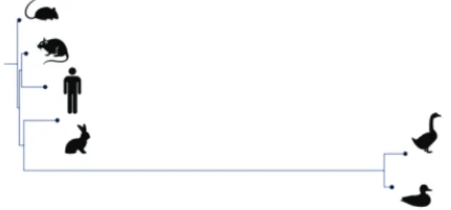
Pre-melanin-concentrating hormone (PMCH) gene encodes a preprotein. Proteolytical process generates dif- ferent proteins (i.e. MCH, neuropeptide-glutamic acid-iso- leucine, or neuropeptide-glycine-glutamic acid). In mammals MCH is a 19 amino acid peptide, the N-terminus is extended by two additional amino acids, with a highly conserved loop structure. In the central nervous system (CNS), in rodents and human the PMCH mRNA sequences have a high degree of homology with 90% overall nucleo- tide identity (Figure 2).
MCH is limited to the magnocellular neurons in the lateral hypothalamus and the zona incerta (Figure 3.).
These neurons have monosynaptic connections through- out the brain, projecting to the cortex, amygdala, nucleus accumbens, olfactory tubercle and brainstem nuclei [2, 3].
Overexpression of MCH may enhance food intake, contrib- utes to reduced glucose tolerance and provokes insulin resistance. In the last decade several research groups investigated the potential effects of MCH receptor (MCHR) antagonists on food intake regulation [4]. According to
the results MCHR antagonists may be beneficial against obesity, anxiety and depression [3, 5]. A novel promising MCHR1 antagonist SNAP-7941 caused less food intake and body weight [6].
MCH has two G-coupled receptors, MCHR1 and MCHR2.
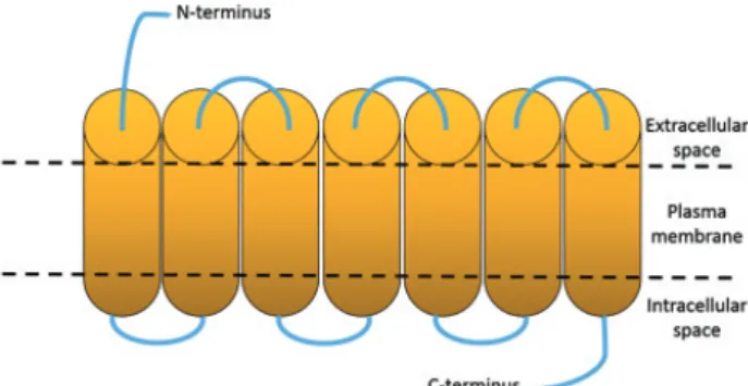
MCHR1 is 353 amino acids long, has seven transmembrane domains, an aspartic acid–arginine–tyrosine (DRY) motif at the end of the third intracellular loop and three potential glycosylation sites at the N-terminus (Figure 4.). MCHR1 gene is localized on chromosome 22q13.3. The receptor is highly conserved among mammals, sequence homol- ogy between human and mouse is 95%; while between human and rat is 96%. When MCH binds to the receptor it suppresses the forskolin-stimulated cAMP activation and increases intracellular Ca2+ level [2, 7].
MCHR1 can couple to Gi, G0 and Gq proteins, however the interaction is stronger with Gi and Gq. It stimulates protein kinase C (PKC), phospholipase C (PLC) and other extracellular-signal-regulated kinase pathways (Figure 5.). In the CNS it has diverse effects. Gi-coupled receptors are known to inhibit voltage-gated Ca2+ channels (VGCCs) and to activate K+-inward rectifying channels. MCH inhibit Ca2+ currents through the N-, P-, and with lesser degree Figure 1: Brain regions involved in food intake regulation (Human
hemisphere, midsagittal view).
Figure 2: Phylogenetic tree based on the amino acids sequence of melanin-concentrating hormone (MCH). [Length of lines shows the degree of difference in 6 species compared to the sequence of mouse MCH].
Figure 3: Melanin-concentrating hormone immunopositive (brown) cells on formalin fixed paraffin embedded rat brain slides, haema- toxylin counterstain. At low magnification (Panel A – 40x) there are strong immunoreactions in the hypothalamic area (circle). At higher magnification (Panel B - 400x) the reaction is limited to the cytoplasm (arrow).
the shell of the nucleus accumbens, an area involved in reward-related behaviour. Furthermore, MCHR1 is highly expressed in the amygdala and hippocampus suggesting its role in the regulation of emotions (i.e. fear or anxiety)
and humans. MCHR2 activation increases the intracellular Ca2+ level and it couples to Gq-proteins [2].
In the ob/ob mouse model, the PMCH expression increases two-to three-fold with fasting. Leptin treatment blunts the fasting-induced increase of PMCH mRNA in both the wild type and ob/ob mice. The intracerebroven- tricular administration of 5 µg MCH to rats led to rapid increase of chow consumption. Compared with control group, MCH-treated animals eat two-three-fold more over a six-hour period. Feeding can also be induced by injec- tion of MCH directly into the paraventricular nucleus.
Repetitive intramuscular injection of MCH into rats over a one-week period did not lead to obesity, whereas chronic infusion into the lateral ventricle led to both hyperphagia and weight gain [2, 8, 9]. These studies experimentally confirmed that MCH is essential to fasting response, but Figure 4: Structure of melanin-concentrating hormone receptor 1
(MCHR1).
Figure 5: Signalling pathways of melanin-concentrating hormone receptor 1 (MCHR1) (left panel) and MCHR2 (right panel). MCH binding to receptors leading to G protein coupling (Gs, Gi, Gq). While, Gi activation causing decreased amount of cAMP (red arrows), Gq resulting in increased mitogen-activated protein kinase (MAPK) activity (blue arrows) via Ras and elevation of intracellular calcium level (mediated by inositol trisphosphate (IP3) and phospholipase C (PLC) activation). This cascade induces changes in cell proliferation and gene transcrip- tion. [Protein kinase A=PKA; protein kinase C=PKC; diacylglycerol=DAG; MAPK kinase=MEK] (Adapted from Presse et al. [24])
unbalanced expression contributes to positive energy balance leading to health problems.
Furthermore, the cytokine receptor interleukin-6 alfa (IL-6Rα) is co-expressed with MCH and orexin in human and mouse hypothalamus in the hypothalamic, periforni- cal, dorsal and posterior areas, dorsomedial nucleus and in the zona incerta. In addition, MCH and orexin positive neurons contain IL-6Rα, suggesting that immune mecha- nisms may also be involved in the orchestration of energy balance. [10].
The ventromedial nucleus of hypothalamus (VMH) is a satiety centre and a main target of leptin which inhibits feeding, increases energy expenditure and finally causes weight loss [11].
The lateral hypothalamic area (LHA) is the feeding centre. It has an important role in mediating hyperphagia which is induced by hypoglycaemia [12].
The Dorsomedial hypothalamic nucleus (DMH) has connection to VMH and the LHA and it integrates the information from these nuclei [13].
The ventromedial aspect of the arcuate nucleus, which produces the orexigenic peptides neuropeptide Y (NPY) and agouti-related peptide (AGRP), contains glu- tamic acid decarboxylase (GAD) and vesicular gamma-am- inobutyric acid (GABA) transporter (VGAT). Surprisingly, in the LHA orexin-producing neurons express vesicular glutamate transporter 1 (VGLUT1) or VGLUT2, but not GAD, whereas some MCH cells contain GAD [14–16]. The hypothalamus is the primary CNS region to control energy balance, but is also associated with thirst, reproduction, temperature, hormonal balance and biological rhythms.
Lesions of lateral hypothalamus produce aphagia, adipsia and weight loss. As a compensatory mechanism MCH expression increases during fasting and encourages food intake [2, 15–18].
Experimentally MCH administration leads to rapid and robust feeding response, while chronic infusions results in mild obesity. MCH expression changes in states of altered energy balance, such as fasting and obesity.
Silencing either PMCH or MCHR genes leads to a lean phe- notype [2]. As we discussed MCH antagonists inhibit both feeding and diet-induced obesity [3–5].
In mammals, the widespread projections of MCH neurons in the lateral and basal hypothalamus suggest the complex role of peptide in the regulation of feeding behaviour, fluid intake, stress response, reproduction, arousal and sensory-motor integration [19].
3 The MCH system in avians
In avians, the diencephalon is highly developed and implicated in numerous physiological processes. A phy- logenetic novelty is the very accurate thermoregulation centre located in bird’s hypothalamus, which is important to maintain the continuously high body temperature of avians. The diencephalon is divided into three parts like in the other vertebrates i.e. the thalamus, hypothalamus and epithalamus. The hypothalamus is a part of the reticular formation. It contains different secretory neurons which have roles in the neuroendocrine regulation [20].
Cardot et al. detected MCH immunopositivity in five species’ brain: Leghorn cocks, Guinea hens, quails, gos- lings and ducks [21]. Neuronal perikarya were strongly positive in hypothalamus mainly in the periventricular hypothalamic nucleus in all species except the cocks, where only few perikarya showed immunoreactivity.
Fibres and nerve terminals were very thin and sparse, especially in ducks and quails. In LHA immunopositivity were found in the periventricular nucleus, the ventricular nucleus and the dorsomedial nucleus. A lot of fibers had positive immunoreaction in the medial area of the thala- mus. Although most of them were thin, some were long and unramified.
Regarding the brainstem, innervation by MCH pos- itive neurons was observed in all species in the rostral part of the pons and metencephalon, along the fourth ventricle, in the locus coeruleus, the locus subcoeruleus, the oral pontine reticular nucleus and the linear caudal nucleus. In the medulla oblongata some immunopositive fibers were detected in the medial vestibular nucleus, in the raphe nucleus, the posterior area of the caudal pontine reticular nucleus and the vestibular nuclei. In birds the MCH system, especially in the lateral and basal hypothalamus, has an important role in the limbic system and it coordinates the vegetative and endocrine functions as well as the emotional behaviour [21]. The non-mam- malian vertebrates studies have identified several mam- malian MCHR1/MCHR2-like receptors. According to the predictions seven mammalian MCHR-like receptor exist (MCHR1-MCHR7) in different species such as zebrafish or coelacanths [22]. However, our knowledge on MCHRs in avians is limited due to the absence of experimen- tal studies. In a recent paper Cui et al. have identified a MCHR1-like receptor (chicken-MCHR4) and a pseudo MCHR2 in chickens [23]. The cMCHR4 is activated by chicken MCH (cMCH) and induces different signalling
the complex regulatory role of MCH system in birds. In addition, fasting increases cMCH mRNA level in chicken hypothalamus indicating its essential function in con- trolling avian energy balance [23].
4 Conclusion
MCH is highly conservative neuropeptide in mammals and avians, which plays a crucial role in the regulation of food intake and affectivity. A development of selective MCH system modulators could become promising thera- peutic options in the treatment of obesity and obesity-re- lated diseases (i.e. diabetes) or depression. Furthermore, in agriculture the enhancement of endogenous MCH levels in livestock may contribute to higher and more eco- nomical meat yield. However, detailed morphological and physiological analysis of MCH system in different species is essential to examine the potential effects of novel phar- macological agents on energy homeostasis.
Acknowledgements: Supported by the National Brain Research Program, Hungary 2017-1.2.1-NKP-2017-00002;
GINOP-2.3.2-15-2016-00043; AGR_PIAC_13-1-2013-0008 (TH); ÚNKP-17-3 New National Excellence Program of the Ministry of Human Capacities and EFOP-3.6.3-VE- KOP-16-2017-00009 (JB).
Conflict of interest statement: Authors state no conflict of interest.
References
[1] Hopkins M., Blundell J., Halford J., King N., Finlayson G., The Regulation of Food Intake in Humans, Endotext, 2000, 1-15 [2] Pissios P., Maratos-Flier E., Melanin-concentrating
hormone: From fish skin to skinny mammals, Trends Endocrinol. Metab., 2003, 14, 243–248, DOI: 10.1016/
S1043-2760(03)00079-1
[3] Arora S., Anubhuti, Role of neuropeptides in appetite regulation and obesity - A review, Neuropeptides, 2006, 40, 375–401, DOI: 10.1016/j.npep.2006.07.001
[6] Doggrell S.A., Does the melanin-concentrating hormone antagonist SNAP-7941 deserve 3As?, Expert Opin Investig Drugs, 2003, 12, 1035–1038, DOI:
10.1517/13543784.12.6.1035
[7] Chung S., Parks G.S., Lee C., Civelli O., Recent updates on the melanin-concentrating hormone (MCH) and its receptor system: Lessons from MCH1R antagonists, J. Mol. Neurosci., 2011, 43, 115–121, DOI: 10.1007/s12031-010-9411-4 [8] Mantzoros C.S., Qu D., Frederich R.C., Susulic V.S., Lowell
B.B., Maratos- Flier E., et al., Activation of ??3 adrenergic receptors suppresses leptin expression and mediates a leptin-independent inhibition of food intake in mice, Diabetes, 1996, 45, 909–914
[9] Nahon J.L., Presse F., Bittencourt J.C., Sawchenko P.E., Vale W., The rat melanin-concentrating hormone messenger ribonucleic acid encodes multiple putative neuropeptides coexpressed in the dorsolateral hypothalamus,
Endocrinology, 1989, 125, 2056–2065, DOI: 10.1210/
endo-125-4-2056
[10] Schéle E., Fekete C., Egri P., Füzesi T., Palkovits M., Keller É., et al., Interleukin-6 Receptor α is Co-localised with Melanin-Concentrating Hormone in Human and Mouse Hypothalamus, J. Neuroendocrinol., 2012, 24, 930–943, DOI:
10.1111/j.1365-2826.2012.02286.x
[11] Satoh N., Ogawa Y., Katsuura G., Tsuji T., Masuzaki H., Hiraoka J., et al., Pathophysiological significance of the obese gene product, leptin, in ventromedial hypothalamus (VMH)lesioned rats: Evidence for loss of its satiety effect in VMH-lesioned rats, Endocrinology, 1997, 138, 947–954, DOI:
10.1210/en.138.3.947
[12] Bernardis L.L., Bellinger L.L., The lateral hypothalamic area revisited: Ingestive behavior, Neurosci. Biobehav. Rev., 1996, 20, 189–287, DOI: 10.1016/0149-7634(95)00015-1
[13] Elmquist J.K., Ahima R.S., Elias C.F., Flier J.S., Saper C.B., Leptin activates distinct projections from the dorsomedial and ventromedial hypothalamic nuclei, Proc. Natl. Acad. Sci., 1998, 95, 741–746, DOI: 10.1073/pnas.95.2.741
[14] Harthoorn L.F., Sañé A., Nethe M., Van Heerikhuize J.J., Multi-transcriptional profiling of melanin-concentrating hormone and orexin-containing neurons, Cell. Mol.
Neurobiol., 2005, 25, 1209–1223, DOI: 10.1007/s10571-005- 8184-8
[15] Guyon A., Conductier G., Rovere C., Enfissi A., Nahon J.L., Melanin-concentrating hormone producing neurons:
Activities and modulations, Peptides, 2009, 30, 2031–2039, DOI: 10.1016/j.peptides.2009.05.028
[16] Meister B., Neurotransmitters in key neurons of the hypothalamus that regulate feeding behavior and body weight, Physiol. Behav., 2007, 92, 263–271, DOI: 10.1016/j.
physbeh.2007.05.021
[17] Griffond B., Risold P.Y., MCH and feeding behavior-interaction with peptidic network, Peptides, 2009, 30, 2045–2051, DOI:
10.1016/j.peptides.2009.07.008
[18] Mouri T., Takahashi K., Kawauchi H., Sone M., Totsune K., Murakami O., et al., Melanin-concentrating hormone in the human brain, Peptides, 1993, 14, 643–646, DOI:
10.1016/0196-9781(93)90158-D
[19] Naufahu J., Cunliffe A.D., Murray J.F., The roles of melanin-concentrating hormone in energy balance and reproductive function: Are they connected, Reproduction, 2013, 146, R141–R150, DOI: 10.1530/REP-12-0385 [20] Nickel R., Schummer A., Seiferle E., The anatomy of the
domestic birds, Springer-Verlag, New York 1977
[21] Cardot J., Griffond B., Risold P.Y., Blähser S., Fellmann D., Melanin-concentrating hormone-producing neurons in birds., J. Comp. Neurol., 1999, 411, 239–56
[22] Yun S., Furlong M., Sim M., Cho M., Park S., Cho E.B., et al., Prevertebrate Local Gene Duplication Facilitated Expansion of the Neuropeptide GPCR Superfamily, Mol. Biol. Evol., 2015, 32, 2803–2817, DOI: 10.1093/molbev/msv179
[23] Cui L., Lv C., Zhang J., Mo C., Lin D., Li J., et al., Charac- terization of melanin-concentrating hormone (MCH) and its receptor in chickens: Tissue expression, functional analysis, and fasting-induced up-regulation of hypothalamic MCH expression, Gene, 2017, 615, 57–67, DOI: 10.1016/j.
gene.2017.03.009
[24] Presse F., Conductier G., Rovere C., Nahon J.-L., The melanin-concentrating hormone receptors: neuronal and non-neuronal functions, Int. J. Obes. Suppl., 2014, 4, S31–
S36, DOI: 10.1038/ijosup.2014.9