NOVEL INNERVATION PATHWAYS IN THE REGULATION OF BASAL FOREBRAIN NEURONS
Ph.D. Thesis
Zsuzsanna Bardóczi
János Szentágothai Doctoral School of Neuroscience Semmelweis University
Supervisor: Imre Kalló, M.D., Ph.D.
Official reviewers: István Ábrahám, Ph.D., D.Sc., Habil Zsuzsanna Várnainé Tóth, Ph.D.
Head of the Final Examination Committee: András Csillag, M.D., Ph.D., D.Sc.
Members of the Final Examination Committee: Ádám Dénes, M.D., Ph.D.
Dobolyi Árpád, Ph.D., D.Sc., Habil
Budapest
2018
2
TABLE OF CONTENTS
List of abbreviations ... 5
1. Introduction ... 8
1.1. Organization of basal forebrain (BF) ... 8
1.2. Cholinergic neurons in the BF ... 9
1.2.1. Distribution and projection of cholinergic neurons in the BF ... 9
1.2.2. Basic operation modes of BF cholinergic neurons ... 10
1.2.3. BF cholinergic neurons as major targets of ascending reticular activating system (ARAS)... 11
1.2.4. Potential role of cholinergic output to forebrain centers ... 12
1.3. GnRH neurons in the BF ... 15
1.3.1. General morphology of the GnRH neurons ... 15
1.3.2. Distribution of GnRH cell bodies ... 16
1.3.3. Hypothalamo-pituitary-gonadal (HPG) axis... 17
1.3.3.1. Operation modes (pulsatile and surge) of the GnRH neurons and the underlying electrophysiological events in GnRH neurons ... 18
1.3.3.2. The “GnRH Pulse generator” – Intrinsic vs. extrinsic features of the network ... 19
1.3.3.3. The surge mode of GnRH secretion – Integration of estrogen- and circadian signals in the regulation of GnRH secretion ... 20
1.3.4. Kisspeptin (KP) neurons forming a major input to GnRH neurons ... 22
1.3.4.1. KP neurons in the rostral periventricular area of the third ventricle (RP3V) – role in surge generation ... 22
1.3.4.2. KP neurons in the arcuate nucleus (Arc) – the extrinsic pulse generator ... 23
1.3.5. Projection areas of GnRH neurons - neuronal output to preoptic/hypothalamic centers ... 24
1.4. Potential role of a glycinergic input to BF neurons ... 28
2. Aims…… ... 30
3. Materials and Methods ... 31
3.1. Mouse brain samples ... 31
3.1.1. Brain tissue collected for immunohistochemical processing ... 31
3.1.2. Mouse models used in the different experiments ... 31
3.1.3. Acute slices collected for electrophysiological examination ... 32
3.2. Human brain tissue samples ... 33
3.3. Preparation of mouse and human sections for light microscopic studies ... 33
3.4. Preparation of mouse and human sections for electron microscopic studies ... 34
3
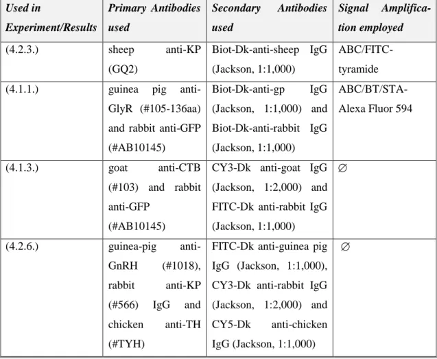
3.5. Single- and multiple labeling of tissue antigens for confocal
analyses ... 35
3.6. Single- and dual labeling for light- and electron microscopy ... 36
3.7. Tract-tracing to identify glycinergic afferents to BF neurons ... 39
3.8. Immunohistochemical controls ... 40
3.9. Microscopy and data analysis ... 41
3.9.1. Correlated light- and electron microscopy ... 41
3.9.2. Confocal laser microscopy ... 41
3.9.3. Mapping and quantification ... 42
4. Results…. ... 43
4.1. Examination of glycinergic input to BF neurons ... 43
4.1.1. Subnucleus and cell-specific appearance of glycine receptors (GlyRs) in the BF ... 43
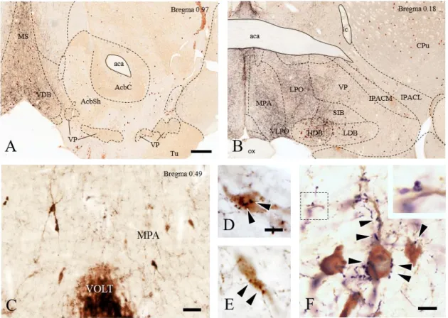
4.1.2. Distribution of glycinergic (GLYT2-IR) fibers in the BF and their appositions to GnRH and cholinergic neurons ... 44
4.1.3. Localization of glycinergic neurons projecting to the BF ... 49
4.1.4. GLYT1-IR astroglial processes in the vicinity of GnRH and cholinergic neurons ... 51
4.1.5. Collaborative studies on the membrane effects of glycine in GnRH- and cholinergic neurons... 53
4.2. Characterization of GnRH projections and their target cells in mice and humans ... 55
4.2.1. Ultrastructure of GnRH-IR processes in mice ... 55
4.2.2. Analysis of KP contacts in mice ... 56
4.2.3. Analysis of KP contacts in human ... 57
4.2.4. Confocal microscopic analysis of GnRH-IR processes forming appositions on KP- and/or tyrosine hydroxylase (TH)-IR neurons in the RP3V and Arc ... 59
4.2.5. Identifying synaptic targets of GnRH axons ... 60
4.2.6. Hormonal and lactation-related effects on the GnRH input to hypothalamic KP- and TH-IR neurons ... 62
4.2.6.1. Circadian effect on GnRH input to KP neurons ... 62
4.2.6.2. Effect of lactation on GnRH input to KP- and/or TH-IR neurons ... 63
5. Discussion ... 66
5.1. Glycinergic input of BF neurons ... 66
5.1.1. GlyRs are distributed throughout the BF ... 66
5.1.2. Role of glycinergic (GLYT2-IR) afferents in the BF... 67
5.1.3. Origin of glycinergic input to the BF ... 67
5.1.4. Role of the presence of the GLYT1-IR astroglial processes in the BF ... 68
5.1.5. Direct glycine responsiveness of BF cholinergic, but not GnRH neurons ... 69
4
5.2. Characterizing GnRH efferents and their target cells in mice and
humans ... 70
5.2.1. Ultrastructural features of GnRH processes in mice ... 70
5.2.2. The importance of KP-KP contacts in mice and human ... 71
5.2.3. GnRH axons target KP-IR neurons in both the RP3V and the Arc ... 73
5.2.4. TH-IR neurons represent the second major neuronal population targeted by GnRH afferents... 74
5.3. Hormonal- and lactation-related effects on the GnRH input of KP- and TH-IR neurons ... 75
5.3.1. Possible plasticity of GnRH input to KP-and TH-IR neurons in lactating animals ... 75
5.3.2. Possible plastic change of the GnRH afferents to KP neurons at different circadian stages ... 76
6. Conclusions... 78
7. Summary ... 82
8. Összefoglalás ... 83
9. References ... 84
10. List of publications ... 113
10.1. List of publications underlying the thesis ... 113
10.2. List of other publications ... 114
11. Acknowledgements ... 115
5
List of abbreviations
ACh - Acetylcholine
AChR - Acetylcholine receptor AD - Alzheimer’s disease
AHA - anterior hypothalamic area Arc - arcuate nucleus
AVPV - anteroventral periventricular nucleus BF - basal forebrain
BLA - basolateral nucleus of the amygdala cAMP - cyclic adenosine monophosphate ChAT - choline acetyltransferase
CNS - central nervous system CTB - cholera toxin B
DA - dopamine
DAB - diaminobenzidine DBB - diagonal band of Broca Dyn - dynorphin
E2 - 17β-estradiol EA - extended amygdala EC - entorhinal cortex
EGFP - enhanced green fluorescent protein ERα - estrogen receptor alpha
ERβ - estrogen receptor beta FSH - follicle-stimulating hormone GABA - gamma-aminobutyric acid
GABA-A - gamma-aminobutyric acid A receptor GABA-B - gamma-aminobutyric acid B receptor GlyR - Glycine receptor
GLYT1 - glycine transporter, type 1 GLYT2 - glycine transporter, type 2 GnRH - gonadotropin-releasing hormone GP - globus pallidus
6
HDB - horizontal limb of the diagonal band of Broca HPG - Hypothalamo-pituitary-gonadal
icv - intracerebroventricular INF - Infundibular nucleus InfS - Infundibular stalk IR - immunoreactive
Kiss1R - Kisspeptin receptor
KNDy - Kisspeptin/Neurokinin B/Dynorphin KP - Kisspeptin
LC - locus coeruleus
LDT - laterodorsal tegmental nuclei LH - luteinizing hormone
LHA - lateral hypothalamus LTP - long term potentiation
mAChRs - muscarinic acetylcholine receptor MBH - mediobasal hypothalamus
MCH - melanin-concentrating hormone MCPO - magnocellular preoptic nucleus MPA - medial preoptic area
MS - medial septum
nAChRs - nicotinic acetylcholine receptor NBM - nucleus basalis of Meynert
Ni-DAB - nickel-diaminobenzidine NK3R - neurokinin B receptor NKB - neurokinin B
NMDA - N-methyl-D-aspartate NREM - non-rapid eye movement OB - olfactory bulb
OVLT - organum vasculosum of the lamina terminalis OVX - ovariectomized
PBS - phosphate buffered saline PFA - paraformaldehyde
PFC - prefrontal cortex
7 PHDA - periventricular hypophysial dopaminergic PPT - pedunculopontine
REM - rapid eye movement RMg - raphe magnus nucleus
RP3V - rostral periventricular area of the third ventricle POA - preoptic area
SCN - suprachiasmatic nucleus SFO - subfornical organs
SGI-NiDAB - silver gold intesified nickel-diaminobenzidine Si - substantia innominata
TH - tyrosine hydroxylase
THDA - tuberohypophysial dopaminergic TIDA - tuberoinfundibular dopaminergic TMN - tuberomammillary nucleus
VAChT - vesicular acetylcholine transporter VDB - vertical limb of the diagonal band of Broca VIP - vasoactive intestinal peptide
VLPO - ventrolateral preoptic nucleus VP - ventral pallidum
ZT - zeitgeber time
8 1. Introduction
1.1. Organization of basal forebrain (BF)
The BF is located close to the medial and ventral surfaces of the cerebral hemispheres, to the front of and below the striatum. The main regions of the BF are: medial septum (MS), ventral pallidum (VP), diagonal band nuclei, substantia innominata (Si)/extended amygdala (EA), nucleus basalis of Meynert (NBM) and peripallidal regions. The BF contains a heterogeneous mixture of cell types: neuropeptide containing neurons [1]
such as Gonadotropin-releasing hormone (GnRH)-immunreactive (IR) neurons, a heterogeneous collection of cholinergic, gamma-aminobutyric acid (GABA)-ergic, glutamatergic projection neurons, and various interneurons [2]. This highly complex brain region has been implicated in the regulation of reproduction, cortical activation, attention, motivation, memory, and neuropsychiatric disorders (i.e. Alzheimer’s disease (AD), Parkinson’s disease, schizophrenia, drug abuse) [3-11].
GnRH neurons form a unique cell population in the BF in that they are born in the olfactory placod and migrate during the embryonic development into the hypothalamus and BF areas [12]. In contrast, the BF cholinergic projection neurons and the other cell types, including GABAergic projection neurons and subsets of cortical and striatal GABAergic interneurons originate within the primordium of the ventral telencephalon (subpallium) [13].
According to the classical concept of the ascending arousal system [14], a major branch of the complex pathways from the rostral pons and caudal midbrain reaches the hypothalamus and the BF to activate these brain regions. The possible existence of an ascending inhibitory (glycinergic) pathway and local action of glycine in the BF have been indicated in previous studies from our laboratory [15, 16]; the expression of the glycine receptor (GlyR) alpha 1 subunit in GnRH neurons strongly suggests that glycine can directly influence GnRH neurons via GlyR. Also, we have confirmed the presence of membrane glycine transporter type 1 (GLYT1) and glycine transporter type 2 (GLYT2) in the BF regions indicating a role for glycine in the regulation of local neuronal populations.
The first part of my Ph.D. thesis addresses the issue of whether the GnRH and/or cholinergic neurons are targets of glycine signaling in the BF.
9 1.2. Cholinergic neurons in the BF
1.2.1. Distribution and projection of cholinergic neurons in the BF
Acetylcholine (ACh) is synthesized in nerve terminals from choline and acetyl coenzyme A by the cytoplasmic enzyme choline acetyltransferase [17] and transported into synaptic vesicles by the vesicular acetylcholine transporter (VAChT) [18]. Using antisera against ChAT represents a reliable marker for the study of cholinergic neurons in the central and peripheral nervous systems. Five main neuronal groups that contain the majority of central cholinergic neurons have been identified.
These include: (i) the efferent cranial nerve nuclei and motoneurons of the spinal cord;
(ii) the parabrachial complex; (iii) the brainstem reticular complex; (iv) the neostriatal complex; and (v) the medial basal forebrain [19]. In my Ph.D. thesis, the focus is on the input of the BF cholinergic neurons.
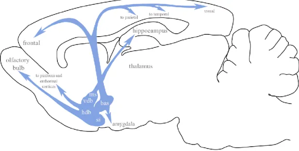
Fig. 1. Schematic representation of the basal forebrain cholinergic neurons and their projection areas. bas=nucleus basalis; ms=medial septum; vdb=vertical diagonal band nucleus; hdb=horizontal diagonal band nucleus; si=substantia innominate. Modified from [20].
Mesulam and his colleagues subdivided the BF cholinergic neurons into four major subgroups, which they designated Chl-Ch4. Ch1 subgroup consist of the medial septal cholinergic neurons. Ch2 subgroup contain the cholinergic neurons, which are located
10
in the vertical limb of the diagonal band (VDB). The Chl and Ch2 groups collectively provide the major cholinergic innervation of the hippocampus. The group of cholinergic neurons in the horizontal limb of the diagonal band (HDB) and magnocellular preoptic nucleus (MCPO) are termed as Ch3 subgroup of cholinergic neurons. The neurons from the Ch3 mainly project to the olfactory bulb, piriform, and entorhinal cortices.
Cholinergic neurons within the VP, Si/EA, globus pallidus (GP), internal capsule, and nucleus ansa lenticularis, collectively termed the Ch4 subgroup, project to the basolateral amygdala, and innervate the entire neocortex according to a rough medio- lateral and antero-posterior topography (Fig.1.) [1, 21]. Cholinergic neurons in the MS/VDB, HDB andMCPO also innervate the orexin/hypocretin neurons in the lateral hypothalamus [22, 23].
1.2.2. Basic operation modes of BF cholinergic neurons
In the central nervous system (CNS), the BF cholinergic neurons release acethylcholine (ACh), which binds to the appropriate receptors on the postsynaptic target cells. The ACh effects depend on the presence of the AChR subtype(s) on the target, the cellular localization of the AChRs and the multiplicity of downstream signaling cascades that can be activated. AChRs are categorized based on their sensitivity to plant alkaloids and their binding capacity for muscarine or nicotine (mAChRs & nAChRs). Muscarinic AChRs are metabotropic receptors that bind ACh and transduce their signaling via acti- vation of heterotrimeric G proteins that, in turn, affect the opening, closing, and kinetics of (primarily) K+, Ca2+ and non-selective cation channels. In contrast to ACh interac- tions with mAChRs, binding of ACh to nicotinic AChRs results in the direct gating of non-selective cation channels. Twelve different types of nAChR subunits have been identified in the brain (α2–10 and β2–β4) [24].
Based on previous studies, ACh release is characterized classically as slow and tonic [25]. The anatomically diffuse cholinergic system and early microdialysis experiments that documented ambient levels of ACh at micromolar concentrations in brain tissue [25] suggest the “volume” mode of transmission.
This hypothesis has now been substantially revised by Sarter & colleagues [26]. Using more rapid assays for ACh release and new approaches for selective activation of
11
cholinergic neurons and their terminal fields, they find evidence for a faster and more focal ACh release and downstream signaling than previously considered [27, 28].
Záborszky and his colleagues supported these findings with a detailed electrophysiological characterization of the cholinergic neurons in the NBM. They identified two populations: a more excitable, early firing population that show spike frequency adaptation and a less excitable, late firing population that could maintain low frequency tonic firing [29]. The two phenotypes of cholinergic neurons may provide the cellular basis for two different modes of signaling: fast and focal and slow and paracrine. Taken together, it appears that these modes of ACh release play important roles in different aspects of information processing [28, 30].
1.2.3. BF cholinergic neurons as major targets of ascending reticular activating system (ARAS)
Forebrain activation and cortical arousal/waking behavior are thought to be critically influenced by ascending pathways deriving from the brainstem [31-38]. The role of the upper brainstem in forebrain arousal was demonstrated fifty years ago by Moruzzi and Magoun [39]. However, the specific structures that activate the forebrain have only been described recently.
The ascending arousal system has two major branches. One pathway originates from cholinergic cell groups in the upper pons, the pedunculopontine (PPT) and laterodorsal tegmental nuclei (LDT) and reaches the thalamus where it activates the thalamic relay neurons that are crucial for transmission of information to the cerebral cortex. The neurons in the PPT/LDT show most rapid firing during wakefulness and rapid eye movement (REM) sleep; the REM stage is characterized by concomitant cortical activation, loss of muscle tone in the body and active dreams [40]. These cholinergic cells are much silent during non-REM (NREM) sleep, when cortical activity is slow.
The other branch circumvents the thalamus and activates the cerebral cortex to facilitate the processing of inputs from the thalamus. This pathway originates from various monoaminergic cell groups, including the tuberomammillary nucleus (TMN) containing histamine, the A10 cell group containing dopamine (DA), the dorsal and median raphe nuclei containing serotonin, and the locus coeruleus (LC) containing noradrenaline. The input to the cerebral cortex is enhanced by lateral hypothalamic peptidergic neurons
12
(containing melanin-concentrating hormone (MCH) or orexin/hypocretin), and BF neurons (containing ACh or GABA). The monoaminergic neurons, which are part of this pathway, increase their firing rate during wakefulness, decrease firing activity during NREM sleep and stop altogether during REM sleep [41-43]. Orexin neurons in the LHA are, similarly, most active during wakefulness [44-46], whereas MCH neurons are active during REM sleep [47]. Many BF neurons, including most cholinergic neurons, are active during both wake and REM sleep [48].
However, other pathways also exist which, in turn, promote sleep. During the 1980s and 1990s, investigators found that a group of neurons located in the ventrolateral preoptic nucleus (VLPO) send outputs to all of the major cell groups in the hypothalamus and brainstem that participate in arousal [49]. The VLPO neurons are primarily active during sleep, and contain the inhibitory neurotransmitters, galanin and GABA [50-52].
They send output to monoaminergic neurons in the TMN, the A10 cell group, the raphe cell groups and the locus coeruleus LC. They also innervate the LHA, including the perifornical (PeF) orexin neurons, and interneurons of the brainstem cholinergic cell groups, the (PPT) and (LDT). The LC and DR play an important role in gating REM sleep [47, 53]. The VLPO neurons also innervate the histaminergic neurons, which are involved in the transitions between arousal and NREM sleep [53-55].
1.2.4. Potential role of cholinergic output to forebrain centers
ACh is reponsible for attention [56, 57], arousal [58-60], learning and memory [61, 62]
and the sleep-wake cycle [60, 63, 64]. It is thought that the effect of ACh depends on its target areas [56, 65].
In rats, cholinergic neurons that project to the cerebral cortex are dispersed throughout the BF within the nuclei of the diagonal band of Broca (DBB), MCPO, Si and GP [66, 67]. They compose the extrathalamic relay from the brainstem activating system to the cerebral cortex [39, 68], where they potently excite cortical neurons and stimulate corti- cal activation [69-71]. Release of ACh is in close association with cortical activation during the states of waking and paradoxical sleep [72-75]. The cholinergic neurons are more active during wakefulness and REM sleep (wake/REM active) than during NREM sleep, as are the glutamatergic and parvalbumin-positive GABAergic neurons, which are also distributed in the BF. Optogenetic activation of these neurons rapidly induces
13
wakefulness, contrasting with somatostatin-positive GABAergic neurons, the stimula- tion of which promotes NREM sleep [76]. However, chemogenetic activation of BF cholinergic or glutamatergic neurons in behaving mice has no effect on total wakeful- ness. In contrast, similar chemogenetic activation of BF GABAergic neurons produces sustained wakefulness and high frequency cortical rhythms [77].
The cholinergic neurons, which are located in the NBM and DBB, project to the prefrontal cortex (PFC) [78-80]. The PFC is an integral node in circuits underlying attention and ACh modulates these processes. Attention consists of two separate processing streams: goal or cue driven attention is known “top down” while sensory driven attention is known “bottom up” [81, 82]. Basically, top down attention is considered as voluntary, or “feed-back” driven, thus incoming sensory information is processed by higher cortical areas. However, bottom up attention is considered as involuntary, or “feedforward”, thus sensory information is fed forward and up to the cortex [81, 82]. Enzyme selective microelectrode studies confirmed that ACh in the PFC modulates the cue detection and cue triggered changes in goal-oriented behavior [83, 84]. Taken together, both the beginning and the end of the attention loop are mediated by ACh and initiate the top down control over downstream sensory cortical areas.
Cholinergic neurons also modulate the sensory cortex related to attention. During the attentional performance of a behavioral task, ACh decorrelates intracortical noise in sensory cortices, which is often measured as “desynchronization” or decreased power of low frequency local field potential (LFP). Decorrelation increases the response reliability of sensory cortex neurons to the appropriate stimuli [85, 86].
Cholinergic innervation from the MS and DBB to the hippocampus plays an important role in the formation of spatial memories: elevated ACh level have been confirmed by microdialysis in the hippocampus during performance of various memory tasks [87-89].
At the circuit level, several lines of studies of memory suggest that ACh, acting via both nicotinic and muscarinic AChRs (nAChRs and mAChRs), is important for the initiation of long-term potentiation (LTP), a synaptic substrate of memory. In the hippocampus cholinergic signaling both promotes LTP and regulates cognition associated oscillatory activity. Theta rhythm phase can both regulate the possibility that LTP is initiated and determine whether stimulation will generate synaptic potentiation or depression [90].
Oscillations are known to isolate the signal of encoding from retrieval actions, the
14
differentiation that is crucial for memory as the status of these oscillations at the onset of a behavioral task presumes learning success [91].
The medial septal cholinergic neurons also send axons to the entorhinal cortex (EC).
The EC plays an important role in processing and conveying spatial information to the hippocampus. Optogenetic studies revealed that the septal cholinergic neurons modulate EC neurons in layer specific manner, via nAChRs and mAChRs. Selective lesions of septal cholinergic neurons or their optogenetic activation have indicated that ACh plays an important role in regulating theta rhythmic activity in the hippocampus, thereby augmenting the dynamics of memory encoding [92-95].
Furthermore, a dense projection of the BF cholinergic neurons reaches the basolateral nucleus of the amygdala (BLA), which is a subcortical limbic structure [2]. While ACh mediates the spatial memory in the hippocampus, it consolidates the emotionally salient memories in the amygdala [20, 96]. Optogenetically stimulated cholinergic neurons in the amygdala enhance emotionally salient memories, and optogenetic inhibition decreases them: both nAChRs and mAChRs play a role in these processes [97].
Cholinergic neurons, which are lying in the HDB, innervate the olfactory bulb (OB) [98, 99]. The layered architecture of the OB, and its function as a relay between odor input and higher cortical processing, make it possible to examine how sensory information is processed at synaptic and circuit levels. The OB receives also strong neuromodulatory inputs, in part from BF cholinergic system. Cholinergic axons regulate the activity of various cells and synapses within the OB, especially the numerous dendro dendritic synapses, resulting in highly variable responses of OB neurons to odor input that is dependent on the behavioural state of the animal. Behavioral, electrophysiological, anatomical, and computational studies provide evidence for the involvement of mAChRs and nAChRs in the actions of ACh in the OB [100].
Taken together, BF cholinergic neurons represent a heterogeneous cell population (Ch1- Ch4; [21]) with different projection areas, and they have been implicated in arousal, sleep-wake cycle, attention, memory and olfactory processes. Thus, it is very important to define which BF cholinergic subgoups are regulated by glycine.
15 1.3. GnRH neurons in the BF
1.3.1. General morphology of the GnRH neurons
GnRH is synthesized and secreted from a relatively small population of neurons, the majority of which is located in mice and rats in the preoptic area (POA) of the hypothalamus; these cells represent the central regulators of reproductive functions and fertility. GnRH neurons do not develop from the neural tube; they originate from the nasal placodes and migrate prenatally along the olfactory sensory axons to the cribriform plate (E13.5) and then, to the BF areas (E16.5). From birth through adulthood, these neurons do not form a well-defined nucleus, but are scattered from the rostral POA to the caudal hypothalamus [12].
The number of GnRH neurons in mice is low (⁓800) [101]. Most GnRH neurons exhibit a fusiform, bipolar morphology with two processes, which originate from opposite sides of the soma. Unipolar or multipolar morphology also appear in a smaller percentage [102]. Furthermore, the GnRH processes have also dendritic spines, filopodium-like structures and cilia. Campbell and colleagues examined 45 biocytin-filled GnRH neurons in mice and they observed the highest density of dendritic spines on the proximal part (mostly on the first 50 µm) of the dendrite, which likely indicates the location of the majority of excitatory synaptic inputs [102-104]. Recently, a relatively high density of afferents were detected on distal processes of GnRH neurons in the vicinity of median eminence [105]. Approximately half of the GnRH neurons have filopodia, which are involved in synaptogenesis by locating and guiding appropriate axons back to the dendrite [102, 106-108]. GnRH neurons possess also multiple cilia [109, 110], which contain signaling molecules, including certain G protein-coupled receptors (GPCRs); these receptors may sense different neuromodulators in the extracellular space. The cilia of GnRH neurons express Kisspeptin receptor (Kiss1R), suggesting that the cilia may also play a role in kisspeptin (KP)-mediated increases in GnRH neuron firing rate [111].
GnRH neurons have long processes (over 1000 µm), which mostly project to the ME.
Between 50% and 70% of all GnRH neurons throughout the BF project to the median eminence [112-114]. Interestingly, these processes have a spike initiation site and conduct action potentials to the median eminence. They also receive simultaneously and integrate synaptic inputs along the complete length of the processes, thus regulating the
16
excitability of the neuron. These processes have axon- and dendrite-like properties, thus nowadays they are termed as „dendrons” [115].
Varicose axons, in addition to these dendrons, have also been observed [116]. Often they emanate from the thick dendron-like processes, and form synapses in the rostral periventricular area of the third ventricle (RP3V) and arcuate nucleus (Arc) as we have shown [117].
1.3.2. Distribution of GnRH cell bodies
The GnRH neurons are distributed from the olfactory bulbs to the medial septal nuclei and POA, ventral aspects of the anterior hypothalamic area (AHA) and mediobasal hypothalamus (MBH). The pattern of the distribution forms an inverted “Y”: the rostral- most midline GnRH cell bodies in the MS dividing at the beginning of the third ventricle within the rostral POA to form the two arms of the inverted “Y” that extend back into the ventral AHA and MBH. Although this continuum can be found in all mammals, there are species differences in the distribution of GnRH neurons along this pathway [109]. In rats, one group of the GnRH neurons is distributed in septal areas as well as in the medial septal nucleus, and in the vertical and horizontal limbs of the diagonal band of Broca and near to the organum vasculosum of the lamina terminalis (OVLT). Another group of GnRH-neurons is found in the medial preoptic area.
Furthermore, fewer GnRH neurons are located in the periventricular portion of the preoptic nucleus, in the bed nucleus of the stria terminalis and in the lateral preoptic area. Caudal to the septal-preoptic area, GnRH perikarya are observed in the anterior and ventrolateral hypothalamus, but absent from the medial basal hypothalamus/Arc.
Some can be found also in different parts of the hippocampus (indusium griseum, CA3 and CA1 fields of Ammon's horn) and in the piriform cortex [116].
In human and monkeys, GnRH cell bodies reside more caudally, as they are most concentrated in the ventral and basal hypothalamus. They extend their processes ventrally to the median eminence and infundibular stalk and caudally to the mammillary complex [118].
17
1.3.3. Hypothalamo-pituitary-gonadal (HPG) axis
Most of the GnRH processes terminate in the external zone of median eminence, where GnRH is secreted in the hypothalamo-hypophysial portal bloodstream, which carries it to the anterior pituitary gland. GnRH acts on the GnRH receptors, which are present on the gonadotrope cells and stimulates the release of luteinizing hormone (LH) and follicles stimulating hormone (FSH).
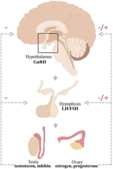
Fig. 2. The schematic drawing of the HPG axis. Hypophysiotropic GnRH is released by the hypothalamus and stimulates the secretion of gonadotropins (LH and FSH) from the hypophysis. LH and FSH act on the gonads and the gonadal steroids and peptides regulate the functions of the HPG axis by negative (in males and females) and positive (only in females) feedback mechanisms. (FSH: follicle stimulating hormone, GnRH:
gonadotropin-releasing hormone, LH: luteinizing hormone). Modified from [119].
18
Low-frequency pulses of GnRH result in release of FSH, whereas high-frequency pulses of GnRH preferentially trigger LH release [120]. Thus, the gonadotropes convert the hypothalamic signal (GnRH) to a systemic response (the release of FSH and LH) regulating fertility. LH and FSH are released into the systemic circulation and act on the gonads. In males, LH stimulates testosterone production from Leydig cells, whereas FSH stimulates the spermatogenesis in the Sertolli cells, which produce inhibin. In females, LH promotes the ovulation as well as corpus luteum formation and function, whereas FSH triggers ovarian follicle formation as well as estrogen secretion. The gonadal steroids (estrogen, progesterone and testosterone) and the peptide hormone (inhibin) exert feedback effects, acting centrally to influence GnRH secretion and at the pituitary to influence gonadotrope responsiveness to GnRH (Fig. 2.) [121]. In both males and females, the gonadal steroids exert negative feedback effects on the hypothalamo-pituitary unit. In reproductively active females, preceding the time of ovulation, estradiol feedback switches from negative to positive action, triggering the LH surge.
1.3.3.1. Operation modes (pulsatile and surge) of the GnRH neurons and the underlying electrophysiological events in GnRH neurons
In humans, the reproductive cycle, called the menstrual cycle, lasts approximately 28 days, while in rodents this cycle, called the estrous cycle, lasts approximately 4-5 days.
The estrous cycle in mice and rats can be divided into 4 stages: proestrus, estrus, metestrus and diestrus [122].
For most of the cycle (during estrus, metestrus and diestrus), the secretion pattern of GnRH is pulsatile characterized by small amplitude and hourly release. The plasma circulation of LH and FSH and plasma concentration of estradiol (E2) are low [123].
The pulsatility of GnRH release is essential for the synthesis and secretion of pituitary gonadotropins and hence the maintenance of normal reproductive function in mammals [124]. Preceding the time of ovulation, at the end of the follicular phase (in proestrus), this pattern turns into a non-pulsatile surge with large amplitude, a few hours lasting release [125-128]. The GnRH surge in turn, evokes the pituitary LH surge that subsequently initiates ovulation. Similarly, in various laboratory animal species, the
19
secretion pattern of GnRH correlates with the pulsatile secretion of LH in the peripheral blood [129-132].
A subpopulation of GnRH neurons exhibit burst firing in acute brain slice preparations.
Another population is silent, whereas a further smaller cell group exhibits continuous activity [133, 134]. Bursts are comprised of two to several action potential spikes and intraburst frequency varied from ~2–25 Hz. Interburst period varies from a few to many seconds both within and among cells [134-138]. These patterns are observed in gonadectomized as well as intact male and female mice throughout the estrous cycle [133, 134, 139]. During burst firing GnRH neurons use typical combinations of T-type calcium currents (IT), calcium-activated potassium currents of the afterhyperpolarization (IKCa), persistent sodium currents and sodium currents of the afterdepolarization (IsADP) and hyperpolarization-activated non-specific cation currents (Ih) [130, 140-142].
During the positive and negative feedback effects of estradiol, synaptic and intrinsic changes are observed in the GnRH neurons. Using the ovariectomized (OVX) and estradiol (E2) treated model, Moenter and her colleagues have shown that during negative feedback, the GnRH neuron activity is suppressed, with reduced GABAergic and glutamatergic transmission and reduced whole-cell calcium current in the background [143-145]. In contrast, during positive feedback, GnRH neuron activity is increased together with the GABAergic transmission and whole-cell calcium currents [143, 144].
1.3.3.2. The “GnRH Pulse generator” – Intrinsic vs. extrinsic features of the network
The pulsatile release of GnRH is essential for normal reproductive function and fertility.
The term ‘GnRH pulse generator’ has been used to describe the central neuroendocrine oscillator since the 1980s. Since the Arc contains the largest population of GnRH neurons in primates it was postulated that the network of GnRH neurons establish the endogenous pulse-generating mechanism [146-148]. Studies on immortalized GnRH neurons [149-152] also supported this view by revealing that cultured GnRH cells are capable of synchronized oscillation of intracellular calcium ion concentrations ([Ca2+]i) at a frequency similar to that of pulsatile GnRH release [153, 154]. However, the
20
evidence for inherent pulsatility of GnRH neurons does not necessarily exclude the hypothesis for the existence of an external GnRH pulse generator.
Results of several rodent experiments employing deafferentations and lesions indicated that the GnRH pulse generator is localized in the Arc [155-158]. This was rather suprising since no or very few GnRH cells are located in the rat or mouse Arc. Ohkura and his colleagues examined the effect of various types of hypothalamic deafferentation on the LH secretion in ovariectomized rats. They found that anterior, anterolateral or complete hypothalamic deafferentation did not affect the frequency of LH pulses.
However, when these investigators cut off the anterior part of the Arc from the MBH, the LH pulses became irregular, indicating that an extrinsic GnRH pulse generator is located in the MBH [159].
The discovery of the role of KP and its receptor, Kiss1R, in hypogonadotropic hypogonadism [17, 160] attracted the attention of the neuroendocrinologist to study the exact role of the KP system in the function of GnRH neurons. There are two major populations of KP neurons located in the POA occupying the anteroventral periventricular nucleus (AVPV) and the Pe (recently called as the rostral periventricular area of the third ventricle; RP3V) and the Arc (see more details in the next chapter), respectively. Despite the evidence that GnRH neurons can synchronize their activity autonomously, the fact that Kiss1R mutations impair reproductive function suggest that GnRH neurons are not the only players. Similarly to the Arc lesions [158], the intra-Arc infusion of a Kiss1R antagonist [161] can also interrupt the pulsatile secretion of LH suggesting that KP neurons, constitute at least a part of the neural substrate of the GnRH pulse generator.
Although GnRH neurons are likely able to independently generate a synchronous rhythm, a GnRH pulse generator existing within the Arc is responsible for fine-tuning the oscillatory activity of GnRH neurons in response to diverse regulatory signals [124, 162, 163].
1.3.3.3. The surge mode of GnRH secretion – Integration of estrogen- and circadian signals in the regulation of GnRH secretion
In rodents, interaction of the estradiol and circadian inputs triggers the LH surge. The LH surge is timed to specific hours of the day, it occurs late afternoon (beginning 1.5 h
21
before the lights off and lasts between 16.00-19.00) in nocturnal species [125, 164, 165]. Using OVX+E2 models, electrophysiological recordings in the afternoon (4–7:30 p.m.) have shown an increased mean firing rate and instantaneous firing frequency of GnRH neurons compared with cells recorded in the morning (from 10 a.m. to 1:30 p.m.). OVX alone caused no time-of-day differences. These findings provide evidence that the estradiol and circadian inputs act together on LH release and differences in pattern of GnRH neuron firing may reflect the switch in estradiol action and underlie GnRH surge generation [125].
While nocturnal rodents have their LH surge in the afternoon of proestrus, humans exhibit the LH surge in the early morning. In both species, this coincides with the beginning of the active phase [166].
The circadian information is conveyed by the suprachiasmatic nucleus (SCN). The ventrolateral part of the SCN sends vasoactive intestinal peptide (VIP)-positive afferents to GnRH neurons [167-169] and ∼40% of GnRH neurons express the VIP receptor (VPAC2) [170]. It is important to note that electrophysiological examinations have shown that VIP neurons have a time-of-day- and estradiol-dependent excitatory effect on a subpopulation of GnRH neurons; the mean firing rates of approximately half of GnRH neurons are increased by VIP from ovariectomized, estradiol-treated female mice but only when recorded between 14:00 and 16:00 h [171]. In addition, the SCN delivers the circadian information to the GnRH neurons via an indirect pathway. The SCN afferents innervate the AVPV neurons, which express ERα [167, 172] and the cyclic adenosine monophosphate (cAMP) levels within the AVPV neurons change with the circadian rhythm in rat [173]. Using tract tracing and anatomical studies, Vida and her colleagues identified vasopressin-containing axons of SCN origin in apposition to KP-immunoreactive (IR) neurons in the RP3V [174]. As RP3V KP neurons express ERα, they may integrate both the circadian and the estrogenic signals, and convey these information to GnRH neurons.
For most of the cycle estradiol has a suppressive, negative feedback effect on gonadotropin secretion. However, in proestrus, this negative effect switches to a positive feedback, which induces the GnRH surge. Estrogens’ feedback actions are mediated predominantly via ERα [175-180], since null mutant mice for these receptors do not show normal regulation of the GnRH/LH axis or LH surges [181] and are infertile [182].
22
ERα is absent from the GnRH neurons [183]. Therefore, the ERα- signal to GnRH neurons is likely indirect and mediated by ERα positive afferents. Such afferent neurons are located in RP3V [184], as well as within the MBH [180]. The phenotypes of these ER-expressing cells in the RP3V may be GABA [185, 186], glutamate [187] or both amino acid transmitters[188] as well as neuropeptides such as neurotensin [189] or KP [190-192].
The absence of ERα from GnRH neurons, however, does not exclude the possibility for estogens to influence directly the function of GnRH neurons. They express functional ERβ receptors (ERβ mRNA [193, 194] and protein [195, 196] as it was shown for rats and mice [194]). ERβ in these cells was implicated in rapid nongenomic estrogen effects on intracellular signaling [197-199]. Studies have shown that estradiol rapidly phosphorylates cAMP response element binding protein (CREB) [200], increases intracellular calcium concentrations [201] and changes the firing rate [201-203] the inhibitory or excitatory effects reported was dependent upon the dose of estradiol applied [202].
1.3.4. Kisspeptin (KP) neurons forming a major input to GnRH neurons
Since its discovery in 2003, there is a growing number of studies about the role of KP in regulation of GnRH function. Kiss1R can directly regulate GnRH neurons and complete KP- or Kiss1R deletion in transgenic mice results in infertility associated with hypogonadotropism and absence of puberty and ovarian cyclicity in adulthood [17, 160, 191, 204-206]. As mentioned in the previous chapter, their major subpopulations are located in the Arc and the RP3V. More than 90% of KP neurons in both the RP3V and Arc express ERα, [207], which suggests that they play important roles in estradiol feedback regulation of GnRH release.
1.3.4.1. KP neurons in the rostral periventricular area of the third ventricle (RP3V) – role in surge generation
Estradiol differentially regulates KP expression of the two KP cell populations; in the RP3V [208-212] ovariectomy reduces and estradiol-replacement increases KP expression. KP neurons exhibit estradiol-regulated currents determining their intrinsic excitability [213]. On the basis of cFos expression, RP3V KP neurons are activated at
23
the time of surge onset in parallel with that of the GnRH neurons [190-192].
Furthermore, the RP3V KP neurons send projections that innervate GnRH-IR neurons [214, 215] thus, they have the capacity to convey critically important estrogen- dependent signals to GnRH neurons. It has been hypothesized that the RP3V KP cell population mediates the positive-feedback effect of estrogen on GnRH neurons and thereby contributes to the generation of the GnRH surge [177, 180, 190, 191, 216-218].
These areas also contain tyrosine hydroxylase immunoreactive (TH-IR) neurons, which play location-specific roles in the neuroendocrine control of both the luteinizing hormone and prolactin secretion, as well as in sexually motivated behaviors.
Furthermore, in the RP3V, KP neurons co-localize with TH. The presence of TH in this neuronal population indicates dopamine synthesis in these neurons [219-221].
1.3.4.2. KP neurons in the arcuate nucleus (Arc) – the extrinsic pulse gene- rator
In contrast to the RP3V, in the Arc ovariectomy increases and estradiol reduces KP expression [208-212]. Investigations have shown that the postovariectomy increase in LH secretion is blunted in GPR54 knockout mice [222] as well as in rats in which KP neurons of the Arc have been deleted nucleus-specifically with saporin [223]. Based on these observations, it was speculated that KP neurons in the Arc mediate the negative feedback of estrogen on GnRH secretion [223] and influence the episodic release of GnRH.
In addition, KP cells co-express NKB and Dyn in the Arc; therefore, they were named
‘KNDy’ neurons (Kisspeptin/Neurokinin B/Dynorphin) [224, 225]. Recent studies suggested that KNDy neurons in the Arc play an important role in driving episodic GnRH secretion. More than 90% of these neurons receive input from other KNDy neurons, thus forming an interconnected network which enables them to fire synchronously [163, 226]. KNDy neurons communicate with each other via NKB and its receptor (NK3R), and also Dyn and its receptor (Dyn/κ-opioid receptor), whereas they do not express Kiss1R. KNDy neurons contain NK3R and administration of an NK3R agonist increased multiple-unit activity [47] [163]. In contrast to NKB, Dyn dampens and eventually terminates KNDy neural activity and thus, GnRH pulse. KNDy axons directly innervate the perikaryon and dendrites of GnRH neurons [214] and
24
communicate primarily via KP/Kiss1R signaling. Indeed, the majority of GnRH neurons express Kiss1R [227-229] and respond to KP with increased neuronal activity [230]. The optogenetic activation of Arc KP neurons in male and in diestrous and ovariectomized mice, evokes LH pulses [231].
The hypothalamic dopaminergic neurons maintain an inhibitory control on prolactin- secreting cells in the hypophysis [232]; this tonic effect is important for the pulsatile secretion of GnRH, since a reduction of dopamine secretion during lactation results in an increased prolactin level, and consequently, suspension of the pulsatile secretion of GnRH [233, 234]. In contrast to the RP3V KP neurons, the Arc KP and TH cells appear to form completely distinct subpopulations. The preoptic TH-IR neurons termed as the A14-15 dopaminergic cell groups whereas dopaminergic neurons in the Arc form the A12 cell group. These dopaminergic neuron populations establish local connections, as well as distinct terminal fields in the pituitary gland. Based on the rostro-caudal distribution of the dopaminergic perikarya and their terminal fields, three anatomically and functionally different systems (i.e. tuberoinfundibular dopaminergic (TIDA), tuberohypophysial dopaminergic (THDA) and periventricular hypophysial dopaminergic (PHDA) are distinguished. TIDA neurons are located mostly in the dorsomedial part of the Arc (A12) and project to the external zone of the median eminence where dopamine is released into the perivascular space surrounding the capillary loops of the pituitary portal system. THDA neurons are found in the rostral Arc (A12) between the previous two cell groups and project to both the intermediate and the neural lobe of the pituitary gland. The PHDA neurons (A14-A15) are located in the AVPV and the Pe and terminate in the intermediate lobe. Furthermore, in the Arc, the KP neurons receive a massive innervation from TH cells (and vice versa), which may contribute to the suspension of the pulse generator during lactation [235, 236].
1.3.5. Projection areas of GnRH neurons - neuronal output to preoptic/hypothalamic centers
GnRH neurons form the final common pathway for the hypothalamic neuronal circuitry that regulates gonadal functions. However, GnRH not only drives the anterior pituitary gland functions that govern ovarian steroidogenesis, follicular maturation and ovulation, but also acts as a neuromodulator within the brain [118, 237].
25
The majority of the axons originating from the GnRH cells in the medial septal and preoptic-suprachiasmatic regions form the septo-preoptico-infundibular tracts. These axons terminate mainly in the median eminence to control pituitary release of gonadotropins. Furthermore, approximately 20% of all rostral POA GnRH neurons project to the OVLT and 85% of these projections may be involved in the GnRH surge mechanism (Fig.3. A-B) [238]. Thus, the majority of the GnRH processes project beyond the blood–brain barrier and sense the blood constituents such as hormones, glucose or other metabolic substrates [239]. Besides the GnRH processes, the OVLT receives dense innervation from axons containing several other different neuropeptides and neurotransmitter, such as thyrotropin-releasing hormone, somatostatin, dopamine, norepinephrine, serotonin, ACh, oxytocin and vasopressin [240]. It is possible that the GnRH neurons match fertility levels in the OVLT to the homeostatic status of the individual [241, 242]. However, the exact role of GnRH-IR fibers in the OVLT remains to be clarified.
The GnRH neurons send also processes to the subfornical organs (SFO), which is another circumventricular organ [238]. The SFO is located in the roof of the third ventricle below the fornix. It is known to be a critical regulator of fluid homeostasis and also contributes to blood pressure regulation [243-245]. However, the exact role of GnRH processes in the SFO is unknown.
26
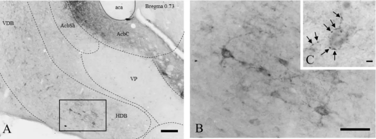
Fig.3. Distribution and projection of GnRH neurons in mouse brain. The GnRH cell bodies are mainly located in OVLT-medial preoptic area [246] region (A) and send processes to the main terminal fields, the OVLT and the external zone of median eminence (ME) (B). Schematic drawing illustrates that the GnRH neurons, from the OVLT-MPA region, project to the median eminence and send axonal branches to the RP3V and Arc forming putative synaptic connections with neuronal populations (C).
Modified from [247].
In the medial septal-preoptic regions, the GnRH cells send fibers in dorsal direction, gives off collaterals and returns to its own or to other perikarya apparently to form axosomatic connections. This observation may provide the morphological basis for a connected syncytium, which might help synchronize the GnRH neuron population.
Furthermore, the GnRH neurons, from the septal-preoptic and suprachiasmatic regions, project also through classical efferent pathways such as the stria terminalis, stria
27
medullaris thalami, stria longitudinalis medialis and lateralis, fimbria hippocampi, fasciculus retroflexus, medial forebrain bundle, and posterior commissure and target several other brain regions. Fibers from the medial septal and preoptic regions pass through the dorsomedial hypothalamus and compose a periventricular subependymal GnRH network that serves as the origin of the extrahypothalamic projections. Fibers pass through the Arc, as well [248]. In my studies, we focused on these fibers forming synaptic contacts on different target cell population in the RP3V and Arc (Fig.3. C).
Scattered GnRH fibers are seen in the mammillary complex, raphe nuclei, medial nucleus of the amygdala, medial habenular nuclei, and ventral tegmental area. In addition to these nuclei, the GnRH processes make up a dense plexus around the aqueductus cerebri in the mesencephalic central gray. Moreover, GnRH neurons from the MS, the nucleus of diagonal band, the olfactory tubercle and the medial surface of the hemispheres reach the medial layers of the olfactory bulb. GnRH neurons also send axons to several areas outside the nervous tissue such as the subarachnoidal space or the ventricular lumen [248].
There is evidence that central administration of GnRH can inhibit the reproductive axis in sheep [249, 250] and rats [251, 252]. Electrophysiological recording of spontaneously active neurons of the Arc revealed that GnRH (10 nM-10 pM) administered adjacent to the infundibular recess significantly altered the firing frequency of unidentified neurons. Predominantly excitatory responses were detected [253]. Results of these previous studies suggest that the GnRH neurons can influence directly the function of certain neurons in the AVPV and the Arc.
Previous anatomical studies have identified putative sites for central actions of GnRH.
GnRH-IR axon projections are detected in the preoptic area (i.e. AVPV) [116, 118, 254]
and in the MBH [118, 254], including the Arc (Fig.3. C).
These previous findings prompted us to study, whether GnRH neurons project to the KP- and TH-IR cells and form synaptic connections with them. Using a correlated light- and electron microscopic immunohistochemical approach, we examined the putative connections between GnRH processes and KP- and TH-IR neurons in both the RP3V and the Arc.
28
1.4. Potential role of a glycinergic input to BF neurons
In the central nervous system, the two main inhibitory amino acids are glycine and γ- aminobutyric acid (GABA). These neurotransmitters activate the strychnine sensitive glycine receptors and GABAA receptors, respectively, which permit chloride influx through the postsynaptic membrane to hyperpolarize postsynaptic neurons. GABAergic neurotransmission is almost ubiquitous in the mammalian CNS, whereas glycinergic neurons are mainly restricted to the spinal cord and brainstem [255, 256]. It is therefore not surprising that our knowledge on the contribution of GABAergic neurons to defined neuronal circuits is much more detailed than the available information about glycinergic neurons. This has changed when the glycin transporter transgenic animals have been generated [257]. GLYTs, depending on their location, have distinct functions at gly- cinergic synapses. GLYT2 provides glycine for the refilling of presynaptic vesicles of glycinergic neurons [258], whereas GLYT1 ensures the removal of glycine from the synaptic cleft into glial cells, leading to the termination of glycine-mediated neuro- transmission. In addition, GLYT1 is also present in certain glutamatergic neurons and regulates the concentration of glycine at excitatory synapses containing NMDA recep- tors, which are known to require glycine as a coagonist [259]. Using the GLYT2-GFP mice and immunohistochemistry, we detected the presence of membrane GLYT1 and GLYT2 in the mouse BF, suggesting a potential role for glycine in this region [15].
Only the use specific antisera against GLYT2, which is a reliable marker for glycinergic neurons in the CNS [260] and is concentrated primarily in the glycinergic fibers, has made it possible to visualize the projections. Zeilhofer and his colleagues generated the bacterial artificial chromosome (BAC) transgenic mice that express enhanced green fluorescent protein (EGFP) specifically in glycinergic neurons under the control of the GLYT2 promoter [257]. This transgenic mouse line made the mapping of the cell bod- ies, thus the source of glycinergic projections to BF areas, possible.
Glycine has a complex role in the central nervous system. Glycine acts on strychnine- sensitive glycine receptors (GlyRs), which mostly cause Cl- influx, hyperpolarizing thereby the neuron to inhibit its activity.
Glycine is also able to act as an excitatory neurotransmitter. On the one hand, in the developing CNS, the intracellular Cl- concentration is high compared to the extracellular space. Binding to the GlyRs causes Cl- to spill out from the cell, causing a strong depo-
29
larization and neurotransmitter release, instead of hyperpolarization [261]. Studies have shown that, this phenomenon exists also in mature neurons [22, 262]. Glycine binding to the GlyR generates depolarizing response instead of hyperpolarization due to the in- creased intracellular Cl- concentration [263]. On the other hand, glycine also acts on the N-methyl-D-aspartate (NMDA) receptor as a coagonist and, as such, facilitates excitato- ry neurotransmission [264].
Our previous studies confirmed the presence of the GlyR alpha 1 subunit mRNA in GnRH neurons by microarray examinations [16]. Furthermore, GnRH neurons express functional ionotropic GABA-A receptors [265-269] and GABA-B [270] receptors, but the response of GnRH neurons to activation of GABA receptors is controversial. Mature GnRH neurons maintain high intracellular chloride concentrations, which can result in excitatory responses to GABA-A-R activation in adult mice [265, 267] and rats [271, 272]. However, many studies suggest that GABA exerts an inhibitory effect on GnRH/LH release [273-275]. Most GnRH neurons (about 80-100%) express α-amino-3- hydroxy-5-methyl-4-isoxazolepropionic acid [246] receptors, whereas only a small subpopulation (∼20%) have NMDA receptors [136, 268, 276, 277].
The BF cholinergic neurons receive GABAergic input [278] and they are inhibited by GABA via GABA-A receptors [279]. Furthermore, the cholinergic neurons also contain NMDA receptor subunits [280].
Taken together, these observations raise the possibility that glycine acts directly on GnRH and cholinergic neurons. One of the possible effects is, depolarization or hyperpolarization via GlyR (depending on the intracellular Cl- concentrations). The other possibility is that, glycine binds to NMDA receptors as coagonist and excites the GnRH and cholinergic neurons.
Thus, using immunohistochemistry we addressed whether the GnRH and cholinergic neurons contain the GlyR and receive direct input from GLYT2-IR fibers. We examined the presence of GLYT1-IR profiles in the vicinity of GnRH and cholinergic neurons and we also tested the direct effect of glycine on these neurons by electrophysiological recordings.
30 2. Aims:
2.1. Investigating potential target cells of glycine in the BF
2.1.1. GlyR in GnRH and cholinergic neurons
2.1.2. GLYT2-IR afferents to GnRH and cholinergic neurons
2.1.3. Source of glycinergic fibers in the BF
2.1.4. GLYT1-IR astrocytic processes in the vicinity of GnRH and cholinergic neurons.
2.1.5. Membrane potential properties of GnRH and cholinergic neurons in the presence of glycine
2.2. Characterization of GnRH efferents and their target cells in mice and humans
2.2.1. Ultrastructural features of GnRH processes in mice
2.2.2. KP-KP contacts in mice and human
2.2.2. Effects of circadian, hormonal and lactation-related changes on the GnRH input to KP- and TH-IR neurons in mice
31 3. Materials and Methods
3.1. Mouse brain samples
3.1.1. Brain tissue collected for immunohistochemical processing
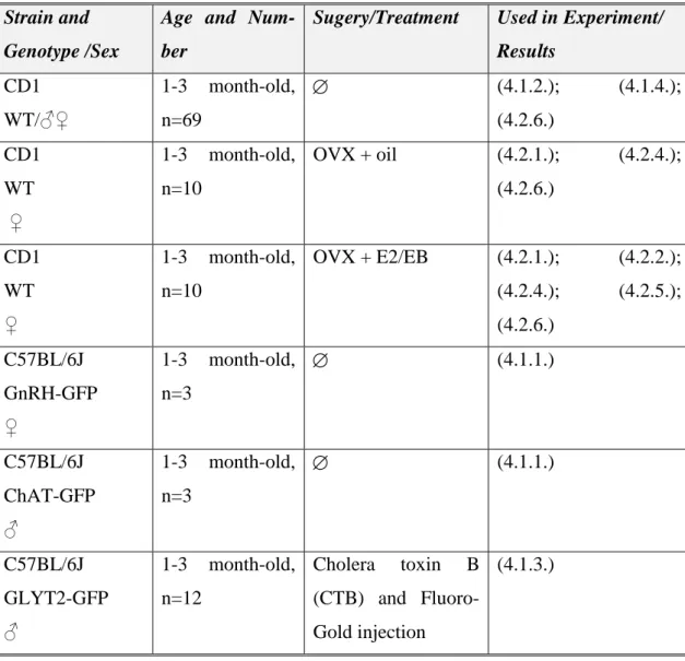
Wild-type (CD1, Charles River) and transgenic mice (1–3 month-old, 25–30 g body weight) were housed under controlled lighting (12 h light/dark cycle; lights on at 7:00 A.M.), and temperature (22°C) conditions with access to food and water ad libitum. The list of animal models used in the different experiments is summarized in Table I. be- low. Five to six virgin animals were kept in a single cage, whereas pregnant and post- partum mothers were individually housed. A group of animals were ovariectomized (OVX, day 0) and 7 days later (day 7) implanted subcutaneously with a capsule (ID 1.57 mm, OD 3.18 mm) containing either 17β-estradiol (0.625 μg in 20 μl sunflower oil; OVX+E2) or vehicle (OVX+Oil) [125]. Three days after implantation (day 10), mice were colchicine-treated (intracerebroventricularly 40 μg in 4 μl 0.9% saline) and 24 h later (day 11) they were sacrificed at either zeitgeber time [281] 4–5 or ZT11–12;
these times include, respectively, the negative and positive feedback phases of oestro- gen’s effects on LH release [125]. Surgery was performed on animals under deep anes- thesia induced by an intraperitoneally injected cocktail of ketamine (25 mg/kg body weight), Xylavet (5 mg/kg body weight), and Pipolphen (2.5 mg/kg body weight) in saline. All studies were performed with permission from the Animal Welfare Commit- tee of the Institute of Experimental Medicine (No. 2285/003), the Debrecen University (No. 6/2011/DE MÁB and 5/2015/DEMÁB), the Eötvös Loránd University (PEI/001/37-4/2015) and in accordance with legal requirements of the European Com- munity (Decree 86/609/EEC).
3.1.2. Mouse models used in the different experiments
For our experiments, we used different mouse models and surgeries, which are summa- rized in Table I. below.
32
Table I. Mouse models, surgeries and their experiments.
Strain and Genotype /Sex
Age and Num- ber
Sugery/Treatment Used in Experiment/
Results CD1
WT/♂♀
1-3 month-old, n=69
(4.1.2.); (4.1.4.);
(4.2.6.) CD1
WT ♀
1-3 month-old, n=10
OVX + oil (4.2.1.); (4.2.4.);
(4.2.6.)
CD1 WT
♀
1-3 month-old, n=10
OVX + E2/EB (4.2.1.); (4.2.2.);
(4.2.4.); (4.2.5.);
(4.2.6.) C57BL/6J
GnRH-GFP
♀
1-3 month-old, n=3
(4.1.1.)
C57BL/6J ChAT-GFP
♂
1-3 month-old, n=3
(4.1.1.)
C57BL/6J GLYT2-GFP
♂
1-3 month-old, n=12
Cholera toxin B (CTB) and Fluoro- Gold injection
(4.1.3.)
3.1.3. Acute slices collected for electrophysiological examination
For electrophysiological experiments, we used two mouse line. The technical details are summarized in Table II. below.
Table II. The mouse acute brain slices.
Strain and Genotype/Sex
Age and Number
Surgery/
Treatment
Used in
Experiment/Results C57BL/6J
GnRH-GFP pro- estrus ♀
1-3 month-old, n=3
(4.1.5.)
C57BL/6J ChAT-GFP ♂
1-3 month-old, n=26
(4.1.5.)
33 3.2. Human brain tissue samples
Human hypothalami were collected at autopsy from the 1st Department of Pathology and Experimental Cancer Research, Semmelweis University, Budapest, Hungary, and the Department of Pathology, Saint Borbála Hospital, Tatabánya. Three-four hours after death, the brains were removed from the skull of a 77-year-old (SKO5) and a 71-year- old (SKO8) female subject and a 55-year-old male individual (SKO7) who died from causes not linked to brain diseases (SKO5, SKO8: heart failure; SKO7: pulmonary em- bolism). Ethic permissions were obtained from the Hungarian Medical Research Coun- cil /ETT TUKEB 33268-1/2015/ EKU (0248/15) and 31443/2011/EKU (518/PI/11)/
and from the Regional and Institutional Committee of Science and Research Ethics of Semmelweis University (SE-TUKEB 251/2016), in accordance with the Hungarian Law (1997 CLIV and 18/1998/XII.27. EÜM Decree/).
3.3. Preparation of mouse and human sections for light microscopic studies The mice to be used for light- and confocal microscopic studies, were perfused trans- cardially, first with phosphate buffered saline (PBS) solution (10 ml 0.1M PBS; pH 7.4) and then with PBS containing 4% paraformaldehyde [237] (100 ml 4% PFA in 0.1M PBS). The brains were post-fixed in 2% PFA/PBS solution for 24h at 4 °C, cryoprotect- ed overnight in 25% sucrose. Serial 30-μm thick coronal sections were cut with a Leica SM 2000R freezing microtome (Leica Microsystems, Nussloch Gmbh, Germany). The sections were divided into three sequential pools and stored in antifreeze solution (30%
ethylene glycol; 25% glycerol; 0.05 M phosphate buffer; pH 7.4) at –20 °C until use.
Hypothalamic tissue blocks were dissected from the brain of three postmenopausal women (aged 53-88) within 24 h after death. The subjects had no history of neurologi- cal or endocrine disorders. The tissues were rinsed briefly with running tap water and then, immersion-fixed with 4% PFA in 0.1 M PBS (PBS; pH 7.4) for 10 days. The tis- sue blocks were infiltrated with 20% sucrose and cut serially either at 30 or at 100 μm thickness with a freezing microtome.
After the endogenous peroxidase activity had been quenched with 0.5% H2O2 (10 min), sections were permeabilized with 0.5% Triton X-100 (catalog #23,472-9, Sigma- Aldrich; 20 min). Finally, 2% normal horse serum (NHS) was applied (for 20 min) to reduce nonspecific antibody binding. Subsequent treatments and interim rinses in PBS
34
(3X for 5 min) were performed at room temperature, except for the incubations in the primary antibody or fluorochrome conjugates, which took place at 4°C.
3.4. Preparation of mouse and human sections for electron microscopic studies Mice were perfused first with PBS (10 ml, 0.1M; pH 7.4), then a mixture of 2% PFA and 4% acrolein. Brains were postfixed in 2% PFA/PBS solution for 24h at 4 °C. 30-μm thick coronal sections were cut with a Leica VTS-1000 Vibratome (Leica Microsys- tems, Wetzlar, Germany) and treated with 1% sodium borohydride (30 min), 0.5% H2O2
(15 min) and permeabilized with three freeze-thaw cycles, as described previously [174].
Human tissue samples from elderly subjects were shown previously to contain high lev- els of KP immunoreactivities ((Hrabovszky, 2014; Rometo, Krajewski, Lou Voytko, &
Rance, 2007). The internal carotid and vertebral arteries were cannulated, and the brains were perfused first with physiological saline (1.5 L for 30 min) containing 5 mL Na- heparin (5000 U/mL), followed by a fixative solution (3–4 L for 2.0–2.5 h) containing 4% PFA, 0.05% glutaraldehyde, and 0.2% picric acid in 0.1 M phosphate buffer (PB;
pH = 7.4). The hypothalami were dissected out and postfixed overnight in 4% PFA without glutaraldehyde. Fifty-micrometer-thick coronal sections were prepared from the hypothalami with a Leica VTS-1000 Vibratome (Leica Microsystems, Wetzlar, Germa- ny).
2% normal horse serum (NHS) was applied (for 20 min) to reduce nonspecific antibody binding. Subsequent treatments and interim rinses in PBS (3X for 5 min) were per- formed at room temperature, except for incubation in the primary antibody which took place at 4°C.
After immunohistochemical detection of tissue antigens, the labelled mice and human sections were treated with 1% osmium tetroxide (1 h) and 2% uranyl acetate (in 70%
ethanol; 40 min), dehydrated in an ascending series of ethanol and acetonitrile, and flat- embedded in TAAB 812 medium epoxy resin between glass microscope slides pre- coated with a liquid release agent (#70880; Electron Microscopy Sciences, Fort Wash- ington, Pa., USA). The resin was allowed to polymerize at 56 °C for 2 days.