The possible role of estradiol and estrogen receptor alpha in TGF-β induced type II epithelial-
mesenchymal transition and the following regeneration in mesenteric mesothelial cells
PhD thesis
dr. Petra Balogh MD
Doctoral School of Molecular Medicine Semmelweis University
Supervisor: Dr. Anna L. Kiss C.Sc Official reviewers:
Dr. András Kiss Ph.D Dr. Kinga Molnár Ph.D
Head of the Final Examination Committee:
Dr. László Tretter D.Sc
Members of the Final Examination Committee:
Dr. Zsuzsanna Darvas Ph.D Dr. Péter Löw Ph.D
Budapest, 2014
2
Table of Contents
1. LIST OF ABBREVIATIONS……..………..…………...4
2. INTRODUCTION……….6
2.1 Epithelial-mesenchymal transition………...6
2.1.1 TGF-β superfamily………...7
2.1.2 The TGF-β signaling events………....8
2.1.3 Clathrin-mediated endocytosis enhances the signaling..……….9
2.1.4 Caveola-mediated endocytosis attenuates TGF-β signaling……….11
2.1.5 Multivesicular body formation………..13
2.1.6 Non-Smad signaling pathways………..15
2.2 The role of ER-α and estradiol in EMT………18
2.2.1 ER-α as a potential molecular modulator of TGF-β signaling…...18
2.2.2 Extragonadal estradiol: renewal of a traditional belief……….19
2.3 The role of autophagy in tissue remodelling………...21
3. OBJECTIVES………...23
4. MATERIALS AND METHODS………....25
5. RESULTS………...32
5.1 The morphological and biochemical characterization of TGF-β induced EMT in mesothelial cells in vivo...32
5.1.1 The ultrastructural evidences of type II EMT...32
5.1.2 Inflammatory cytokines and TGF-β are released into the peritoneal cavity upon Freund’s adjuvant treatment in vivo….….36 5.1.3 The morphological detection and subcellular distribution of the main canonical TGF-β signaling molecules in mesothelial cells……….38
5.1.4 En route to multivesicular bodies: the possible role of caveolar internalization in TGF-β signaling pathway………...42
5.2 The possible role of ER-α and estradiol in EMT………...49
5.2.1 Estrogen receptor alpha expression and its subcellular distribution in mesothelial cells………..49
5.2.2 The changes in the expression of ER-α upon inflammatory evets....51
3
5.2.3 The intersection of ER-α and TGF-β pathway
at the level of caveolae……….……52
5.2.4 Extragonadal estradiol (E2) is detected in the peritoneal cavity under in vivo inflammatory circumstances………...56
5.3 The role of autophagy in tissue remodelling 5.3.1 The role of autophagy in the retrieval of simple squamous morphology of mesothelial cells following acute inflammation….……….57
5.3.2 The possible inducer of autophagy: Extragonadal estradiol is in the focus……….……...60
6. DISCUSSION………...63
7. CONCLUSION.………...72
8. SUMMARY………....73
9. SUMMARY IN HUNGARIAN (ÖSSZEFOGLALÁS)………..……...75
10. BIBLIOGRAPHY..………..………...77
11. LIST OF PUBLICATIONS.………...95
12. ACKNOWLEDGEMENT.………...96
4
1. List of Abbreviations
4A androstendione
AMPK AMP-activated protein kinase BMP bone morphogenetic protein CD63 cluster of differentiation 63 DHEA dehidroepiandrosterone DNA deoxyribonucleic acid E2 estradiol
E2-S estradiol-sulfate
EDTA ethylenediaminetetraacetic acid disodium salt EEA1 early endosome antigene-1
EGF epidermal growth factor
EMT epithelial-mesenchymal transiton ER –α estrogen-receptor alpha
ERK extracellular signal-regulated kinase
ESCRT endosomal sorting complexes required for transport EST estradiol sulfate
FGF fibroblast growth factor FGF fibroblast growth factor
FYVE zink finger domain named after the four cysteine.rich proteins:Fab1, YOTB, Vac1, EEA1
GDF growth differentiation factor
GDNF glial cell-line derived neurotrophic factor GPI glycophosphatidylinositol
GPR30 G protein coupled receptor 30 IGF insulin-like growth factor IL interleukin
ILV intraluminal vesicle JNK c-Jun N-terminal kinase
LC3 microtubule-associated protein light chain 3 MAPK mitogen-activated protein kinase
5
MH Mad Homology
mRNA messenger ribonucleic acid mTOR mammalian target of rapamycin MVB multivesicular body
NES nuclear export signal NLS nuclear localization signal NPS nuclear pore signal
PI3K phosphatidylinozitol-3-kinase PKC protein kinase C
qRT-PCR quantitative real-time polymerase chain reaction SARA Smad anchored for receptor activation
Smad Sma and Mad homology domains
T testosteron
TGF-β transforming growth factor beta TNF tumor necrosis factor
6
2. Introduction
2.1 Epithelial-mesenchymal transition
Epithelial-mesenchymal transition (EMT) is a biological process that allows a polarized epithelial cell to undergo several biochemical and morphological changes to obtain a mesenchymal phenotype (Kalluri&Weinberg 2009). Epithelial cells typically form an uniform array, often a monolayer. The cells are adhered to each other via cell- cell junctions providing a tight connection between the neighbouring cells. Epithelial cells furthermore are polarized meaning that apical and basolateral surfaces are responsible for different cellular functions. Mesenchymal cells, in contrast are elongated, spindle-shaped, apolar cells and are capable of locomotion as they are lacking intercellular adhesions (Lee et al 2006). This capacity for plasticity, the process of EMT was first described by Elisabeth Hay who also depicted the basic differences of the cellular actions observed during embryogenesis and tumorigenesis (Hay 2005).
Since then three subtypes of EMTs have been distinguished with different functional consequences. Besides epithelial-mesenchymal transition during embryogenesis (type I) and tumorigenesis (type III), type II EMT is associated with wound healing, tissue regeneration and organ fibrosis (Sodek et al 2012, Yanez Mó et al 2003). It has been demonstrated that upon inflammation many cells (monocytes/macropaghes, fibroblasts) can trigger type II EMT through secretion of growth factors such as transforming growth factor-beta (TGF-β) or epidermal growth factor (EGF). Most prominent among these cells are the macrophages and activated resident fibroblasts that accumulate at the site of injury and release these growth factors (Kalluri&Weinberg 2009, Lee et al 2006, Strutz et al 2002) (Fig. 1). TGF-β was first described to induce EMT via the canonical Smad 2/3-dependent pathway and meanwhile it became evident that the cellular actions of the cytokine can be modulated by other Smad-independent signaling pathways like MAP kinase pathways (Zavadil&Böttinger 2005, Derynck&Zhang 2003, Massagué 1998).
7 2.1.1 TGF-β superfamily
TGF-β superfamily consists of numerous groups of cytokins that regulate a diverse set of cellular processes. Besides the TGF-β isoforms (TGF-β₁, TGF-β₂ and TGF-β₃), further members of the family include the bone morphogenetic proteins (BMP), inhibin, myostatin, Nodal, GDF, GDNF, MIS (Müllerian Inhibiting Substance), each with different roles in cell differentiation, apoptosis, cell migration and adhesion during embryogenesis and in adult tissues (Massagué 1998). TGF-β has also dual role depending on the cell type and the environment. While it is able to suppress cell growth in epithelial and hemopoetic cells by inducing G1 arrest, it also initiates cell proliferation and differentiation in mesenchymal cells. The cellular processes that regulate the morphological plasticity of a cell and result in phenotypic change is known as epithelial-mesenchymal transition (EMT) (Shook&Keller 2003). The universal role of TGF-β in the different types of EMTs is unambigous as well as the biochemistry of the signaling is well characterized. It is less clear, however in which cellular/cytoplasmic compartments the molecules along the downstream pathway are accommodated and how their localization changes with the dynamics of the signaling.
Another question of great interest is whether different compartments can be involved in regulating the pathway and if so, whether they can promote or suppress the signaling events?
8
Figure 1. Inflammation and growth factors like TGF-β promote mesenchymal transition.
Epithelial-mesenchymal transition takes place during development, and is also proposed to play a role in cancer invasion and generation of myofibroblasts that contribute to fibrosis. In response to injury, inflammatory cells and activated fibroblasts produce growth factors such as TGF-β and FGF-2, as well as matrix metalloproteinases and chemokines. TGF-β and other factors trigger signaling cascades in epithelial cells that lead to a change from an epithelial to a mesenchymal phenotype (Source: Kalluri&Weinberg 2009, Strutz et al 2002).
2.1.2 The TGF-β signaling events
TGF-β, the prototype of the family, signals through its cell surface serine/threonin kinase receptors. Functionally and structurally type I and type II TGF-β receptors (TβR-I, TβRII) can be distinguished. The type II receptor is considered a constitutively active kinase (activated by autophosphorylation) while type I receptor contains a special GS domain the phosphorylation of which leads to the activation of the receptor. In the prototypic TGF-β pathway, ligand binds to type II receptor and induces the formation of a heterotetrameric receptor complex within which TβR-II transphosphorylates and activates the type I receptor and the activated TβR-I initiates the Smad signal transduction pathway (Shi&Massagué 2003, Massagué 1998, Wrana et al 1994).
The Smad proteins are subgrouped based on their structural and functional differences.
The i) receptor-regulated (R)-Smad proteins are Smad 2,3 that are the only substrates for type I receptor kinases while further members of this group include Smad 1,5,8, that are phosphorylated by the activated BMP receptors. After phosphorylation, thus activation, the R-Smads associate with ii) common mediator (Co)-Smad protein, Smad4. They form oligomeric complexes and are transported to the nucleus to regulate the transcription of target genes together with other nuclear cofactors. The members of the third class of Smads act as negative regulators of the signaling pathway: iii) inhibitory (I)-Smads include Smad6 and Smad7 proteins (Shi&Massagué 2003, Moustakas et al 2001, Zhang et al 2001).
Recent findings have demonstrated that accessory proteins interact with type I, type II receptors and Smad proteins (Moustakas et al 2001). An example is SARA (Smad
9
anchor for receptor activation) that facilitates the association of R-Smads with TGF-β receptor at the plasma membrane, though it is predominantly localized and bound to phosphatidylinositol 3-phosphate (PtdIns3P) rich early endosomes (Di Guglielmo et al 2003, Tsukazaki et al 1998). Furthermore, some data suggest that SARA can interact with cell surface TβRs and in this way protects the complex from degradation (Chen 2009, Chen at al 2007, Gillooly et al 2001, Itoh et al 2002).
Ligand binding to its cell surface receptors means not only the beginning of the signaling events through Smads, but also triggers internalization of both ligand and receptors (Chen 2009, Chen at al 2007, Le Roy&Wrana 2005, Itoh et al 2002, Gillooly et al 2001). The receptor internalization is required for the initiation of downstream signaling. There are two main endocytic pathways through which the TGF-β ligand- receptor complex can be internalized (Di Guglielmo et al 2003). One of them is the well-characterized clathrin-mediated endocytosis and a less clear pathway is the lipid/caveola-mediated endocytosis. Both types of pathways are used for the internalization of TβRs. It is already clear that via different internalization routes cells can control the number of surface-receptors and this is crucial for regulating the signaling, receptor turnover, the magnitude and duration of the events (Chen 2009).
2.1.3 Clathrin-mediated endocytosis enhances the signaling
Internalization of most cell surface receptors is mediated by short specific sequences in their cytoplasmic domain. Tyrosine-containing sequences and di-leucin- based motifs function as internalization signals for clathrin-dependent endocytosis.
These sequences can directly bind to the endocytic machinery and play important role in cargo enrichment on the clathrin-coated pit as well as in vesicle formation (Bonifacino&Traub 2003, Bonifacino&Lippincott-Schwartz 2003). Such internalization signals have also been identified in TGFβ receptors. Both TβRI and TβRII appear to be rapidly internalized upon ligand binding. After receptor-ligand internalization in clathrin coated vesicles, the complex is targeted into EEA1 (early endosome antigene-1) positive endosomes. These compartments promote the signal transduction by recruiting the FYVE domain-containing proteins (like SARA). The C-terminal phosphorylation of R-
10
Smads bounded to endosomal membranes leads to their dissociation from both SARA and receptor (Di Guglielmo et al 2003, Hayes at al 2002, Penheiter et al 2002).
Afterwards the phosphorylated R-Smads can bind to Smad4 (Xu et al 2000) forming the oligomeric complex that can enter the nucleus to regulate target genes in association with other coactivators and corepressors. It is important to emphasize that although clathrin-mediated endocytosis of TβRs can enhance Smad-mediated TGFβ signaling, it is still debated whether this process is required for the signaling (Hayes et al 2002, Penheiter et al 2002). Thus, early endosomes (EE) are important not only in the sorting of internalized cargo proteins and receptors but by providing a special microenvironment through recruiting the signaling molecules (Hayes et al 2002), they presumably play role in defining the activity of signaling events as well.
After dissociation, TGF-β receptors can either recycle back to the plasma membrane in recycling endosomes (Mitchell et al 2004) or be degraded in the downstream endosomal compartments.
The shuttling of TGFβ-induced Smad complexes between the cytoplasm and the nucleus is strictly regulated. The R-Smad and Co-Smad proteins have conserved Mad- homology 1 (MH1) and MH2 domains connected by a linker domain, while the I-Smads are lack a distinct MH1 domain. The R-Smads and Co-Smad have a nuclear localization sequence (NLS) in their Mad-homology 1 (MH1) domain while their MH2 domain contains nuclear export signal (NES) and nuclear pore signal (NPS) as well (Heldin&Moustakas 2012) (Fig. 2). Phosphorylated Smad3 was shown to interact with importin-β1 of the nuclear pore and enters into the nucleus in a GTPase dependent manner (Xiao et al 2003, Kurisaki et al 2001).
11
Figure 2. Functional domains of Smad proteins. The R-Smad (Smad2/3) and Co-Smad (Smad4) proteins have conserved Mad-homology 1 (MH1) and MH2 domains connected by a linker domain, while the I-Smad (Smad7) lacks a distinct MH1 domain. The MH1 domain contains DNA-binding site (except for Smad2) and nuclear localization signal (NLS) and mediates interactions with different transcription factors to stabilize the nuclear Smad complex.
MH2 domain is higly conserved among all Smads and is responsible for receptor interaction, the nucleocytoplasmic shuttling of Smad proteins (NPS, NES) and also mediates the formation of Smad oligomer complexes and the interaction of other proteins such as SARA. The linker region contains phosphorylation sites allowing crosstalks with other signaling pathways and binds ubiquitin ligases (Smurf proteins) via the PY motif. (Source: Balogh et al 2013).
2.1.4 Caveola-mediated endocytosis attenuates TGF-β signaling
Another internalization route into the cell is via caveolin-1 positive vesicles. It is known that TβRs are accommodated in caveolin-1 containing lipid rafts of the plasma membrane and use this internalization route as well. Caveolae are small omega or flask- shape plasma membrane invaginations that play important role in many cellular functions including endocytosis, signal transduction, cellular growth control and apoptotic cell death. The main protein components of caveolae are the scaffolding
12
proteins termed caveolin-1,-2,-3 (Couet et al 1997, Lisanti et al 1994). Complex events lie behind the regulation of the internalization pathway through caveolae and the intermediate compartments are still less clear. Earlier data showed that caveolae internalize into the cell, and form so-called caveosomes that were supposed not to communicate with other cellular compartments. According to this idea caveosomes would represent an individual cytosolic compartment the content of which may avoid lysosomal degradation (Nicols 2002, Pelkmans et al 2001). Recent data, however, have shown that caveolin-positive vesicles can also associate with early endosomes (Parton&Simons 2007) or might continuously remain in connection with the plasma membrane (Kiss&Botos 2009). Meanwhile, however the terminology ’caveosome’ as a distinct cellular compartment was repealed (Parton&Howes 2010). Caveolar endocytosis presumably provides another possible way for sequestering receptors (Le Roy&Wrana 2005). Several lines of evidence support the idea that receptor-ligand internalization via the (non-classical) caveolar pathway turns off the TGFβ signaling events by targeting the receptor-ligand complex to lysosomal and/or proteasomal degradation (Chen 2009, Le Roy&Wrana 2005, Di Guglielmo et al 2003). This receptor degradation plays an important role in controlling the amount of receptors on the plasma membrane. The possible pathways targeting the signaling molecules towards degradation are not entirely known and most of the papers avoid the detailed discussion of these routes. The inhibitory Smad (I-Smad), Smad7 is one of the main regulator in the degradative events. I-Smad inhibits TGFβ signaling through multiple mechanisms as a decoy substrate forming a stable complex with receptors to prevent recruitment of R- Smads (Nakao et al 1997, Hayashi et al 1997) and also disrupts the functional Smad- DNA complex formation (Zhang et al 2007). Smad7 exerts its negative effects at the level of the plasma membrane by competing with R-Smads for the receptor and also by recruiting the E3 ubiquitin ligase Smurf1/2 proteins to the active TβRs (Ebisawa et al 2001, Kavsak et al 2000, Nakao et al 1997) to promote receptor ubiquitination and degradation (Heldin&Moustakas 2012). (Smad7 has a putative NLS in its N-terminal region and resides in the nucleus in non-stimulated cells. In response to TGFβ stimulus, Smad7 leaves the nucleus in complex with the ubiquitin ligases, Smurf1/2 (Heldin&Moustakas 2012, Ebisawa et al 2001, Kavsak et al 2000, Itoh et al 1998). The interaction of Smad7 and Smurf proteins with activated TβRs targets the complex to
13
lipid rafts/caveolae and in this way the caveola-mediated endocytosis could promote receptor turnover and the termination of signaling (Di Guglielmo et al 2003).
TGFβ receptors after receptor ubiquitination have been shown to be degraded by both lysosomal and proteasomal machineries (Kowanetz et al 2008, Kavsak et al 2000).
Though limited data are available on factors controlling the proteasomal degradation of TGFβ receptors. Recent data showed that a GPI-anchored protein, CD109 functions as a TGFβ co-receptor, associates with caveolin-1, promotes the caveolar localization of the TGFβ receptors and might regulate its proteasomal degradation (Bizet et al 2011).
However, it is not clear how the caveolar endocytic machinery can drive receptors to the proteasomal pathway.
Besides TβRs, the stability of Smad proteins and caveolin is also controlled by ubiquitination suggesting the regulatory role of different ubiquitination signals. While poliubiquitination is a sign that directs the cargos to proteasomes, mono/multiubiquitination is a signal for the entry of proteins via the endocytic pathways. Thus, ubiquitination, indeed plays essential role both in signal transduction and also to determine the way of degradation towards proteasomes or towards multivescular body/late endosomes (Mukhopadhyay&Riezman 2007, Ciechanover 2005).
2.1.5 Multivesicular body formation
The lysosomal degradation of internalized cargos via caveolar endocytosis includes multivesicular body (MVB) formation and the early endosomes are the clue compartments of this process as well. Internalized cargo proteins are targeted first to early endosomes (Hayer et al 2010) indicating that MVB formation starts at the level of this cellular compartment. MVBs are formed when limiting membrane of endosomes invaginates and buds into the lumen of the organelles. (Gruenberg&Maxfield 1995, Felder et al 1990). A subset of membrane proteins within the limiting membrane of the endosomes are sorted into these invaginating vesicles and this sorting requires the inclusion of a 350kDa complex, called ESCRT-1 (endosomal sorting complexes required for transport). The membrane of early endosome (EE) contains the ESCRT complex that recognizes and binds ubiquitinated cargos and initiates the transport of the
14
cargos to late endosomes/multivesicular bodies. MVB sorting into intraluminal vesicles (ILV) and the subsequent lysosomal degradation of cell surface receptors is therefore a critical mechanism for regulating the signaling events (Katzmann et al 2001).
Furthermore, the asymmetric compositon of the limiting membrane of these endosomal compartments presumably provide a platform for generating unique signals as well (Hanson&Cashikar 2012) (Fig. 3). Hence, early endosomes play a central role not only promoting the TGF-β pathway, but it is likely to be important intermediate cytosolic compartments that help to attenuate the signaling as well.
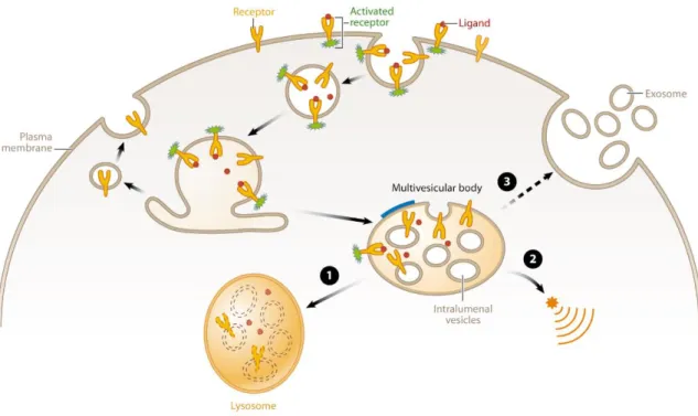
Figure 3. Multivesicular body (MVB) function. Schematic diagram showing the general functions of multivesicular bodies. Receptors (yellow) are activated (denoted in green) upon binding to their ligands (red) and then enter the cell by endocytosis. Receptors may be recycled back to the plasma membrane or may be targeted to intralumenal vesicles (ILVs) in the MVB.
Decreasing pH in the early endosome, MVB and lysosome are indicated by different shades of orange. The thick blue line on the MVB limiting membrane indicates the characteristic flat bilayered coat. Fusion of MVBs with lysosomes leads to degradation of ILV content (1). MVBs also provide a platform for the generation of unique signals (2). Finally, MVBs can fuse with the plasma membrane to release ILVs in the form of exosomes (3). (Source: Hanson&Cashikar 2012)
15 2.1.6 Non-Smad signaling pathway
Besides the (canonical) Smad-pathway, TGFβ activates other non-Smad signaling pathways such as ERK, JNK, p38 MAP kinase pathways in a cell-specific and context-dependent manner. MAPK pathways help and complete the process of TGFβ induced epithelial-mesenchymal transition, although the mechanism by which TGFβ activates these pathways and their biological consequences are poorly characterized (Moustakas&Heldin 2005, Derynck&Zhang 2003). MAPK cascade is composed of several protein kinases that specifically phosphorylate and activate each other. The elements of the cascade are organized in levels that are termed MAP kinase kinase kinase (MAPKKK), MAP kinase kinase (MAPKK) and MAP kinase (MAPK). The activation of MAPK leads to its translocation to the nucleus where MAPK phosphorylates and activates its targets, e.x transcription factors. It is well known that MAPK pathways transmit extracellular signals to the nucleus to regulate different cellular processes (Chuderland&Seger 2005, Adachi et al 2000, Chen et al 1992) (Fig.
4).
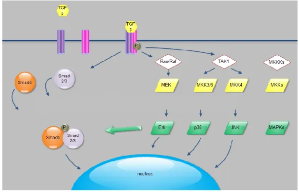
Figure 4. Summary of Smad-dependent and Smad-independent pathways that play role in TGF-β induced epithelial-mesenchymal transition. The formation of TGF-β ligand- receptor
16
complex activates Smad2/3 proteins that form oligomer complexes with Smad4 and enter the nucleus exerting their effects on target genes. Besides this signaling route, TGF-β ligand- receptor complex activates MAPK pathways (ERK, p38, JNK) that not only carry different extracellular signals towards the nucleus and contribute to the activity of transcription, but MAP kinases also modify the activity of Smad-proteins. The balance between activation of Smad proteins and MAPK pathways defines the cellular responses to TGF-β. For more details, see text. (Source: Balogh et al 2013)
However, it has recently been described that non-Smad signaling proteins (the elements of MAPK cascade) take part in the physiological responses of TGFβ as well by other different mechanisms: i) they can directly modify the activity of Smad proteins by e.x phosphorylation (p38 MAP kinase and JNK kinase have been reported to phosphorylate Smad2/3 and suppress their activity (Kamaraju&Roberts 2005, Mori et al 2004). ii) They can directly interact or be phosphorylated by TβRs, hence a parallel signaling is initiated that might antagonise Smad pathway or iii) non-Smad proteins can directly be modulated by Smads that transmit signals to other pathways (Moustakas&Heldin 2005). Emerging new data reflects the complexity of how the Smad- and non-Smad pathways are interconnected. The ERK MAPK phosphorylates the MH1 domain of Smad2 and blocks its nuclear translocation, thus transcriptional output. TGF-β induced JNK can also phosphorylate Smad3 and induces its translocation to the nucleus (Kretzschmar et al 1999, Engel et al 1999). The role of regulation of TβRs (phosphorylation, ubiquitilation, sumoylation) is also necessary to be elucidated (Kang&Derynck 2009). The phosphorylation of TβRII on tyrosine can contribute to the activation of TGFβ-induced p38 MAPK pathway and also the tyrosine phosphorylation of TβRI is necessary for the initiation of ERK MAPK pathway in response to TGFβ stimulus (Galliher&Schiemann 2007).
Both Smad and MAPK signaling induced by TGF-β work together in a complex cellular network and the subcellular compartmentalization of both Smad and non-Smad proteins might have a role to define signaling activity, thus cellular answers. The signaling elements of MAPK pathways (MAPKKKs, MAPKKs,) are found at the plasma membrane and in the membrane of endosomes, while the activated MAPKs are bound solely to endosomal membranes. Thus, endosomes are crucial cytoplasmic
17
compartments; as they create a platform and a special environment for the signaling molecules, they can orchestrate the spatial and temporal regulation of different signaling routes (Zehorai 2010, Taub et al 2007).
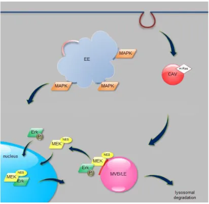
By now it is accepted that TGFβ induced Smad activation occurs in both lipid rafts/cavolae and non-lipid rafts, but a recent observation suggests that activation of MAPK in lipid rafts/cavolae is specially required for TGFβ induced EMT (Zuo&Chen 2009). The role of raft compartments and endosomes is best characterized in the Raf- MEK-ERK pathway. Raf kinases are localized near the plasma membrane in the cytoplasm through interactions with different anchoring and scaffolding proteins or lipid compounds (Galmiche et al 2008). MEKs are localized in the cytoplasm of resting cells due to their nuclear export signal (NES). They shuttle between the cytoplasm and the nucleus constantly and they serve as cytoplasmic anchors for ERK. With the help of adaptor protein p18, MEKs are localized in the lipid rafts of late endosomes indicating the importance of endosomal compartments. Upon stimulation, ERK dissociates from MEK and through the formation of homodimers ERKs enter the nucleus by active transport mechanism, while as a monomer it can enter the nucleus by passive diffusion.
The nuclear export of ERKs is mediated by a MEK dependent active transport mechanism due to their nuclear export signal (Zehorai et al 2010, Taub et al 2007, Adachi et al 2000, Adachi et al 1999, Fukuda et al 1997a, Fukuda et al 1996) (Fig. 5).
18
Figure 5. The subcellular localization of ERK MAP kinase pathway. Activated MAPKKK (H-Ras) is localized in lipid rafts/caveolae (CAV). MEK proteins are bound to caveolin-1 positive lipid domains of late endosomal membranes through adaptor proteins and MEKs serve as anchors for ERK. Upon stimulation, ERK dissociates from MEK and enters the nucleus by passive transport and is relocated to the cytoplasm with the help of MEK proteins that has nuclear export signal (NES). For more details, see text. EE: early endosome, CAV: caveola, MVB: multivesicular body, LE: late endosome, P: phosphorylation (Source: Balogh et al 2013)
2.2 The role of ER-α and estradiol in EMT
2.2.1 ER-α as a potential molecular modulator of TGF-β signaling
Recently, estrogen receptor alpha (ER-α) has been suggested as another player in the molecular mechanism of EMT (Guttila et al 2012, Ye et al 2010, Planas- Silva&Waltz 2007). ER-α and TGF-β have opposing roles in cell proliferation and differentiation of epithelial cells. Their regulatory pathways intersect and ER-α blocks the TGF-β pathway at different cellular levels inside the nucleus as well as in the cytoplasm and plasma membrane. Both transcription factors have a prominent role in
19
maintaining a controlled signaling that is essential for cell and tissue homeostasis and both act in a cell-specific and context-dependent manner (Band&Laiho 2011, Ito et al 2010). For long, estrogen receptors (ER-α, ER-β) have been considered exclusively as transcription factors acting inside the nucleus (Beato et al 1995, Tsai&O’Malley 1994).
However, the discovery of its membrane-associated form and the ER–mediated transcription in the absence of its ligand generally changed this concept (Driggers&Segards 2002, Levin 2002). The theory of a hormone-independent ER-α activation that can serve as a mechanism to amplify growth factor pathways (Hall et al 2001) has also been accepted by now.
Besides the nuclear and cytoplasmic pool of ER-α, it has been proved that a small percentage of the receptor (5-10%) resides in the cell membrane and can elicit both genomic and non-genomic responses by activating multiple protein kinase cascades that include MAPK, protein kinase C, Src kinase and PI3K (Levin 2009, Song&Santen 2006, Song et al 2005, Simoncini et al 2004, Razandi et al 1999, Migliaccio et al 1996, Pietras&Szego 1977). There are also data that indicate the role of estrogen-receptor (ER) α as a negative regulator of TGF-β pathway by increasing the degradation of nuclear Smad proteins. ERα forms a protein complex with Smad3/4 and ubiquitin ligases in the nucleus and enhances the degradation of the transcription complex by the ubiquitin-proteasome system (Ito et al 2010).
2.2.2 Extragonadal estradiol: renewal of a traditional belief
The natural ligand of ER-α, estrogen is considered an important morphogen.
Meanwhile the concept about ER receptors has largely changed, the renewal of the theory about their ligand effects was essential as well. Besides their gonadal synthesis some articles reported extragonadal estradiol (E2) production ex. in adipocytes, osteoblasts (Bruch et al 1992), chondrocytes, vascular endothelial cells (Bayard et al 1995), aortic smooth muscle cells (Murakami et al 1998), brain tissue (Labrie et al 1997). Thus E2 is no longer solely an endocrine factor, but produced in several extragonadal sites it has the potential to exert its biological effects locally acting as a paracrine or intracrine factor (Simpson et al 2000, Labrie et al 1998, Labrie et al 1997).
20
The final step in physiological synthesis of 17β-estradiol is aromatization of precursor testosterone by cytochrome P450 aromatase. Aromatase enzyme is essential to convert androgens to estrogens and principally, these extragonadal sites are dependent on circulating C19 steroids (DHEA, 4A) as well as testosteron for E2 biosynthesis (Fig. 6).
Another important feature is that E2 synthesized within these sites is probably active only at a local tissue level, but the high concentrations achieved presumably exert significant biological influence in loco (Simpson et al 1999). Besides the sufficient substrate concentrations, aromatase activity is regulated by several factors in adipocytes.
Most important, macrophage-derived pro-inflammatory cytokines such as IL-6, IL-11, TNF-α were reported to upregulate the expression of the enzyme (Simpson et al 2000) that is consistent with other results demonstrating increased aromatase activity in inflammation (Morris et al 2011).
One among the possible cellular processes in which extragonadal estradiol may have regulatory role is autophagy. Recently sex steroids have been described as potential players in the induction of this degradative pathway under inflammatory conditions (Yang et al 2013, Barbati et al 2012).
vessel
adipocyte
21
Figure 6. Intracrinology of estrogen in adipose tissue. In premenopausal women, estrogen (E2) functions as a circulating hormone. Conversely, in men and in postmenopausal women, E2 is locally synthesized from androgenic precursors such as testosteron (T), androstendione (4A), or dehydroepiandrosterone (DHEA) in extragonadal sites such as breast, brain, bone, fat.
Cellular estrogenic output depends on 1) the ER signaling and sensitivity, 2) the activity of enzymes such as aromatase involved in the biosynthesis of E2 from androgenic precursors, and 3) the inactivation of E2 in E2 sulfate (E2-S), by EST. (Source: Mauvais-Jarvis 2012)
2.3 The role of autophagy in tissue remodelling
Autophagy is a biological process that allows cells to control the number and turnover of intracytoplasmic organelles while maintaining viability upon stress stimuli (Kroemer et al 2010, Levine&Klionsky 2004). The characteristic components of this intracellular degradative pathway are the autophagosomes containing cytoplasmic components targeted into lysosomes for degradation (Levine&Kroemer 2008, Xie&Klionsky 2007). Autophagy is highly conserved across the species, from yeast to mammmals. The identification of autophagy-related Atg proteins and several non-Atg proteins helped to reveal better the regulatory events of the process. Three types of autophagy can be distinguished: besides i) macroautophagy (referred to as ’autophagy’
further on) that is best characterized, ii) microautophagy and iii) chaperon-mediated autophagy have been described as well (Mizushima 2007).
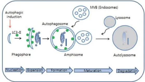
The morphological steps of autophagosome formation consists of sequential events including the exfoliation of a double isolating membrane that is further expanding and maturing resulting in a double membrane bound vacuole. The autophagosome fuses with endosomal compartments (MVB/late endosome) creating the so called amphisome.
The ultimate fusion of the amphisome with lysosomes results in a single-membrane bound autophago-lysosome in order to degrade the engulfed substances (Mizushima et al 2008, Klionsky 2007, Levine&Klionsky 2004, Ohsumi 2001) (Fig. 7). Several studies have demonstrated that autophagy has a role both in physiological as well as pathophysiological processes (Mizushima 2005). Besides the pivotal role of autophagy in tissue remodelling (Gajewska et al 2013, Mizushima&Komatsu 2011), recent studies demonstrated that autophagy may play essential role in inflammatory processes and
22
impact the outcome of disease progression (Choi&Ryter 2011, Levine et al 2011).
There is a complex reciprocal relationship between the autophagy pathway/proteins and immunity/inflammation; the autophagy proteins function in both the induction and suppression of immune and inflammatory responses, and immune and inflammatory signals function in both the induction and suppression of autophagy (Levine et al 2011).
Figure 7. The cellular processes during autophagy. Autophagic process follows distinct stages: nucleation (formation of phagophore), expansion (autophagosome formation), maturation (fusion of autophagosome with MVB (multivesicular body)/lysosome, degradation (acidification). Once autophagy is induced by autophagic stimuli such as inhibition of mTOR, phagophore (isolation membrane) begin to be formed and then cytosolic components are sequestered by autophagosomes characterized by LC3-II-positive double membrane structure.
Endosome such as MVB or lysosome can be fused with autophagosome to form amphisome or autolysosome, respectively. In final step, cytosolic components are degraded in autolysosome.
(Source: Lee 2012)
23
3. Objectives
I. Our previous light microscopical results showed that Freund’s adjuvant treatment induces remarkable phenotypic changes in mesothelial cells.The first experiments of our studies were directed to certify in detail whether intraperitoneal Freund’s adjuvant administration leads to epithelial- mesenchymal transition in mesenteric mesothelial cells in vivo by inducing acute inflammation. According to data from literature, TGF-β has an universal role in EMT. We were also interested in revealing whether the cytokine plays role upon inflammation in our system. The following questions were addressed:
1. What are the ultrastructural changes that can be observed in mesothelial cells upon the inflammation and the following regeneration?
2. Can inflammatory cytokines (IL-1, IL-6) be detected in the peritoneal fluid upon Freund’s adjuvant treatment?
3. Does TGF-β have any roles in the inflammation-induced epithelial-mesenchymal transition? If so, how the level of its peritoneal secretion correlates with the inflammatory events?
4. Can the elements of the canonical TGF-β signaling pathway be detected in mesothelial cells? If so, what are the cellular compartments along which the signaling molecules (TβRII/Smad7) are accommodated in correlation with the inflammatory events?
5. Does caveolar internalization have a role in the dynamics of TGF-β signaling?
II. Recently, ER-α has been suggested as another player in the molecular mechanism of EMT and has opposing roles with TGF-β in a cell and context-dependent manner. We previously showed that mesothelial cells can assume a macrophage character by expressing ED1 (macrophage marker)
24
and may serve as a source of peritoneal macrophages upon treatment. Since macrophages are well-known to express estrogen-receptor α (ER-α), we were interested in whether the receptor might be present in mesothelial cells and has a role in TGF-β induced EMT. Based upon our previous results and the data from literature we considered to answer the following questions:
1. Do mesothelial cells express ER-α upon steady state and inflammatory conditions? If so, what is the subcellular distribution of the receptor?
2. Does the level of ER-α expression change during the inflammation?
3. Do ER-α and elements of the TGF-β pathway meet in any cytoplasmic compartments (caveolae, multivesicular bodies)?
4. Is there any natural ligand of ER-α in the peritoneal fluid and if so, how the level of its secretion correlate with the kinetics of inflammation and/or regeneration?
III. The detected and prolonged secretion of extragonadal estradiol raised the question about its possible role in tissue remodelling, regeneration. Among the several factors sex steroids have recently been described to induce autophagy and help in tissue remodelling, thus our interest turned towards examining whether possible estradiol-induced autophagy is present in mesothelial cells following acute inflammation and the process might contribute to the morphological re-establishement of the mesothelium. The following questions were addressed:
1. Is autophagy present and may play role in the removal of cytoplasmic organelles following inflammation? If so, how the rate of autophagy correlates with the inflammatory events and the secreted E2 concentrations?
2. Does extragonadal estradiol have pivotal role in inducing autophagy? Are there any extracellular signals (TNFα) that are present and aid or overwrite the effects of estradiol in our system?
25
4. Materials and methods
Material and Treatment
Rat mesentery (peritoneum of small intestine) was isolated from control and Freund’s adjuvant (Sigma-Aldrich, Steinheim, Germany) treated Sprague-Dawley male rats (200–400 g) (Female rats were used in one experiment, see in Chapter II/1/3). 1 ml complete Freund’s adjuvant was injected into the peritoneal cavity. One day (D1), two days (D2), three (D3), five (D5), six (D6) eight (D8) and eleven (D11) days following intraperitoneal injections, prior to sample removal the animals were anaesthetized and sacrificed by decapitation.
Reagents
Primary antibodies used for the morphological experiments were: rabbit polyclonal anti- caveolin-1 antibody (Transduction Laboratories), rabbit polyclonal anti-EEA1 antibody (Abcam), rabbit polyclonal anti-Smad7 antibody (Epitomics). Rabbit polyclonal anti estrogen-receptor alpha antibody (H-184): sc-7207) and rabbit polyclonal TGF-βRII antibody (C-16)-R: sc-220-R were purchased from Santa Cruz. Biotinylated anti-rabbit IgG was used as a secondary antibody at a dilution 1:200 (Vector Laboratories Inc, Burlington, CA). For confocal microscopy, streptavidin Alexa Fluor 488 and 555 conjugates, goat anti-rabbit IgG Alexa Fluor 555 (MolecularProbes, Leiden, the Netherlands) were all used at a dilution 1:200. The nuclei were stained with DAPI (Vector Laboratories Inc, Burlington, CA). For immunogold labeling, protein A conjugated to 10- (1: 50) and 15-nm (1:50) gold particles was manufactured and purchased from Cell Microscopy Centre, Utrecht, The Netherlands.
Besides the above described primary antibodies we further applied in the biochemical experiments anti-TGF-β antibody, polyclonal anti-LC3B antibody purchased from Cell Signaling. Polyclonal anti-pErk1/2 was the generous gift of dr. Márk Oláh and monoclonal anti-tErk1/2 was purchased from Life Technologies. Polyclonal anti-actin antibody and polyclonal anti-rab7 antibody were obtained from Sigma-Aldrich and polyclonal anti-Cd63 antibody was ordered from Santa Cruz. The below inserted table summarizes the sources and applied dilutions of primary antibodies (Table 1).
26
Table 1. Primary antibodies applied for the experiments.
Antibody Source Host Applied dilutions
anti-caveolin-1 BD, Transduction Laboratories, Lexington, KY
rabbit polyclonal ICC 1:200 IEM 1:40 IP 1/400 µl anti-EEA1 Abcam, Cambridge,
UK
rabbit polyclonal ICC 1:200 IEM 1:50 anti-TGFβRII
(C-16)-R: sc-220- R
Santa Cruz Biotech Inc. , Santa Cruz, CA
rabbit polyclonal ICC 1:200 IEM 1:60 IP 2/400 µl anti-Smad7
(C-terminal)
Epitomics, Burlingame, CA
rabbit polyclonal ICC 1:20 anti-ER-α
(H-184): sc-7207
Santa Cruz Biotech Inc. , Santa Cruz, CA
rabbit polyclonal ICC 1:200 IEM 1:50 WB 1:300 anti-LC3B Cell Signaling
Technology, Inc., Danvers, MA
rabbit polyclonal ICC 1:200 WB 1:200 anti-pERK1/2 Cell Signaling
Technology, Inc., Danvers, MA
rabbit polyclonal WB 1:200
anti-tERK1/2 Life Technologies mouse monoclonal
WB 1:200 anti-actin Sigma-Aldrich, Co.,
St. Louis, MO
rabbit polyclonal WB 1:5000 anti-rab7 Sigma-Aldrich, Co.,
St. Louis, MO
rabbit polyclonal WB 1:1000 anti-CD63
(H-193): sc- 15363
Santa Cruz Biotech Inc. , Santa Cruz, CA
rabbit polyclonal WB 1:500
anti-TGF-β Cell Signaling Technology, Inc., Danvers, MA
rabbit polyclonal WB 1:1000
Tissue fixation for light and electron microscopy
Mesentery was isolated from both control and Freund’s adjuvant-treated animals. The samples were fixed either in a 1:1 mixture of 2 % glutaraldehyde (GA in 0.2 M cacodylate buffer) and 2 % OsO₄ (Os) in distilled water (30 min, on ice) or freshly prepared 4 % paraformaldehyde (PFA) in 0.1 M PB (2-4 h, room temperature). The samples were washed in cacodylate buffer and PBS, respectively and the adipose tissue
27
was removed from the mesentery. The GA-Os fixed material was proceeded to electron microscopic embedding, while the PFA-fixed samples were used for immunocytochemistry.
Conventional Electron Microscopy
The GA-Os fixed samples were washed in 0.1 M cacodylate buffer three times, dehydrated with ethanol and stained with 1 % uranyl acetate (in 70 % ethanol for 1h, room temperature) prior to araldite embedding. Ultrathin sections were contrasted with uranyl acetate and lead citrate. The samples were analyzed in a Hitachi H-7600 (Tokyo, Japan) transmission electron microscope.
Preparation of semithin and ultrathin cryosections
The PFA fixed samples were stored in 1 % paraformaldehyde (in 0.1 M PB) at 4 °C until further processing. For semithin cryosectioning and immunolabeling, the fixed samples were washed twice in phosphate-buffered saline (PBS), once in 0.02 M glycine/PBS and infiltrated gradually with gelatine solutions of increasing concentrations (2 %, 5 %, 12 % in PB) at 37 °C for 30 min each. The samples were oriented in liquid gelatine and cut into small blocks. For cryoprotection, the blocks were infiltrated with 2.3 M sucrose at 4 °C overnight and afterwards mounted on metal pins, frozen and stored in liquid nitrogen. For preparing semithin and ultrathin cryosections we used Leica Ultracut S ultramicrotome equipped with cryo-attachment (Vienna, Austria). The pickup solution was a 1:1 mixture of 2.3 M sucrose and 1.8 % methylcellulose.
Immunolabeling for light and electron microscopy
0.5 μm semithin sections mounted on microscopic slides were incubated with 0.02 M glycine in PBS for 15 min and they were blocked in PBS containing 1% BSA. Primary antibodies were applied in 1% BSA containing buffer in a humidified chamber at 4 °C (overnight). Prior to a 1 h incubation with the secondary antibodies in a humidified chamber at room temperature, the samples were washed three times with PBS. Sections were then rinsed again with PBS, stained with DAPI, placed under coverslips and
28
visualized in a Bio-Rad (Ontario, Canada) Radiance 2100 Rainbow confocal microscope.
For cryosectioning and immuno-EM, the fixed tissues were further processed as described (Slot&Geuze 2007). In brief, ultrathin cryosections prepared at −110 °C were transferred to copper grids by pickup with a 1:1 mixture of 2.3 M sucrose in PBS and 1.8% methylcellulose. For immunogold labeling, the grids were incubated first on 2%
gelatine/PBS at 37 °C, blocked with 0.02 M glycine/PBS and subsequently incubated with primary antibodies followed by protein A-gold. The sections were contrast stained with 2% uranyl acetate/oxalate, pH 7, followed by 0.4% uranyl acetate pH 4 and 1.8%
methylcellulose and dried. The cryosections were analyzed in a Hitachi H-7600 (Tokyo, Japan) transmission electron microscope at 80 kV.
Morphometry and statistical analysis
Twelve to fifteen electron micrographs were taken from three-three parallel biological samples per group. The surface/volume ratio was calculated according to the method described by Weibel et al (Weibel et al 1996). Results were expressed in number of autophage vacuoles per surface area of cell. Statistical analysis was calculated by ANOVA method and Tukey’s HSD test using Statistica 11 software (StatSoft Inc., Tulsa, OK).
Isolation of mesenteric mesothelial cells
Prior to removal the mesentery, we first washed the abdominal cavity with 2 ml PBS and then rinsed the removed mesentery in clean buffer for 10 minutes. Subsequently, it was placed into 0.2 % collagenase (in DMEM, Sigma-Aldrich Corp., St Louis, MO).
The solid remnants (adipose tissue, connective tissue) were then removed and the samples were washed three times in PBS by centrifugations at 1000 rpm, for 10 min at room temperature. The pellets were then placed into liquid nitrogen for 10 minutes and further stored at -80 °C until use. (qRT-PCR results confirmed the mesothelial phenotype of the retrieved cell population.)
29
Collection of peritoneal fluid and trunk blood, Chemiluminescence immunoassay To determine the secretion level of 17β-estradiol in the peritoneal fluid (PF), six to eight samples per group were collected from male animals by washing the peritoneal cavity with 2 ml PBS. The PF samples were centrifuged at 1000 rpm, for 10 min at 4 °C and the supernatants were stored at -20 °C until use for analysis. To determine the estradiol (E2) concentrations in the plasma, we collected the trunk blood of the animals following decapitation. The collected blood was centrifuged at 1500 rpm, for 20 min at 4°C. The gained serum samples were stored until use in a NaCl (0.9%) and EDTA (5%) containing solution at -20 °C. Elecsys Estradiol II assay on Cobas E411 (Basel, Switzerland) was used to measure and determine the plasma and peritoneal hormone concentrations. The significance was tested by the ANOVA method and Tukey’s HSD test using Statistica 11 software (StatSoft Inc., Tulsa, OK).
RNA isolation and quantitative RT-PCR
Total RNA was extracted from isolated mesothelial cells with RNeasy tissue mini kit (QIAGEN Inc., Chatsworth, CA). For qualitative and quantitative analysis of RNA preparations, RNA 6000 Nano Chip kits in an Agilent 2100 Bioanalyzer (Agilent Technologies Inc., Santa Clara, CA) was used according to the manufacturer’s instructions. Following RNA isolation, RNAs obtained were transcribed into cDNAs using SuperScript™ III First-Strand Synthesis SuperMix (Life Technologies, Carlsbad, CA), according to the manufacturer’s instructions. From each samples, 0.5-1 μg of total RNA was converted into cDNA. Real-time PCR was performed using Taqman Universal Master Mix II, No UNG and the 7900HT Real-Time PCR System (Life Technologies, Carlsbad, CA), with the following parameters: 50 °C for 2 min and 95 °C for 10 min, followed by 40 two-step cycles at 95 °C and at 60 °C for 1 min. Applied Biosystems pre-designed TaqMan® Gene Expression Assays were used for real-time PCR (Il6 Rn00561420_m1, Il1a Rn00566700_m1, Il1b Rn00580432_m1, Esr1 Rn00664737_m1, Tnf. Rn00562055_m1, GAPD.4352338E and B2m Rn00560865_m1). The qRT-PCRs were executed in triplicate using TaqMan probe synthesized by Applied Biosystems). SDS 2.3 (Applied Biosystems, Foster City, CA) and RQ manager 1.2 softwares (Applied Biosystems, Foster City, CA) were applied for calculation of the threshold cycle (Ct) values in each sample.
30
Expression level was calculated by the ddCt method, and fold changes (FC) were obtained using the formula 2-ddCt. Computed internal control corresponding to the geometric mean of Ct values of the two housekeeping genes (GAPD and B2m) was used for the ddCt calculation. Gene expression levels at different time points were compared to controls using ANOVA and t-test with a statistical software SPSS version 19 (SPSS Inc., Chicago, IL, USA).
Immunoprecipitation
Immunoprecipitation was performed by standard methodologies. Briefly, isolated mesothelial cells from D3-D5 samples were lysed in 1 ml solubilization buffer (50 mM Tris-HCl, pH 7.5, 150 mM NaCl, 1 mM EDTA, 1 mM Na3VO4, 1 mM NaF, 10 % glycerol, 0.5 % Nonidet P40, 0.1 mM PMSF, 10 µg/ml aprotinin. The lysates were then incubated with specific antibodies, anti-caveolin-1 and anti-TGF-βRII for 5 h at 4°C.
Immune complexes were formed by addition of protein-A-Sepharose 4B (Sigma) and incubated for 1 h at 4°C. The immune complexes were then sedimented by centrifugation at 12,000 G, followed by 4-5 washes in lysis buffer. Bound proteins were solubilized and analyzed on SDS-PAGE, followed by immunoblotting.
Western blotting
The pellets of isolated mesothelial cells were dissolved in 1ml lysis buffer containing 50 mM Tris-HCl pH 7.5, 150 mM NaCl, 2 mM EDTA, 200 mM Na3VO4, 1 mM NaF, 1 % Nonidet P-40 and protease inhibitor mixture (Complete, Mini, Roche). Insoluble material was removed by centrifugation with 1000 rpm for 10 min. The protein contents were determined by the Bradford method (Bradford 1976) and diluted to a concentration of 0.5 mg/ml. Afterwards the samples were mixed with the same amount of reducing Tris-SDS buffer (Tris 0.5 M pH 6.8, 10 % glycerol, 2% SDS, 0.00125 % bromophenol blue, 0.5 % mercapto-ethanol) and heated at 95 °C for 4 min. 20 µl per well from the samples were loaded onto an Acrylamide/Bis gel and separated by electrophoresis.
After separation, proteins were transferred onto a nitrocellulose membrane (Amersham, GE Healthcare Biosciences, Pittsburgh) in a buffer containing Tris–Glycine pH 8.3, 0.1
% SDS and 20 % methanol. Aspecific reactions were blocked by placing the membrane
31
at room temperature for 2 h in PBS-Tween (0.5 M PBS, Tween 0.05 %) containing 5 % skim milk powder. The membranes were then incubated with primary antibodies diluted in PBS-Tween containing 0.5 % bovine serum albumine at 4 °C for overnight. After washing in PBS-Tween the membranes were treated with species-specific peroxydase- conjugated secondary antibodies (Amersham, GE Healthcare Biosciences, Pittsburgh) for 1 h at room temperature. The labeled protein bands were visualized by the ECL Plus chemiluminenscence method and developed onto high performance chemiluminescence film (Amersham, GE Healthcare Biosciences, Pittsburgh). Relative optical densities were measured using the ImageJ software (U. S. National Institutes of Health, Bethesda, Maryland) and the results of three to four independent experiments were compared and statistically analyzed. The significance was tested by the ANOVA method and Tukey’s HSD test using Statistica 11 software (StatSoft Inc., Tulsa, OK).
32
5. Results
5.1 The morphological characterization of TGF-β induced EMT in mesothelial cells in vivo
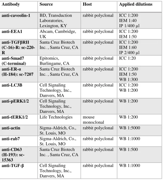
5.1.1 Ultrastructural evidences of type II EMT
In untreated, control animals mesothelial cells form a continuous simple squamous epithelial layer on both sides of the mesentery. As observed in the electron microscope, the cytoplasm of these cells was rather poor in cell organelles and the cells were dominated by the nucleus. A continuous basal membrane separated the mesothelial cells from the underlying connective tissue and maintained the integrity of the mesothelial layer. Caveolae (caveolin-containing lipid rafts seen as flask-shape invaginations of the cell membrane) were abundantly present on the basolateral and apical surfaces of the cells (Fig. 8 A).
Our previous light microscopical data revealed that mesothelial cells lose their epithelial character (decreased cytokeratin and E-cadherin expression) upon Freund’s adjuvant treatment and assume a mesenchymal phenotype by expressing vimentin (Katz et al.
2012). Upon inflammatory stimuli (Freund’s adjuvant treatment) remarkable changes appeared in the ultrastructure of mesothelial cells as well. The dynamics of these changes showed a culmination on the second to third day (D2/D3) after treatment. From the fifth day (D5) on the tissue started to recover and the repairment was morphologically accomplished by the eleventh day (D11).
Characteristic ultrastructural changes were the disintegration of basal membrane and transformation of the squamous mesothelial cells into individual cuboidal-shaped cells in two days after treatment (Fig. 8 B). By the fifth day, the cells developed numerous lamellar processes and became spindle-shaped (Fig. 8 C). The cytoplasmic compartments were more prominent: an increased number of mitochondria, polyribosomes, numerous vesicles and a growing number of multivesicular bodies (MVBs) could be observed in parallel with the inflammatory events of the surrounding tissue from D3. A striking observation was the presence of thickened, electron-dense
33
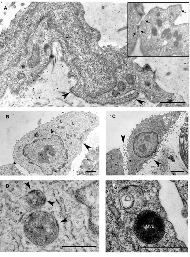
domains on the limiting membrane of MVBs with a fine coat of medium electron density on the cytosolic side (Fig. 8 D). These specific membrane domains (referred from now on as MVB plaques) disappeared by the later stages of the MVB maturation process (by D5) and could not be observed in the membrane of intermediate forms between MVB and lysosome (Fig. 8 E). By D11 mesothelial cells retrieved their flat morphology and became integrated with the underlying connective tissue by the rearrangement of the basal lamina (Fig. 9 A). Furthermore, the neighbouring mesothelial cells came into contact with each other via reassembling intercellular adhesions (tight juctions, adherens junction) thus completing an uniform mesothelial layer again (Fig. 9 B).
34
Figure 8. Fine structural aspects of EMT in mesothelial cells upon Freund’s adjuvant treatment A Non-treated (control) cells are flat and form a continuous layer on the basal lamina (arrowheads). Caveolae are abundantly present in the control cells seen as small flask shape invaginations of the plasma membrane (insert box, arrows). B Upon inflammation, at D2 mesothelial cells progressively lose the connection with the underlying connective tissue as the
35
basal lamina becomes discontinuous and disintegrates (arrowhead). The cytoplasm of mesothelial cells contains lipid droplets (L) that are probably identical with internalized Freund’s adjuvant oil droplets. C On the fifth day of treatment, mesothelial cells assume a spindle-shape morphology with several villar or lamellar processes (arrowheads) on their surface. D An increasing number of multivesicular bodies (MVB) appear from D3. Note specific membrane domains of the organelles (arrowheads) showing an increased density and apposition of a coat on the cytosolic side (MVB plaques). E The MVB plaques disappear by the later stages of MVB maturation, by D5. Scale bars: (A) 1µm, insert 400nm (B) 2µm (C) 2.9µm (D) 500 nm, (E) 1μm.
Figure 9. Electron micrographs showing mesothelial cells eleven days after Freund’s adjuvant treatment. A Note the continuous basal lamina (arrows) and the flat morphology of mesothelial cell. B Reassembly of intercellular junctions (tight or adherens junctions) (dashed arrow) could be observed between the neighbouring mesothelial cells at this time. Bars indicate 833nm
36
5.1.2 Inflammatory cytokines and TGF-β are released into the peritoneal cavity upon Freund’s adjuvant treatment in vivo
In addition to the observed ultrastructural changes, it was of primary importance to see if Freund’s adjuvant treatment induces inflammatory responses in our in vivo system at a molecular level as well. To verify this, we determined the expression levels of pro-inflammatory cytokines in mesothelial cells as they are well-known to be involved in immune responses and inflammatory processes. The results of quantitative RT-PCR showed that mRNA expression levels of interleukin type 1alpha and type 1beta and also interleukin 6 increased in mesothelial cells in response to Freund’s adjuvant treatment (Fig. 10 A). The elevated mRNA levels of these cytokines correlated with the dynamics of the observed morphological changes during the inflammatory events: expression levels of mRNAs had a peak on D3 followed by a significant downregulation that could be observed from the fifth day indicating the termination of the inflammatory response.
TGF-β has an universal role in EMT. While the observed morphological changes seemed identical with the steps of EMT, it was pivotal to see whether TGF-β plays role in our system as well. The Western blot data proved that TGF-β was secreted into the peritoneal cavity upon inflammation and showed a peak between D2 and D3 (Fig. 10 B- C). The alterations in the secretion of the cytokine were in accordance with the observed morphological changes as well as with the expression levels of inflammatory cytokines indicating its role in EMT.
37
Figure 10. Expression levels of proinflammatory cytokines in mesothelial cells and peritoneal secretion of TGF-β in response to Freund’s adjuvant treatment. A The mRNA expression levels of IL-1alpha (IL1a), IL-1beta (IL1b) and IL-6 increase significantly until the third day where expression levels reach a maximum. Further on a decline was observed and reached a similar level measured at the beginning of inflammation induction. B The secretion of
38
TGF-β (measured by Western blot using Pan-TGF-β antibody, 46kDa) in the peritoneal cavity follows the same pattern. C The relative levels of secreted TGF-β were measured by densitometry. The asterisks show significant differences from the control group (p <0,05).
M: male F: female
5.1.3 The morphological detection and the subcellular distribution of the main canonical TGF- β signaling molecules in mesothelial cells
To map whether the elements of TGF-β signaling are present in mesothelial cells, we selected major downstream molecules (TβRII&Smad7) that are considered indispensable either in the promotion or in the termination of the signaling.
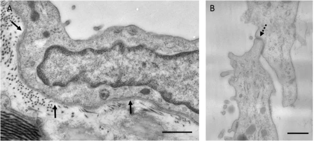
Upon ligand binding, TGF-βRII is responsible for the transmission and promotion of the signaling. We determined the subcellular localization of the receptor by using immunocytochemical approach: our confocal microscopical results showed that the receptor was located both along the plasma membrane as well as inside the cytoplasm.
Under steady state conditions, intracellular receptor labeling appeared in early endosome antigen-1 (EEA1) positive compartments (Fig. 11 A-C). By D3 the majority of the detected TβRII labeling occured inside the cytoplasm (Fig. 11 D-F) and was predominantly found in vesicular (punctate) structures overlapping substantially with EEA1. By D5 numerous large colocalization puncta could still be identified in the cytoplasm of mesothelial cells (Fig. 11 G-I).
39
Figure 11. TGF-βRII is abundantly present and mainly accommodated in early endosome antigen-1 positive compartments in control and treated mesothelial cells. Semithin frozen sections labeled with antibodies directed against TβRII (red) and early endosome antigen-1 (EEA1) (green). A-C In control mesothelial cells, TβRII could be detected either along the plasma membrane or in EEA1 positive early endosomes (arrowheads).
D-F By D3, the receptor labeling entirely fulfilled the cytoplasm where it was found in vesicular structures overlapping substantially with EEA1 (arrowheads). G-I By D5, colocalization of TGFβRII and EEA1 was still pronounced in the cytoplasm of mesothelial cells (arrowheads). Nuclei were stained with DAPI, bars indicate 10µm.
Since caveola-mediated internalization of TβRs presumably enhance the termination of TGF-β signaling, it was of great interest to see whether the receptor shows colocalization with caveolin-1 positive lipid domains and/or caveolae at any time of the inflammation in mesothelial cells in vivo.
We carried out double immunolabeling on semithin frozen sections and we found that in control mesothelial cells TβRII could not be detected together with caveolin-1 (Fig. 12 A-C). At the peak time of inflammation (D3), however TβRII abundantly appeared all
40
over the cytoplasm of mesothelial cells and several overlapping puncta (indicating colocalization) could be observed between the two markers (Fig. 12 D-F). With the progression of inflammation, by D5 substantial cytosolic co-labeling between TβRII and caveolin-1 was present in mesothelial cells (Fig. 12 G-I).
Figure 12. The occurence of TβRII in caveolin positive lipid domains/caveolae upon inflammation. Semithin frozen sections immunolabeled with antibodies directed against TβRII (red) and caveolin-1 (green). A-C In untreated mesothelial cells, TβRII appeared separately from caveolin-1 labeling. D-F By the peak time of inflammation (D3), the receptor labeling was found all over the cytoplasm overlapping with caveolin-1 (arrowheads). G-I By D5, several orange (colocalization) puncta could be identified at the periphery (arrowheads) as well as in the cytoplasm (arrows) of the cells. Nuclei were stained with DAPI, bars indicate 10µm.
Among the inhibitory Smad proteins, Smad7 has a pivotal role in the termination of TGF-β signaling. Since there were data showing that the active TβR complex binding Smad7 is targeted and located in caveolin containing lipid domains of the plasma membrane, we also tried to approve this in our system. To obtain data about the presence and localization of the protein we carried out double immunolabeling on semithin frozen sections. Smad7 protein was barely detectable morphologically in mesothelial cells. However on D3 samples the protein was expressed and could be observed both inside the cytoplasm as well as along the plasma membrane. Whenever
41
we could detect Smad7 at the plasma membrane, it showed colocalization with caveolin-1 (Fig. 13 A-C)
Figure 13. Subcellular distribution of Smad7. A-C Immunofluorescence labeling with antibodies directed against Smad7 (green) and caveolin-1 (red). Smad7 labeling could be detected in mesothelial cells from D3 when it appeared both inside the cytoplasm and along the plasma membrane. The plasma membrane protein labeling showed colocalizaton (orange) with caveolin-1 (arrowheads). Bar indicates: 10µm.